Báo cáo y học: "Dynamic correlation between CTL response and viral load in primary human immunodeficiency virus-1 infected Koreans" ppt
Bạn đang xem bản rút gọn của tài liệu. Xem và tải ngay bản đầy đủ của tài liệu tại đây (434.29 KB, 7 trang )
RESEARC H Open Access
Dynamic correlation between CTL response and
viral load in primary human immunodeficiency
virus-1 infected Koreans
Gab Jung Kim
†
, Hak Sung Lee
†
, Kee-Jong Hong, Sung Soon Kim
*
Abstract
Background: HIV-1 specific cytotoxic T lymphocytes (CTLs) have an important role as antiviral effector cells for
controlling HIV-1 infection.
Methods: To investigate CTL response during the early stage of HIV infection, we measured immunity-related
factors including CD4
+
T cell counts, CD8
+
T cell counts, HIV-1 RNA viral loads and IFN-g secretion according to
CTL response in 78 selected primary HIV-1-infected Koreans.
Results: The CTL response was strongly induced by HIV-1 specific Gag and Nef peptides (p = 0.016) compared with
induction by Tat or Env peptides. These results suggest that the major antiviral factors inducing strong HIV-specific
CTL responses are associated with the Gag and Nef viral regions in primary HIV-1 infected Koreans. The relationship
between viral load and CTL response showed varying correlations with time following HIV infection. CTL response
was inversely correlated with viral loads at preseroconversion stage I (r = -0.224 to -0.33) and changed to a positive
correlation at the preseroconversion stage II (r = 0.132 to 0.854). Finally, it changed to an inverse correlation again
after seroconversion until a viral set point was established on serological profiling (r = -0. 195 to -0.407).
Conclusions: These findings demonstrate a dynamic correlation between viral load and subsequent CTL responses
during early HIV infection.
Background
Human immunodeficiency virus type 1 (HIV-1) specific
CD8
+
T cells play a key role in the control of viral repli-
cation during HIV-1 infection. The cytotoxic T lympho-
cyte (CTL) response is mainly measured at the early
stage of infection and its a ppearance coincides with a
rapid fall in plasma viremia during the early stage of
infection with HIV-1 [1]. One of the well-characterized
effector functions of CD8
+
T cells in the control of vire-
mia is interferon gamma (IFN-g) secretion. IFN-g
secreted by CD8
+
T cells inhibits the viral replication
through induction of antiviral proteins and host immune
responses that kill infected cells. Therefore, strong CTL
responses a re often associated with better virus control
and slower disease progression during the early stage of
HIV infection [2].
Analyses of epitopes or epitope-rich immunodominant
regions inducing HIV-specific CTLs are likely the best
way for pro tection against infection, and offer preferable
approaches for vaccine development [3]. In particular,
the HIV peptides Gag a nd Nef have been suggested a s
being more frequently recognized than Env and Pol in
subjects during the early stage of HIV infection [4].
However,therehavebeendiver se reports about the
induction of specific CTL responses by H IV peptides
and it is still debatable if a role of each peptide can be
constantly associated with immunogenic reactions with
time during the early stages of HIV infection. Some
results have shown that the frequencies of IFN-g -secret-
ing cells during HIV infection were positively correlated
with viral load [5-7], while other studies indicated that
the level of IFN-g induced by CTL responses was nega-
tively correlated with viral load [8,9]. Furthermore, some
other reports suggested that there was no significant
correlation b etween virus-specific T cell responses and
HIV-1 viral load [10-12]. Thus, th e relationship between
* Correspondence:
† Contributed equally
Division of AIDS, Center for Immunology and Pathology, Korea National
Institute of Health, Seoul, Korea
Kim et al. Virology Journal 2010, 7:239
/>© 2010 Kim et al; licensee BioMed Central Ltd. This is an Open Access article distributed under the terms of the Creative Commons
Attribu tion License ( which permits unrestricted use, distribution, and reproduction in
any medium , provided the original work is properly cited.
immune components and virological events in HIV-1
infection remains controversial. The factors leading to
different conclusions from each research group might
include varying parameters of each study population,
such as ethnic differences, clinical status or the different
genetic traits of HIV strains [10,13].
To understand the c haracteristics of HIV-specific
CTL responses in the control of virus replication dur-
ing the early stage of HIV infection, we i nvestigated
the correlation between HIV-1 RNA viral load and
HIV-1-specific CTL responses through measurement
of IFN-g secretion in response to overlapping peptide
stimulation in peripheral blood mononuclear cells
(PBMCs) from Korean subjects with primary HIV
infection (PHI). These constituted a homogeneous eth-
nic group [14] and were infected with the distinct
HIV-1 K orean clade B [15].
Methods
Study samples
The Korean National Institute of Health (KNIH) per-
formed final confirmation tests for the samples. These
were identified as being from subjects with an indeter-
minate HIV status or who were suspected as having an
acute HIV infection, through a hospital or local public
health and environment institute (IPHE), as part of the
national HIV testing strategy. Among the samples
referred to KNIH for confirmation, we selected 78 sub-
jects identified by serological testing as being at an HIV-
1 preseroconversion stage or at t he seroconversion
stage. These patients were followed up to confirm their
HIV infection status using antibody detection. The sam-
ples were analyzed for CD4
+
T cel l counts, CD8
+
T cell
counts and HIV RNA v iral load. We collected the sub-
jects’ epidemiological data and treatment history from
hospitals or public health centers. All of these patients
were antiretroviral therapy naive.
Flow cytometry analysis of CD4
+
and CD8
+
T cell
subpopulations
The numbers of CD4
+
T cells and CD8
+
T cells in
PBMCs were counted after blood w as taken from the
subjects using tubes with EDTA anticoagulant. A CD4-
FITC/CD8-PE/CD3-PC5 monoclonal antibody mix
(Beckman Coulter, F ullerton, CA, USA) was added to
100 μL of each specimen and incubated for 15 min at
room temperature in the dark. IgG1-FITC/IgG1-PE/
IgG1-PC5 (Beckman Coulter) was used as an isotype
control. Red blood cells were removed using Immuno-
prep™ reagent lysis solution (containi ng 1.5% formalde-
hyde, Beckman Coulter) after incubation. Finally, the
stained cells were analyzed using a Cytomics FC500
flow cytometry system (Beckman Coulter).
Quantitative analysis of HIV-1 RNA
Using a Nuclisens Easy H IV-1 system (BioMe rieux,
Durham, NC, USA), HIV-1 RNA in each patient’ s
plasma was quantified. Plasma was stored at -70°C after
isolation from HIV-1 infected blood. After mixing ali-
quots of plasma (200-2000 μL) with lysis buffer (con-
taining guanidine thiocyanate and Triton X-100,
BioMerieux) by vigorous shaking for 30 min at 37°C,
nucleic acids were absorbed by inverted shaking with
50 μL silica for 10 min. We used 20 μL aliquots of cali-
brators including the HIV-1 Gag gene (Qa, Qb, Qc) as
an internal control. Pure HIV-1 RNA was isolated using
NucliSens Extracter (BioMerieux.) Isolated HIV-1 RNA
was amplified using the Nucleic Acid Sequence Based
Amplification method and amplicons derived from a
single strand HIV-1 RNA were collected from the
amplified products. Collected HIV-1 amplicons were
quantified using a NucliSens electrochemiluminescence
reader (BioMerieux) after hybridization.
Measurement of CTL responses using an IFN-g enzyme-
linked immunosorbent spot (ELISPOT) assay
Cryopreserved PBMCs were thawed and cultured in
RPMI 1640 medium containing 10% fetal b ovine serum
(FBS) and 1% penicillin/streptomycin (Gibco, Grand
Island, NY, USA) for 24 h at 37°C. Cultured P BMCs
were counted using trypan blue vital staining (Gibco) at
the beginning of the IFN-g ELISPOT assay. IFN-g
precoated plates (Mabtech, Stockholm, Sweden) were
blocked w ith culture medium containing 10% FBS, and
1.5 × 10
5
PBMCs w ere added to each well. Then, HIV
specific peptides (Gag p17, Gag p24, Tat, Env gp120 or
Nef), dissolved in DMSO with a final concentration of
5 μg/mL, were added. The concentration of DMSO was
always less than 0.5% after dilution. The peptides were
overlapped each other by 10 mer amino acids (from the
National Institute of Biological Standards and Control,
UK). Phytohemagglutinin (5 μg/mL, Sigma-Aldrich,
St.Louis,MO,USA)andCD3antigen(100ng/mL;
Mabtech) were added as positive controls. Plates were
incubated under 5% CO
2
in air at 37°C for 24 h, and
developed using alkaline phosphate-conjugated mono-
clonal antibody 1-B6-1 and NBT/BCIP substrate
(Mabtech) for 15 min at room temperature. Spot forma-
tion was analyzed using an ELISPOT Reader (Immuno-
spot S5 Micro Analyzer, Cellular Technology Ltd.,
Cleveland, OH, USA). Results are expressed as spot-
forming cells (SFC)/10
6
PBMCs = 10
6
× [(SFC number/
well)/(number of cells/well)].
Statistical analysis
Statistical analyses were performed using SAS software
version 9.1(SAS Institute Inc., Cary, NC, USA).
Kim et al. Virology Journal 2010, 7:239
/>Page 2 of 7
Spearman’s rank correlation test was used to determine
any correlation between the HIV-1-specific CTL
response and HIV RNA viral load.
Results
Baseline characteristics in Korean subjects with PHI
We selected 78 subjects with PHI: 72 men and 6 women
with a mean age of 35.3 years. The main transmission
route of 44 subjects was recorded as sexual contact: 61%
by heterosexual transmission and 39% by homosexual
transmission. Of the subjects, 45 had an HIV presero-
conversion status in that only antigen was detected,
without any antibody at the initial tests. The other 33
individuals w ere identified as indeterminate by western
blot testing. However, all subjects turned out to be sero-
positive in the follow-up tests. The mean duration
between receiving the first referred sample and the fol-
low-up sample was about 51 days (Table 1).
Only 50 of the 78 subj ects showed positive responses
in the ELISPOT assay. These CTL responders were
divided into four groups according to the serological
profile and the interval between the initial and follow-
up visits. This was based on the detection of HIV-1 spe-
cific antigen and antibodies in plasma. In group I (pre-
seroconversion group I, n = 7), HIV RNA viral load and
HIV-1-specific enzyme immunoassay (EIA) antigen
levels were extremely high and HIV-1 antibodies were
weakly detectable. In group II (preseroconversion group
II, n = 12), the HIV-1 specific EIA antigen value was
decreased and antibodies were detectable by the EIA
system and the western blot pattern was indeterminate.
In group III (seroconversion group I, n = 13), HIV-1
specific EIA antigen was undetec table, antibody values
were high and western blots were completely positive.
Group IV consisted of 18 subjects who had also
undergone s eroconversion. The HIV-1 specific antigen
and antibody profile of this group showed a similar pat-
tern to group III. The follow-up durations of groups I,
II and III were within 2 weeks, 1 month and 2 months
of the first visit, respectively. For the follow-up samples,
the mean values were 356 cells/mm
3
(range 129-765)
for CD4
+
T cell count, 1,464 ce lls/mm
3
(range 406-
5,937) for CD8
+
T cell co unt and l og
10
5.08 copies/mL
(range 1.40-6.82) for HIV-1 RNA viral load (Table 2).
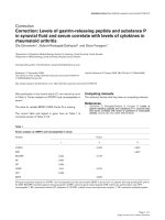
CTL response induced by HIV-specific peptides
For the ELISPOT assay results, only 50 (64%) of these
78 subjects with newly diagnosed PHI showed IFN-g
spot formation in comparison with the positive controls.
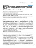
The HIV-specific CTL response in these responders was
strongly induced by Gag and Nef peptides (Fig. 1). Spot
formation induced by Gag p17, p24 and Nef peptides
was strongly enhanced while that for Env or Tat peptide
was not (p = 0.016). Cell viability reflected the effects of
CTL response. The responder group showed higher cell
viability than the nonr esponder group in the IFN-g ELI-
SPOT assay: 79.6% for nonresponders and 89.2% for
responders (p < 0.001; data not shown).
Correlation between CTL response and HIV viral load
Correlation analysis between CTL response and viral
load in 50 CTL responders demonstrated that RNA viral
load during the PHI period did not correlate with CTL
responses (Fig. 2). HIV-specific CTL response induced
by HIV specific peptides showed a slightly positive cor-
relation with HIV RNA viral load with r = 0.153 for
Gag p17 (p = 0.347), r = 0.01 for Gag p24 (p = 0.949)
and r = 0.036 for Env (p = 0.827), respectively. However,
there was no significant correlation between the CTL
response induction by Tat or Nef peptide and RNA viral
load.
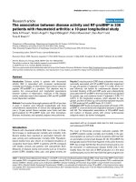
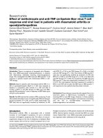
Analysis of the CTL response and viral load in the
four subgroups divided according to the follow-up dura-
tion demonstrated significant results based on the time
course of infection. Fig. 2 shows th e correlations of viral
load and CTL response to each epitope in subjects
divided into four gr oups based on their serological
profile and e lapsed time following HIV infection. For
groups I (preseroconversion group I) and IV (serocon-
version group II), the HIV-specific CTL responses to
the five investigated peptides correlated inversely with
the viral loads (r = -0.224 to -0 .330 for group I and
r = 0.195 to -0.407 for group IV). In contrast, the CTL
response correla ted positively with vi ral load in group II
(preseroco nvers ion group II; r = 0.132-0.530) and group
III (seroconversion group I; r = 0.561-0.854). As men-
tioned above, patterns of correlation between viral load
and CTL responses appeared to be transiently associated
in the course of natural HIV-1 infections.
Table 1 Serological characteristics of the 78 study
subjects with primary HIV-1 infection
Sample
Characteristics Initial sampling Follow-up
Serological status
EIA
(mean, range)
Antigen ratio (OD/
CO)
13.140
(0.243~30.612)
3.379
(0.063~24.793)
Antibody ratio(OD/
CO)
1.910
(0.288~14.135)
9.504
(0.159~19.608)
PA
(reactivity, No.)
Reactive 20 66
Non-reactive 58 12
WB
(band pattern,
No.)
Negative 45 6
Indeterminate 29 22
Positive 4 50
EIA, Enzyme Immunoabsorbent Assay. PA, Particle Agglutination. WB, Western
Blot. OD/CO, ratio of optical density to cut-off value.
Kim et al. Virology Journal 2010, 7:239
/>Page 3 of 7
Table 2 Baseline characteristics of cytotoxic T lymphocyte (CTL) responses in Korean subjects with primary HIV
infection
Characteristics
Serological profile Immunological profile
Group At initial sampling At follow-up* CD4
+
T cell
(cells/mm
3
)*
CD8
+
T cell
(cells/mm
3
)*
Viral load
(log
10
copies/mL)*
Total
(n = 50)
356
(129-610)
1,464
(366-5,937)
5.09
(2.53-7.84)
I
(n = 7)
EIA Ag+ EIA Ag+, EIA Ab+/-, PA+/-, WB+/- 299
(131-507)
1,322
(581-2,190)
6.08
(5.30-7.48)
II
(n = 12)
EIA Ag+ EIA Ag +/-, EIA Ab+, PA+, WB+ 339
(209-614)
1,547
(520-4,955)
4.91
(3.59-6.48)
III
(n = 13)
EIA Ag+, EIA+/-, PA-, WB+/- EIA Ag-, EIA Ab+, PA+, WB+ 342
(129-765)
1,784
(366-5,937)
4.82
(3.40-6.20)
IV
(n = 18)
EIA Ab+, PA+/-, WB+/- EIA Ag-, EIA Ab+, PA+, WB+ 393
(165-610)
1,216
(446-2,432)
5.07
(2.53-6.82)
* Measured at the time of HIV-1-specific CTL analysis
Group I (preseroconversion group I): subjects were HIV seronegative at the initial sampling time and the follow-up duration was within 2 weeks of first referral.
Group II (Preseroconversion group II): subjects were HIV seronegative at the initial sampling time and the follow-up duration was within 1 month of first referral.
Group III (Seroconversion group I): subjects were starting to become seropositive at the initial sampling time and the follow-up duration was within 2 months of
the first referral.
Group IV (Seroconversion group II): subjects were HIV seropositive at the initial sampling time or starting to become seropositive.
Figure 1 CTL responses for HIV -1 infected Koreans. Interferon gamma (IFN-g) production was measured by enzyme-link ed immunosorbent
spot (ELISPOT) assay after treatment with overlapping peptides. Gag p17, Gag p24 and Nef induced higher levels of IFN-g than Tat or Env
gp120. The solid horizontal bars represent mean values for each group.
Kim et al. Virology Journal 2010, 7:239
/>Page 4 of 7
Discussion
Studies on the CTL response have reported that CD8
+
T
lymphocytes have an important role in controlling viral
replication f ollowing PHI [2,16,17]. Generally, the HIV-
specific CTL response to control H IV replication is
influenced by various factors such as malfunction of
immune cells affected by apopto tic events, modification
of T cell surface antigens, changes in cytokine secretion,
reduced expression of MHC (HLA) classes, mutation or
in characteristic changes to the HIV antigen loci [18].
The characteristics of HIV-specific immune response s
and the parameters of HIV infection in Asian popula-
tions including Koreans are not fully understood,
although there are many reports on Caucasian and Afri-
can populations [19-21]. Therefore, we investigated the
relationship between HIV-specific CTL response and
viral replication in these Korean subjects with PHI.
Most of them were infected by a distinct strain of HIV-
1 subtype B monoclade and their genetic background
was comparatively homogeneous [14].
In our study, CTL responses induced by Gag p17, Gag
p24 and Nef peptides were significantly higher than
when induced by Env or Tat peptides (p = 0.016; Fig. 1).
Many reports have demo nstrated that Gag-specific T
cell-mediated immune responses might be especially
important to control viral load, consid ering the correla-
tion between viral protein levels and CTL response in
adults and children among diverse ethnic groups
[22,23]. Furthermore, highly induced Nef-specific CTL
responses correlated with high viral loads in the plasma
[24]. These studies suggest that the HIV Gag and Nef
peptides might be major factors inducing epitope-speci-
fic CTL responses in subjects with PHI. We detected
HIV-specific CTL responses in only 50 of the 78 sub-
jects in this series. The responder group demonstrated
higher cell viability than th e nonresponder group (89.2%
vs 79.6%; p < 0.001). One of the determi ning factors for
detection of CTL response is the composition of over-
lapping peptide sets. The peptides we used f or the
assessment of responses consisted of only five epitopes:
Gag p17, Gag p24, Tat, Env and Nef. Therefore, we
need to further investigate the induction capacity of
other epitopes to understand the detailed mechanism of
HIV-1-specific CTL response in Koreans.
Previous studies have suggested that the virus-specific
CTL response developed in patients with PHI is respon-
sible for the initial control of viral replication [2,25,26].
However, our results demonstrated that the CTL
responses and viral load in Korean subjects with PHI
did not show a c onstant correlation and there is still
controversy about this correlation. Furthermore, the
reasons for the differences in these findings remain
unclear. Thus, we attempted to identify the reason for
this controversial correlation in Koreans with PHI.
Figure 2 Correlation between HIV-1 specific T cell response and plasma viral load in subjects with HIV primary infection. Each column
represents change of correlation value between HIV-1 specific CTL responses and viral load. CTL responses were performed using overlapping
HIV-1 peptides: Gag p17 (spotted bars), Gag p24 (horizontal dashed bars), Tat (white bars), Env gp120 (black bars) and Nef (dashed bars).
Kim et al. Virology Journal 2010, 7:239
/>Page 5 of 7
Based on our studies, one of the possible reasons for
differences in the reports could be the duration following
infection in subjects with PHI. We found that the HIV-
specific CTL response was transiently associated with
plasma viral load through successive clinical stages after
HIV infection. In fact, the CTL response to HIV-specific
peptides did not show obvious correlation to viral load in
the 50 responders (Table 2). However, the co rrelation
between CTL response and viral load in divided sub-
groups demonstrated different results based on the clini-
cal status of the subjects. In detail, the CTL response was
inversely correlated with HIV viral load in group I,
presumed to be in an acute stage of infection, showing
viral load abruptly rising without the production of HIV-
1-specific antibody. While this correlation was changed
to positive in groups II and III (identified as preserocon-
version stage and initial seroconversion stage), it was
negative again in group IV subjects who had undergone
seroconversion. Therefore, we speculate that the CTL
response is insufficiently activated to control viral repli-
cation during the preseroconversion stage. After this
stage, the correlation changed from negative to positive
because the CTL response was increasing to control the
elevated viral load. During the period from seroconver-
sion to the viral set point when virus concentration is
maintained, the correlations between viral replication
and the host immune response changed dynamically
because CTL responses and viral load were linked during
disease progression. That is, each individual can reveal a
different correlation between CTL responses a nd viral
load even during PHI. This implies that the clinical stage
of each subject is an important factor for the HIV-1 spe-
cific CTL response to control virus replication and for its
correlation with viral loa d. Moreover, the host’s immune
response might not be maintained constantly before the
viral set point is established. Musey et al. also reported
an alternating correlation during the early infection per-
iod before the viral set point was established within
6 months after seroconversion [27,28]. Therefore, a long-
itudinal study during a period between infection and viral
set point should be performed to identify a pattern of
alternating correlation between CTL responses and viral
load in subjects with PHI.
In conclusion, we identified the Gag and Nef peptides
as important HIV-1 specific CTL epitopes in regulating
HIV-1 replication in this Korean population with homo-
geneous ethnic cha racteristics during PHI. We also
found alternating correlations between HIV-1 viral load
and HIV-1-specific CTL responses. The genetic back-
ground of the population might be an important factor
for vaccine efficacy, particularly when limited epitope-
specific vaccine designs are used. Thus, our results may
help to improve the selection of antigen for the design
of future HIV-1 vaccines in Korea.
Acknowledgements
HIV peptides were obtained from the Centralized Facility for AIDS Reagents
supported by European Union Program EVA/MRC (contact QLKZ-CT-1999-
00609) and the UK Medical Research Council, which were originally provided
by Dr. H. C. Holmes (National Institute of Biological Standards and Control).
This research was supported by an intramural grant from the National
Institute of Health, Korea (2004-N51002-00).
Authors’ contributions
HS lee and GJ Kim carried out experiments and drafted the manuscript. KJ
contributed to the revising the manuscript. SS Kim participated in the
design of the study. All authors have read and approved the manuscript.
Competing interests
The authors declare that they have no competing interests.
Received: 22 June 2010 Accepted: 16 September 2010
Published: 16 September 2010
References
1. Appay V, Papagno L, Spina CA, Hansasuta P, King A, Jones L, Ogg GS,
Little S, McMichael AJ, Rowland-Jones SL: Dynamics of T cell responses in
HIV infection. J Immunol 2002, 168:3660-3666.
2. Cao J, McNevin J, Holte S, Fink L, Corey L, McElrath MJ: Comprehensive
analysis of human immunodeficiency virus type 1 (HIV-1)-specific
gamma interferon-secreting CD8+ T cells in primary HIV-1 infection. J
Virol 2003, 77:6867-6878.
3. Livingston BD, Newman M, Crimi C, McKinney D, Chesnut R, Sette A:
Optimization of epitope processing enhances immunogenicity of
multiepitope DNA vaccines. Vaccine 2001, 19:4652-4660.
4. Dalod M, Dupuis M, Deschemin JC, Goujard C, Deveau C, Meyer L, Ngo N,
Rouzioux C, Guillet JG, Delfraissy JF, Sinet M, Venet A: Weak anti-HIV CD8
(+) T-cell effector activity in HIV primary infection. J Clin Invest 1999,
104:1431-1439.
5. Betts MR, Ambrozak DR, Douek DC, Bonhoeffer S, Brenchleey JM,
Casazza JP, Koup RA, Picker LJ: Analysis of total human immunodeficiency
virus (HIV)-specific CD4(+) and CD8(+) T-cell responses: relationship to
viral load in untreated HIV infection. J Virol 2001, 75:11983-11991.
6. Buseyne F, Chenadec JL, Corre B, Porrot F, Burgard M, Rouzioux C,
Blanche S, Mayaux MJ, Riviere Y: Inverse correlation between memory
Gag-specific cytotoxic T lymphocytes and viral replication in human
immunodeficiency virus-infected children. J Infect Dis 2002,
186:1589-1596.
7. Trabattoni D, Piconi S, Biasin M, Rizzardini G, Migliorino M, Seminari E,
Boasso A, Piacentini L, Villa ML, Maserati R, Clerici M: Granule-dependent
mechanisms of lysis are defective in CD8 T cells of HIV-infected,
antiretroviral therapy-treated individuals. AIDS 2004, 18:859-869.
8. Patke DS, Langan SJ, Carruth LM, Keating SM, Sabundayo BP, Margolick JB,
Quinn TC, Bollinger RC: Association of Gag-specific T lymphocyte
responses during the early phase of human immunodeficiency virus 1
infection and lower virus load set point. J Infect Dis 2002, 186:1177-1180.
9. Thakar MR, Patke D, Lakhashe SK, Bhonge L, Kulkarni SV, Tripathy SP,
Gupte N, Brookmeyer R, Quinn TC, Paranjape RS, Bollinger RC: Consistent
subtype specific anti-HIV type 1 T lymphocyte responses in Indian
subjects recently infected with HIV type 1. AIDS research human
retroviruses 2002, 18:1389-1393.
10. Addo MM, Yu XG, Rathod A, Cohen D, Eldridge RL, Strick D, Johnston MN,
Corcoran C, Wurcel AG, Fitzpatrick CA, Feeney ME, Rodriguez WR, Basgoz N,
Draenert R, Stone DR, Brander C, Goulder PJR, Rogenberg ES, Altfeld M,
Walker BD: Comprehensive epitope analysis of human immunodeficiency
virus type 1 (HIV-1)-specific T-cell responses directed against the entire
expressed HIV-1 genome demonstrate broadly directed responses, but
no correlation to viral load. J Virol 2003, 77:2081-2092.
11. Dalod M, Dupuis M, Deschemin JC, Sicard D, Salmon D, Delfraissy JF,
Venet A, Sinet M, Guillet JG: Broad, intense anti-human immunodeficiency
virus (HIV) ex vivo CD8(+) responses in HIV type 1-infected patients:
comparison with anti-Epstein-Barr virus responses and changes during
antiretroviral therapy. J Virol 1999, 73:7108-7016.
12. Gea-Banacloche JC, Migueles SA, Martino L, Shupert WL, McNeil AC,
Sabbaghian MS, Ehler L, Prussin C, Stevens R, Lambert L, Altman J,
Hallahan CW, Bernaldo de Quiros JCL, Connors M: Maintenance of large
Kim et al. Virology Journal 2010, 7:239
/>Page 6 of 7
numbers of virus-specific CD8+ T cells in HIV-infected progressors and
long-term nonprogressors. J Immunol 2000, 165:1082-1092.
13. Wang S, Zhuang Y, Zhai S, Zhao S, Kang W, Li X, Yu XG, Walker DD,
Altfeld MA, Sun Y: Association between HIV Type 1-specific T cell
responses and CD4+ T cell counts or CD4+: CD8+ T cell ratios in HIV
Type 1 subtype B infection in China. AIDS research human retroviruses
2006, 22:780-787.
14. Kim TG, Han H, Lim BU, Kim WI, Kim SM: Distribution of HLA Class I alleles
and haplotypes in Korea. J Kor Med Sci 1993, 8:180-186.
15. Kim YB, CHO YK: Monophyletic clade of HIV-1 subtype B in Korea:
evolutionary pressure or single introduction ? AIDS Research Human
Retroviruses 2003, 19:619-623.
16. Jin BX, Bauer DE, Tuttleton SE, Lewin S, Gettie A, Blanchard J, Irwin CE,
Safrit JT, Mittler J, Weinberger L, Kostrikis LG, Zhang L, Perelson AS, Ho DD:
Dramatic rise in plasma viremia after CD8(+) T cell depletion in simian
immunodeficiency virus-infected macaques. J Exp Med 1999,
189(6):991-998.
17. Schmitz JE, Kuroda MJ, Santra S, Sasseville VG, Simon MA, Lifton MA, Racz P,
Tenner-Racz K, Dalesandro M, Scallon BJ, Ghrayeb J, Forman MA,
Montefiori DC, Rieber EP, Letvin NL, Reimann KA: Control of viremia in
simian immunodeficiency virus infection by CD8+ lymphocytes. Science
1999, 283(5403):857-860.
18. Gulzar N, Copeland KF: CD8+ T-cells: function and response to HIV
infection. Current HIV research 2004, 2:23-37.
19. Rinaldo C, Huang XL, Fan ZF, Ding M, Beltz L, Logar A, Panicali D,
Mazzara G, Liebmann J, Cottrill M, Gupta P: High levels of anti-Human
Immunodeficiency Virus Type 1(HIV-1) memory cytotoxic T-lymphocyte
activity and low viral load are associated with lack of disease in HIV-1
-infected long-term nonprogressors. J Virol 1995, 69(9):5838-5842.
20. Novitsky V, Gilbert P, Peter T, McLane MF, Gaolekwe S, Rybak N, Thior I,
Marlink R, Lee TH, Essex M: Association between virus-specific T-cell
responses and plasma viral load in human immunodeficiency virus type
1 subtype C infection. J Virol 2003, 77:882-890.
21. Bouscarat F, Levacher-Clergeot M, Dazza MC, Strauss KW, Giraed DM,
Ruggeri C, Sinet M: Correlation of CD8 lymphocyte activation with
cellular viremia and plasma HIV RNA levels in asymptomatic patients
infected by Human Immunodeficiency Virus Type 1. AIDS Res Hum
Retroviroses 1996, 12(1):17-24.
22. Edwards BH, Bansal A, Sabbaj S, Bakari J, Mulligan MJ, Goepfert PA:
Magnitude of functional CD8+ T-cell responses to the gag protein of
human immunodeficiency virus type 1 correlates inversely with viral
load in plasma. J Virol 2002, 76:2298-2305.
23. Masemola AM, Mashishi TN, Khowry G, Bredell H, Paximadis M,
Mathebula T, Barkhan D, Puren A, Vardas E, Colvin M, Zijenah L,
Katzenstein D, Musonda R, Allen S, Kumwenda N, Taha T, Gray G,
McIntyre J, Karim SA, Sheppard HW, Gray CM, HIVNET 029 Study Team:
Novel and promiscuous CTL epitopes in conserved regions of Gag
targeted by individuals with early subtype C HIV type 1 infection from
southern Africa. J Immunol 2004, 173:4607-4617.
24. Bouscarat F, Levacher M, Landman R, Muffat-Joly M, Girard PM, Saimot AG,
Brun-Vezinet F, Sinet M: Changes in blood CD8+ lymphocyte activation
status and plasma HIV RNA levels during antiretroviral therapy. AIDS
1998, 12:1267-1273.
25. Koup RA, Safrit JT, Cao Y, Andrews CA, McLeod G, Borkowsky W, Farthing C,
Ho DD: Temporal association of cellular immune responses with the
initial control of viremia in primary human immunodeficiency virus type
1 syndrom. J Virol 1994, 68:4650-4655.
26. Oxenius A, Price DA, Trkola A, Edwards C, Gostick E, Zhang HT,
Easterbrook PJ, Tun T, Johnson A, Waters A, Holmes EC, Phillips RE: Loss of
viral control in early HIV-1 infection is temporally associated with
sequencial escape from CD8+ T cell responses and decrease in HIV-1
specific CD4+ and CD8+ T cell frequencies. J Infect 2004, 190:713-721.
27. Musey L, Hughes J, Schacker T, Shea T, Corey L, McElrath MJ: Cytotoxic-T-
cell responses, viral load, and disease progression in early human
immunodeficiency virus type 1 infection. N Engl J Med 1997,
337(18):1267-1274.
28. Mellors JW, Kingsley LA, Rinaldo CR Jr, Todd JA, Hoo BS, Kokka RP, Grapta P:
Quantitation of HIV-1 RNA in plasma predicts outcome after
seroconversion. Ann Intern Med 1995, 122:573-579.
doi:10.1186/1743-422X-7-239
Cite this article as: Kim et al.: Dynamic correlation between CTL
response and viral load in primary human immunodeficiency virus-1
infected Koreans. Virology Journal 2010 7:239.
Submit your next manuscript to BioMed Central
and take full advantage of:
• Convenient online submission
• Thorough peer review
• No space constraints or color figure charges
• Immediate publication on acceptance
• Inclusion in PubMed, CAS, Scopus and Google Scholar
• Research which is freely available for redistribution
Submit your manuscript at
www.biomedcentral.com/submit
Kim et al. Virology Journal 2010, 7:239
/>Page 7 of 7