Báo cáo y học: " An RNA replication-center assay for high content image-based quantifications of human rhinovirus and coxsackievirus infections" pps
Bạn đang xem bản rút gọn của tài liệu. Xem và tải ngay bản đầy đủ của tài liệu tại đây (2.48 MB, 13 trang )
RESEA R C H Open Access
An RNA replication-center assay for high content
image-based quantifications of human rhinovirus
and coxsackievirus infections
Andreas Jurgeit
1
, Stefan Moese
3
, Pascal Roulin
1,2
, Alexander Dorsch
1
, Mark Lötzerich
1
, Wai-Ming Lee
4
,
Urs F Greber
1*
Abstract
Background: Picornaviruses are common human and animal pathogens, including polio and rhinoviruses of the
enterovirus family, and hepatits A or food-and-mouth disease viruses. There are no effective countermeasures
against the vast majority of picornaviruses, with the exception of polio and hepatitis A vaccines. Human
rhinoviruses (HRV) are the most prevalent picornaviruses comprising more than one hundred serotypes. The
existing and also emerging HRVs pose severe health risks for patients with asthma or chronic obstructive
pulmonary disease. Here, we developed a serotype-independent infection assay using a commercially available
mouse monoclonal antibody (mabJ2) detecting double-strand RNA.
Results: Immunocytochemical staining for RNA replication centers using mabJ2 identified cells that were infected
with either HRV1A, 2, 14, 16, 37 or coxsackievirus (CV) B3, B4 or A21. MabJ2 labeled-cells were
immunocytochemically positive for newly synthesized viral capsid proteins from HRV1A, 14, 16, 37 or CVB3, 4. We
optimized the procedure for detection of virus replication in settings for high content screening with automated
fluorescence microscopy and single cell analysis. Our data show that the infection signal was dependent on
multiplicity, time and temperature of infection, and the mabJ2-positive cell numbers correlated with viral titres
determined in single step growth curves. The mabJ2 infection assay was adapted to determine the efficacy of anti-
viral compounds and small interfering RNAs (siRNAs) blocking enterovirus infections.
Conclusions: We report a broadly applicable, rapid protocol to measure infection of cultured cells with
enteroviruses at single cell resolution. This assay can be applied to a wide range of plus-sense RNA viruses, and
hence allows comparative studies of viral infection biology without dedicated reagents or procedures. This
protocol also allows to directly compare results from small compound or siRNA infection screens for different
serotypes wi thout the risk of assay specific artifacts.
Background
The family of picornaviridae comprises a wide variety of
human and animal pathogens [1]. Notable members of
the twelve genera are the enteroviruses, such as polio-
virus, the causative agent for poliomyelitis, which
affected millions of people before bro ad vaccinations
became available in the last decades. Within the picor-
navirus subgenera, the number of serotypes per species
varies from three in the case o f poliovirus up to more
than one hundred for human rhinoviruses (HRV). HRVs
are the main cause of common cold [2], and for recur-
ring infections in humans [3]. HRV infections lead to
severe exacerbations in patients with asthma or chronic
obstructivepulmonarydisease[4].HRVscomprisespe-
cies A, B and C [2]. Twelve HRVs from species A bind
to the minor receptors from the low density lipoprotein
(LDL) receptor family, and the other 61 A-members as
well as the B-viruses bind to in tercellular adhesion
molecule 1 (ICAM-1) for infection [5]. The receptor(s)
for the HRV-C serotypes are unknown. The enterotropic
coxsackieviruses (CV) can cause myocarditis, pancreati-
tis and meningitis. The hepatitis A hepatovirus i s
* Correspondence:
1
Institute of Molecular Life Sciences, University of Zurich, Winterthurerstrasse
190, CH-8057 Zurich, Switzerland
Full list of author information is available at the end of the article
Jurgeit et al. Virology Journal 2010, 7:264
/>© 2010 Jurgeit et al; licensee BioMed Central Ltd. Thi s is an Open Access a rticle dist ribut ed under the term s of the Creativ e Commons
Attribution License ( which permits unrestricted use, distribution, and reproduction in
any medium, provided the original work is properly cited.
responsible for mild forms of human hepatitis. An
example of a non-human picornavirus is the foot-and-
mouth disease virus of the apthovirus genus, which
induces lesions in cloven-hoof animals, such as cattle,
swine, goat, sheep and buffalo, and is the cause for tre-
mendous economic losses, as experienced during the
last outbreak in England in 2001 [6].
Picornaviruses are small, non-enveloped RNA viruses
with an icosahedral capsid of about 28-30 nm in dia-
meter [7], and a single strand, plus-sense RNA genome,
which is in case of enteroviruses about 7.2 to 8.45 kb
[8].Thegenomeencodesasinglepolyproteinthatis
proteolytically processed by viral proteases into struc-
tural and non-structural proteins. The replication of
picornaviruses takes place in the cytoplasm in close
associati on with endo-m embranes containing single-and
multi-membrane vesicles and complex membranous
structures of various sizes [9]. Cytoplasmic membranes
are well known to support the replication of plus-sense
RNA viruses, for example the alphavirus Semliki Forest
virus [10-12], the rubivirus rubella virus [13,14], the
enterovirus poliovirus [15], or t he flaviviruses hepatitis
C, Dengue and West Nile viruses [16-18], where it is
referred to as membranous web. Membrane associated
replication structures are thought to protect the repli-
cating viral RNA from anti-viral factors recognizing
double-strand RNA (dsRNA), and may provide a scaf-
fold for anchoring and locally concentrating the viral
replication complexes. Since its establishment requires
de novo lipid synthesis, it may represent an anti-viral
target, as suggested from work with drosophila C v irus,
a dicistronic virus, which is in many ways similar to
picornaviruses, for example, encoding a polyprotein by a
single positive-strand RNA genome, or using cap-
independent, internal ribosome entry site-dependent
translation [19,20].
The replication process of viruses has been a target for
classical anti-viral agents directed against proteases,
polymerases or integrases in the case of human immu-
nodeficiency syndrome viruses (HIV) or hepatitis C
viruses (HCV) [reviewed in [21]]. Enterovirus inhibitors
have been developed against the HRV protease 3C [22]
or the capsid uncoating mec hanism [f or example, pleco-
naril, [23]]. Alternative approaches against host factors
that support viral replication include d protein kinases
involved in virus entry, such as the serine/threonine
kinase PAK1 for echoviruses, adenoviruses or vaccinia
virus [24-28], as well as tyrosine kinases for coxsackie-
virus B3-RD [29] or microbial pathogens [for a review,
see [30]]. To enhance the identification of anti-viral
agents, standardized infection assays should be devel-
oped for cultured cells as a basis for high throughput
screening projects.
Here we describe a simple immunofluorescence-based
infection protocol to quantitatively assess infection of
cultured cells with enteroviruses, using the mouse
monoclonal anti-dsRNA antibody J2 [mabJ2, [31]]. It
recognizes dsRNA duplexes larger than about 40 bp and
was used earlier to d etect replicating HCV genomes in
distinct cytoplasmic foci [32], or RNA replication inter-
mediates from the groundnut roset te v irus RNA-depen-
dent RNA polymerase [31]. The cytoplasmic foci
recognized by mabJ2 are similar to foci recognized by
an anti-dsRNA serum in rubella virus or Semliki Forest
virus-infected cells [13,33]. We found that the appear-
ance of mabJ2-positive dsRNA replication centers in
HRV or coxsackievirus infected cells correlated with the
emergence of capsid protein e pitopes and infectious
virus titer, and the mabJ2 assay was applicable for pro-
totypic high throughput, image-based siRNA and small
compound screens.
Results
Double-strand RNA replication centers identify HRV and
coxsackievirus infected cells
We first tested if the formation of dsRNA-positive repli-
cation centers can be used as an assay for infection of
HeLa cells strain Ohio (herein referred to as HeLa) with
HRV or CV. HeLa cells are widely used to isolate and
study HRVs and other enteroviruses [34]. Cells were
infected at low multiplicity of infection (moi 0.2-0.4)
with HRV1A, 14, 16, 37 or CVB3 or B4, and co-stained
by double label immunofluorescence for dsRNA using
mabJ2, and newly synthesized viral proteins using
mabR16-7-Alexa488 (conj ugated with Alexa488 dye) or
a rabbit polyclonal antibody raised against purified cap-
sid proteins (Fig. 1A). MabR16-7 had been raised against
HRV16 and recognized VP2 from both HRV16 and 1A
[35]. As expected, all cells positive for newly synthesized
viral protein were also positive for dsRNA detected by
mabJ2, and replication foci had a subcellular localization
similar to c ytoplasmic foci, w hich had been reported
earlier as replication centers in picornavirus-infected
cells [15,36]. Performing a similar exper iment with the
mabK1, detecting dsRNA >40bp, gave identical results,
although with lower signal intensity (data not shown).
We hence used mabJ2 for all following experiments.
Attempts to detect incoming viral particles by mabJ2
failed, although incoming HRV16 have been successfully
visualized with mabR16-7, detecting a capsid epitope
(data not shown). This was in agreement with the
notion that mabJ2 detects long duplexes of double-
strand structures of the replicating RNA rather than
genomic RNA, that is, most likely duplexes of postive
and negative-strand RNAs [31,32]. Biochemical assays
estimated the numbers of negative-strand RNA copies
Jurgeit et al. Virology Journal 2010, 7:264
/>Page 2 of 13
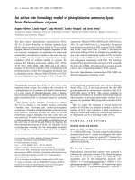
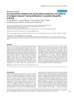
Figure 1 MabJ2 detects viral replication-induced dsRNA in high content image based assays. (A) Cells with dsRNA replication centers are
positive for newly synthesized viral protein. HeLa cells were infected with the indicated HRV or CV serotypes, fixed and stained with mabJ2 (red)
or capsid specific antibodies (green). CVB3, CVB4, HRV37 and HRV14 were stained with a rabbit polyclonal serum (rpc); HRV1A and 16 were
stained by mabR16-7 covalently labelled with Alexa488 (R16-7-488). Magnification 60×; scale bar 20 μm. (B) Appearance of dsRNA replication
centers is moi dependent. Example overview of a 96 multiwell plate of HeLa cells infected with serial dilutions of indicated HRV or CV serotypes.
Imaging by automated microscopy was with 10× magnification. One out of nine images per well is shown for each condition. dsRNA replication
centers (green) and DAPI stained nuclei (blue) are shown. Scale bar 100 μm. (C) An example for automated fluorescence image analysis to score
infection of HeLa cells with HRV16 (moi 0.3) with raw images on the left and an image processed and pseudocolored with a Matlab algorithm
on the right side. Scale bar 100 μm. (D) Example for the quantification of moi dependent fraction of infected cells (infection index) of the
experiment shown in (B), and analysis by the scoring algorithm presented in (C). More detailed characterisations (time, dose) of this assay are
shown in the subsequent figures.
Jurgeit et al. Virology Journal 2010, 7:264
/>Page 3 of 13
in poliovirus infected HeLa cells to about 1000 per cell
at the log phase of replication, corresponding to a few
percent of the total viral RNA [37]. Since poliovirus
replicates to higher levels than HRV in HeLa cells as
determined, for example, in single step growth curves
(WML, unpublished), we suggest that our image-based
assay detects less than 1000 dsRNA molecules per cell.
Although it might be possible to correlate the mabJ2
signal intensity wit h the viral RNA load per cell, this
would require higher resolution image acquisition and
quantitative measurements, and hence would reduce the
throughput of the assay, and require orders of magni-
tude more data to be processed, which would limit the
utility of this assay for screening purposes.
To test if the mabJ2 assay is useful for high-content,
image-based infection screens, we infected HeLa cells
with serial dilutions of different HRV and CV serotypes
in multiwell plates, followed by staining with mabJ2 and
countersta ining of the cell nuclei with 4′,6’-diamidin-2-
phenylindol (DAPI, Fig. 1B). Non-infected cells did not
show detectable signals from mabJ2, while cells inocu-
lated with HRV1A, 2, 14, 16, 37 or CVB3 or B4 showed
dose-dependent mabJ2 signals. Infected ce lls were quan-
tified using a custom-written Matlab routine. This algo-
rithm scored cells as infected, if t he DAPI signal
overlapped with a thresholded infection marker, which
were either the newly synthesized viral protein or
dsRNA replication centers (Fig. 1C, and additional file 1,
Fig. S1). This analysis did not discriminate between
“weak” and “intense” infection signals, but rather scored
cells as infected if certain criteria were met (see details
described i n the methods section and additional file 1,
Fig. S1). The analysis confirmed that the mabJ2 infection
assay was robust and specific for HRV1A, 2, 14, 16 and
CVB3, B4 infections in a dose-dependent manner
(Fig. 1D).
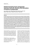
For a biological v alidat ion of the ma bJ2 assay, we per-
formed a receptor interference experiment using the
mouse monoclonal antibody mab15.2L to block the
binding site o f major HRV serotypes 14, 16 and 37 and
CVA21 o n the intracellular adhesion molecule 1
(ICAM-1) [38-40]. As expected, for ICAM-1 tropic
HRVs and CVA21, receptor blocking led to a >90%
decrease of infection, whereas minor group HRVs and
CVB3, which use t he low density lipoprotein (LDL)-
receptor or coxsackievirus adenovirus receptor (CAR),
respectively [41,42], were not affected (Fig. 2). Note that
a lo w amount of mabJ2 signal (appro ximately 5%) was
detected in non-infected cells treated with the mouse
anti-ICAM-1 antibody, but not in non-antibody treated
cells, and hence represents the reactivity of the second-
aryanti-mouseantibody(see addit ional file 2, Fig. S2).
We conclude that the mabJ2 replication center assay is
reliable and has a good signal-to-noise ratio.
Towards high content image based infection screening
To determine optimal conditions for high content infec-
tion assays we performed time course a nd titration
experiments with HRV1A, 2, 14, 16 and 37 and CVB3
and B4. As expected from the initial experiments (see
Fig. 1B, D), the dsRNA infection assay scored a time-
and dose-dependent increase of the infection index for
HRV16andCVB3(Fig.3A,B),andalsofortheother
viruses (additional file 2, Fig. S2). We found that an
infection at low moi (less than 0.5) for 7 h at 37°C was
optimal for HRVs and C Vs. Longer infection times led
to cytopathic effects and loss of infected cells from the
culture dish. Notably, HRV infections were similar or
even more efficient at 37°Ccomparedtoat33.5°C,
whereas CVB3 and B4 infections were attenuated at
33.5°C (Fig. 3A, B, and additional file 3, Fig. S3). The
strong attenuation o f C Vs at 33.5°C was expected. The
good growth characteristics of HRVs at 37°C was consis-
tent wi th recent data showing that HRVs replicate well
at core body temperature [43,44] and are associated
with lower respiratory tract i nfections [3,35,45,46 ]. In
addition, the dsRNA mabJ2 assay detected increasing
infection rates in time course experiments with all the
five HRVs and both coxsackieviruses (additional file 4,
Fig. S4), further confirming the specificity of the assay.
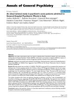
WenextaskedifthemabJ2replicationsignalfrom
HRV1A and 16 correlated with viral titers produced in
the infected cells. We found a strong correlation
between the number of infected cells detected by mabJ2
in the producer cells (dubbed ‘ infectio n’) and infectious
virus production by the infected cells, as determined by
single step growth curves yielding more than 30-fold
higher titers than inoculum (Fig.3C).Thisisinclose
agreements with reports from the literature [47]. We
conclude that mabJ2-positive cells produce infectious
particles confirming that the image based dsRNA infec-
tion assay can also be used for h igh throughput full
cycle infection assessments.
The RNA replication assay for studies with antiviral
compounds
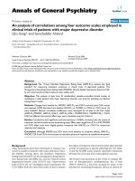
We next tested the performance of the mabJ2 dsRNA
detection assay with the HRV and CV entry inhibitor
pleconaril [23]. Pleconari l binds in the hydrophobic
pocket of the capsid protein VP1 of several entero-
viruses [48], and thereby prevents conformational
changes in the capsid that enable RNA release upon
receptor-mediated endocytosis. The concentration for
50% inhibition (IC50) of pleconaril in our dsRNA-based
infection assay ranged from 0.01 μg/ml for the highly
sensitive CVB4 up to 0.05 μg/ml to 0.1 μg/ml for the
majority of HRVs (Fig. 4A, color code as in panel B).
Our CVB3 strain was resistant to pleconaril in accor-
dance with data from the literature [48].
Jurgeit et al. Virology Journal 2010, 7:264
/>Page 4 of 13
To test if the dsRNA infection assay can be used to
determine at which step of the viral life cycle a particu-
lar compound blocks infection, we performed successive
compound addition experiments. Cells were treated with
pleconaril either prior to infection or at defined time
points post infection (pi). Pleconaril strongly inhibited
infection only when added at early time points (up to
about 45 min) post infection (pi) (Fig. 4B), in agreement
with the notio n that it inhibits the en try and conversion
steps of the capsid prior to release of the RNA genome,
but not genome replication [49].
To address if the dsRNA replication assay responded
to downstream replicatio n blocking agents, we treated
cells with guanidine-HCl, which blocks the enteroviral
protein 2C and specifically prevents the initiation of
negative-strand RNA synthesis but not translation of the
polyprotein [50-53]. All five HRVs (1A , 2, 14, 16, 37)
and CVB3 and B4 were sensitive to the highest concen-
tration of guanidine-HCl tested (20 mM), but HRV1A
and HRV16 were not inhibited by interm ediate concen-
trations of 2 mM (Fig. 4C), which c ould be related to
the close genetic relationship of HRV1A and 16 [5]. The
lowest concentration of guanidine (0.2 mM) i nhibited
HRV14 and 37, but none of the other viruses, which
may also reflect the genetic diversity of the 2C protein
[see for example, [5]]. Consistent with guanidine inhibi-
tion of replication but not upstream processes of infec-
tion, we found that 2 mM guanidine blocked the
appearance of dsRNA mabJ2 epitopes when added up to
120 min pi for CVB3, and up to 240 min pi for the
slower replicating and highly guanidine-sensitive HRV14
(Fig. 4D). The guanidine insensitive HRV1A and 16
remained rather unaffected by guanidine in the time
course experiment confirming the results from the
dose-dependent pre-incubation experiment (Fig. 4C).
Together, these data illustrate that the dsRNA image-
based replication assay is applicable for screening o f
smallanti-viralcompoundsanddeterminingthetime
point of their maximal efficacy in the viral replication
cycle.
Application of the RNA replication assay for image-based
siRNA screens
siRNA profiling in cultured cells has been widely used to
identify host factors with potential therapeutic impact for
anti-viral or an ti-microbial interference, but the re were
only a few genes comm only identified in the different
screens. To reduce some of the technical variables for
siRNA screenings in viral infections, we evaluated the
mabJ2 infection assay for its applicability in high content
image-based siRNA infection screens with a prototype
library of 137 host factors, and a set of defined controls
targeting the HRV genome, that is, three siRNA oligos
per target, a total of 490 individual data points including
scrambled siRNAs and-non-treated controls. Infection of
HeLa cells with HRV14 was scored by mabJ2 staining
and a rabbit polyclonal antibody against structural pro-
teins of HRV14 (W.M. Lee, unpublished). Inspection of
the primary imaging data revealed a strong correlation of
the extent of infection determined by staining for newly
Figure 2 ICAM-1 receptor blocking antibodies abolish the formation of dsRNA replication centers by major group HRVs and CVA21.
HeLa cells were pre-incubated with anti-ICAM-1 mab15.2L for 30 min and infected with indicated HRV and CV serotypes. Infection was
quantified by the mabJ2 anti-dsRNA antibody using automated image acquisition and analysis. Fold infections relative to untreated control cells
are indicated in arbitrary units (AU). The means including standard errors of the mean (SEM) from four independent infections are shown.
Example images for HRV1A (A, B) and HRV16 (C, D) are shown, scale bar 25 μm.
Jurgeit et al. Virology Journal 2010, 7:264
/>Page 5 of 13
synthesized viral protein or the dsRNA replication cen-
ters (Fig. 5A, B). Likewise, comparing the log2 infection
indices between three independent siRNA screens of
HRV16-infected HeLa cells showed strong correlations
(R2 > 0.9) among the three independent replica screens
using both a viral capsid specific antibody (mabR16-7)
and the dsRNA infection assay (Fig. 5C). These data
demonstrate that mabJ2 can be employed for detection
of RNA replication centers in high throughput image-
based infection screens.
Figure 3 Appe arance of dsRNA replication centers is t ime, dose and temperature dependent and correlates with emergence of
infectious titres. (A, B) The time and dose dependencies of HRV16 and CVB3 infections at 33.5°C (blue) or 37°C (red) were determined using
the mabJ2 dsRNA infection assay in HeLa cells by either infection for 300 to 700 min, or with two fold serial dilutions of inocula. (C) To
determine the correlation of mabJ2 dsRNA staining with viral titre production, HeLa cells were infected with HRV1A or 16 for 16 h (infection,
blue) with serial dilutions of inocula. Newly synthesized particles were released from in parallel treated cells by three freeze/thaw cycles and
inoculated on naïve HeLa cells to obtain single step growth curves (red). Infection was scored using automated image analysis. Means and SEMs
of one representative triplicate are shown.
Jurgeit et al. Virology Journal 2010, 7:264
/>Page 6 of 13
The RNA replication center assay detects infection of
non-transformed human WI-38 fibroblasts
Finally, we also tested if mabJ2 recognized HRV-infected
WI-38 primary human lung fibroblasts. We readily
detected mabJ2-positive cells inoculated with the two
minor group serotypes HRV1A and HRV2 ( Fig. 6A).
HRV1A and HRV2 infections were dependent on the
temperature and inoculum dose, as indicated by analyses
at 7 and 8 h pi (Fig. 6B, C). In addition, both infections
were strongly attenuated by an inhibitor of the vacuolar
ATPase, bafilomycin A1, in a dose-dependent manner
with an IC50 of 1 nM [Fig. 6D, E, [54]]. These data
were in agreement with earlier reports showing that
infectious cell entry of minor group HRVs, as s hown
with HRV2, was dependent on low endosomal pH [55],
and that both HRV1A and HRV2 were readily inacti-
vated by low pH solutions in vitro [data not shown, and
[56]]. To our surprise, however, the major group viruses
HRV14 as well as CVB3 and B4 did not le ad to detect-
able formation of mabJ2-positive replication centers in
WI-38 cells up to 8 h pi, even at high moi (100-1000
times higher than for HeLa cells), while HRV16, HRV37
and CVA21 gave low levels of mabJ2 signals (Suppl.
Fig. 5). These data show that mabJ2 detects subtle
differences in infection levels in cultured cells.
Discussion
Comprehensive studies of the vast number of entero-
virus serotypes and their cell biological mechanisms of
infection are a key foundation for developing new anti-
viral therapies. Progress in this area has been limited by
the lack of reagents to detect infection of all the sero-
types, and hence it has remained difficult to stringently
compare the infection mechanisms from different viru s
serotypes or families.
Here we present a dsRNA replication center assay that
canbeusedtodetectinfectionsbyabroadrangeof
enteroviruses in HeLa cells, that is, five human rhino-
virus and three coxsackievirus s erotypes. In the case of
the minor HRV serotypes HRV1A and HRV2 the a ssay
also detected infection of primary human lung WI-38
fibroblasts. The assay is applicable for high content
Figure 4 Formation of dsRNA replication centers can be inhibited by pleconaril or guanidine-HCl. HeLa cells were either pre-incubated
with different concentrations of pleconaril (A), or pleconaril [0.5 μg/ml] was added at indicated time points before or after infection (0 min) (B).
The same types of experiments were done with guanidine-HCl (C, D guanidine HCl [2 mM]). Infections with indicated HRV or CV serotypes
occurred at 37°C for 7 h, and were scored by automated analysis of mabJ2. Means and SEMs of one representative triplicate are shown.
Jurgeit et al. Virology Journal 2010, 7:264
/>Page 7 of 13
Figure 5 The mabJ2 dsRNA replication assay is compatible with high content image based siRNA infection screens. ( A) Overview
montage of an example siRNA screening plate. HeLa cells were infected with HRV14 and stained with a rabbit polyclonal antibody (rpc, green)
raised against purified viral capsid, mabJ2 recognizing dsRNA (red) and nuclei (DAPI, blue). One out of nine images per well is shown for each
siRNA, which are not specified here. (B) Examples close-ups from wells treated with HRV-targeting (HRV siRNA), no siRNA, or scrambled siRNA,
followed by staining as described in (A). Merged colors are shown above, single channel micrographs are in black and white. Scale bars 100 μm.
(C) Normalized HRV16 infection index (log2 transformed) determined by automated microscopy/analysis from three independent siRNA screens.
Infection was measured either by mabR16-7 recognizing a VP2 epitope or mabJ2 recognizing replicated dsRNA.
Jurgeit et al. Virology Journal 2010, 7:264
/>Page 8 of 13
screening, and infection readouts are time, dose and
temperature-dependent.
Importantly, our assay is compatible with siRNA
screening approaches, which have received considerable
attention in the last few y ears, due to the promise to
uncover much of the so far hidden host functions that
support viral infections. Recently genome wide or subge-
nomic screens have been published for a variety of viral
pathogen s, including HIV [57-59], HCV [6 0,61], dengue
virus [62], West Nile virus [63], influenza virus [64-68],
human papillomavirus [69] and vaccinia virus [70]. The
multiple screens for HIV, influenza virus and HCV,
however, identified only very few overlapping gene s for
the individual viruses. Reasons for such findings have
been attributed to the biological n ature of cells and
viruses, including virus strain differences, cell line differ-
ences, cell context-dependent effects and redundancies
of host factors. Among the technical reasons for the low
Figure 6 MabJ2 detects HRV1A and 2 infections of diploid human lung airway cells. (A) Example images of WI-38 non-transformed primary
human embryonic diploid airway cells inoculated with HRV1A or HRV2 and stained for dsRNA replication centers using mabJ2 (green) and nuclei
(DAPI, blue) 7 h pi. Scale bar 100 μm. (B, C) WI-38 cells were inoculated with serial dilutions of HRV1A or HRV2 for 7 or 8 h at 33.5°C (blue) or 37°C
(red), and infection was quantified by the mabJ2 dsRNA infection assay using automated image acquisition/analysis. The infection index is plotted
in arbitrary units (AU), where 1 means all cells infected. (D) WI-38 cells were pre-treated with increasing concentrations of bafilomycin A1 (BafA1) for
30 min, and infected with HRV1A or HRV2 for 7 h. Quantification by the mabJ2 dsRNA infection assay was by automated image acquisition/analysis
and the means (n = 3) and SEMs of the normalized infection index relative to DMSO carrier control infected cells are plotted.
Jurgeit et al. Virology Journal 2010, 7:264
/>Page 9 of 13
levels of overlapping hits from the published screens are
also the different sources and efficacies of siRNAs,
which depended on the manufacturer, or whether single
siRNAs or siRNA pools were used. In addition, the dif-
ferent hit scoring algorithms, including post-processing
filters and variable accounts for toxicity and specificity,
hit ranking algorithms, or consideration of hit assign-
men t to prev iously known functional networks of cellu-
lar pathways can contribute to different hit lists from
siRNA s creens. Last but not least, the assays for infec-
tion are not st andardized, that is, different types of
infection assays cover variable phases of the viral repli-
cation cycle with variable efficacies and, hence, detection
sensitivities and hit identifications are poorly informed.
Our data support the notion that mabJ2 detects repli-
cating dsRNA in infected cells rather than genomic
RNA from incoming virus particles. MabJ2 is hence use-
ful to measure viral replic ation. We suggest that mabJ2
(or any similar antibody) can be used to detect infec-
tions of any positive-strand RNA virus that is actively
replicating. It may even be used to detect dsRNA from
certain DNA virus infections [71]. These findings and
the fact that mabJ2 detects dsRNA with high sensitivity
in solid support based assays [31] open a path t owards
standardized and reproducible infection assays, and pos-
sibly clinical diagnostics.
Our dsRNA replication assay was validated at several
levels. The dsRNA r eadout correlated with single step
growth curves, w hereby the infectious titers produced
per cell were similar t o values reported in the literature,
that is, in the range of 40 plaque forming units per cell
[47]. We have al so validated the assay with two proof of
concept chemical compounds known to block entero-
virus infections, the capsid binding component pleco-
naril [23,72] and the 2C protein inhibitor guanidine
[50]. While pleco naril was an entry inhibitor with a half
maximal inhibition time of about 25 to 30 min, guani-
dine blocked infection until 2 to 4 h pi, reflecting the
different modes of action of these compounds. Hence,
our dsRNA replication assay in the image-based high
content format may prove usef ul also for screening o f
small chemical libraries against viral infections.
Conclusions
The mabJ2 RNA replication assay has proven to be a
reliable procedure to study enterovirus infections on a
systematic level opening new doors for comparative
genomic and chemical studies. It fulfils requirements
such as robustness, good signal-to-noise ratio and prac-
tical usability, making it broadly and systematically
applicable for high content infection assays for entero-
viruses, and possibly other plus-sense RNA v iruses. The
assay covers steps required for virus entry, translation
and RNA replication, and can be extended to a full
replication cycle assay. It is based on a commercially
available m ouse monoclonal antibody, which is readily
accessible for both academic and commercial labora-
tories. The assay also offers a way to carry out mechan-
istic studies with many different serotypes, including
emerging picornaviruses, and hence identify serotype
independent requirements for picornavirus infection.
Methods
Cell culture and virus production
HeLa cervical carcinoma cells strain Ohio (from L. Kai-
ser; Central Laboratory of Virology, University Hospital
Geneva, Switzerland) and primary human embryonic
lung WI-38 cells [American Type Culture Collection,
[73]] were cultured in Dulbecco ’s Modified Eagle Med-
ium (Sigma-Aldrich) supplemented with L-glut amine
(Sigma-Aldrich), non-essential amino acids (Sigma-
Aldrich) and 10% fetal calf serum (FCS, Sigma-Aldrich)
at 37°C and 5% CO
2
in a humidified incubator. In all
experiments passage numbers were kept at a maximum
of 25 post thawing. For infection experiments in 96 well
imaging plates (Matrix ) 14,000 cells were split in a total
of 100 μl the day before the experiment. HRV serotypes
1A and16 were provided by W.M. Lee (Department of
Pediatrics, School of Medicine and Public Health, Uni-
versity of Wisconsin, Madison, Wisconsin, USA), HRV2,
14 and 37 were from L. Kaiser and CVB3, B4 and A21
were from T. Hyypiä (Department of Virology, Univer-
sity of Turku, Finland).
BothHRVsandCVsweregrowninHeLacells.
Briefly, cells were inoculated with a cell lysate stock
from the respective serotypes at 33.5°C (HRV) or 37°C
(CV) over night in infection media (IM/FC-DMEM sup-
plemented with L-glutamine, 30 mM MgCl
2
and 2%
FCS). When CPE was visible in 80-90% of the cells,
media was removed and cells harvested by scraping and
pelleting, lysed by 3 freeze/thaw cycles and centrifuged
at 2500 × g for 10 min. Aliquots of t he supernatants
containing stock virus were stored at -80°C. All sero-
types used in this study were analyzed by reverse tran-
scriptase-polymerase chain reaction and diagnostic
sequencing of the 5’ UTR and/or capsid regions and
found to be virtually identical with the published
sequences. For details, see additional files 5, 6, 7, 8.
Infections and immunocytochemistry
Viruses where added to cells in infection media/BSA
(DMEM supplemented with L-glutamine, 30 mM MgCl
2
and 0.2% BSA, Sigma-Aldrich). For all the compound
and siRNA experiments, moi was chosen such that
approximately 20 to 40% of the cells were infected at 7
h pi. Cells were fixed by adding 1/3 volume of 16%
para-formaldehyde d irectly to the cells in culture media.
Fixation was for either 15 min at room temperature or
Jurgeit et al. Virology Journal 2010, 7:264
/>Page 10 of 13
long term storage at 4°C. Cells were washed with PBS,
PBS/25 mM NH
4
Cl and PBS, permeabilized with 0.2%
Triton X-100 (Sigma-Aldrich) and washed twice with
PBS and blocked with PBS containing 1% BSA (Fraction
V, Sigma). Antibodies detecting viral protein antigens
were used as follows: for HRV1A and HRV16 mabR16-7
[35], for HRV2 mab8F5 [ 74], for HRV14, 37 and CVB3,
B4 the rabbit polyclonal antisera (rpc, W.M. Lee, unpub-
lished). MabJ2 and K1 used to detect dsRNA of infected
cells [31,71] were obtained from English & Scientific
Consulting (Bt. Szirák, Hungary). F ixed and permeabi-
lized cells were incubated at room temperature for 1 h
with diluted mabJ2 in PBS/1%BSA (0.33 μg/ml which
corresponded to a 1:1500 dilution of the 0.5 mg/ml anti-
bod y). Cells were washed twice with PBS and incubated
with Alexa-fluor labelled secondary antibodies (Invitro-
gen) at 0.2 μg/ml for 1 h. Nuclei were stained with
DAPI, and cells on coverslips mounted in mounting
media (Dako), or the 96 well imaging plates were stored
at 4°C in PBS/NaN
3
.
Automated image acquisition and data analysis
Automated image acquisition was performed with an
ImageXpress Micro (Molecular Devices) equipped with
a CoolSNAP HQ 12bit greyscale camera (Roper Scienti-
fic) and 10×/NA 0.5 objective (Nikon). Routinely, 9-20
images per 96 well were acquired leading to an average
of 5000-12000 cells analyzed per well. For high resolu-
tion images, an Olympus IX81 equipped with a 60×/1.4
NA. objective and oil immersion was used. Image over-
lays were made using MetaXpress (Molecular Devices)
and ImageJ (NIH Image, />image/). Images were analyzed using a custom written
Matlab routine. Briefly, a canny edge algorithm was
used to identify areas of all the nuclei stained with
DAPI [75] and infected cells stained for newly synthe-
sized viral protein or replicating dsRNA were identified
by a user-defined thresholding method scoring staining
intensity and size. If the overlap of the nuclear and
infection signals exceeded a u ser defined threshold, a
cell was scored as infected. Data analysis was performed
using Prism (version 5.01, Graphpad), and data for dif-
ferent serotypes were plotted in the order of HRV1A, 2,
14, 1 6, 37, and CVB3, B4 as infection indices (fraction
of i nfected cells per total cell number, indicated as arbi -
trary units) unless stated otherwise.
ICAM-1 receptor blocking and compound assays
HeLa cells were pre-incubated with mouse monoclonal
anti-ICAM-1 antibody mab15.2L (Santa Cruz) at 37°C at
a concentration of 0.5 μg of antibody in 50 μlofinfec-
tion medium/BSA per 96 well for 1 h, followed by infec-
tion for 7 h and staining for dsRNA replication centers.
For compound assays cells were pre-incubated f or 30
min with compounds diluted in infection medium/BSA
prior to virus ad dition. Virus diluted in infection med-
ium/BSA was added to the cells at 37°C for 7 h, and
cells were fixed and immunostained. All compounds
were dissolved in dimethyl sulfoxid (DMSO, cell cul ture
grad e, Sigma-Aldrich) and the respective concen trations
of DMSO were used as controls. Pleconaril was a kind
gift from 3-V Biosciences and guanidine-HCl was
bought from Sigma-Aldrich.
siRNA screens
For siRNA experiments, siRNA oligos (Qiagen) were
spotted in OptiMEM-I (Gibco) at a final concentration
of 50 nM in 96 well imaging plates (Matrix). Lipofecta-
mine 2000 (Invitrogen)/OptiMEM-I was added to a total
volume of 25 μl, and 3000 HeLa cells were seeded into
each 96 well in a total of 100 μl per well. Transfected
cells were incubated for 65 h, followed by infection at
37°C for 7 h and fixation/staining as indicated above.
Specific siRNA oligos directed against the structural
protein VP4 (termed HRV siRNA) were designed
according to the specific genomic sequence of the parti-
cular serotype [76].
Additional material
Additional file 1: Fig. S1. Automated image analysis details. The matlab
scoring algorithm (1) detects edges of the nuclei (A, DAPI) and infection
(B, immunostaining) channels using a canny edge algorithm and user
defined thresholds and forms areas by closing the edges. (2) Areas
below or above a set size-threshold are excluded from both channels
(A2, B2) leading to the final total cell (A3) and infection (B3) mask.
Merging of both masks leads to the final result indicating infected and
not infected cells (as shown in Fig. 1C). Scale bar corresponds to 100 μm.
Additional file 2: Fig. S2. Dose and temperature dependent formation
of dsRNA replication centers of HRV1A, 2, 14, 37 or CVB4 infected HeLa
cells. The dose dependencies of HRV1A, 2, 14, 37 and CVB4 infections at
33.5° (blue) or 37°C (red) were determined for the mabJ2 dsRNA
infection assay in HeLa cells by two fold serial dilutions of inocula.
Infection was scored using automated image acquisition/analysis. Means
and SEMs of one representative triplicate are shown.
Additional file 3: Fig. S3. Time and temperature dependent formation
of dsRNA replication centers of HRV1A, 2, 14, 37 and CVB4 and A21
infected HeLa cells. The time dependencies of of HRV1A, 2, 14, 37 and
CVB4 and A21 infections at 33.5°C (blue) or 37°C (red) were determined
for the mabJ2 dsRNA infection assay in HeLa cells by infection for 300 to
700 min. Infections were scored using automated image analysis. Means
and SEMs of one representative triplicate are shown.
Additional file 4: Fig. S4. MabJ2 dsRNA replication center assay in
normal human lung airway cells. (A) Example images of WI-38 non-
transformed primary human embryonic diploid airway cells inoculated
with the indicated HRV and CV serotypes and stained for dsRNA
replication centers using mabJ2 (green) and nuclei (DAPI, blue) 7 h pi.
Scale bar 100 μm. (B) WI-38 cells were inoculated with serial dilutions of
the indicated HRV and CV serotypes for 7 or 8 h at 33.5°C (blue) or 37°C
(red), and infection was quantified by the mabJ2 dsRNA infection assay
using automated image acquisition/analysis. The infection index is
plotted in arbitrary units (AU), where 1 means all cells infected.
Additional file 5: Table S1. List of primers for diagnostic sequencing of
HRV and CV serotypes.
Jurgeit et al. Virology Journal 2010, 7:264
/>Page 11 of 13
Additional file 6: Table S2. Top results of Blastn alignments of HRV and
CV diagnostic PCR products.
Additional file 7: Table S3. DNA sequences of reverse transcribed PCR
products from five HRV and two CV serotypes.
Additional file 8: Supplemental references.
Acknowledgements
We are grateful to M. Kikkert (Molecular Virology Laboratory, Department of
Medical Microbiology, Leiden, The Netherlands) for providing mabJ2 for
initial experiments, Dr. T. Hyypiä for coxsackievirus strains and advi ce, Dr. ’sC.
Tapparel and L. Kaiser for advice in diagnostic sequencing and providing
HRV2, 14, 37 and HeLa-Ohio cells, and Qian Feng (Department of Medical
Microbiology, Radboud University Nijmegen) for comments on the
manuscript.
Author details
1
Institute of Molecular Life Sciences, University of Zurich, Winterthurerstrasse
190, CH-8057 Zurich, Switzerland.
2
Molecular Life Sciences Graduate School,
ETH and University of Zurich, Switzerland.
3
3-V Biosciences GmbH, Schlieren,
Switzerland & Menlo Park, CA, USA.
4
Department of Pediatrics, School of
Medicine and Public Health, University of Wisconsin, Madison, Wisconsin,
USA.
Authors’ contributions
AJ set up and optimized the assay and performed all experiments
documented by figures; UFG had the initial idea to test mabJ2 in high
content infection screening; SM provided the Matlab code for analysis of
infection experiments; AD, PR, AJ and ML designed and performed the
diagnostic sequencing of HRVs and CVs; WML provided essential antibodies
and protocols for virus growth, UFG & AJ wrote the manuscript.All authors
have read and approved the final manuscript.
Competing interests
The project was in part financially supported by a grant from 3-V
Biosciences Inc (Zurich, Switzerland, and Menlo Park, CA, USA), the Swiss
National Science Foundation, the Swiss SystemsX.ch initiative, grant InfectX
and the Kanton Zurich to UFG. The funders had no role in study design,
data collection and analysis or preparation of the manuscript. UFG is a
founder of 3-V Biosciences, and UFG and SM are shareholders of 3-V
Biosciences.
Received: 19 August 2010 Accepted: 11 October 2010
Published: 11 October 2010
References
1. Whitton JL, Cornell CT, Feuer R: Host and virus determinants of
picornavirus pathogenesis and tropism. Nat Rev Microbiol 2005, 3:765-776.
2. Mackay IM: Human rhinoviruses: the cold wars resume. J Clin Virol 2008,
42:297-320.
3. Gern JE: The ABCs of rhinoviruses, wheezing, and asthma. J Virol 2010,
84:7418-7426.
4. Traves SL, Proud D: Viral-associated exacerbations of asthma and COPD.
Curr Opin Pharmacol 2007, 7:252-258.
5. Palmenberg AC, Spiro D, Kuzmickas R, Wang S, Djikeng A, Rathe JA, Fraser-
Liggett CM, Liggett SB: Sequencing and analyses of all known human
rhinovirus genomes reveal structure and evolution. Science 2009,
324:55-59.
6. Thompson D, Muriel P, Russell D, Osborne P, Bromley A, Rowland M,
Creigh-Tyte S, Brown C: Economic costs of the foot-and-mouth disease
outbreak in the United Kingdom in 2001. Rev Sci Tech 2002, 21:675-687.
7. Rossmann MG, Johnson JE: Icosahedral RNA virus structure. Annu Rev
Biochem 1989, 58:533-573.
8. Racaniello V: Picornaviridae: the viruses and their replication. In Fields
Virology. Edited by: Knipe D, Howley P. Philadelphia, PA, USA: Wolters
Kluwer, Lippincott Williams , 5 2007:1:795-838.
9. Dales S, Eggers HJ, Tamm I, Palade GE: Electron Microscopic Study of the
Formation of Poliovirus. Virology 1965, 26:379-389.
10. Grimley PM, Berezesky IK, Friedman RM: Cytoplasmic structures associated
with an arbovirus infection: loci of viral ribonucleic acid synthesis. J Virol
1968, 2:1326-1338.
11. Friedman RM, Levin JG, Grimley PM, Berezesky IK: Membrane-associated
replication complex in arbovirus infection. J Virol 1972, 10:504-515.
12. Froshauer S, Kartenbeck J, Helenius A: Alphavirus RNA replication occurs
on the cytoplasmic surface of endosomes and lysosomes. J Cell Biol 1988,
107:2075-2086.
13. Lee JY, Marshall JA, Bowden DS: Characterization of rubella virus
replication complexes using antibodies to double-stranded RNA. Virology
1994, 200:307-312.
14. Magliano D, Marshall JA, Bowden DS, Vardaxis N, Meanger J, Lee JY:
Rubella virus replication complexes are virus-modified lysosomes.
Virology 1998, 240:57-63.
15. Bienz K, Egger D, Rasser Y, Bossart W: Intracellular distribution of
poliovirus proteins and the induction of virus-specific cytoplasmic
structures. Virology
1983, 131:39-48.
16. Mackenzie J: Wrapping things up about virus RNA replication. Traffic
2005, 6:967-977.
17. Gosert R, Egger D, Lohmann V, Bartenschlager R, Blum HE, Bienz K,
Moradpour D: Identification of the hepatitis C virus RNA replication
complex in Huh-7 cells harboring subgenomic replicons. J Virol 2003,
77:5487-5492.
18. Welsch S, Miller S, Romero-Brey I, Merz A, Bleck CK, Walther P, Fuller SD,
Antony C, Krijnse-Locker J, Bartenschlager R: Composition and three-
dimensional architecture of the dengue virus replication and assembly
sites. Cell Host Microbe 2009, 5:365-375.
19. King L, Moore N: Evidence for the presence of a genome linked protein
in two insect picornaviruses, cricket paralysis virus and Drosophila C
virus. FEMS Lett 1988, 50:41.
20. Cherry S, Kunte A, Wang H, Coyne C, Rawson RB, Perrimon N: COPI activity
coupled with fatty acid biosynthesis is required for viral replication. PLoS
Pathog 2006, 2:e102.
21. Sturmer M, Doerr HW, Gurtler L: Human immunodeficiency virus: 25 years
of diagnostic and therapeutic strategies and their impact on hepatitis B
and C virus. Med Microbiol Immunol 2009, 198:147-155.
22. Patick AK: Rhinovirus chemotherapy. Antiviral Res 2006, 71:391-396.
23. Pevear DC, Tull TM, Seipel ME, Groarke JM: Activity of pleconaril against
enteroviruses. Antimicrob Agents Chemother 1999, 43:2109-2115.
24. Van den Broeke C, Radu M, Chernoff J, Favoreel HW: An emerging role for
p21-activated kinases (Paks) in viral infections. Trends Cell Biol 2010,
20:160-169.
25. Kalin S, Amstutz B, Gastaldelli M, Wolfrum N, Boucke K, Havenga M,
DiGennaro F, Liska N, Hemmi S, Greber UF: Macropinocytotic uptake and
infection of human epithelial cells with species B2 adenovirus type 35. J
Virol 2010, 84:5336-5350.
26. Mercer J, Helenius A: Vaccinia virus uses macropinocytosis and apoptotic
mimicry to enter host cells. Science 2008, 320:531-535.
27. Liberali P, Kakkonen E, Turacchio G, Valente C, Spaar A, Perinetti G,
Bockmann RA, Corda D, Colanzi A, Marjomaki V, Luini A: The closure of
Pak1-dependent macropinosomes requires the phosphorylation of
CtBP1/BARS. Embo J 2008, 27:970-981.
28. Amstutz B, Gastaldelli M, Kälin S, Imelli N, Boucke K, Wandeler E, Mercer J,
Hemmi S, Greber UF: Subversion of CtBP1 controlled macropinocytosis
by human Adenovirus serotype 3. EMBO J 2008, 27:956-966.
29. Coyne CB, Bergelson JM:
Virus-Induced Abl and Fyn Kinase Signals Permit
Coxsackievirus Entry through Epithelial Tight Junctions. Cell 2006,
124:119-131.
30. Backert S, Feller SM, Wessler S: Emerging roles of Abl family tyrosine
kinases in microbial pathogenesis. Trends Biochem Sci 2008, 33:80-90.
31. Schonborn J, Oberstrass J, Breyel E, Tittgen J, Schumacher J, Lukacs N:
Monoclonal antibodies to double-stranded RNA as probes of RNA
structure in crude nucleic acid extracts. Nucleic Acids Res 1991,
19:2993-3000.
32. Targett-Adams P, Boulant S, McLauchlan J: Visualization of double-
stranded RNA in cells supporting hepatitis C virus RNA replication. J Virol
2008, 82:2182-2195.
33. Bowden DS, Pedersen JS, Toh BH, Westaway EG: Distribution by
immunofluorescence of viral products and actin-containing cytoskeletal
filaments in rubella virus-infected cells. Arch Virol 1987, 92:211-219.
Jurgeit et al. Virology Journal 2010, 7:264
/>Page 12 of 13
34. Couch R: Rhinoviruses. In Laboratory diagnosis of viral infections. Edited by:
Lennette EH. New York, NY: Marcel Dekker, Inc; 1992:.
35. Mosser AG, Brockman-Schneider R, Amineva S, Burchell L, Sedgwick JB,
Busse WW, Gern JE: Similar frequency of rhinovirus-infectible cells in
upper and lower airway epithelium. J Infect Dis 2002, 185:734-743.
36. Lanke KH, van der Schaar HM, Belov GA, Feng Q, Duijsings D, Jackson CL,
Ehrenfeld E, van Kuppeveld FJ: GBF1, a guanine nucleotide exchange
factor for Arf, is crucial for coxsackievirus B3 RNA replication. J Virol 2009,
83:11940-11949.
37. Novak JE, Kirkegaard K: Improved method for detecting poliovirus
negative strands used to demonstrate specificity of positive-strand
encapsidation and the ratio of positive to negative strands in infected
cells. J Virol 1991, 65:3384-3387.
38. Greve JM, Davis G, Meyer AM, Forte CP, Yost SC, Marlor CW, Kamarck ME,
McClelland A: The major human rhinovirus receptor is ICAM-1. Cell 1989,
56:839-834.
39. Jedrzejas MJ, Miglietta J, Griffin JA, Luo M: Structure of a monoclonal anti-
ICAM-1 antibody R6.5 Fab fragment at 2.8 A resolution. Acta Crystallogr D
Biol Crystallogr 1995, 51:380-385.
40. Bella J, Rossmann MG: ICAM-1 receptors and cold viruses. Pharm Acta Helv
2000, 74:291-297.
41. Hofer F, Gruenberger M, Kowalski H, Machat H, Huettinger M, Kuechler E,
Blass D: Members of the low density lipoprotein receptor family mediate
cell entry of a minor-group common cold virus. Proc Natl Acad Sci USA
1994, 91:1839-1842.
42. Shafren DR, Williams DT, Barry RD: A decay-accelerating factor-binding
strain of coxsackievirus B3 requires the coxsackievirus-adenovirus
receptor protein to mediate lytic infection of rhabdomyosarcoma cells.
Journal of Virology 1997, 71:9844-9848.
43. Papadopoulos NG, Sanderson G, Hunter J, Johnston SL: Rhinoviruses
replicate effectively at lower airway temperatures. Journal of Medical
Virology 1999, 58:100-104.
44. Schroth MK, Grimm E, Frindt P, Galagan DM, Konno SI, Love R, Gern JE:
Rhinovirus replication causes RANTES production in primary bronchial
epithelial cells. Am J Respir Cell Mol Biol 1999, 20:1220-1228.
45. Gern JE, Galagan DM, Jarjour NN, Dick EC, Busse WW: Detection of
rhinovirus RNA in lower airway cells during experimentally induced
infection. Am J Respir Crit Care Med 1997, 155:1159-1161.
46. Hayden FG: Rhinovirus and the lower respiratory tract. Rev Med Virol 2004,
14:17-31.
47. Fiala M: Plaque formation by 55 rhinovirus serotypes. Appl Microbiol 1968,
16:1445-1450.
48. Schmidtke M, Hammerschmidt E, Schuler S, Zell R, Birch-Hirschfeld E,
Makarov VA, Riabova OB, Wutzler P: Susceptibility of coxsackievirus B3
laboratory strains and clinical isolates to the capsid function inhibitor
pleconaril: antiviral studies with virus chimeras demonstrate the crucial
role of amino acid 1092 in treatment. J Antimicrob Chemother 2005,
56:648-656.
49. Shepard DA, Heinz BA, Rueckert RR: WIN 52035-2 inhibits both
attachment and eclipse of human rhinovirus 14. Journal of Virology 1993,
67:2245-2245.
50. Rightsel WA, Dice JR, Mc AR, Timm EA, Mc LI Jr, Dixon GJ, Schabel FM Jr:
Antiviral effect of guanidine. Science 1961, 134:558-559.
51. Loddo B, Ferrari W, Brotzu G, Spanedda A: In vitro inhibition of infectivity
of polio viruses by guanidine. Nature 1962, 193:97-98.
52. Pincus SE, Diamond DC, Emini EA, Wimmer E: Guanidine-selected mutants
of poliovirus: mapping of point mutations to polypeptide 2C. J Virol
1986, 57:638-646.
53. Pincus SE, Wimmer E: Production of guanidine-resistant and -dependent
poliovirus mutants from cloned cDNA: mutations in polypeptide 2C are
directly responsible for altered guanidine sensitivity. J Virol 1986,
60:793-796.
54. Bowman EJ, Siebers A, Altendorf K: Bafilomycins: a class of inhibitors of
membrane ATPases from microorganisms, animal cells, and plant cells.
Proc Natl Acad Sci USA 1988, 85:7972-7976.
55. Brabec M, Baravalle G, Blaas D, Fuchs R: Conformational changes, plasma
membrane penetration, and infection by human rhinovirus type 2: role
of receptors and low pH. J Virol 2003, 77:5370-5377.
56. Gern JE, Mosser AG, Swenson CA, Rennie PJ, England RJ, Shaffer J,
Mizoguchi H: Inhibition of rhinovirus replication in vitro and in vivo by
acid-buffered saline. J Infect Dis 2007, 195:1137-1143.
57. Brass AL, Dykxhoorn DM, Benita Y, Yan N, Engelman A, Xavier RJ,
Lieberman J, Elledge SJ: Identification of host proteins required for HIV
infection through a functional genomic screen. Science 2008, 319:921-926.
58. Zhou H, Xu M, Huang Q, Gates AT, Zhang XD, Castle JC, Stec E, Ferrer M,
Strulovici B, Hazuda DJ, Espeseth AS: Genome-scale RNAi screen for host
factors required for HIV replication. Cell Host Microbe 2008, 4:495-504.
59. Konig R, Zhou Y, Elleder D, Diamond TL, Bonamy GM, Irelan JT, Chiang CY,
Tu BP, De Jesus PD, Lilley CE, et al: Global analysis of host-pathogen
interactions that regulate early-stage HIV-1 replication. Cell 2008,
135:49-60.
60. Li Q, Brass AL, Ng A, Hu Z, Xavier RJ, Liang TJ, Elledge SJ: A genome-wide
genetic screen for host factors required for hepatitis C virus
propagation. Proc Natl Acad Sci USA 2009, 106:16410-16415.
61. Coller KE, Berger KL, Heaton NS, Cooper JD, Yoon R, Randall G: RNA
interference and single particle tracking analysis of hepatitis C virus
endocytosis. PLoS Pathog
2009, 5:e1000702.
62. Sessions OM, Barrows NJ, Souza-Neto JA, Robinson TJ, Hershey CL,
Rodgers MA, Ramirez JL, Dimopoulos G, Yang PL, Pearson JL, Garcia-
Blanco MA: Discovery of insect and human dengue virus host factors.
Nature 2009, 458:1047-1050.
63. Krishnan MN, Ng A, Sukumaran B, Gilfoy FD, Uchil PD, Sultana H, Brass AL,
Adametz R, Tsui M, Qian F, et al: RNA interference screen for human
genes associated with West Nile virus infection. Nature 2008, 455:242-245.
64. Hao L, Sakurai A, Watanabe T, Sorensen E, Nidom CA, Newton MA,
Ahlquist P, Kawaoka Y: Drosophila RNAi screen identifies host genes
important for influenza virus replication. Nature 2008, 454:890-893.
65. Karlas A, Machuy N, Shin Y, Pleissner KP, Artarini A, Heuer D, Becker D,
Khalil H, Ogilvie LA, Hess S, et al: Genome-wide RNAi screen identifies
human host factors crucial for influenza virus replication. Nature 2010,
463:818-822.
66. Konig R, Stertz S, Zhou Y, Inoue A, Hoffmann HH, Bhattacharyya S,
Alamares JG, Tscherne DM, Ortigoza MB, Liang Y, et al: Human host factors
required for influenza virus replication. Nature 2010, 463:813-817.
67. Shapira SD, Gat-Viks I, Shum BO, Dricot A, de Grace MM, Wu L, Gupta PB,
Hao T, Silver SJ, Root DE, et al: A physical and regulatory map of host-
influenza interactions reveals pathways in H1N1 infection. Cell 2009,
139:1255-1267.
68. Brass AL, Huang IC, Benita Y, John SP, Krishnan MN, Feeley EM, Ryan BJ,
Weyer JL, van der Weyden L, Fikrig E, et al: The IFITM proteins mediate
cellular resistance to influenza A H1N1 virus, West Nile virus, and
dengue virus. Cell 2009, 139:1243-1254.
69. Smith JA, White EA, Sowa ME, Powell ML, Ottinger M, Harper JW,
Howley PM: Genome-wide siRNA screen identifies SMCX, EP400, and
Brd4 as E2-dependent regulators of human papillomavirus oncogene
expression. Proc Natl Acad Sci USA 2010, 107:3752-3757.
70. Moser TS, Jones RG, Thompson CB, Coyne CB, Cherry S: A Kinome RNAi
Screen Identified AMPK as Promoting Poxvirus Entry through the
Control of Actin Dynamics. PLoS Pathog 2010, 6:e1000954.
71. Weber F, Wagner V, Rasmussen SB, Hartmann R, Paludan SR: Double-
stranded RNA is produced by positive-strand RNA viruses and DNA
viruses but not in detectable amounts by negative-strand RNA viruses. J
Virol 2006, 80:5059-5064.
72. Florea NR, Maglio D, Nicolau DP: Pleconaril, a novel antipicornaviral
agent. Pharmacotherapy 2003, 23:339-348.
73. Hayflick L, Moorhead PS: The serial cultivation of human diploid cell
strains. Exp Cell Res 1961, 25:585-621.
74. Skern T, Neubauer C, Frasel L, Grundler P, Sommergruber W, Zorn M,
Kuechler E, Blaas D: A neutralizing epitope on human rhinovirus type 2
includes amino acid residues between 153 and 164 of virus capsid
protein VP2. J Gen Virol 1987, 68(Pt 2):315-323.
75. Canny J: A Computational Approach to Edge-Detection. Ieee Transactions
on Pattern Analysis and Machine Intelligence 1986, 8:679-698.
76. Phipps KM, Martinez A, Lu J, Heinz BA, Zhao G: Small interfering RNA
molecules as potential anti-human rhinovirus agents: in vitro potency,
specificity, and mechanism. Antiviral Res 2004, 61:49-55.
doi:10.1186/1743-422X-7-264
Cite this article as: Jurgeit et al.: An RNA replication-center assay for
high content image-based quantifications of human rhinovirus and
coxsackievirus infections. Virology Journal 2010 7:264.
Jurgeit et al. Virology Journal 2010, 7:264
/>Page 13 of 13