báo cáo khoa học: " Identification of three wheat globulin genes by screening a Triticum aestivum BAC genomic library with cDNA from a diabetes-associated globulin" doc
Bạn đang xem bản rút gọn của tài liệu. Xem và tải ngay bản đầy đủ của tài liệu tại đây (1.62 MB, 11 trang )
BioMed Central
Page 1 of 11
(page number not for citation purposes)
BMC Plant Biology
Open Access
Research article
Identification of three wheat globulin genes by screening a Triticum
aestivum BAC genomic library with cDNA from a
diabetes-associated globulin
Evelin Loit
1
, Charles W Melnyk
1,3
, Amanda J MacFarlane
1,2,4
,
Fraser W Scott
1,2
and Illimar Altosaar*
1
Address:
1
Department of Biochemistry, Microbiology and Immunology, Faculty of Medicine, University of Ottawa, Ottawa, Canada,
2
Chronic
Disease Program, Ottawa Hospital Research Institute, Ottawa, Canada,
3
Department of Plant Sciences, University of Cambridge, Cambridge, UK
and
4
Bureau of Nutritional Sciences, Food Directorate, Health Canada, Ottawa, Canada
Email: Evelin Loit - ; Charles W Melnyk - ; Amanda J MacFarlane - amanda_macfarlane@hc-
sc.gc.ca; Fraser W Scott - ; Illimar Altosaar* -
* Corresponding author
Abstract
Background: Exposure to dietary wheat proteins in genetically susceptible individuals has been
associated with increased risk for the development of Type 1 diabetes (T1D). Recently, a wheat
protein encoded by cDNA WP5212 has been shown to be antigenic in mice, rats and humans with
autoimmune T1D. To investigate the genomic origin of the identified wheat protein cDNA, a
hexaploid wheat genomic library from Glenlea cultivar was screened.
Results: Three unique wheat globulin genes, Glo-3A, Glo3-B and Glo-3C, were identified. We
describe the genomic structure of these genes and their expression pattern in wheat seeds. The
Glo-3A gene shared 99% identity with the cDNA of WP5212 at the nucleotide and deduced amino
acid level, indicating that we have identified the gene(s) encoding wheat protein WP5212. Southern
analysis revealed the presence of multiple copies of Glo-3-like sequences in all wheat samples,
including hexaploid, tetraploid and diploid species wheat seed. Aleurone and embryo tissue
specificity of WP5212 gene expression, suggested by promoter region analysis, which
demonstrated an absence of endosperm specific cis elements, was confirmed by
immunofluorescence microscopy using anti-WP5212 antibodies.
Conclusion: Taken together, the results indicate that a diverse group of globulins exists in wheat,
some of which could be associated with the pathogenesis of T1D in some susceptible individuals.
These data expand our knowledge of specific wheat globulins and will enable further elucidation of
their role in wheat biology and human health.
Published: 17 July 2009
BMC Plant Biology 2009, 9:93 doi:10.1186/1471-2229-9-93
Received: 19 January 2009
Accepted: 17 July 2009
This article is available from: />© 2009 Loit et al; licensee BioMed Central Ltd.
This is an Open Access article distributed under the terms of the Creative Commons Attribution License ( />),
which permits unrestricted use, distribution, and reproduction in any medium, provided the original work is properly cited.
BMC Plant Biology 2009, 9:93 />Page 2 of 11
(page number not for citation purposes)
Background
Wheat has a primary position in the human diet, and
together with maize and rice provides more than 60% of
the calories and proteins consumed by the world popula-
tion [1]. For the majority of the population, ingestion of
wheat does not stimulate an immune response. However,
in some genetically susceptible individuals, wheat pro-
teins induce an acute mucosal inflammatory response
known as celiac disease [2-4] or Baker's asthma [5,6]. Data
are also accumulating that dietary wheat proteins pro-
mote an inflammatory response in the gut mucosa of
patients with autoimmune Type 1 diabetes (T1D) [7-10].
A wheat storage globulin has been associated with the
development of T1D [11]. This protein was identified by
screening a wheat cDNA expression library with polyclo-
nal antibodies from diabetic BB rats, a model of spontane-
ous autoimmune T1D [11]. Antibody reactivity to the
gene product of one cDNA clone WP5212 was shown to
correlate with pancreatic islet damage and inflammation
in diabetic BB rats. The WP5212 cDNA shared 90% nucle-
otide identity with a 1387 nucleotide region of the
sequence M81719 which was annotated in the NCBI data-
base as a wheat 7S globulin sequence, and which has sub-
sequently been attributed to barley [12]. It also shares
100% identity with a Triticum aestivum assembled tran-
script (TA61374_4565) designated as a homologue to
barley globulin Beg1 precursor.
7S globulin proteins have been previously characterized
in barley at the cDNA level and in maize at the protein
level [13,14]. A single homologous gene encodes both
globulins, Beg1 in barley and Glb1 in maize [13,15]. In
wheat, Globulin 1, Triticin and Globulin 2 have been
described [16-18]. Other storage proteins have also been
shown to be immunomodulatory. Specifically, various
vicilins have been identified as major allergens in a variety
of foods, including peanut (Ara h 1), cashew (Ana o 1),
walnut (Jug r 2), and soybean (Gly m Bd 28K) [19-21].
In an effort to identify the gene(s) encoding WP5212, we
screened a wheat Glenlea genomic library using WP5212
cDNA as a probe. This approach enabled us to identify
and characterize the three novel wheat genes Glo-3A, Glo-
3B and Glo-3C that encode 7S globulins.
Results
Identification of WP5212-like DNA sequences from the
hexaploid wheat genome
A total of 25 positive BAC clones were identified by
screening 24 high-density filters of the hexaploid wheat
Triticum aestivum cultivar Glenlea BAC library (3.1× hap-
loid genome coverage) with the WP5212 specific probe.
Twenty-two positive clones were confirmed to contain
WP5212-like inserts through PCR analysis. Secondary
screening by DNA sequencing confirmed three unique
sequences.
Sequencing of WP5212-like sequences from positive BAC
clones
Out of 22 candidates, three unique clones, 1333A17,
895N14, and 1324M8 were chosen to be sequenced.
Sequencing primers were designed based on conserved
regions in cDNAs from WP5212, barley Beg1 [GenBank:
M64372
] and maize Glb1 [GenBank: M24845] deter-
mined by sequence alignment. Sequencing was conducted
by primer walking. Obtained DNA sequences were assem-
bled into contigs of 4406, 3671 and 1457 bp nucleotides
for 1333A17, 895N14 and 1324M8, respectively.
Sequence characterization and open reading frame
identification
Contigs from clones 1333A17 and 895N14 contained full
genomic sequences for a globulin gene as determined by
open reading frame analysis. The globulin gene open
reading frame starts at base pair 1213 in BAC clone
1333A17 and at base pair 309 in BAC clone 895N14. For
the 1324M8 clone, 1457 bp ORF was delineated, but no
stop codon was identified. However, a partial 5' coding
sequence was identified, starting at position 109 in
1324M8 contig.
No similarity to previously identified wheat globulin
genes was found, thus the specific genes were named Glo-
3A, Glo-3B and Glo-3C in the order they were identified
(originating from BAC clones 1333A17, 895N14, and
1324M8, respectively). All three sequences have been
entered into GenBank [GenBank: FJ439135
, FJ439136
and FJ439134]. Putative open reading frames, the tran-
scription start sites and the polyadenylation signal
sequences were identified (Figure 1A). All sequences con-
tain five to seven exons and four to six introns.
Comparisons between the cDNA clone WP5212 and the
coding region of Glo-3A showed 99% identity at the nucle-
otide level. Translated sequence alignment with the pre-
dicted amino acid sequence of cDNA clone WP5212
resulted in 99% identity: 583 identical, 3 conserved (G5A,
A7V, R102H) and 2 non-conserved (R43Q, A549T)
amino acids out of 588 total amino acid residues (Figure
2). Similarly, 99% identity was shared between Glo-3A
and the assembled wheat transcript [TIGR:
TA61374_4565], composed of 230 T. aestivum ESTs,
known to be 100% identical to WP5212. Importantly,
taken together these results demonstrate that the correct
terminology for the WP5212 homologue in wheat (previ-
ously called Glb1 homologue) should now be named
Glo-3A.
BMC Plant Biology 2009, 9:93 />Page 3 of 11
(page number not for citation purposes)
The alignment of Glo-3B with Glo-3C indicated that both
genes share 88% and 70% identity at the nucleotide and
amino acid level, respectively. Glo-3B and Glo-3C share
91% and 95% identity with the Glo-3A sequence at the
nucleotide level, and 74% and 93% identity at the amino
acid level. The deduced translation product identified two
cupin domains in Glo-3A (at 136–260 and 341–508) and
one in Glo-3B (299–425) and Glo-3C (150–264). Cupin
domains are common features among vicilins. They are
important for nutrition and also play a role in immunoac-
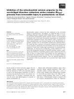
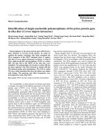
Glo-3A, Glo-3B and Glo-3C putative gene structures and promoter elements for Glo3-AFigure 1
Glo-3A, Glo-3B and Glo-3C putative gene structures and promoter elements for Glo3-A. (A) The gene structures
for Glo-3A, Glo-3B and Glo-3C. Full boxes represent the exons. TSS and (A)n represent the transcriptional start site and polyade-
nylation sequences, respectively. Red arrows represent RT-PCR primers WPF1 and WPR1. (B) Glo-3A promoter region
sequence and regulatory elements. Nucleotide positions start one nucleotide upstream of the start of translation at +1 (ATG).
Putative regulatory elements are indicated: black underlined (CAAT element), boxed (ABRE element), dotted underlined
(DOF core recognition sequence), bold neat (prolamine-box), grey underlined (Arr1), bold wave underlined (T-box), grey
wave underlined (C-box), bold double underlined (pyrimidine box), dashed underlined (GATA-box), shaded (RY repeat), dou-
ble underlined (E-box).
BMC Plant Biology 2009, 9:93 />Page 4 of 11
(page number not for citation purposes)
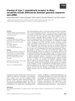
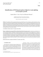
Alignment of Glo-3 amino acid sequences with WP5212Figure 2
Alignment of Glo-3 amino acid sequences with WP5212. Identical amino acids are shaded black. Similar amino acids are
shaded grey.
BMC Plant Biology 2009, 9:93 />Page 5 of 11
(page number not for citation purposes)
tivation [22,23]. Proteins expressed from Glo-3A and Glo-
3B would be 66.3 kDa and 56.9 kDa, and have predicted
pIs of 7.7 and 7.5, respectively.
Promoter identification and regulatory elements
The 1200 bp of the 5' flanking sequence of Glo-3A were
analyzed to identify a potential promoter using TSSP, a
plant promoter recognition program t
berry.com and PlantProm database [24]. A putative pro-
moter region was indicated to be between -897 and -43
upstream of the ATG start codon. The promoter region
was analyzed for potential cis-acting elements using the
PLACE database [25]. Multiple putative cis-acting ele-
ments were identified (Figure 1B and see Additional file
1). The promoter region includes potential binding sites
for transcription factors, such as bZIP and DOF. Presence
of the abscisic acid response element ABRE suggests the
expression of Glo-3A is hormone-regulated. Several ele-
ments related to tissue specific expression were also
found, including E-box, RY repeat, Pyrimidine box, C-
box, T-box, and a Prolamin-box.
Transcriptional activity
The presence of Glo-3A mRNA in T. aestivum cv. Glenlea
seeds 16 days post anthesis demonstrates that this gene is
actively transcribed. Primers that correspond to highly
conserved regions of Glo-3A, Glo-3B, Glo-3C, barley Beg1
and maize Glb1, so they could bind to a transcript from
any of the three sequences, were used to identify Glo-3
transcripts. Since the 3' end of Glo-3C was not recovered
during primer walking, we designed our primers on the
assumption that the 3' region of Glo-3C was similar to the
other Glo-3 genes and, if transcribed, would be amplified
during RT-PCR analysis. A cDNA fragment of 900 bp,
sequenced from the RT-PCR product, was 100% identical
with the Glo-3A predicted cDNA sequence in the studied
region and corroborated the predicted intron-exon struc-
tures (Data not shown). However, RT-PCR products for
Glo-3B and Glo-3C were not observed. Transcriptional
activity of Glo-3A was further supported by BLAST analysis
of wheat EST databases. At least 729 EST sequences from
the hexaploid wheat T. aestivum shared high similarity
with Glo-3A and Glo-3B and 250 ESTs with Glo-3C (see
Additional file 2). Among the total of 740 ESTs matching
to Glo-3A, 722 ESTs were from the developing or mature
seed. Relatively few, only 25, ESTs were from T. aestivum
Glenlea developing seed library 15 DPA, the same cultivar
and sampling time used for constructing the genomic
library screened in this study and RT-PCR analysis, respec-
tively. With respect to tissue-specific Glo-3 gene expres-
sion, eight EST sequences from T. turgidum durum seedling
library were identified. Also, a highly homologous EST
clone [GenBank: BQ802077
] from T. monococcum EST ver-
nalized apex library was found. Mapped ESTs (from Chi-
nese Spring deletion lines) indicated the location of Glo-
3A to be on the 4AL and/or 4BS chromosome [Grain-
Genes: BE590748].
Gene Family Size: determining Glo-3 gene copy number in
wheat
Wheat cultivars were screened by Southern blot to identify
possible genetic lines that might be devoid of WP5212-
like proteins. DNA was extracted from cultivars represent-
ing all ploidy levels: T. aestivum AC Barrie (AABBDD
genome), T. aestivum Glenlea (AABBDD genome), T. aes-
tivum Chinese Spring (AABBDD genome), T. aestivum
Spelta (AABBDD genome), T. turgidum durum Kyle (AABB
genome), T. turgidum dicoccum (AABB genome), T. mono-
coccum monococcum (AA genome), Aegilops speltoides (BB
genome), and Ae. tauschii (DD genome). A 650 bp frag-
ment, amplified from the highly conserved region among
all of the three BAC clones was used as a hybridization
probe. All of the cultivars examined contained Glo-3
genes. Based solely on the number of bands, there are at
least two copies of Glo-3 genes in A, B and D diploid
genomes and at least four homologous copies in tetra-
ploid and hexaploid genomes (Figure 3).
Immunolocalization
Immunofluorescence labeling using rabbit anti-WP5212
antibodies revealed the localization of corresponding
globulin protein in the aleurone layer and embryo, but
not the endosperm of wheat seed sections (Figure 4A, C).
Observed fluorescence in wheat seed coat is attributable
to background non-specific binding. WP5212 was shown
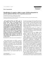
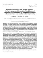
Southern blot analysis of diploid, tetraploid and hexaploid wheatFigure 3
Southern blot analysis of diploid, tetraploid and hexa-
ploid wheat. Genomic DNA was digested with Xba I and
Xho I and hybridized to Glo-3-specific probe.1-T. aestivum cv.
AC Barrie, 2-T. aestivum cv. Glenlea, 3-T. aestivum cv. Chi-
nese Spring, 4-T. aestivum cv. Spelta, 5-T. turgidum durum cv.
Kyle, 6-T. turgidum diccoccum, 7-T. monococcum monococcum,
8-Aegilops speltoides, 9-Ae. tauschii, 10-Nicotiana tabaccum. 1
kb Plus marker sizes are shown in base pairs. Ploidy levels
and genomes are indicated underneath the figure.
BMC Plant Biology 2009, 9:93 />Page 6 of 11
(page number not for citation purposes)
to share immunodominant epitopes to the peanut aller-
gen Ara h1. The same antibodies bound proteins from
peanut cotyledon (Figure 4C). WP5212 pre-immune
serum did not localize to the embryo, endosperm or aleu-
rone layer in wheat or cotyledons in peanut (Figure 4B, D,
F).
Discussion
We identified three new globulin genes in Glenlea cultivar
of hexaploid wheat, Glo-3A, Glo-3B and Glo-3C. These
genes share a high degree (73–93%) of nucleotide
sequence identity, with occasional amino acid substitu-
tions and indels at specific regions. One of the three, Glo-
3A, was identified as the genomic sequence corresponding
to the WP5212 cDNA sequence [11] sharing 99% identity
at the amino acid level. These data confirm that WP5212
is expressed in commercial wheat. The five amino acid dif-
ference between WP5212 from AC Barrie and Glo-3A from
Glenlea could be explained by natural genetic variation
due to their origin from two distinct cultivars. The cDNA
clone WP5212 was isolated from the AC Barrie cultivar
and the Glo-3A gene was identified from the Glenlea cul-
tivar. Conservation of storage protein genes in wheat is
common. Comparison of puroindoline gene sequences
from Triticum and Aegilops taxa identified an average of
98.4% identity within one taxonomic group [26].
DNA hybridization studies using diploid, tetraploid and
hexaploid wheat samples confirmed the presence of Glo-
3-like genes at all ploidy levels. The restriction enzymes
Xba I and Xho I do not have restriction sites within any
globulin sequence. Therefore, these Southern blots are
assumed to represent individual copies of Glo-3 genes.
Multiple bands of different intensities were observed on
the fluorograph for all of the studied wheat cultivars (Fig-
ure 3). Our results indicate the presence of multiple copies
of Glo-3 genes in all genomes analyzed (Figure 3). The
presence of homologous copies in each ploidy level indi-
cates that a Glo-3 sequence has been present during the
evolution of all of the wheat lines examined. Additionally,
since only one probe designed to recognize one region
was used, a broader range of probes may provide a truer
measure of Glo-3 copy number. The data suggest that the
likelihood of identifying or selecting for a Glo-3 globulin-
negative wheat variety from existing breeding stocks is
low. Although BAC screening of the hexaploid wheat
Glenlea identified three unique Glo-3 genes, it is possible
that other homologous sequences exist. There is evidence
to support an association between wheat intake and T1D,
but the basis of this association and the link to specific
molecules remains an open question [7-9]. Nonetheless,
the present study using a reverse genetics approach, dem-
onstrates the presence of three novel wheat globulins that
are potentially antigenic in patients with T1D and that are
present in commercial wheats. Further extensive sequence
variance analysis would be required before tools such as
siRNA gene silencing could be applied to silence the
expression of Glo-3 genes.
In polyploid plants, usually only one homologous
sequence is transcribed, and the redundant copies are
silenced [27]. Of the three genomic globulin copies, RT-
PCR analysis showed that the sequence of Glo-3A is tran-
scribed 16 days post anthesis (DPA), a time when 7S vici-
lins are known to be deposited in developing dicot seeds
[28-32]. Similarly, Beg1 expression has been shown via
Northern analysis to start at 15 DPA in barley grain and
continues until the maturity of the seed [13]. Glo-3B and
Glo-3C were not found to be expressed in this study at 16
DPA, but further analysis could show their expression at
alternate time points.
Immuno-localization of WP5212 in developing wheat grain (cv AC Barrie) and peanut cotyledon (cv Valencia)Figure 4
Immuno-localization of WP5212 in developing wheat
grain (cv AC Barrie) and peanut cotyledon (cv Valen-
cia). (A) Wheat aleurone (al) cells were positively stained
while endosperm (es) remained unstained when sections
were stained with WP5212 antibodies. (B) Wheat aleurone
layer and endosperm treated with preimmune serum (PIS).
Green staining in seed coat (sc) shows unspecific staining. (C)
Wheat embryonic (em) tissue stained with WP5212 antibod-
ies. (D) Wheat embryonic cells treated with PIS (E) Peanut
cotyledon (cot) stained with WP5212 antibodies. (F) Peanut
cotyledon treated with PIS. Green (examples indicated with
arrowheads) indicates positive staining.
BMC Plant Biology 2009, 9:93 />Page 7 of 11
(page number not for citation purposes)
Screening of GenBank EST libraries with Glo-3 genes
returned over 700 highly similar ESTs (see Additional file
2). The three datasets exhibit similar results – the same
ESTs were identified by all three Glo-3 genes, which are
attributed to their high degree of identity. The smaller
number in the Glo-3C dataset is due to the missing 3' end,
since ESTs are biased toward the 3'end. Among the others,
25 ESTs from the 15 DPA Glenlea library were found,
whereas no matches were found from the 5 DPA library of
the same source. This supports the prediction that Glo-3
genes in T. aestivum Glenlea are expressed 15 DPA. How-
ever, screening 22 different libraries (see Additional file 2)
identified a small yet significant number of ESTs detected
in very early stages of seed development, pre-anthesis
flower tissues (5 ESTs) and 7 DPA seeds (6 ESTs). Taken
together, the presence of more than 700 corresponding
EST sequences in the wheat Triticum EST database pro-
vides further evidence that Glo-3 genes are indeed actively
transcribed in a temporally controlled manner.
The majority of the matching ESTs (98%) were isolated
from the developing or mature seed. Interestingly, 8 ESTs
from T. turgidum durum seedling EST library were found to
be 99% identical within a 957 bp region, which indicates
that Glo-3 genes could be expressed in tissues other than
seed. To delineate the tightness of spatio-temporal gene
regulation requires further experiments to determine the
expression patterns of these particular globulins in non-
seed tissues of monocots.
Screening the mapped EST database with Glo-3A identi-
fied a clone previously mapped to the 4AL and/or 4BS
chromosome [Genbank: BE590748
]. In addition, another
EST clone [GenBank: BQ802077
] from T. monococcum, the
A genome progenitor, was found through a GenBank Triti-
cum EST database search. These findings suggest that at
least one active copy of Glo-3A is located on chromosome
4AL/BS. However, the EST databases of T. monococcum
contain 11,190 ESTs, Ae. speltoides 4,315 ESTs and Ae.
tauschii only 116 ESTs, which makes the in silico transcrip-
tional analysis of Glo-3 genes from different genomes lim-
ited or premature.
Putative regulatory elements were identified in the 5'
upstream sequence of Glo-3A by searching the PLACE
database [25] (Figure 1B and see Additional file 1). The
prolamin-box located -412 on the negative strand is
required for endosperm specific expression, but has been
shown to be inactive without a concomitant GCN4 ele-
ment. This element was not observed in the promoter
region of the Glo-3 genes supporting our immunolocaliza-
tion data showing that the WP5212 protein does not
localize to the endosperm [33].
The presence of an ABRE element, the ABscisic acid
Responsive Element from the early methionine-labeled
Em gene of wheat [34], at 149 nucleotides 5' to the start
codon suggests that Glo-3A expression could be regulated
by abscisic acid (ABA). Abscisic acid is a hormone that has
been shown to regulate maize Glb1 expression [35]. Maize
Glb1 synthesis and accumulation are positively regulated
by ABA through suppressing germination and degrada-
tion over the course of embryogenesis [36]. These results
suggest that ABA influences storage globulin accumula-
tion by initiating synthesis, suppressing degradation, and
inhibiting precocious germination.
E-box and RY elements, also found within the promoter
region, could be responsible for embryo-specific expres-
sion in wheat as shown in Arabidopsis using phas promoter
[37]. Also, it has been shown that mutations in the RY
repeat abolish the seed specific expression [38]. Taken
together, the presence of the specific promoter regulatory
elements observed in the Glo-3 genes suggests not only
seed-specific gene expression, but describes an active gene
that is specifically expressed in the aleurone layer and the
embryo.
The EST analysis also indicates that Glo-3 expression is
mostly limited to seed tissue (see Additional file 2).
Indeed, immunolocalization studies confirmed that
expression of Glo-3 was restricted to the aleurone layer
and embryo within the wheat grain (Figure 4A–D). This is
consistent with the observation that the essential
endosperm specific regulatory elements, namely the DNA
binding site GCN4, are not present in the Glo-3A pro-
moter region (Figure 1B). Interestingly, a similar staining
pattern was noted in peanut cotyledons (Figure 4E–F).
WP5212 was originally shown to share amino acid
homology with the peanut allergen Ara h I [11], and our
data indicate that antibodies raised against WP5212 also
recognize proteins in peanuts. Therefore, we propose that
these immunomodulatory proteins could share common
antigenic epitopes.
Based on solubility, we speculated that the WP5212 pro-
tein could be one of many normal trace contaminants
found in wheat gluten and this was demonstrated by 2D
gel electrophoresis [11]. Gluten consists of gliadins and
glutelins, and these proteins are expressed in the
endosperm and are not present in aleurone tissue [39,40].
It is of interest to determine whether WP5212 is present in
white flour, which is derived from the endosperm, and in
industrial gluten, which is produced from whole wheat.
Conclusion
WP5212 was first identified by probing a wheat cDNA
library with antibodies from diabetic rats. The goal of the
current work was to identify WP5212-like genes. Three
new globulin gene sequences, Glo-3A, Glo-3B and Glo-3C,
from hexaploid wheat were identified and characterized.
Glo-3A was shown to be the genomic counterpart of the
BMC Plant Biology 2009, 9:93 />Page 8 of 11
(page number not for citation purposes)
cDNA clone WP5212 and is located on chromosome 4 in
the wheat genome. As more full-length sequences become
available for the Glo-3 genes and proteins, it will be possi-
ble to establish their evolutionary relationship. The Glo-
3A gene was actively transcribed and its protein product
localized to the seed coat, aleurone and embryo tissue in
the developing seed. We did not identify a Glo-3-negative
cultivar suggesting that this gene is evolutionarily con-
served. These studies have identified the Glo-3A gene that
shares 99% identity with the cDNA of a WP5212 protein,
previously associated with autoimmune T1D and have
identified a new globulin gene family that consists of Glo-
3A and at least two other genes, Glo-3B and Glo-3C. These
findings, identification of the genes whose homologues
code for wheat proteins potentially associated with the
pathogenesis of T1D, prompt consideration since they are
present in the germ and bran layer of commercial wheats,
which are significant nutritional components of the
human diet.
Methods
Plant material and preparation of high molecular weight
DNA
Wheat Triticum aestivum cv. AC Barrie seeds were provided
by Agriculture and Agri-Food Canada, Indian Head
Research Farm and Seed Increase Unit (Indian Head, SK).
T. aestivum cv. Glenlea, T. aestivum cv. Chinese Spring, T.
aestivum cv. Spelta (CDC Bavaria), T. turgidum durum cv.
Kyle (CN42944), T. turgidum diccoccum (CItr 3686), T.
monococcum monococcum, Aegilops speltoides (PI 542261),
and Ae. tauschii (WGRC2375) seeds were obtained from
USDA National Small Grains Research Facility (Idaho).
Seeds were grown under standard greenhouse conditions,
until shoots were 15–20 cm long. Large-scale purification
of DNA was performed as described previously [41].
Briefly, leaves were cut and ground immediately in liquid
nitrogen. Thirty mL of 65°C extraction buffer (100 mM
Tris-HCl (pH 8.0), 50 mM EDTA (pH 8.0), and 500 mM
NaCl) was added to 20 mL of frozen tissue in 50 mL fal-
con tubes and the contents were shaken. Two millilitres of
20% sodium dodecyl sulphate (SDS) was added to each
tube and the tubes were shaken vigorously. The samples
were incubated at 65°C for 10 min. Ten mL of 5 M potas-
sium acetate was added to each tube, the tubes were
shaken vigorously and placed on ice for 20 min. Samples
were clarified by centrifugation at 9000 rpm for 20 min at
4°C in a Beckman JA-12 rotor (Beckman Coulter, Fuller-
ton, California) followed by filtration of the supernatants
through one layer of sterile Medicom gauze into sterile 50
mL tubes. RNAse A (10 mg/mL stock) was added at a con-
centration of 2 μg/mL sample. After ethanol precipitation,
the DNA was removed from each tube using a glass Pas-
teur pipette. The DNA was washed once in 70% ethanol,
dried, and dissolved in TE buffer (10 mM Tris-HCl (pH
8.0) – 1 mM EDTA (pH 8.0)) at a concentration of
approximately 5–10 mg/mL.
Restriction digests and Southern blotting
DNA samples were digested with restriction enzymes, Xba
I and Xho I in buffer supplied by the manufacturer (Invit-
rogen). Gels were alkaline blotted onto Hybond N+
(Amersham, Piscataway, N.J.) membranes using a stand-
ard capillary transfer setup with glass plates for support.
Hybridization studies – BAC library screen and Southern
blots
The Glenlea BAC library, kindly supplied by Dr. Cloutier
from AAFC-Winnipeg, contains 656,640 clones with an
estimated 3.1× haploid genome coverage and has been
gridded onto 24 high-density filters [42]. A 459 bp probe
(amplified using primers 5'AAAAAGCAGGCTTTCGAC-
GAAGTGTCCAGG 3'and 5'AGAAAGCTGGGTTGCCCAA-
GAGACTACCCA 3') for the BAC library screening and 650
bp probe (amplified using primers Glb09F and Glb09R)
for the Southern blot, consisting of the partial coding
regions of WP5212 cDNA clone [11] was labeled with [α-
32
P]dCTP using Ready-to-Go DNA labeling beads (Amer-
sham, Baie d'Urfé, QC) following the manufacturer's
instructions and used to screen the filters and membranes.
Hybridization was performed as described by Nilmalgoda
and co-workers [42]. In short, hybridization buffer was
prepared as described by Church and Gilbert (1984), but
without BSA. The blots were pre-hybridized overnight at
65°C. The probe was denatured by 10 min boiling, and
added to the hybridization tubes. Hybridization was car-
ried out overnight at 65°C. Filters were washed at 65°C in
increasingly stringent buffers (2 × sodium chloride/
sodium citrate SSC, 0.1% SDS to 0.1× SSC, 0.1% SDS)
until counts were approximately 1000 cpm. The following
PCR conditions were used for probe preparation: 1× PCR
buffer (Invitrogen), 1.5 mmol MgCl
2
/L (Invitrogen), 0.2
mmol/L each of the four dNTPs (Invitrogen), 15 pmol
each forward and reverse primers, 1.2 U Taq polymerase.
The amplification reaction: 94°C for 3 min, followed by
35 cycles at 94°C for 30 s, 60°C for 30 s, and 72°C for 30
s, and a final extension at 72°C for 5 min before cooling
to 4°C. The PCR products were purified using the QIAEX
II gel extraction kit (Qiagen, Mississauga, ON) according
to the manufacturer's instructions.
RT-PCR
RT-PCR was performed following Invitrogen's protocol.
In short, total RNA was isolated from ground seeds (100
mg aliquots) of T. aestivum cv. Glenlea, collected 16 days
post anthesis. RNA was extracted and purified using RNe-
asy plant mini kit (Qiagen, Mississauga, ON) according to
the manufacturer's instructions. The first strand cDNA was
synthesized using the First-Strand Synthesis System for
RT-PCR from Invitrogen according to the manufacturer's
protocol. The RT-PCR product was amplified with WPF1
and WPR1 primer pair (design based on three Glo-3
sequences [GenBank: FJ439134
-FJ439136], barley Beg1
[GenBank: M64372
] and maize Glb1 [GenBank: M24845]
BMC Plant Biology 2009, 9:93 />Page 9 of 11
(page number not for citation purposes)
(see Additional file 3). Expected RT-PCR product sizes on
mRNA were 1177 bp and 949 bp for Glo-3A and Glo-3B,
respectively. The same primer pair would amplify PCR
products of 1562 bp for Glo-3A and 1604 bp of Glo-3B on
a DNA template. RT-PCR products were cloned into a
plasmid vector pGEM-T Easy (Promega, Madison, WI),
and their nucleotide sequences were determined using
WPR1, WPR2, WPF1, Glb1R, Glb02F and Glb08F primers
(see Additional file 3).
Sequencing
All sequencing was performed at the Ottawa Hospital
Research Institute, using Applied Biosystems 3730 DNA
Analyzer. Primers used for sequencing are shown in Addi-
tional file 3. The sequences identified in this paper have
been deposited to GenBank [GenBank: FJ439134
;
FJ439135
; FJ439136].
Sequence analysis
The contig sequences were analyzed using the NCBI ORF
finder /> and
FGENESH 3.0 alpha
to deter-
mine the predicted protein sequence. The conceptually
translated proteins were aligned using ClustalX and
BioEdit programs [43]. Regulatory elements from the pro-
moter region were identified using the PLACE database
/>[25]. BLASTn analyses
were performed using NCBI databases [44], Plant Tran-
script Assembly database />plantta_blast.cgi, and wheat mapped EST database (http:/
/wheat.pw.usda.gov/GG2/blast.shtml; all URLs last
viewed 22.03.09). EST abundance for each Glo-3 mRNA
was measured using MegaBlast against GenBank "non-
mouse and non-human EST database limited to Triticum
(total of 1,106,281 sequences, 22.03.09). EST sequences
with minimum identity of 86% and minimum E value of
1e-07 were chosen. Presence of the conserved domains
was screened with NCBI CDD database http://
www.ncbi.nlm.nih.gov/Structure/cdd/cdd.shtml[45].
WP5212 polyclonal serum production
Antigenic WP5212 peptides were predicted by Sigma-
Genosys (The Woodlands, TX) technical services (propri-
etary software). Two WP5212 specific peptides were syn-
thesized for the purpose of polyclonal WP5212 antisera
production (Sigma-Genosys). Peptide 1, a 77% pure 16
residue peptide (CRDTFNLLEQRPKIAN), was conjugated
to the carrier protein Keyhole-limpet hemocyanin for
immunization. Peptide 2 was a 51% pure 15 residue pep-
tide (RGDEAVEAFLRMATA). The purity was determined
by mass spectral and HPLC analyses performed by Sigma-
Genosys.
The polyclonal antibody production was performed by
Sigma-Genosys. Pre-immune serum was collected from
two rabbits, after which they were each co-immunized
with 200 μg of peptides 1 and 2 in Complete Freund's
Adjuvant (day 0). The rabbits were co-immunized with
100 μg of peptides 1 and 2 in Incomplete Freund's Adju-
vant on days 14, 28, 42, 56 and 70. Production bleeds
from both rabbits were collected on day 40, 63 and 77.
WP5212-specific polyclonal antibody production was
assessed by 1D SDS-PAGE and Western blotting of recom-
binant WP5212 isolated from baculovirus infected insect
cell lysate (data not shown). Production bleeds were com-
pared to pre-immune serum samples to confirm antibody
production.
LR White sectioning
All fixation and embedding of seeds was performed at
4°C. For London Resin White (LR White) sectioning:
mature seeds were fixed in a 4% paraformaldehyde, 0.1 M
potassium phosphate buffer (pH 7) for two days. Seeds
were placed in two changes of 0.1 M phosphate buffer pH
7 for two hours each, and cut with a razor blade into very
thin (2 mm) slices that were placed directly in 70% (v/v)
ethanol for one day. Seeds were placed sequentially in
85% ethanol, 100% ethanol and a mixture of 20, 40, 60,
80% (v/v) ethanol: unpolymerized Medium LR White
(Sigma-Aldrich, Oakville, ON) for a minimum of two
hours per change. Seeds were moved to 100% unpolym-
erized LR White, and three changes of resin were per-
formed, with a minimum of 12 hours between each
change. Finally, seeds were moved to 0.5 ml Beem (Fisher
Scientific, Ottawa, ON) capsules containing unpolymer-
ized LR White, and cast at 60°C in a dry oven for four days
to polymerize the resin. Polymerized LR White containing
seeds were mounted and sectioned with a LKB Ultrami-
crotome. 2 μm thick sections were placed on Fisher Super-
frost Plus slides (Fisher Scientific, Ottawa, ON), and
heated on a hot plate for one minute to adhere the sec-
tions to the slide.
Immunofluorescent staining
Mounted sections of wheat AC Barrie and peanut Valencia
seeds were washed three times for 10 minutes each in PBS.
For primary and secondary immunostaining, the slides
were washed three times for 10 minutes each in PBS. Sec-
tions were incubated with the primary antibody for one
hour at room temperature in a humidity chamber. A
1:1000 (wheat embryo and peanut cotyledon) or 1:5000
(wheat aleurone and endosperm) dilution of the WP5212
antibody was incubated with the sections. After the pri-
mary antibody incubation, slides were rinsed three times
for 10 minutes each in PBS. 20 μl of the 1:400 dilution of
anti-rabbit conjugated Alexa Fluor antibody in PBS was
pipetted onto each slide and incubated at room tempera-
ture for one hour. Slides were rinsed three times for 10
minutes each in PBS and dried. A drop of Prolong
®
Gold
antifade reagent (Invitrogen, Burlington, ON) was added
to each section, 0.2 mm cover slips (Fisher Scientific,
Ottawa, ON) were overlayed, and slides were dried over-
BMC Plant Biology 2009, 9:93 />Page 10 of 11
(page number not for citation purposes)
night at room temperature in the dark. Slides were stored
at -20°C. Fluorescent and phase contrast images were vis-
ualized using a Zeiss Axiophot microscope (Oberkochen,
Germany) equipped with an Olympus DP70 CCD cam-
era. Imaging software, MetaMorph (MetaMorph Imaging
Systems, Sunnyvale, California), was used to capture
images.
Abbreviations
(T1D): Type 1 diabetes; (globulin-3): Glo-3; (EST):
expressed sequence tag; (DPA): days post anthesis.
Authors' contributions
EL performed all experiments (with the exception of
immunostaining), analyzed data, contributed all figures
and tables except figure 4, and drafted the first manu-
script. CWM performed immunostaining of wheat seeds
(figure 4). AJM provided the WP5212 antibodies and
edited the manuscript. FWS participated in the study
design and edited the manuscript. IA designed the study,
contributed to data analysis, and edited the manuscript.
All authors read and approved the final manuscript.
Additional material
Acknowledgements
We are grateful to Dr. Cloutier from AAFC-Winnipeg for kindly providing
us with the Glenlea BAC library. Monsanto and a Natural Sciences and Engi-
neering Research Council of Canada Collaborative Research Organization
grants to IA are gratefully acknowledged. FWS thanks Juvenile Diabetes
Research Foundation and Canadian Institutes for Health Research for fund-
ing. AJM was the recipient of an Ontario Graduate Scholarship. EL thanks
CIDA for an AUCC/PPT fellowship. Drs. George Fedak and Selma Gulbitti-
Onarici and Melissa McNulty provided invaluable assistance. We thank the
reviewers for constructive suggestions.
References
1. Gill BS, Appels R, Botha-Oberholster AM, Buell CR, Bennetzen JL,
Chalhoub B, Chumley F, Dvorak J, Iwanaga M, Keller B, et al.: A
workshop report on wheat genome sequencing: Interna-
tional Genome Research on Wheat Consortium. Genetics
2004, 168(2):1087-1096.
2. Berti C, Roncoroni L, Falini ML, Caramanico R, Dolfini E, Bardella MT,
Elli L, Terrani C, Forlani F: Celiac-related properties of chemi-
cally and enzymatically modified gluten proteins. J Agric Food
Chem. 2007, 55(6):2482-2488.
3. Schuppan D, Hahn EG: Biomedicine – Gluten and the gut – Les-
sons for immune regulation. Science 2002,
297(5590):2218-2220.
4. Shan L, Molberg O, Parrot I, Hausch F, Filiz F, Gray GM, Sollid LM,
Khosla C: Structural basis for gluten intolerance in Celiac
sprue. Science 2002, 297(5590):2275-2279.
5. Baur X, Posch A: Characterized allergens causing bakers'
asthma. Allergy 1998, 53(6):562-566.
6. Bittner C, Grassau B, Frenzel K, Baur X: Identification of wheat
gliadins as an allergen family related to baker's asthma. J
Allergy Clin Immunol 2008, 121(3):744-749.
7. Ziegler AG, Schmid S, Huber D, Hummel M, Bonifacio E: Early
infant feeding and risk of developing type 1 diabetes-associ-
ated autoantibodies. JAMA 2003, 290(13):1721-1728.
8. Norris JM, Barriga K, Klingensmith G, Hoffman M, Eisenbarth GS,
Erlich HA, Rewers M: Timing of initial cereal exposure in
infancy and risk of islet autoimmunity. JAMA 2003,
290(13):1713-1720.
9. Lefebvre DE, Powell KL, Strom A, Scott FW: Dietary proteins as
environmental modifiers of type 1 diabetes mellitus. Annu Rev
Nutr 2006, 26:175-202.
10. Auricchio R, Paparo F, Maglio M, Franzese A, Lombardi F, Valerio G,
Nardone G, Percopo S, Greco L, Troncone R: In vitro-deranged
intestinal immune response to gliadin in type 1 diabetes.
Dia-
betes 2004, 53(7):1680-1683.
11. MacFarlane AJ, Burghardt KM, Kelly J, Simell T, Simell O, Altosaar I,
Scott FW: A type 1 diabetes-related protein from wheat (Triti-
cum aestivum) – cDNA clone of a wheat storage globulin,
Glb1, linked to islet damage. Journal of Biological Chemistry 2003,
278(1):54-63.
12. Kriz AL: 7S globulins of cereal. In Seed Proteins Edited by: Shewry
PR, Casey R. Dordrecht, The Netherlands: Kluwer Academic Publish-
ers; 1999:477-498.
13. Heck GR, Chamberlain AK, Ho THD: Barley embryo globulin-1
gene, beg1 – characterization of cDNA, chromosome map-
ping and regulation of expression. Mol Gen Genet 1993, 239(1–
2):209-218.
14. Kriz AL: Characterization of Embryo Globulins Encoded by
the Maize Glb Genes. Biochemical Genetics 1989, 27(3–
4):239-251.
15. Belanger FC, Kriz AL: Molecular characterization of the major
maize embryo globulin encoded by the glb1 gene. Plant Physiol
1989, 91(2):636-643.
16. Gomez L, Sanchezmonge R, Salcedo G: A family of endosperm
globulins encoded by genes located in group 1 chromosomes
of wheat and related species. Mol Gen Genet 1988,
214(3):541-546.
17. Singh NK, Shepherd KW, Langridge P, Gruen LC: Purification and
biochemical characterization of triticin, a legumin-like pro-
tein in wheat endosperm. J Cereal Sci 1991, 13(3):207-219.
18. Anderson OD, Rausch C, Moullet O, Lagudah ES: The wheat D-
genome HMW-glutenin locus: BAC sequencing, gene distri-
bution, and retrotransposon clusters. Funct Integr Genomics
2003, 3(1–2):56-68.
19. Burks AW, Cockrell G, Stanley JS, Helm RM, Bannon GA: Recom-
binant Peanut Allergen Ara-H-I Expression and Ige Binding
in Patients with Peanut Hypersensitivity. J Clin Invest 1995,
96(4):
1715-1721.
20. Wang F, Robotham JM, Teuber SS, Tawde P, Sathe SK, Roux KH: Ana
o 1, a cashew (Anacardium occidental) allergen of the vicilin
Additional file 1
Putative cis elements present in the promoter sequence of Glo-3A. A
table listing all of the cis elements present within Glo-3A promoter
sequence.
Click here for file
[ />2229-9-93-S1.doc]
Additional file 2
Summary of ESTs resulting from Triticum BLAST analysis. A table
providing information about all of the available wheat EST sequences that
have similarities to Glo-3 sequence.
Click here for file
[ />2229-9-93-S2.doc]
Additional file 3
Glo-3 gene specific primers designed using consensus sequences from
Glo-3A, Glo-3B, Glo-3C, full length Beg1 (barley), maize Glb1
(M24845) and cDNA clone WP5212. The list of all of the primers used
for sequencing.
Click here for file
[ />2229-9-93-S3.doc]
Publish with BioMed Central and every
scientist can read your work free of charge
"BioMed Central will be the most significant development for
disseminating the results of biomedical research in our lifetime."
Sir Paul Nurse, Cancer Research UK
Your research papers will be:
available free of charge to the entire biomedical community
peer reviewed and published immediately upon acceptance
cited in PubMed and archived on PubMed Central
yours — you keep the copyright
Submit your manuscript here:
/>BioMedcentral
BMC Plant Biology 2009, 9:93 />Page 11 of 11
(page number not for citation purposes)
seed storage protein family. J Allergy Clin Immunol 2002,
110(1):160-166.
21. Tsuji H, Hiemori M, Kimoto M, Yamashita H, Kobatake R, Adachi M,
Fukuda T, Bando N, Okita M, Utsumi S: Cloning of cDNA encod-
ing a soybean allergen, Gly m Bd 28K. Biochim Biophys Acta 2001,
1518(1–2):178-182.
22. Dunwell JM, Purvis A, Khuri S: Cupins: the most functionally
diverse protein superfamily? Phytochemistry 2004, 65(1):7-17.
23. Mills EN, Jenkins J, Marigheto N, Belton PS, Gunning AP, Morris VJ:
Allergens of the cupin superfamily. Biochem Soc Trans 2002,
30:925-929.
24. Shahmuradov IA, Gammerman AJ, Hancock JM, Bramley PM, Solo-
vyev VV: PlantProm: a database of plant promoter sequences.
Nucleic Acids Res 2003, 31(1):114-117.
25. Higo K, Ugawa Y, Iwamoto M, Korenaga T: Plant cis-acting regu-
latory DNA elements (PLACE) database: 1999. Nucleic Acids
Res 1999, 27(1):297-300.
26. Simeone MC, Gedye KR, Mason-Gamer R, Gill BS, Morris CF: Con-
served regulatory elements identified from a comparative
puroindoline gene sequence survey of Triticum and Aegilops
diploid taxa. J Cereal Sci 2006, 44(1):21-33.
27. Wendel JF: Genome evolution in polyploids. Plant Mol Biol 2000,
42(1):225-249.
28. Wright DJ, Boulter D: Characterization of vicilin during seed
development in Vicia faba (L). Planta 1972, 105(1):60-65.
29. Millerd A: Biochemistry of legume seed proteins. Annu Rev Plant
Physiol Plant Molec Biol 1975, 26:53-72.
30. Goldberg RB, Barker SJ, Perezgrau L: Regulation of gene-expres-
sion during plant embryogenesis. Cell 1989, 56(2):149-160.
31. Thomas TL: Gene expression during plant embryogenesis and
germination – an overview. Plant Cell 1993, 5(10):1401-1410.
32. Ellerstrom M, Stalberg K, Ezcurra I, Rask L: Functional dissection
of a napin gene promoter: Identification of promoter ele-
ments required for embryo and endosperm-specific tran-
scription. Plant Mol Biol 1996, 32(6):1019-1027.
33. Wu CY, Washida H, Onodera Y, Harada K, Takaiwa F: Quantitative
nature of the Prolamin-box, ACGT and AACA motifs in a
rice glutelin gene promoter: minimal cis-element require-
ments for endosperm-specific gene expression. Plant J 2000,
23(3):415-421.
34. Marcotte WR, Russell SH, Quatrano RS: Abscisic acid-responsive
sequences from the em gene of wheat. Plant Cell 1989,
1(10):969-976.
35. Kriz AR, Wallace MS, Paiva R: Globulin gene expression in
embryos of maize viviparous mutants – evidence for regula-
tion of the glb1-gene by ABA. Plant Physiol 1990, 92(2):538-542.
36. Rivin CJ, Grudt T: Abscicic acid and the developmental regula-
tion of embryo storage proteins in maize. Plant Physiol 1991,
95(2):358-365.
37. Chandrasekharan MB, Bishop KJ, Hall TC: Module-specific regula-
tion of the beta-phaseolin promoter during embryogenesis.
Plant J 2003, 33(5):853-866.
38. Fujiwara T, Beachy RN: Tissue-specific and temporal regulation
of a beta-conglycinin gene – roles of the RY repeat and other
cis-acting elements. Plant Mol Biol. 1994, 24(2):261-272.
39. Van Herpen T, Riley M, Sparks C, Jones HD, Gritsch C, Dekking EH,
Hamer RJ, Bosch D, Salentijn EMJ, Smulders MJM, et al.: Detailed
analysis of the expression of an alpha-gliadin promoter and
the deposition of alpha-gliadin protein during wheat grain
development. Ann Bot 2008, 102(3):331-342.
40. D'Ovidio R, Masci S: The low-molecular-weight glutenin subu-
nits of wheat gluten. J Cereal Sci 2004, 39(3):321-339.
41. Wight CP, Tinker NA, Kianian SF, Sorrells ME, O'Donoughue LS,
Hoffman DL, Groh S, Scoles GJ, Li CD, Webster FH, et al.
: A molec-
ular marker map in 'Kanota' × 'Ogle' hexaploid oat (Avena
spp.) enhanced by additional markers and a robust frame-
work. Genome 2003, 46(1):28-47.
42. Nilmalgoda SD, Cloutier S, Walichnowski AZ: Construction and
characterization of a bacterial artificial chromosome (BAC)
library of hexaploid wheat (Triticum aestivum L.) and valida-
tion of genome coverage using locus-specific primers.
Genome 2003, 46(5):870-878.
43. Thompson JD, Gibson TJ, Plewniak F, Jeanmougin F, Higgins DG: The
CLUSTAL_X windows interface: flexible strategies for mul-
tiple sequence alignment aided by quality analysis tools.
Nucleic Acids Res 1997, 25(24):4876-4882.
44. Altschul SF, Madden TL, Schaffer AA, Zhang JH, Zhang Z, Miller W,
Lipman DJ: Gapped BLAST and PSI-BLAST: a new generation
of protein database search programs. Nucleic Acids Res 1997,
25(17):3389-3402.
45. Marchler-Bauer A, Anderson JB, Cherukuri PF, DeWweese-Scott C,
Geer LY, Gwadz M, He SQ, Hurwitz DI, Jackson JD, Ke ZX, et al.:
CDD: a conserved domain database for protein classifica-
tion. Nucleic Acids Res 2005, 33:D192-D196.