Điều trị Gãy xương pot
Bạn đang xem bản rút gọn của tài liệu. Xem và tải ngay bản đầy đủ của tài liệu tại đây (215.12 KB, 9 trang )
Salvage of Failed Treatment of Hip Fractures
George J. Haidukewych, MD, and Daniel J. Berry, MD
Abstract
With contemporary techniques of open
reduction and internal fixation, most
femoral neck and intertr ochanteric hip
fractures heal uneventfully. Neverthe-
less, because the number of hip frac-
tures is large and continually increas-
ing, a small percentage of patients
experience nonunion or early fixation
failure.
1,2
Unfavorable fracture pat-
terns, poor implant placement, and
poor bone quality all increase the like-
lihood of failure of fracture fixation.
1,3,4
Effective salvage is important because
patients typically are severely disabled.
The main management options are re-
vision internal fixation (with or with-
out bone grafting) and prosthetic re-
placement. The choice of salvage
method depends on whether the frac-
ture occurred at the femoral neck or
at the intertrochanteric level. Treatment
is then individualized, according to
physiologic age, activity level, remain-
ing bone quality, viability of the fem-
oral head, and status of the hip joint
articular surface.
Preoperative Evaluation
Although most nonunions with failed
fixation devices and persistent frac-
ture instability are easy to diagnose,
occasionally nonunion can be subtle
and difficult to recognize. Several
months after internal fixation, patients
may present with persistent pain and
difficulty with ambulation. Radio-
graphs may demonstrate settling of
the fracture or backing out of hard-
ware (Fig. 1, A). Alho et al
5
reviewed
the radiographic signs that predict
failure in patients with internally fixed
femoral neck fractures; they consid-
ered 3 months to be the critical time
for prognosis. Change in fracture po-
sition by 10 mm, change in screw po-
sition by 5%, backing out of the
screws by 20 mm, and perforation of
the femoral head each correlated with
a high rate of revision. When plain ra-
diography is equivocal, computed to-
mography (CT) can help determine
whether bony union has occurred
(Fig. 1, B). Usually, revision is con-
sidered for acute failure of fracture
fixation, unacceptable fracture align-
ment, or established fracture non-
union. Although 3 months is a rea-
sonable time to expect union in most
patients, fixation failure may be ev-
ident much earlier; in some patients,
however, especially those with radio-
graphic evidence of progressive but
incomplete healing, a longer period
of observation may be necessary.
In evaluating any patient with
failed internal fixation of a hip frac-
ture, occult infection should be con-
sidered as a potential cause of the fail-
ure. Prudent preoperative evaluation
includes complete blood count with
manual dif fer ential count, erythrocyte
sedimentation rate, and C-reactive
Dr. Haidukewych is Orthopaedic Traumatologist
and Adult Reconstructive Surgeon, Florida Or-
thopedic Institute, Tampa, FL. Dr. Berry is Pro-
fessor of Orthopaedics, Mayo Clinic College of
Medicine, and Consultant, Orthopaedic Surgery,
Mayo Clinic, Rochester, MN.
Neither Dr.Haidukewych nor the department with
which he is affiliated has received anything of val-
ue from or owns stock in a commercial company
or institution related directly or indirectly to the
subject of this article. Dr. Berry or the department
with which he is affiliated has received research
or institutional support from DePuy , Zimmer, and
Stryker. Dr. Berry or the department with which
he is affiliated has received royalties from DePuy.
Reprint requests: Dr. Haidukewych, Florida Or-
thopedic Institute, 13020 Telecom Parkway, Tem-
ple Terrace, FL 33637.
Copyright 2005 by the American Academy of
Orthopaedic Surgeons.
Typically, patients with failed internal fixation of a hip fracture have marked pain
and disability. These patients may present treatment challenges. Salvage is tailored
to the anatomic site of the nonunion, the quality of the remaining bone and articular
surface, and patient factors such as age and activity level. In younger patients with
either a femoral neck or intertrochanteric fracture nonunion with a satisfactory hip
joint, treatment typically involves revision internal fixation with or without osteotomy
or bone grafting. In older patients with poor remaining proximal bone stock or a
badly damaged hip joint, conversion to hip arthroplasty can restore function effec-
tively and reduce pain. For femoral head salvage procedures, choosing a fixation de-
vice and accurate preoperative planning are the major challenges in decision mak-
ing. For conversion to arthroplasty, the major challenges are assessing the need for
acetabular resurfacing, selecting the femoral implant, and managing the greater tro-
chanter. Technical challenges include broken hardware, deformity, and femoral bone
defects. Attention to technical details can minimize potential complications.
J Am Acad Orthop Surg 2005;13:101-109
Vol 13, No 2, March/April 2005 101
protein level. Aspiration of the non-
union site does not need to be per-
formed routinely because it is tech-
nically difficult to obtain an adequate
specimen, and the reliability of the re-
sults of such aspirates has not been
well documented. Intraoperative tis-
sue from the nonunion site is obtained
for frozen-section histology. When
there is evidence of infection, all
hardware should be removed, deep
cultures obtained, and necrotic tis-
sue débrided; antibiotic-impr egnated
polymethylmethacrylate beads or spa-
cers may be placed. If arthroplasty is
contemplated as the final method of
reconstruction, then a Girdlestone
resection with placement of an
antibiotic-impregnated spacer may be
considered when the femoral head is
thought to be infected. The definitive
reconstruction is then performed af-
ter a period of organism-specific in-
travenous antibiotic administration.
A staged approach is usually prefer-
able when infection is present, wheth-
er arthroplasty or an attempt to sal-
vage the femoral head is planned.
Symptomatic malunion is uncom-
mon following hip fracture. Howev-
er, shortening of the femoral neck,
shortening through the intertrochan-
teric area, and malunion of the great-
er trochanter all can occur after hip
fracture. Any of these can lead to
limb-length discrepancy or adverse
hip biomechanics, resulting in limp
or pain. In most cases, moderately
suboptimal hip biomechanics are ac-
cepted as the trade-off to gain good
bone apposition in a stable position
and fractur e union. Little information
is available about the options for sal-
vage of a severe malunion; most data
have been gathered from the treat-
ment of neglected intertrochanteric
hip fractures. In one series of 48 treat-
ed hips,
6
corrective osteotomy was
recommended for symptomatic inter -
trochanteric malunions in younger
patients, wher eas older patients were
treated with hip arthroplasty. More
studies are needed to determine the
ideal methods to prevent and salvage
malunions after hip fracture.
Generally, the viability of the fem-
oral head can be assessed with plain
radiographs, using the radiographic
criteria described for osteonecrosis.
7
If necessary, bone scintigraphy or
magnetic resonance imaging (when
titanium implants are present) can be
useful.
7
However, such additional im-
aging modalities are rarely required
because in the younger patient with-
out collapse of the femoral head, ev-
ery attempt is made to preserve the
femoral head, even if small areas of
avascular bone are present.
When evaluating the patient with
a failed hip fracture, certain patient-
specific issues also should be ad-
dressed. When osteosynthesis is at-
tempted, tobacco use in any form
should be discontinued. Achieving
optimal medical and nutritional sta-
tus, especially in elderly, debilitated
patients, also is critical.
Salvage of Failed Femoral
Neck Fractur es
Young Patients
Usually, femoral neck fractur e non-
unions in physiologically young pa-
tients are treated with methods de-
signed to salvage the femoral head and
preserve the hip joint. Preserving the
femoral head is preferable to prosthet-
ic replacement. The most common
techniques used for femoral neck non-
unions in young patients fall into two
categories: those designed to improve
the mechanical environment at the
fracture site (ie, valgus-producing os-
teotomies) and those designed to im-
prove the biologic environment of the
nonunion site by bone grafting (non-
vascularized, free vascularized, or
muscle pedicle–type grafts).
7
The Mey-
ers quadratus femoris pedicle graft,
the most widely studied graft, pro-
vides a vascularized local bone graft
to improve the biology at the nonunion
site.
8-10
Its use may be indicated when
there is loss of bone stock posterior-
ly or when patients have well-aligned
fractures with low shear angles. Sev-
eral series have evaluated individu-
al methods of bone grafting for fem-
oral neck nonunions
8,9,11-17
(Table 1).
The indications for these techniques
have yet to be fully elucidated; how-
ever, they may be useful for neglect-
ed fractures, failed fixation attempts,
or well-aligned nonunions with os-
teonecrosis. The clear superiority of
any of the bone grafting choices is un-
substantiated by the curr ent literature.
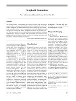
Figure 1 A, Anter oposterior radiograph demonstrating femoral neck nonunion in a 35-year-
old woman. She continued to have groin pain with ambulation for more than 1 year post-
operatively. Note the varus alignment and the backing out of the cannulated screws. B, Coro-
nal CT scan demonstrating nonunion.
Salvage of Failed Treatment of Hip Fractures
102 Journal of the American Academy of Orthopaedic Surgeons
Valgus intertrochanteric osteoto-
mies can convert shear forces at the
nonunion site to compressive forces,
which then promote fracture healing
(Fig. 2). Marti et al
18
reported on a se-
ries of 50 patients (mean age, 53 years)
who were treated with valgus inter-
trochanteric osteotomy for femoral
neck nonunion. Eighty-six percent of
nonunions united in a mean of 4
months. Of the 22 patients who had
radiographic evidence of osteonecro-
sis (without collapse) at the time of
osteotomy, only 3 (14%) showed pro-
gressive collapse of the femoral head
requiring hip replacement. Anglen
19
reported on a series of 13 patients fol-
lowed up for a mean of 25 months af-
ter valgus osteotomy for failed inter-
nal fixation of a femoral neck fracture.
All fractures healed, and 11 of the 13
patients (85%) had good to excellent
results. Later, two patients (15%) un-
derwent arthroplasty because of os-
teonecrosis.
Ballmer et al
20
reported on a series
of 17 patients with nonunions of the
femoral neck treated with valgus-
producing osteotomies. Twelve of 17
(71%) healed with one procedure.
Three patients required revision fix-
ation but eventually healed, increas-
ing the overall union rate to 88%. Three
patients (18%) had progressive os-
teonecrosis and required hip arthro-
plasty. Thus, even with areas of os-
teonecrosis, the results of salvage of
the femoral head can be good. When
segmental collapse of the femoral head
is present, valgus osteotomy is rare-
ly a satisfactory alternative because
the results are then less predictable.
Additionally, the osteotomy deforms
the proximal femur, which may make
later revision to total hip arthr oplasty,
if needed, more difficult.
Wu et al
21
compared the use of a
sliding compression screw with and
without subtrochanteric valgus os-
teotomy for femoral neck nonunions
in 32 patients (mean age, 38 years).
All of the nonunions healed at a mean
of 4.6 months. Even though there
were fewer complications in the
nonosteotomy group, the authors rec-
ommended valgus osteotomy for pa-
tients with shortening of more than
Table 1
Bone Grafting Techniques for Nonunion of the Femoral Neck: Summary of Results
Study
No. of
Patients
Mean
Follow-Up
(mo)
Mean
Age
(yrs)
No. (%)
Preoperative
Osteonecrosis Type of Graft
No.
(%)
Fracture
Union
No. (%)
Progression
of Osteo-
necrosis
No. (%)
Converted
to Total
Hip Ar-
throplasty
LeCroy et al
11
22 85 29 16 (73) stage I
and II,
6 (27) stage III
and higher
Free vascularized
fibula
20 (91) 13 (59) 2 (9)
Nagi et al
12
40 59 35 8 (20) Free vascularized
fibula
38 (93) 7 (18) 3 (8)
Hou et al
13
5 24 24 0 Iliac crest pedicle
(deep circumflex
iliac artery)
5 (100) 0 0
Leung and
Shen
14
15 60-84* 38 0 Iliac crest pedicle
(deep circumflex
iliac artery)
15
(100)
1 (7) 1 (7)
Nagi et al
15
26 29 39 4 (15) Autograft fibula
nonvascularized
25 (96) 0 0
Baksi
8
56 35 42 34 (61) stage
I and II
Quadratus femoris
muscle-pedicle
42 (75) 2 (4) Not stated
Meyers et al
9
32 14
followed
>1 yr
16-79* Not stated Quadratus femoris
muscle pedicle
23 (72) Not
stated
Not stated
Bonfiglio and
Voke
16
77 60 31-79* 77 (100) Autogenous tibial
strut, nonvascu-
larized
72 (94) Not
stated
Not stated
Henderson
17
77 69
followed
to union
46 Not stated Autograft fibula or
tibia, nonvascu-
larized
46 (67) Not
stated
Not stated
* Mean not stated; therefore, the range is given.
George J. Haidukewych, MD, and Daniel J. Berry, MD
Vol 13, No 2, March/April 2005 103
1.5 cm because the valgus osteotomy
helps gain leg length.
Although studies of valgus os-
teotomy have focused on union rates
and progression of osteonecrosis, lit-
tle has been written about clinical func-
tion after such salvage procedur es. Re-
cently, Mathews et al
22
evaluated the
functional outcome in 15 patients with
valgus-producing osteotomies for fem-
oral neck nonunions at a mean of 4
years after sur gery. Although fracture
union without progression of osteone-
crosis was achieved in most patients,
a persistent limp was common, prob-
ably caused by loss of femoral offset
and abductor moment arm (Fig. 2).
Most femoral neck nonunions in
younger patients result primarily from
mechanical, not biologic, factors. The
original fractures and subsequent non-
unions typically have high shear an-
gles (Pauwels type III
18
), have become
shortened, and are aligned in varus.
The preferred salvage operation there-
fore should be the valgus-producing
intertrochanteric osteotomy.
The technique of valgus-pr oducing
intertrochanteric osteotomy has been
well described.
23
It involves convert-
ing a vertically oriented fracture to a
more horizontal orientation, thus min-
imizing the shear forces at the frac-
ture site and promoting union. The
recommended horizontality of the
nonunion after osteotomy should be
approximately 20° to 30°.
18
Thus, the
size of the intertrochanteric wedge re-
moved would be calculated as the dif-
ference between the curr ent nonunion
verticality and the desired horizon-
tality. For example, a patient with a
70° nonunion verticality would have
a 40° to 50° wedge resected from the
intertrochanteric region to properly
reposition the proximal fragment.
Fracture shear angles may be quite dif-
ficult to measure accurately because
of leg rotation and should be measured
from a line perpendicular to the fem-
oral shaft.
19
These osteotomies should be per-
formed on a fracture table that allows
excellent fluor oscopic visualization of
the pr oximal femur. Car eful preoper-
ative templating is performed to de-
termine the appropriate blade plate
angle. Blade plates with multiple an-
gles are available, and the selected an-
gle of the plate should allow excel-
lent fixation of the proximal fragment
and the appropriate neck shaft angle
after correction. After the original
hardware is removed, the proximal
femur is prepared with the seating
chisel to accept the blade plate before
the osteotomy is performed (Fig. 2,
C). It is important to mark the correct
leg rotation, usually with Kirschner
wires in the proximal and distal frag-
ments or before making the osteoto-
my. The chisel that creates a path for
the blade is seated to the appropri-
ate depth and is then removed.
The osteotomy is then performed
parallel to the chisel tract, taking care
to leave at least 2 c m of bone between
the inferior aspect of the blade tract
and the superior aspect of the os-
teotomy. This minimizes the chance
of fracture o f this inferior bony bridge
Figure 2 A, Early postoperative anteroposterior radiograph following valgus-producing intertrochanteric osteotomy. Note the medializa-
tion of the femoral shaft, which should be minimized if possible. B, Femoral neck nonunion with the typical foreshortening and verticality
of the nonunion site. C, Appropriate seating of the chisel in the proximal fragment based on templating. To avoid fixation failure, it is im-
portant to leave sufficient bone between the planned blade plate and the osteotomy. In this situation, the intertrochanteric wedge size re-
moved is planned to allow horizontal orientation of the nonunion site. D, Nonunion verticality has been decreased from 70° (panel B) to
approximately 30°. Note the lateralization of the femoral shaft and fixation with the angled blade plate.
Salvage of Failed Treatment of Hip Fractures
104 Journal of the American Academy of Orthopaedic Surgeons
(Fig. 2, C). Commercially available pro-
tractors are available for exact calcu-
lation of the intertrochanteric wedge.
These are typically placed along the
anterior femur, and a fluoroscopic im-
age is taken (Fig. 3). Kirschner wires
are then used to mark the appropri-
ate wedge trajectory, and the os-
teotomy is performed with a saw. It
is important to cool the saw with pe-
riodic irrigation because the bone in
this anatomic region can be dense, and
thermal necrosis could occur.
After the appropriate wedge has
been removed, a blade plate of appro-
priate length and angle is impacted
into the femoral head. A secondary
proximal screw is placed below the
blade; then, distal screws are placed
in the usual fashion (Fig. 2, D). Good
compression across the osteotomy site
usually results as the distal screws are
placed because of the osteotomy obliq-
uity. Care should be taken to keep the
bone both proximal and distal to the
osteotomy well aligned on the later-
al view to avoid creating a deformi-
ty that would be difficult later to con-
vert to a hip arthroplasty. It is wise
to place bone graft at the osteotomy
site by morcellizing the cancellous
bone from the resected wedge and
placing this along the osteotomy line.
The wound is closed in the usual
layered fashion. Patients should be
cautioned that, although union rates
are high, a persistent limp is common.
The amount of femoral shaft medi-
alization should be minimized when
performing such osteotomies. This
can be accomplished by choosing a
slightly longer blade. When seated to
the appropriate depth, the plate re-
mains lateral, which helps keep the
shaft lateral. Shaft medialization de-
creases offset, thereby decreasing ab-
ductor muscle efficiency and increas-
ing the joint r eactive force. In addition,
excessive shaft medialization may
cause valgus alignment at the knee.
Occasionally, despite all efforts to
preserve the femoral head in the
young patient, there may be no rea-
sonable alternative to hip arthroplas-
ty or hip arthrodesis. For example,
a patient with total collapse of the
femoral head and a nonunion would
not be a good candidate for a joint-
preserving procedure. Hip arthro-
plasty in young patients should be re-
served for those in whom several
well-done attempts to preserve the
joint have failed and for those with
collapse of the femoral head.
Older Patients
Typically, in physiologically older
patients, femoral neck fracture non-
unions are salvaged with hip arthro-
plasty, either hemiarthroplasty or to-
tal hip arthr oplasty. Hemiarthroplasty
has the advantage of being a less ex-
tensive surgery and likely has a lower
risk of instability. In cases of badly
damaged articular cartilage of the hip
(from degenerative arthritis or er osion
because of hardware penetration), to-
tal hip arthroplasty is usually pre-
ferred. When the articular cartilage of
the acetabulum is well preserved, the
decision between hemiarthroplasty
and total hip arthroplasty is at the sur-
geon’s discretion. Scrutiny of preop-
erative radiographs and intraopera-
tive inspection of the acetabular
cartilage may guide decision making.
Either bipolar or unipolar compo-
nents may be used, based on surgeon
preference. A bipolar implant is more
commonly used if total hip arthro-
plasty is not performed because of the
excellent hip stability and low rates
of acetabular er osion it offers. If hemi-
arthroplasty is planned, it is wise to
have total hip arthroplasty compo-
nents available as well because pre-
operative radiographs may underes-
timate the amount of articular surface
damage.
Several important technical issues
must be considered when a total hip
arthroplasty is done for failed femo-
ral neck fracture. The original hard-
ware usually needs to be removed,
thereby leaving a defect in the shaft
of the femur. Also, acetabular bone
quality in patients with femoral neck
nonunion often is very poor because
of disuse osteopenia. Most of these pa-
tients do not have degenerative hip
Figure 3 Anteroposterior fluoroscopic image demonstrating calculation of intertrochan-
teric wedge and placement of Kirschner wires.
George J. Haidukewych, MD, and Daniel J. Berry, MD
Vol 13, No 2, March/April 2005 105
arthritis and so do not have the scle-
rotic subchondral bone typically
present in patients undergoing elec-
tive hip r eplacement for degenerative
arthritis. Therefore, when a cement-
less cup is used, poor press-fit fix-
ation or even acetabular fracture
during implant insertion can occur.
Judicious acetabular r eaming, with an
effort made to preserve the subchon-
dral bone, is recommended. Care
should be taken to avoid forceful ac-
etabular component impaction, and
augmentation of fixation with screws
should be considered. Standard fem-
oral components typically can be used;
however, proximal defects from pri-
or hardware can pose intraoperative
fracture risk during canal pr eparation.
Little has been written about the
results and complications of hip ar-
throplasty for failed tr eatment of fem-
oral neck fractures.
24-28
McKinley and
Robinson
29
reported a matched-pair
series of 214 patients: 107 patients
with failed open reduction and inter-
nal fixation of a femoral neck fracture
were treated with early-salvage ce-
mented total hip arthr oplasty; another
group of 107 patients with fracture
were treated with arthroplasty. The
salvage arthroplasty group had sig-
nificantly higher dislocation rates
(21% versus 8%) and more superficial
infections (P < 0.05) than did the pri-
mary arthroplasty group. Functional
scores and implant survivorship were
inferior for the salvage group, as well.
Mabry et al
30
reported on the long-
term follow-up of 99 patients with
femoral neck nonunions treated with
Charnley hip arthroplasties between
1970 and 1977. The mean age at time
of arthroplasty was 68 years (range,
36 to 92 years). At a mean 12-year
follow-up of 84 patients, 12 had un-
dergone revision arthroplasty. Im-
plant survivorship free of revision for
any reason was 93% at 10 years and
76% at 20 years. Implant survivorship
was better for older patients (age >65
years). Instability occurred in 9% of
patients, half of whom had recurrent
dislocation. Thus, reported results
clearly document the value of total
hip arthroplasty for salvage of fem-
oral neck nonunion in older patients.
The use of larger-diameter femoral
heads and surgical approaches that
reduce dislocation risk may be use-
ful to reduce the risk of dislocation
in this patient population, although
no published data substantiate this
speculation.
Salvage of Failed
Intertrochanteric Hip
Fractures
Young Patients
Nonunion of the intertrochanteric
hip fracture in young patients is un-
common. For those with proximal
bone quality adequate for internal fix-
ation, the most common treatment is
revision internal fixation with select-
ed bone grafting.
31
A fixed-angle de-
vice, such as the angled blade plate
or dynamic condylar screw, is pre-
ferred, usually accompanied by au-
togenous bone grafting. These devic-
es can target the bone in the inferior
region of the femoral head, which
usually has not been violated by pri-
or implants (Fig. 4).
Few studies of intertrochanteric
nonunions have been published.
32,33
Mariani and Rand
34
reported on 1 1 pa-
tients (mean age, 53 years) whose in-
tertrochanteric nonunions were treat-
ed with repeat open reduction and
internal fixation. Nine of 11 (82%)
achieved union at a mean of 6 months.
A variety of implants was used suc-
cessfully, based on the location of re-
maining bone stock in the femoral
head. Wu et al
35
reported on 14 inter-
trochanteric fractures with cutout of
a lag screw of a dynamic hip screw
fixation. All were treated with rein-
sertion of a lag screw inferiorly in the
femoral head, cement augmentation,
and valgus-producing subtrochanteric
osteotomy. All nonunions healed at
a mean of 5 months. Sarathy et al
36
reported on seven patients with in-
tertrochanteric nonunions treated with
valgus osteotomy, medial displace-
ment, and 130° blade plate fixation.
Figure 4 A, Anteroposterior radiograph demonstrating failure of internal fixation of an in-
tertrochanteric fracture 3 weeks postoperatively in a 52-year-old woman. Note the excellent
remaining proximal bone stock. B, Anteroposterior radiograph in another patient demon-
strating salvage with a 95° angled blade plate. Note the fixation targeting the inferior fem-
oral head bone. (Reproduced with permission from Haidukewych GJ, Berry DJ: Salvage of
failed internal fixation of intertrochanteric hip fractures. Clin Orthop 2003;412:184-188.)
Salvage of Failed Treatment of Hip Fractures
106 Journal of the American Academy of Orthopaedic Surgeons
Six of seven healed. Haidukewych and
Berry
31
reported on a series of 20 in-
tertrochanteric nonunions revised with
open reduction and internal fixation
and selected bone grafting. Fixed-angle
devices were used in 75% of cases.
Nineteen of 20 nonunions healed. The
available literature therefore suggests
that a variety of differ ent implants may
be used successfully to salvage the in-
tertrochanteric nonunion as long as
stable fixation of the proximal frag-
ment is obtained.
Older Patients
Most intertr ochanteric hip fracture
nonunions occur in older patients
with poor proximal bone quality and
fail by implant cutout from the fem-
oral head.
1
The decision to perform
revision internal fixation versus pr os-
thetic r eplacement is based on patient
characteristics, fracture pattern, re-
maining bone quality, and status of
the hip joint. In older patients, arthro-
plasty has some advantages because
it allows earlier patient mobilization.
When hip arthroplasty is per-
formed for salvage of failed intertro-
chanteric fractures, specific technical
considerations must be addressed.
The initial decision is whether to per-
form a total hip arthroplasty or a
hemiarthroplasty. It is not uncommon
to have had the cutout of the previ-
ous internal fixation cause secondary
damage to the hip joint. Usually, in
this circumstance or in patients with
markedly sever e preexisting arthritis,
a total hip arthroplasty is performed.
With well-preserved articular carti-
lage, hemiarthroplasty may be con-
sidered. The same advantages and
disadvantages of hemiarthroplasty
versus total hip arthroplasty dis-
cussed for salvage of femoral neck
nonunion also pertain to intertro-
chanteric nonunion.
Defects fr om previous internal fix-
ation devices on the lateral femoral
shaft create stress risers that can lead
to intraoperative fracture of the fe-
mur, particularly with torsion. Pre-
liminary dislocation of the hip before
hardware is removed may reduce fe-
mur fractur e risk in these hips, which
often are quite stiff and can require
much force to dislocate. Frequently,
broken screws are present. It is help-
ful to keep instruments, including tre-
phines and grasping tools, available
to remove broken screws.
Most patients with failed intertro-
chanteric fracture fixation have bone
loss below the standard resection
level for a routine, primary total hip
arthroplasty. Therefore, many need
a calcar-replacing implant to restore
leg length and hip stability. To pre-
vent the chance of subsequent frac-
ture when using longer stems, it is
wise to bypass screw holes in the fe-
mur by two cortical diameters
37
(Fig.
5). Successful femoral component
fixation can be obtained with either
cemented or cementless implants.
For many older patients, cement-
ed fixation is advantageous, particu-
larly when bone quality is poor and
the canal diameter is large. Cement-
ed fixation also allows rapid mobili-
zation in this patient population. If a
cemented stem is chosen, the surgeon
needs to be aware that cement can ex-
trude from the empty screw holes.
38
Bone graft from the resected femoral
head can be used to graft large lat-
eral defects, such as those created by
the barrel of a sliding hip screw.
If a cementless implant is used, ex-
tensively porous-coated stems have
the advantage of providing fixation
in the diaphysis of the femur, bypass-
ing the damaged, deformed, or defi-
cient proximal bone. Intraoperative
fracture is possible with insertion of
large cementless implants, especial-
ly in patients with poor bone with
multiple previous bicortical screw
holes. Intraoperative radiographs af-
ter implant placement are recom-
mended, regardless of the type of
femoral fixation chosen.
Management of the greater tro-
chanter has been problematic and
warrants special discussion. The
greater trochanter may be a separate,
ununited piece of bone, or it may be
Figure 5 A , Anter oposterior radiograph demonstrating intertrochanteric nonunion with cut-
out and poor proximal bone stock in a 78-year-old woman. B, Anteroposterior radiograph
in another patient showing salvage with a long-stem, calcar-replacing bipolar hemiarthro-
plasty and fixation of the greater trochanter.
George J. Haidukewych, MD, and Daniel J. Berry, MD
Vol 13, No 2, March/April 2005 107
malunited, preventing entrance into
the femoral canal for femoral prep-
aration. In these circumstances, the
trochanteric slide technique is pre-
ferred because it r etains the vastus lat-
eralis muscle, greater trochanter, and
abductor muscles as a single sleeve
of tissue. Patients should be coun-
seled in advance that trochanteric
problems relating to either persistent
nonunion or painful trochanteric fix-
ation devices are not infrequent after
such reconstructions.
39
Finally, bone deformity of the prox-
imal femur related to fracture callus,
fracture translation, or malunion of-
ten is pr esent, which increases the risk
of femoral fractur e during canal pr ep-
aration. Shaping of the proximal bone
with a high-speed burr is safer than
performing the same procedure with
a rasp. The tracts of previously placed
fixation devices often are sclerotic and
can deflect reamers or broaches, lead-
ing to proximal fracture or femoral
perforation.
There are few published series on
the results of hip arthroplasty for re-
vision after intertrochanteric non-
unions. Mariani and Rand
34
reported
on nine patients with intertrochan-
teric nonunions treated with hip ar-
throplasty. At an average follow-up
of 6.6 years, all patients had functional
improvement. Stoffelen et al
40
re-
ported on seven hip arthroplasties for
intertrochanteric nonunion. Seventy-
two percent (5 patients) had good to
excellent results. Mehlhoff et al
41
re-
ported on 13 patients followed for a
mean of 34 months; only 5 had good
to excellent r esults. Three patients had
dislocations and two of them required
revision for instability.
More recently, Haidukewych and
Berry
39
reported on 60 patients (mean
age, 78 years) treated between 1985
and 1997 with hip arthroplasty for
failed treatment of intertrochanteric
hip fractures. Thirty-two total hip
arthroplasties and 27 bipolar hemiar-
throplasties were performed. Forty-
four patients were followed for a
mean of 5 years. Two hip arthroplas-
ties were revised for aseptic loosen-
ing at 8 and 10 years. There was one
dislocation. The 7-year survivorship
of the arthroplasties free of revision
for any reason was 100%; 10-year sur-
vivorship was 88%. Importantly, a
calcar-replacing stem or extra long
neck-length stem was needed in 65%
of cases, and long-stemmed implants
were used in a high percentage of pa-
tients, as well. A standard prosthesis
was suitable only in 15% of cases. Se-
rious complications were uncommon,
and most patients’ ambulatory status
and pain were markedly improved.
The most common persistent com-
plaint was discomfort over the great-
er trochanter, which was present in
11% of hips.
Summary
In younger patients, salvage of the
failed hip fracture typically involves
efforts to preserve the hip joint with
internal fixation, whereas in most old-
er patients, prosthetic replacement is
a r eliable salvage option. The location
of the nonunion, physiologic age of
the patient, quality of the remaining
proximal bone, presence of deformi-
ty, status of the hip joint, and viabil-
ity of the femoral head all influence
decision making. Regardless of the
salvage method chosen, attention to
specific technical details can improve
the success rate and reduce the com-
plications of treating these challeng-
ing problems.
The OKO video ″Approaches
to Total Hip Arthr oplasty,″ by Bassam
A. Masri, MD, Philip Mitchell, MD,
and Clive Duncan, MD, is available
at />main.cfm.
References
1. Kyle RF, Cabanela ME, Russell TA,
et al: Fractures of the proximal part of
the femur. Instr Course Lect 1995;44:
227-253.
2. Kyle RF, Gustilo RB, Premer RF: Anal-
ysis of 622 intertrochanteric hip fractures.
J Bone Joint Surg Am 1979;61:216-221.
3. Baumgaertner MR, Solberg BD: Aware-
ness of tip-apex distance reduces fail-
ure of fixation of trochanteric fractures
of the hip. J Bone Joint Surg Br 1997;79:
969-971.
4. Haidukewych GJ, Israel TA, Berry DJ:
Reverse obliquity of fractures of the in-
tertrochanteric region of the femur.
J Bone Joint Surg Am 2001;83:643-650.
5. AlhoA, Benterud JG, Solovieva S: Inter-
nally fixed femoral neck fractures: Ear-
ly prediction of failure in 203 elderly
patients with displaced fractures. Acta
Orthop Scand 1999;70:141-144.
6. Lifeso R, Younge D: The neglected hip
fracture. J Orthop Trauma 1990;4:287-292.
7. Jackson M, Learmonth ID: The treat-
ment of nonunion after intracapsular
fractures of the proximal femur. Clin
Orthop 2002;399:119-128.
8. Baksi DP: Internal fixation of ununited
femoral neck fractures combined with
muscle-pedicle bone grafting. J Bone
Joint Surg Br 1986;68:239-245.
9. Meyers MH, Harvey JP Jr, Moore TM:
The muscle pedicle bone graft in the
treatment of displaced fractures of the
femoral neck: Indications, operative
technique and results. Orthop Clin
North Am 1974;5:779-792.
10. Meyers MH, Harvey P Jr, Moore TM:
Treatment of displaced subcapital and
transcervical fractures of the femoral
neck by muscle-pedicle-bone graft and
internal fixation. J Bone Joint Surg Am
1973;55:257-274.
11. LeCroy CM, Rizzo M, Gunneson EE,
Urbaniak JR: Free vascularized fibular
bone grafting in the management of
femoral neck nonunion in patients
younger than fifty years. J Orthop Trau-
ma 2002;16:464-472.
12. Nagi ON, Dhillon MS, Goni VG: Open
reduction, internal fixation and fibular
Salvage of Failed Treatment of Hip Fractures
108 Journal of the American Academy of Orthopaedic Surgeons
autografting for neglected fracture of
the femoral neck. J Bone Joint Surg Br
1998;80:798-804.
13. Hou SM, Hang YS, Liu TK: Ununited
femoral neck fractures by open reduc-
tion and vascularized iliac bone graft.
Clin Orthop 1993;294:176-180.
14. Leung PC, Shen WY: Fracture of the fem-
oral neck in younger adults: A new meth-
od of treatment for delayed and non-
unions. Clin Orthop 1993;295:156-160.
15. Nagi ON, Gautam VK, Marya SK: Treat-
ment of femoral neck fractur es witha can-
cellous scr ew and fibular graft. J Bone Joint
Surg Br 1986;68:387-391.
16. Bonfiglio M, Voke EM: Aseptic necrosis
of the femoral head and nonunion of
the femoral neck. J Bone Joint Surg Am
1968;50:48-66.
17. Henderson MS: Ununited fracture of
the neck of the femur treated by the aid
of the bone graft. J Bone Joint Surg 1940;
22:97-106.
18. Marti RK, Schuller HM, Raaymakers E:
Intertrochanteric osteotomy for non-
union of the femoral neck. J Bone Joint
Surg Br 1989;71:782-787.
19. Anglen JO: Intertrochanteric osteotomy
for failed internal fixation of femoral
neck fracture. Clin Orthop 1997;341:
175-182.
20. Ballmer FT, Ballmer PM, Baumgaertel
F, Ganz R, Mast JW: Pauwels osteoto-
my for nonunions of the femoral neck.
Orthop Clin North Am 1990;21:759-767.
21. Wu CC, Shih CH, Chen WJ, Tai CL:
Treatment of cutout of a lag screw of a
dynamic hip screw in an intertrochan-
teric fracture. Arch Orthop Trauma Surg
1998;117:193-196.
22. Mathews V, Berry DJ, Trousdale RT,
Cabanela ME: Poster: Clinical and func-
tional results of valgus intertrochanter-
ic osteotomy for femoral neck fracture
nonunion. 70th Annual Meeting Proceed-
ings. Rosemont, IL: American Academy
of Orthopaedic Surgeons, 2003, p 380.
23. Müller ME: Intertrochanteric osteoto-
my: Indication, preoperative planning,
technique, in Schatzker J (ed): The Inter-
trochanteric Osteotomy. New York, NY:
Springer-Verlag, 1984, pp 25-66.
24. Franzen H, Nilsson LT, Stromqvist B,
Johnsson R, Herrlin K: Secondary total
hip replacement after fractures of the
femoral neck. J Bone Joint Surg Br 1990;
72:784-787.
25. Hagglund G, Nordstrom B, Lidgren L:
Total hip replacement after nailing fail-
ure in femoral neck fractures. Arch Or-
thop Trauma Surg 1984;103:125-127.
26. Nilsson LT, Jalovaara P, Franzen H,
Niinimaki T, Stromqvist B: Function af-
ter primary hemiarthroplasty and sec-
ondary total hip arthroplasty in femo-
ral neck fracture. J Arthroplasty 1994;9:
369-373.
27. Tabsh I, Waddell JP, Morton J: Total hip
arthroplasty for complications of prox-
imal femoral fractures. J Orthop Trauma
1997;11:166-169.
28. Turner A, Wroblewski BM: Charnley
low-friction arthroplasty for the treat-
ment of hips with late complications of
femoral neck fractures. Clin Orthop
1984;185:126-130.
29. McKinley JC, Robinson CM: Treatment
of displaced intracapsular hip fractures
with total hip arthroplasty: Compari-
son of primary arthroplasty with early
salvage arthroplasty after failed inter-
nal fixation. J Bone Joint Surg Am 2002;
84:2010-2015.
30. Mabry TM, Prpa B, Haidukewych GJ,
Harmsen WS, Berry DJ: Long-term re-
sults of total hip arthroplasty for fem-
oral neck fracture nonunion. J Bone Joint
Surg Am 2004;86:2263-2267.
31. Haidukewych GJ, Berry DJ: Salvage of
failed internal fixation of intertrochan-
teric hip fractures. Clin Orthop 2003;412:
184-188.
32. Haentjens P, Casteleyn PP, Opdecan P:
Hip arthroplasty for failed internal fix-
ation of intertrochanteric and subtro-
chanteric fractures in the elderly pa-
tient. Arch Orthop Trauma Surg 1994;113:
222-227.
33. Kim Y-H, Oh J-H, Koh Y-G: Salvage of
neglected unstable intertrochanteric
fractures with cementless porous-
coated hemiarthroplasty. Clin Orthop
1992;277:182-187.
34. Mariani EM, Rand JA: Nonunion of in-
tertrochanteric fractures of the femur
following open reduction and internal
fixation: Results of second attempts to
gain union. Clin Orthop 1987;218:81-89.
35. Wu CC, Shih CH, Chen WJ, Tai CL:
Treatment of femoral neck nonunions
with asliding compression screw: Com-
parison with and without subtrochan-
teric valgus osteotomy. J Trauma 1999;
46:312-317.
36. Sarathy MP, Madhavan P, Ravichan-
dran KM: Nonunion of intertrochanter-
ic fractures of the femur. J Bone Joint
Surg Br 1995;77:90-92.
37. Patterson BM, Salvati EA, Huo MH:
Total hip arthroplasty for complications
of intertrochanteric fracture: A techni-
cal note. J Bone Joint Surg Am 1990;72:
776-777.
38. Eschenroeder HC Jr, Krackow KA:
Late onset femoral stress fracture asso-
ciated with extruded cement following
hip arthroplasty. Clin Orthop 1988;236:
210-213.
39. Haidukewych GJ, Berry DJ: Hip arthro-
plasty for salvage of failed treatment of
intertrochanteric hip fractures. J Bone
Joint Surg Am 2003;85:899-905.
40. Stoffelen D, Haentjens P, Reynders P, et
al: Hip arthroplasty for failed internal
fixation of intertrochanteric and subtro-
chanteric fractures in the elderly pa-
tient. Acta Orthop Belg 1994;60:135-139.
41. Mehlhoff T, Landon GC, Tullos HS: To-
tal hip arthroplasty following failed in-
ternal fixation of hip fractures. Clin Or-
thop 1991;269:32-37.
George J. Haidukewych, MD, and Daniel J. Berry, MD
Vol 13, No 2, March/April 2005 109