Chấn thương cánh tay pdf
Bạn đang xem bản rút gọn của tài liệu. Xem và tải ngay bản đầy đủ của tài liệu tại đây (137.53 KB, 10 trang )
Vol 5, No 4, July/August 1997
205
Brachial plexus birth palsy occurs
in 0.1% to 0.4% of live births.
1,2
Most infants with brachial plexus
birth palsy who show signs of
recovery in the first 2 months of life
will subsequently have normal
function. However, infants who do
not recover in the first 3 months of
life have a considerable risk of
long-term limited strength and
range of motion. As the delay in re-
covery extends from 3 months to
beyond 6 months, this risk increases
proportionately, and microsurgery
may be indicated.
Central to the controversy of
treatment of brachial plexus birth
palsies is predicting the natural
history of recovery of the neural
lesion. In general, this depends on
the type of nerve lesion (stretch,
rupture, or avulsion), the level of
injury (partial [i.e., upper, lower,
or mixed] or total), and the sever-
ity of the injury (Sunderland
grades I through V). Many re-
searchers have attempted to
address the predictive value of
physical examination, plain and
interventional radiography, and
electrodiagnostic testing in deter-
mining the severity of injury.
However, it has been difficult to
predict long-term recovery on the
basis of information obtained in
early infancy. At present, the
decision to allow for spontaneous
reinnervation and muscle recovery
or to undertake microsurgical
reconstruction of the injured
plexus remains dependent on the
physical findings. The purpose of
this article is to review the present
knowledge of the natural history
of brachial plexus birth palsies, the
indications for microsurgical inter-
vention during infancy, and the
indicators for tendon transfers and
osteotomies in the child with
chronic plexopathy.
Etiology
Perinatal risk factors include
large size for gestational age,
multiparous pregnancy, pro-
longed labor, and difficult deliv-
ery. Fetal distress may contribute
to relative hypotonia and less
protection of the plexus due to
Dr. Waters is Assistant Professor, Department
of Orthopaedic Surgery, Harvard Medical
School, Boston.
Reprint requests: Dr. Waters, Department of
Orthopaedic Surgery, Harvard Medical School,
Children’s Hospital, 300 Longwood Avenue,
Boston, MA 02115.
Copyright 1997 by the American Academy of
Orthopaedic Surgeons.
Abstract
Most infants with brachial plexus birth palsy who show signs of recovery in
the first 2 months of life will subsequently have normal function. However,
infants who do not recover in the first 3 months of life have a considerable
risk of long-term limited strength and range of motion. As the delay in
recovery extends from 3 months to beyond 6 months, this risk increases pro-
portionately. The presence of a total plexus lesion, a partial plexus lesion
with loss at C5–C7, or Horner’s syndrome carries a worse prognosis.
Microsurgery is indicated for failure of return of function by 3 to 6 months.
The exact timing of intervention is still open to debate. With microsurgical
reconstruction, there is improvement in outcome in a high percentage of
patients. However, the neural lesion is too severe and complex for present
methods of reconstruction to restore normal function. Secondary correction
of shoulder dysfunction with either latissimus dorsi–teres major tendon
transfer or humeral derotation osteotomy is clearly beneficial for patients
with chronic brachial plexopathy, as is reconstruction of supination forearm
contracture with biceps rerouting transfer and/or forearm osteotomy.
Reconstruction of the hand is also indicated for the patient with chronic dis-
ability. All of these procedures improve, but do not completely normalize,
function.
J Am Acad Orthop Surg 1997;5:205-214
Obstetric Brachial Plexus Injuries:
Evaluation and Management
Peter M. Waters, MD
stretch injury during delivery.
Mechanically, shoulder dystocia
in vertex deliveries and difficult
arm or head extraction in breech
deliveries increase the risk of
neural injury.
3
Brachial plexus birth palsy usu-
ally involves the upper trunk (C5
and C6 Erb’s palsy), although there
may be an additional injury to C7.
Less often, the entire plexus
(C5–T1) is involved.
4
In rare in-
stances, the lower trunk (C8-T1) is
most seriously involved. Obstetric
injuries to the upper trunk are gen-
erally postganglionic. The excep-
tion is upper trunk lesions after
breech delivery, which tend to be
preganglionic injuries of C5-C6.
When the lower plexus is involved,
it is usually a preganglionic injury
of C8-T1.
Anatomy
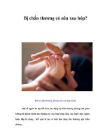
Essential to any discussion regard-
ing the natural history and treat-
ment of a brachial plexus lesion is a
thorough understanding of the
anatomy (Fig. 1). The brachial
plexus most commonly (77% of
cases) receives contributions con-
tiguously from the anterior spinal
nerve roots of C5 to T1. Prefixed
cords (22% of cases) receive an
additional contribution from C4.
The much less common postfixed
cords (1% of cases) receive a contri-
bution from T2.
5
The C5 and C6 nerve roots join
to form the upper trunk; the C7
nerve root continues as the middle
trunk; and the C8 and T1 nerve
roots combine to form the lower
trunk. Each trunk bifurcates into
anterior and posterior divisions.
The posterior divisions of all three
trunks make up the posterior cord.
The anterior divisions of the upper
and middle trunks form the lateral
cord. Finally, the anterior division
of the lower trunk forms the medial
cord. The major nerves of the
upper extremity are terminal
branches from the cords, with the
ulnar nerve arising from the medial
cord, the radial and axillary nerves
from the posterior cord, the muscu-
locutaneous nerve from the lateral
cord, and the median nerve from
branches of the medial and lateral
cords.
To predict outcome, it is impor-
tant to determine whether the
lesion is preganglionic or postgan-
glionic. The ganglion is adjacent to
the spinal cord and contains the
Obstetric Brachial Plexus Injuries
Journal of the American Academy of Orthopaedic Surgeons
206
Fig. 1 Structures of the brachial plexus.
T1
C8
C7
C6
C5
Long thoracic nerve
Spinal nerves Trunks Divisions Cords Branches
Anterior
Anterior
Anterior
Posterior
Posterior
Posterior
Posterior
Lateral
Medial
Dorsal scapular nerve
Suprascapular nerve
Upper
Middle
Lower
Lateral pectoral nerve
Musculocutaneous nerve
Medial antebrachial cutaneous nerve
Medial brachial cutaneous nerve
Medial pectoral nerve
Axillary nerve
Radial nerve
Medial nerve
Ulnar nerve
Upper and lower subscapular nerves
Thoracodorsal nerve
sensory cell body. The motor cell
body is in the spinal cord. Pre-
ganglionic lesions are avulsions
from the cord, which will not spon-
taneously recover motor function.
By assessing the function of several
nerves that arise close to the gan-
glion, careful physical examination
can determine the level of the
lesion. Specifically, the presence of
Horner’s syndrome (sympathetic
chain), an elevated hemidiaphragm
(phrenic nerve), or a winged scapu-
la (long thoracic nerve) raises seri-
ous concern about a preganglionic
lesion, as does the absence of
rhomboid (subscapular nerve),
rotator cuff (suprascapular nerve),
and latissimus dorsi (thoracodorsal
nerve) function.
Classification Systems
A modification of the Mallet clas-
sification system
6
can be used to
define the recovery of upper-trunk
function in infants. It has five sep-
arate categories for global abduc-
tion, global external rotation and
hand-to-neck, hand-to-mouth, and
hand-to-sacrum function. Grad-
ing is on a scale of 0 to 5, with 5
being normal and 0 being no mus-
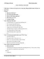
cle contraction. Grades II through
IV are illustrated for each category
in Figure 2. Preliminary studies
on natural history, microsurgical
plexus reconstruction, and sec-
ondary reconstructive shoulder
surgery have used the Mallet clas-
sification. Unfortunately, it does
not measure individual motor
strength or separate joint function
or provide a comparative scoring
system. Its usefulness is primarily
in upper-trunk assessment of
infants. It cannot be used to
assess forearm, wrist, and hand
function.
Michelow et al
7
proposed a
scoring system for surgical indica-
tions for nerve reconstruction of
the infantile brachial plexus.
Scoring is based on return of (1)
shoulder abduction, (2) elbow flex-
ion, (3) extension of the wrist, (4)
extension of the fingers, and (5)
extension of the thumb. A score of
0 to 2 is given for each of those five
motor functions. (A score of 0 rep-
resents no function; a score of 1,
partial function; a score of 2, nor-
mal function.) A total score of less
than 3.5 beyond 3 months of life is
an indication for microsurgery.
There are several other pro-
posed systems of measuring func-
tion and outcome, but none has
been validated or is widely
accepted. The absence of a uni-
form, accepted measure of out-
come makes comparison of results
of natural history, microsurgery,
and reconstructive surgery stud-
ies difficult. Obviously, this is
essential for defining the indica-
tions and results of surgical proce-
dures.
Diagnosis
The most important reason to
define the level and severity of
neural injury is to predict the po-
tential for spontaneous recovery.
Physical examination of the infant
is the most reliable method of
assessing the severity of neural
injury. Spontaneous shoulder,
Peter M. Waters, MD
Vol 5, No 4, July/August 1997
207
Global abduction
Global external
rotation
Hand to neck
Hand on spine
Hand to mouth
<30° 30° to 90° >90°
<0° 0° to 20° >20°
Not possible Difficult Easy
Not possible S1 T12
Marked trumpet
sign
Partial trumpet
sign
<40° of
abduction
Fig. 2 Modification of the Mallet classification for assessing upper trunk function in
young children. Grade I is no function, and grade V is normal function. Grades II, III, and
IV are depicted for each category.
Grade II Grade III Grade IV
elbow, wrist, and finger motion are
evaluated. Provocative testing by
stimulating neonatal reflexes
(Moro, asymmetric tonic neck, and
Votja reflexes) to induce elbow
flexion and wrist and digital exten-
sion is used. The presence or
absence of Horner’s syndrome is
recorded. Serial examinations are
necessary over the first 3 to 6
months of life.
Gilbert and Tassin
6
first pointed
out the importance of monitoring
the return of biceps function as an
indicator of brachial plexus recov-
ery. In their original work, they
found that if normal biceps func-
tion (as determined with the mod-
ified Mallet classification) failed to
return by 3 months of age, the out-
come at 2 years of age was not
normal (Fig. 2). This was general-
ly confirmed by subsequent stud-
ies.
1,7-9
However, Michelow et al
7
found that return of biceps func-
tion at 3 months had a 12% rate of
failure in detecting poor outcome.
By combining return of elbow flex-
ion with return of wrist extension,
digital extension, thumb exten-
sion, and shoulder abduction, they
were able to decrease their error
rate to 5%. In all studies, the pres-
ence of total plexus involvement,
C5–C7 involvement, and/or Hor-
ner’s syndrome meant a poorer
prognosis for spontaneous re-
covery.
Invasive radiologic studies with
myelography, combined myelogra-
phy–computed tomography (CT),
and magnetic resonance (MR)
imaging have been used in an
attempt to distinguish between
avulsions and extraforaminal rup-
tures. Kawai et al
10
compared the
findings obtained with all three
techniques with the operative find-
ings in infants. Myelography had
an 84% true-positive rate, a 4%
false-positive rate, and a 12% false-
negative rate. The addition of CT
to myelography increased the true-
positive rate to 94%. The presence
of small diverticula was only 60%
accurate for an avulsion. However,
the presence of large diverticula or
frank meningoceles was diagnostic.
Magnetic resonance imaging had a
true-positive rate similar to that of
myelographic CT studies and also
had the additional benefit of allow-
ing more distal imaging of the
plexus. These findings agreed with
those of similar studies in adults
with traumatic brachial plexus
lesions.
Electrodiagnostic studies with
electromyography and measure-
ment of nerve-conduction veloci-
ties have also been used in an
attempt to improve the accuracy of
evaluating the severity of the neural
lesion. Unfortunately, the pres-
ence of motor activity in a given
muscle has not been accurate in
predicting an acceptable level of
motor recovery. The absence of re-
innervation at 3 months is indica-
tive of an avulsion, but the pres-
ence of reinnervation seems only
to confuse the clinical picture.
11-14
At present, most clinicians rely
on clinical examination for deter-
mination of the level and severity
of the lesion. The rate and extent
of spontaneous recovery of elbow
flexion, shoulder abduction, and
extension of the wrist, fingers,
and thumb in the first 3 to 6
months of life help predict out-
come.
7
The presence of Horner’s
syndrome indicates a poorer
prognosis.
4,6,7,9,11,12,14
Nonsurgical Treatment
During the period of observation
for neural recovery, passive range
of motion of all joints should be
maintained. This often requires the
assistance of a physical therapist.
In particular, glenohumeral motion
should be maintained by passive
therapy while stabilizing the
scapulothoracic joint. This may
prevent the development of gleno-
humeral capsular tightness or
lessen its severity. Votja tech-
niques attempt to induce the nor-
mal infantile reflexes of elbow flex-
ion and wrist and digital extension
with specific stimulation. It is pos-
tulated that this stimulates reinner-
vation, although supportive data
are limited. Stimulation of the limb
for sensory reeducation has been
advocated.
11,12
Microsurgery
Indications and Timing
Without question, the role and
timing of microsurgery are the
most controversial issues in the
treatment of infants with brachial
plexus injuries. At present, micro-
surgery is performed more com-
monly in Europe, South Africa,
and Asia
4,12,13
than it is in North
America. The original interven-
tions (at the turn of the 20th cen-
tury) were resection of the neuro-
ma and direct repair. Early direct
repair is currently performed only
in Finland.
The present recommendations
for care are transection of the neu-
roma and sural nerve grafting for
extraforaminal ruptures. In the
treatment of upper-trunk ruptures,
grafts are performed from the C5
and C6 roots to the musculocuta-
neous nerve or lateral cord, supra-
scapular nerve, and upper-trunk
posterior division to the posterior
cord. In the case of avulsions,
nerve transfers are performed with
the use of the thoracic intercostals
and/or a branch of the spinal
accessory nerve beyond the point
at which it innervates the trape-
zius. For the treatment of total
avulsions, Gilbert
14
advocates pri-
oritizing microsurgical reconstruc-
tion of the median and ulnar
nerves to reinnervate the hand.
Obstetric Brachial Plexus Injuries
Journal of the American Academy of Orthopaedic Surgeons
208
Unlike adults, infants with brachial
plexopathy may have the potential
to regain hand function after nerve
grafting or transfers.
Although there is an ongoing
debate about the timing of micro-
surgical intervention, the criteria
for use in clinical practice have
been established. Brachial plexus
exploration followed by recon-
struction with sural nerve grafts is
indicated (1) for infants with total
plexopathy, Horner’s syndrome,
and no return of biceps function
at 3 months or a Toronto score
less than 3.5; and (2) for infants
with upper-trunk plexopathy, no
return of biceps function at 3 to 6
months, and a Toronto score less
than 3.5
4,6,7,9,11,12,15
(Fig. 3). Recon-
struction is usually performed
between 3 and 6 months of age,
although the range in various
studies extends from 1 to 24
months.
The problem with reviewing the
results of microsurgery is that very
few patients have had long-term
follow-up and microsurgery has
usually been combined with other
methods of treatment. Gilbert and
Tassin’s original study
6
compared
the data on cases in which micro-
surgery was performed with the
data on cases in which sponta-
neous recovery occurred. In the
cases of C5-C6 lesions, 100% of the
infants treated nonoperatively had
class III recovery (modified Mallet
classification). Of the infants treat-
ed microsurgically, 37% had class
III recovery, and 63% had class IV
recovery. In the cases of C5–C7
lesions, 30% of the infants in the
nonsurgical group had class II
recovery, and 70% had class III
recovery. Of the infants treated
with microsurgery, 35% had class II
recovery; 42%, class III; and 22%,
class IV.
More recently, Gilbert and
Whitaker
4
reported the results of
reconstruction at 2-year follow-up
in terms of modified Mallet scores
for abduction. Of the infants with
C5-C6 reconstructions, 81% had
class III, IV, or V recovery. Of the
infants who underwent total plexus
reconstruction, 64% had class III or
IV recovery.
14
At 5-year follow-up,
after performance of secondary
shoulder reconstructions, these
results improved such that 70% of
the infants with C5-C6 reconstruc-
tions had Mallet class IV or V
abduction recovery.
14
The results
were similar for total plexopathy
reconstructions, in which nerve
grafting for the hand was priori-
tized. At 2-year follow-up, only
25% of patients had grade III or IV
shoulder function; 70% had grade
III, IV, or V elbow function; and
35% had grade III or IV hand func-
tion. With the addition of sec-
ondary tendon transfers and stabi-
lization procedures, 77% had good
shoulder function, and 75% had
good hand function at 6-year follow-
up.
14
Gilbert
14
maintains that mi-
crosurgery not only improves func-
tion in selected patients over what
would be expected from the natur-
al history but also increases the
possibilities for secondary tendon
transfers.
These results are comparable
with the limited natural history
data. Benson et al
8
examined the
data on 142 patients to assess the
natural history of brachial plexopa-
thy. Seventy-one patients had full
recovery by 6 weeks. The other 71
were older than 6 weeks when
biceps function returned. At final
follow-up, 67% had excellent
shoulder function; the results were
good in another 12%, fair in 5%,
and poor in 10%.
Waters
9
addressed the same
issue prospectively and found that
Peter M. Waters, MD
Vol 5, No 4, July/August 1997
209
No Horner’s
syndrome
Horner’s
syndrome
No biceps returnBiceps return
Brachial plexus birth injury
Physical therapy
Observe until age 2
Observe for first 3 months of life for return of shoulder
abduction, elbow flexion, and wrist and finger extension
Biceps return No return
Observe for additional 3
months for biceps return
Microsurgery
Reconstruction of brachial plexus
Fig. 3 Algorithm for treatment of infants with incomplete recovery of neural function.
of 49 infants with no biceps recov-
ery at 3 months, 42 recovered
biceps function by 6 months. In
infants with biceps recovery
between 3 and 6 months, there was
a progressive decrease in Mallet
grades for abduction, external rota-
tion, and hand-to-mouth and hand-
to-neck activities with each succes-
sive month. None of the children
with biceps recovery after 3
months of age had normal function
by Mallet criteria.
Like microsurgery, secondary
shoulder tendon transfers and oste-
otomies significantly improve func-
tion in patients with residual
deficits. In a subgroup of 20 pa-
tients with shoulder reconstruc-
tions,
13
there was a significant
(P<0.0005) improvement for all
Mallet classes. Therein lies the
basis for another of the present
controversies. Clearly, patients
with no biceps function by 6
months or a Toronto score less than
3.5 have a poor prognosis and will
benefit from microsurgical recon-
struction of the plexus.
4,6,8,9
But
how different are patients who
undergo microsurgery at 3 months
from those who recover biceps
function between 3 and 6 months
and undergo secondary reconstruc-
tions? As Gilbert and Whitaker’s
microsurgery results
4
include sec-
ondary procedures, this controver-
sy is presently unresolved. Al-
though there are many believers in
the importance of microsurgical
intervention at 3 months, we know
of no current studies randomizing
entry to treatment protocols that
will answer these questions.
Technique
Standard exposure of the
brachial plexus is performed with
a Z-plasty skin incision extending
from adjacent to the mastoid
process, parallel to the sternocla-
viculomastoid muscle, and across
the clavicle and descending into
the axilla. Supraclavicular expo-
sure of the roots and trunks is per-
formed between the anterior and
middle scalene muscles. In in-
fants, the clavicle is not osteot-
omized, but rather is retracted.
The major nerves are identified
distally after appropriate takedown
of the pectoralis major and minor
muscles. Proximally, the extent of
injury is defined as an avulsion or
extraforaminal rupture for each
nerve root. In the presence of
extraforaminal rupture, proximal
transection of the neuroma is per-
formed. This is generally at the
C5-C6 root or the upper-trunk
level. The viability of the proximal
nerve is confirmed by (1) micro-
scopic inspection of the fascicles,
(2) histologic examination of the
myelin fibers, and (3) peripheral-
to-central somatosensory evoked
potentials or central-to-peripheral
motor stimulation. Sural nerve
grafts from the lower portions of
both legs are placed from the proxi-
mal C5 and C6 roots to the lateral
cord or musculocutaneous nerve,
the suprascapular nerve, and the
posterior division of the upper
trunk to the posterior cord.
4,11,12,15
In the presence of upper-root
avulsions, nerve transfers are nec-
essary. The spinal accessory nerve
beyond the point at which it sup-
plies the trapezius is transferred to
the suprascapular nerve. Thoracic
intercostal nerves (T2–T4) are used
for repair of the musculocutaneous
nerve or lateral cord and the poste-
rior cord.
In the presence of a total plex-
opathy with a combination of C5-
C6 rupture and distal avulsion, the
hand is prioritized. The C5 and C6
nerve roots are used for grafting to
the median nerve and the medial
cord or ulnar nerve. Transfers of
spinal accessory and intercostal
nerves are used for the suprascapu-
lar nerve and the posterior and lat-
eral cords.
Secondary Reconstruction
of Internal Rotation
Contractures of the
Shoulder
Open Reduction for Posterior
Glenohumeral Dislocation
Treatment of posterior gleno-
humeral dislocation varies accord-
ing to the age of the child at diag-
nosis and the extent of glenoid
deformity (Fig. 4).
In rare instances, infants less
than 1 year of age have a posterior
dislocation of the glenohumeral
joint. There is limitation of external
rotation, and the humeral head is
palpably dislocated posteriorly.
Ultrasonography, arthrography,
CT, or MR imaging can be used to
confirm the diagnosis (Fig. 5).
If dislocation is detected in
infancy, open reduction and cap-
sulorrhaphy are indicated. There
must be an anatomic glenoid for
stable reduction of the humeral
head. Simultaneous anterior and
posterior approaches to the gleno-
humeral joint are used. An ante-
rior release and posterior capsu-
lorrhaphy are performed as out-
lined by Troum et al.
16
Whether a
simultaneous latissimus dorsi
transfer should be performed is
unclear. Postoperative immobi-
lization in a spica cast is main-
tained for 4 weeks. Passive and
active exercises for maintaining
Obstetric Brachial Plexus Injuries
Journal of the American Academy of Orthopaedic Surgeons
210
Infantile dislocation
Early recognition,
minimal glenoid deformity
Late recognition,
no glenoid present
Open reduction,
capsulorrhaphy
Humeral derota-
tion osteotomy
Fig. 4 Algorithm for treatment of patients
with infantile dislocation.
range of motion are started imme-
diately thereafter.
If posterior glenohumeral dislo-
cation is detected beyond infancy
and there is marked glenoid defi-
ciency, a humeral derotation oste-
otomy is a more appropriate means
of treatment than open reduction
and capsulorrhaphy.
Tendon Transfers and
Osteotomies
Reconstructive surgery is clearly
beneficial for children with chronic
plexopathy, an internal rotation
contracture, and external rotation
weakness of the shoulder
13,14,17,18
(Fig. 6). The long-standing muscle
imbalance from an upper-trunk
lesion with intact adductors and
internal rotators and weak abduc-
tors and external rotators leads to
progressive glenohumeral deformi-
ty.
13
Early release of the subscapu-
laris muscle origin
19
at 1 year of age
may improve passive external rota-
tion and lessen the risk of progres-
sive glenohumeral subluxation in
infants with a contracture that is
unresponsive to physical therapy.
Anterior release of the pectoralis
major tendon and transfer of the
latissimus dorsi and teres major
muscles is appropriate for patients
with minimal glenohumeral defor-
mity and a debilitating contracture.
Humeral derotation osteotomy is
best for patients with an internal
contracture and advanced gleno-
humeral deformity.
13,18
Subscapularis Release
Release of the origin of the sub-
scapularis muscle
19
may be indicat-
ed when intensive physical therapy
fails to improve an internal rotation
shoulder contracture in an infant.
Therapy should be directed at
increasing the humeroscapular
angle in external rotation by stabi-
lizing the scapulothoracic joint.
11,12
A subscapularis release may be
indicated if there is less than 30
degrees of external rotation in
adduction by 1 year of age.
Carlioz and Brahimi
19
have out-
lined a procedure that exposes the
subscapularis origin posteriorly
along the medial border of the
scapula. A muscle slide is per-
formed to improve passive external
rotation to more than 30 degrees.
Postoperative immobilization in a
shoulder spica cast is maintained
for 3 to 4 weeks.
Anterior Release of Pectoralis Major
and Latissimus Dorsi–Teres Major
Transfer
Anterior release of the pectoralis
major insertion and transfer of the
latissimus dorsi and teres major
muscles to the rotator cuff is indi-
cated for patients with (1) persis-
tent internal rotation contracture,
Peter M. Waters, MD
Vol 5, No 4, July/August 1997
211
A
C
B
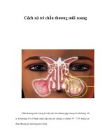
Fig. 5 Images of glenohumeral deformity in patients with chronic
plexopathy associated with an internal rotation contracture of the
shoulder. A, CT scan of an infant with glenohumeral dislocation
before open reduction and capsulorrhaphy at age 9 months. B, MR
image depicts hypoplasia of the glenoid, subluxation of the humer-
al head, and development of a false glenoid. C, CT scan reveals
severe flattening of the humeral head and glenoid associated with
posterior glenohumeral dislocation.
(2) external rotation weakness, (3)
limited abduction, and (4) posterior
subluxation of the glenohumeral
joint without glenoid deformity
(Fig. 6). Transfer can be successfully
performed between the ages of 2 to
7 years, depending on the severity
of glenohumeral deformity.
13,17
In
rare instances, the transfer may be
of insufficient strength to provide
effective abduction and external
rotation; a supplemental derotation
osteotomy of the humerus or shoul-
der arthrodesis may be necessary.
Hoffer et al
17
have outlined an
approach through an anterior inci-
sion in which the pectoralis major
tendon is lengthened at its humeral
insertion. Through a posterior inci-
sion, the latissimus dorsi–teres
major insertion is then transferred
to the greater tuberosity of the
humerus. Others have modified
the approach of Hoffer et al by
leaving the pectoralis major intact,
releasing the teres major, and trans-
ferring only the latissimus dorsi
muscle. Postoperative immobiliza-
tion in a shoulder spica cast in
abduction and external rotation is
maintained for 4 to 6 weeks, fol-
lowed by physical therapy for
transfer education.
Humeral Derotation Osteotomy
The indications for humeral
derotation osteotomy are the same
as those for latissimus dorsi–teres
major tendon transfer except that
patients are selected for osteotomy
if there is more severe glenohumer-
al deformity with flattening of the
glenoid and humeral head. This
presentation is most common in
adolescents.
11,13,18
Anterior humeral exposure is
performed in the distal aspect of
the deltopectoral interval. The pec-
toralis major and deltoid muscle
insertions are identified. Subperi-
osteal dissection is then performed
proximal to the deltoid muscle
insertion. The radial nerve is pro-
tected with this exposure, as it
crosses posterior to the deltoid at
this level. The osteotomy is per-
formed proximal to the deltoid
insertion in transverse fashion.
The distal humerus is positioned
in 30 degrees of external rotation
and is then stabilized with a four-
to six-hole plate across the osteot-
omy. The degree of postoperative
immobilization is dependent on the
age of the patient and the stability
of internal fixation. This can range
from a shoulder spica cast for a
young child to a sling and swathe
for an adolescent. For a child, ther-
apy is begun as soon as the osteot-
omy has healed; for an adolescent,
therapy is begun when hardware
provides sufficient stability.
11,18
Secondary Reconstruction
of Supination Contractures
of the Forearm
It is common to have an elbow flex-
ion and forearm supination con-
tracture in the rare patients with
residual C8-T1 neuropathy and
recovery of C5-C6 function. These
children have intact shoulder
abduction, elbow flexion, and fore-
arm supination and may have
active wrist dorsiflexion and digital
flexion. By surgically correcting
the supination posture and reposi-
tioning the forearm into 20 degrees
of pronation, the affected limb
becomes a better assist (Fig. 7).
When the posture of dorsiflexion
of the wrist is corrected, gravity
assists palmar flexion. The palmar
flexion of the wrist aids digital
extension by tenodesis.
Zancolli
20
advocated rerouting
the biceps insertion to convert the
biceps from a forearm supinator to
a pronator. In the presence of a
supination contracture, simultane-
ous interosseous membrane release
was recommended. However, only
50% of his patients maintained the
correction. Instead, Manske et al
21
recommend osteotoclasis of the
radius and ulna. Most often, some
variant of forearm osteotomy
rather than soft-tissue release is
performed for patients with a con-
tracture.
Obstetric Brachial Plexus Injuries
Journal of the American Academy of Orthopaedic Surgeons
212
Physical therapy
Mild glenohumeral
deformity
Severe glenohumeral
deformity
Latissimus dorsi–teres
major tendon transfer
Humeral derotation
osteotomy
Subscapularis
release
Unresponsive to physical
therapy beyond age 2
Internal rotation contracture
and/or external rotation weakness
Unresponsive to
physical therapy by
1 year of age
Fig. 6 Algorithm for treatment of patients with disabling internal rotation contractures.
Biceps Tendon Transfer
Rerouting of the biceps tendon
insertion to convert its muscle
action from supination to prona-
tion is indicated for patients with
elbow flexion, forearm supination,
and wrist dorsiflexion posturing
from residual C7–T1 weakness and
C5-C6 recovery (Fig. 7). Ideally,
patients will have antigravity wrist
dorsiflexion strength for effective
postoperative wrist tenodesis to aid
finger flexion. In the absence of at
least 60 degrees of passive prona-
tion, a simultaneous or sequential
forearm osteotomy should be per-
formed.
11,18,20
The biceps rerouting transfer fol-
lows the procedure outlined by
Zancolli.
20
A Z-plasty skin incision
is made in the cubital fossa. The
biceps tendon insertion is exposed
laterally to protect the median
nerve and the brachial artery. The
tendon is lengthened in Z fashion.
The distal insertion is rerouted
around the radial neck while pro-
tecting the posterior interosseous
nerve and is sutured to itself to act
as a pronator rather than a supina-
tor. Protective cast immobilization
in 90 degrees of elbow flexion and
20 degrees of forearm pronation is
maintained for 4 to 6 weeks. Active
range-of-motion and strengthening
exercises are begun thereafter.
Forearm Osteotomy
In the absence of forearm pas-
sive pronation, an osteotomy of the
radius alone or of both the radius
and the ulna is performed to cor-
rect the supination deformity.
Manske et al
21
recommend a two-
stage osteoclasis technique. A single-
stage technique can be used if
intramedullary fixation of the
radius and ulna is accomplished
before osteotomy. In the case of a
less severe deformity, a distal radi-
al osteotomy alone with internal
plate fixation can be used. A
simultaneous biceps rerouting pro-
cedure may lessen the risk of recur-
rent deformity with growth.
Summary
Most infants with brachial plexus
birth palsy who show signs of
recovery in the first 2 months of life
should subsequently have normal
function. However, infants who
fail to recover in the first 3 months
of life have a considerable risk of
long-term limited function, espe-
cially about the shoulder. As the
delay in recovery extends from 3
months to beyond 6 months, this
risk increases proportionately. The
presence of a total plexus lesion, a
partial plexus lesion with C5–C7
loss, or Horner’s syndrome all
carry a worse prognosis.
Microsurgery may be indicated
if function does not return in the
first 3 to 6 months of life. The exact
timing of intervention is still open
to debate. With microsurgical
reconstruction, there is improve-
ment in outcome for a high per-
centage of patients. However, the
neural lesion is too severe and
complex for our present methods
of reconstruction to result in nor-
mal function. Secondary recon-
struction of a dysfunctional shoul-
der by means of a latissimus
dorsi–teres major tendon transfer
or humeral derotation osteotomy is
clearly beneficial to patients with
chronic brachial plexopathy, as is
secondary reconstruction of a fore-
arm supination contracture by
means of biceps rerouting transfer
and/or forearm osteotomy. Recon-
struction of the hand is also indi-
cated for patients with chronic dis-
ability. All of these procedures
should improve, but will not com-
pletely normalize, function.
Peter M. Waters, MD
Vol 5, No 4, July/August 1997
213
Supination contracture of the forearm
Intact forearm
passive pronation
Limited forearm
passive pronation
Osteotomy of forearm to
20° pronation with biceps
rerouting procedure
Biceps rerouting
tendon transfer
Fig. 7 Algorithm for treatment of supina-
tion contracture associated with predomi-
nant C7–T1 dysfunction. Intact wrist dorsi-
flexion is important preoperatively.
References
1. Hardy AE: Birth injuries of the
brachial plexus: Incidence and progno-
sis. J Bone Joint Surg Br 1981;63:98-101.
2. Greenwald AG, Schute PC, Shiveley
JL: Brachial plexus birth palsy: A 10-
year report on the incidence and
prognosis. J Pediatr Orthop 1984;4:
689-692.
3. Geutjens G, Gilbert A, Helsen K:
Obstetric brachial plexus palsy associ-
ated with breech delivery: A different
pattern of injury. J Bone Joint Surg Br
1996;78:303-306.
4. Gilbert A, Whitaker I: Obstetrical
brachial plexus lesions. J Hand Surg
[Br] 1991;16:489-491.
5. Lee HY, Chung IH, Sir WS, et al:
Variations of the ventral rami of the
brachial plexus. J Korean Med Sci
1992;7:19-24.
6. Gilbert A, Tassin JL: Réparation
chirurgicale du plexus brachial dans la
paralysie obstétricale. Chirurgie 1984;
110:70-75.
7. Michelow BJ, Clarke HM, Curtis CG,
et al: The natural history of obstetrical
brachial plexus palsy. Plast Reconstr
Surg 1994;93:675-681.
8. Benson LJ, Ezaki M, Carter P, Knetzer
D: Brachial plexus birth palsy: A
prospective natural history study.
Orthop Trans 1996;20:311.
9. Waters P: When is timing of biceps
return of function reliable in patients
with obstetrical brachial plexopathy?
Presented at the annual meeting of the
Pediatric Orthopedic Society of North
America, Phoenix, May 13, 1996.
10. Kawai H, Tsuyuguchi Y, Masada K, et
al: Identification of the lesion in
brachial plexus injuries with root avul-
sion: A comprehensive assessment by
means of preoperative findings, myel-
ography, surgical exploration and
intraoperative electrodiagnosis.
Neuro-Orthop 1989;7:15-23.
11. Doi K: Obstetric and traumatic pedi-
atric palsy, in Peimer CA (ed): Surgery
of the Hand and Upper Extremity. New
York: McGraw-Hill, 1996, vol 2, pp
1443-1463.
12. Narakas AO: Obstetrical brachial
plexus injuries, in Lamb DW (ed): The
Paralysed Hand. Edinburgh: Churchill-
Livingstone, 1987, pp 116-135.
13. Waters P, Smith G: Glenohumeral
deformities in residual brachial plexus
birth palsy. Presented at the annual
meeting of the American Society of
Surgery of the Hand Residents and
Fellows Conference, Nashville, Tenn,
September 29, 1996.
14. Gilbert A: Long-term outcome of micro-
surgery in brachial plexus birth palsy.
Presented at the Obstetrical Brachial
Plexus Meeting, Paris, April 13, 1996.
15. Boome RS, Kaye JC: Obstetric traction
injuries of the brachial plexus: Natural
history, indications for surgical repair
and results. J Bone Joint Surg Br
1988;70:571-576.
16. Troum S, Floyd WE III, Waters PM:
Posterior dislocation of the humeral
head in infancy associated with obstet-
rical paralysis: A case report. J Bone
Joint Surg Am 1993;75:1370-1375.
17. Hoffer MM, Wickenden R, Roper B:
Brachial plexus birth palsies: Results
of tendon transfers to the rotator cuff.
J Bone Joint Surg Am 1978;60:691-695.
18. Goddard NJ, Fixsen JA: Rotation
osteotomy of the humerus for birth
injuries of the brachial plexus. J Bone
Joint Surg Br 1984;66:257-259.
19. Carlioz H, Brahimi L: La place de la
désinsertion interne du sous-scapu-
laire dans le traitement de la paralysie
obstétricale du membre supérieur chez
l’enfant. Ann Chir Infant 1971;12:
159-168.
20. Zancolli EA: Paralytic supination con-
tracture of the forearm. J Bone Joint
Surg Am 1967;49:1275-1284.
21. Manske PR, McCarroll HR Jr, Hale R:
Biceps tendon rerouting and percuta-
neous osteoclasis in the treatment of
supination deformity in obstetrical
palsy. J Hand Surg [Am] 1980;5:
153-159.
Obstetric Brachial Plexus Injuries
Journal of the American Academy of Orthopaedic Surgeons
214