Chấn thương cột sống cổ tử cung pptx
Bạn đang xem bản rút gọn của tài liệu. Xem và tải ngay bản đầy đủ của tài liệu tại đây (323.25 KB, 10 trang )
Journal of the American Academy of Orthopaedic Surgeons
338
More than 1.2 million individuals
participate annually in high school
football. Another approximately
200,000 individuals engage in col-
lege and professional play each
year.
1
It has been estimated that
cervical spine injuries occur in 10%
to 15% of football players, most
commonly in linemen, defensive
ends, and linebackers.
2-4
Injuries
may involve structural elements of
the spine (bones, disks, ligaments)
and/or neural elements (brachial
plexus, nerve roots, spinal cord).
The overwhelming majority of such
injuries are self-limited, and full
recovery can be expected.
5
How-
ever, in one study 50% of college
freshman football players with a
history of previous Òneck injuryÓ
demonstrated radiographic changes
including compression fractures,
neural arch fractures, and abnormal
motion segments.
4
In a National
Collegiate Athletic Association
(NCAA) study of football-related
injuries incurred between 1977 and
1989, 128 players suffered perma-
nent spinal cord injury.
6
Vigilance
is required to detect those injury
patterns that require immediate
evaluation and treatment or long-
term protection.
Clinical Syndromes
Root and Brachial Plexus
Neurapraxia
The most frequent cervical spine
injury in football is neurapraxia of
the nerve roots or brachial plexus.
In one study,
7
half of the members
of a collegiate football squad re-
ported one or more such episodes
during a regular season. Linemen,
defensive ends, and linebackers are
most commonly affected.
2,8
ÒSting-
ersÓ and ÒburnersÓ are the lay terms
applied to this spectrum of injuries.
There is no agreement on the specif-
ic clinical definitions for these sub-
jective entities, which lack dis-
cernible signs. Objective findings
may be subtle. A careful examina-
tion is required to prevent attri-
bution of a burning or stinging sen-
sation to a benign condition when,
in fact, it may be the result of a more
serious problem. Such symptoms,
when present in both upper ex-
tremities, suggest spinal cord, rather
than nerve root or plexus, involve-
ment.
The transient stinging and burn-
ing in neurapraxias arise from com-
pressive or traction injuries to multi-
ple roots or to the brachial plexus.
2,7
The upper trunk of the brachial
plexus is tensioned by a sudden
shoulder depression and concomi-
tant lateral head flexion toward the
unaffected side. With simultaneous
head rotation toward the affected
arm, the neural foramen narrows,
compressing exiting nerve roots.
Neurapraxia may also be caused
by direct compression of the bra-
chial plexus. A poorly fitting, mo-
bile shoulder pad may be pushed
Dr. Thomas is Orthopaedic Surgeon, Naval
Medical Center, San Diego, Calif. Dr.
McCullen is Orthopaedic Spine Surgeon,
Naval Medical Center, San Diego; and Clinical
Instructor of Orthopaedic Surgery, University
of California, San Diego. Dr. Yuan is Professor
of Orthopaedic and Neurological Surgery, State
University of New York, Syracuse.
Reprint requests: Dr. McCullen, Naval
Medical Center, San Diego, 34800 Bob Wilson
Drive, San Diego, CA 92134-5000.
The views expressed in this article are those of
the authors and do not reflect the official policy
or position of the Department of Defense or the
United States Government.
Abstract
Cervical spine injuries have been estimated to occur in 10% to 15% of football
players, most commonly in linemen, defensive ends, and linebackers. The over-
whelming majority of such injuries are self-limited, and full recovery can be
expected. However, the presenting symptoms of serious cervical spine injuries
may closely resemble those of minor injuries. The orthopaedic surgeon frequent-
ly must make a judgment, on the field or later in the office, about the advisability
of returning the athlete to the game. These decisions can have an enormous
impact on the player and his family. Most severe cervical spine injuries share
the common mechanism of application of an axial load to the straightened spine.
Avoiding techniques that employ head-down "spear" tackling and wearing prop-
erly fitted equipment markedly reduce the risk of serious injury.
J Am Acad Orthop Surg 1999;7:338-347
Cervical Spine Injuries in Football Players
Bruce E. Thomas, MD, Geoffrey M. McCullen, MD, and Hansen A. Yuan, MD
Bruce E. Thomas, MD, et al
Vol 7, No 5, September/October 1999
339
into ErbÕs point (in the anterolater-
al portion of the neck, 2 to 3 cm
above the clavicle), compressing
the brachial plexus between the
shoulder pad and the superior me-
dial scapula.
8,9
The athlete may complain of a
Òdead armÓ with shoulder and/or
arm pain as well as transient, unilat-
eral muscle paresis. Symptoms are
self-limited. Burning pain resolves
in seconds to minutes. Strength
usually returns in 24 hours. A vari-
able degree of weakness in the mus-
cles innervated by the upper trunk
of the brachial plexus may last for
up to 6 weeks. Examination of the
cervical spine demonstrates pain-
free full range of motion with no
tenderness or palpable deformity.
5
If symptoms resolve quickly and the
neurologic examination is normal
with full motor strength, the patient
may return to the game. Persistence
of symptoms or lack of a pain-free
range of motion requires further
evaluation, including cervical spine
radiographs. Players should be
restricted from further play until
they have recovered full muscle
strength.
Wearing a thermoplastic total-
contact neck-shoulder-chest ortho-
sis beneath a well-fitting shoulder
pad decreases the severity and
recurrence of compressive brachial
plexus injuries.
8
A U-shaped foam
neck roll may also be effective by
limiting neck motion and prevent-
ing the shoulder pad from being
forced into the neck. Stiff yet com-
fortable thick pads at the base of
the neck provide support against
extension and lateral bending.
Acute Cervical Sprain
Acute cervical sprain, which is
in fact a ligamentous injury with
potential for instability, is the result
of a direct collision. The athlete
complains of a Òjammed neckÓ sen-
sation with pain localized to the
neck without radiation into the
arms. Typically, there is decreased
cervical motion. Reproducible fo-
cal tenderness is indicative of a sig-
nificant bone or soft-tissue injury.
No neurologic deficits are demon-
strable on examination. Individuals
with a history of a collision who
have pain and limited range of
motion should be placed in cervical
immobilization.
The initial radiographic examina-
tion should include anteroposterior,
lateral, and odontoid views of the
cervical spine. Once the acute
symptoms have subsided, flexion-
extension lateral views should be
obtained if the initial static radio-
graphs were normal. In cases of
continuing limitation of motion,
pain, or radicular symptoms, mag-
netic resonance (MR) imaging or
bone scintigraphy may be indicated.
In general, treatment should be
tailored to the degree of severity of
the injury. A collar and analgesic
agents can be used until there is
pain-free full range of motion.
Intervertebral Disk Lesions
Acute traumatic disk herniation
with resultant cord compression
can result in transient quadriplegia
or permanent quadriparesis or
quadriplegia.
10,11
Affected players
experience acute paralysis of all
four extremities and a loss of pain
and temperature sensation. Mag-
netic resonance imaging or the com-
bination of computed tomography
(CT) and myelography can confirm
the diagnosis. Anterior diskectomy
with interbody fusion is warranted
for a patient with persistent radicu-
lar pain or myelopathy.
Cervical spondylolytic changes
without herniation or neurologic
findings are frequent in football
players. In one study,
4
5 of 75 (7%)
college freshman football players
demonstrated an abnormally nar-
row disk space. Early degenerative
changes can be attributed to repeti-
tive loading in the preceding years
of play. An MR imaging study may
demonstrate a bulge without herni-
ation. Treatment is usually nonsur-
gical with activity modification.
Severe spondylolytic changes may
cause (1) uncovertebral joint hyper-
trophy with narrowing of the neu-
ral foramen affecting the exiting
nerve root; and (2) disk-osteophyte
occlusion of the central canal (ac-
quired cervical stenosis).
Transient Quadriplegia
Ladd and Scranton
11
and Torg et
al
12
have separately described the
clinical entity of Òneurapraxia of the
cervical cordÓ with transient quadri-
plegia after an axial load with hyper-
flexion or hyperextension (Fig. 1).
During the 1984 NCAA season, neu-
rapraxia of the cord was reported in
1.3/10,000 players.
12
The symptoms
include bilateral burning pain, tin-
gling, and loss of sensation in the
arms and/or legs. Motor symptoms
vary from mild weakness to com-
plete paralysis. Episodes are tran-
sient, and complete recovery usually
occurs within 10 to 15 minutes but
may take as long as 48 hours. Radio-
graphs are negative for fractures or
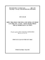
dislocations (Fig. 2) but frequently
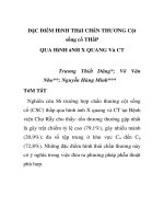
Fig. 1 Due to a pincer mechanism, injury
to the cervical spinal cord may occur dur-
ing extremes of flexion or extension. In
hyperextension, the cord may be com-
pressed between the posteroinferior portion
of the vertebral body above and the antero-
superior lamina of the vertebra below.
Cervical Spine Injuries in Football Players
Journal of the American Academy of Orthopaedic Surgeons
340
show congenital stenosis, Klippel-
Feil syndrome, or evidence of inter-
vertebral disk disease or acquired
stenosis.
12
Maroon
13
has described the
Òburning handsÓ syndrome. This is
believed to be a variant of the cen-
tral cord syndrome. Edema and
vascular insufficiency occur selec-
tively within the medial aspect of
the somatotopically arranged spino-
thalamic tracts.
13,14
Burning dyses-
thesias and paresthesias occur with-
in a glovelike distribution, although
strength, reflexes, and sensation are
maintained. This clinical picture
may be associated with a fracture-
dislocation with or without a de-
tectable radiographic abnormality.
14
In addition to plain radiography,
MR imaging or postmyelography
CT should be performed as part of
the neural evaluation of all players
who demonstrate the signs or symp-
toms of a cord injury.
Cord compression without re-
sidual radiographic abnormality
may occur by means of a momen-
tary pincerlike mechanism, original-
ly described by Penning
15
(Fig. 1).
When the cervical spine is in hyper-
extension, the cord is compressed
between the posteroinferior margin
of the superior vertebra and the
anterosuperior lamina of the subja-
cent vertebra. In addition, infolding
of the posterior longitudinal liga-
ment and the ligamentum flavum
contribute to central canal narrow-
ing. With hyperflexion, a pinching
effect is created between the lamina
of the superior vertebra and the
posterosuperior aspect of the subja-
cent vertebral body. Athletes with
congenital or acquired cervical ste-
nosis are predisposed to cord neura-
praxia with hyperextension or hy-
perflexion loading.
To assess for congenital narrow-
ing, the canal diameter is measured
on a lateral radiograph from the
midpoint of the posterior aspect of
the vertebral body to the nearest
point along the spinolaminar line
(Fig. 3).
16
The normal midsagittal
diameter is 14 to 23 mm. ÒStenosisÓ
is defined on the basis of a diameter
of less than 13 mm. Variations in
technique (e.g., use of different
focus-to-film and object-to-film dis-
tances) and anatomy (e.g., variabil-
ity in the triangular cross-sectional
shape of the canal) often contribute
to inaccurate measurements. To
minimize these errors, Pavlov pro-
posed using a ratio of the segmental
A B
C D
Fig. 2 A 19-year-old player received an axial load to the top of his helmet, which resulted
in complete quadriplegia for approximately 10 minutes. All symptoms resolved rapidly
and completely. Neutral lateral (A) and flexion (B) and extension (C) radiographs showed
no abnormal soft-tissue swelling, no fractures or subluxations, and Pavlov ratios at C3
through C6 of 1.0. Sagittal MR imaging study (D) showed a disk-osteophyte complex at
C6-7. No other degenerative changes, stenosis, or posterior ligamentous disruptions were
noted. The spinal cord displayed no abnormal signal change. Subsequent flexion-extension
radiographs showed no instability. The patient was allowed to participate in contact
sports after demonstrating painless full range of motion.
Bruce E. Thomas, MD, et al
Vol 7, No 5, September/October 1999
341
sagittal diameter of the canal to the
width of the vertebral body.
16
A
ratio of less than 0.8 has been used
to define a developmentally narrow
canal. In one study,
17
that value
was documented in 93% of players
with transient quadriplegia, 12% of
asymptomatic nonathletes, and 48%
of asymptomatic football players.
17
A threefold increase in the inci-
dence of stingers has also been seen
among subjects with a ratio of less
than 0.8, but this difference is con-
sidered to be secondary to forami-
nal, rather than central, stenosis.
2
This ratio must be interpreted
with caution, however, as some
football players with relatively large
vertebral bodies have a low ratio
despite ample canal dimensions.
18
In addition, the ratio may be insen-
sitive if the canal is narrow because
of compression by soft-tissue ele-
ments (disk, ligamentum flavum).
Thus, ÒstenosisÓ cannot be accurate-
ly diagnosed on the basis of bone
measurements alone.
To clarify the risk to players
with this entity, Torg et al
12,17
used
data from the National Football
Head and Neck Injury Registry to
compare groups of males who had
participated in tackle football with
a control group of nonathletes.
Players with cervical canal stenosis
(as determined on the basis of a
canalÐvertebral body ratio of less
than 0.8) were no more susceptible
to neurologic injury than members
of the general population (positive
predictive value, 0.2%).
17
How-
ever, this study should be viewed
with caution because of the previ-
ously discussed problems that may
arise when the Torg ratio is used to
define stenosis. A survey of 177
athletes who had been rendered
quadriplegic by football-related
accidents documented the absence
of antecedent cord symptoms.
12
Therefore, screening with plain
radiography to assess for stenosis
in high school, college, or profes-
sional football players is not rou-
tinely recommended.
12,17,19
There is a subset of players, how-
ever, in whom radiographs may be
predictive of the risk of quadri-
plegia. These players have all regu-
larly employed tackling techniques
involving ÒspearingÓ (i.e., using the
top of the helmet to intentionally
ram an opponent). In addition,
developmental stenosis, loss of the
normal lordotic curve of the cervical
spine, and posttraumatic abnormali-
ties are all demonstrated radio-
graphically. This dangerous con-
stellation has been referred to as
Òspear tacklerÕs spineÓ by Torg et
al
20
and is an absolute contraindica-
tion to participation in football.
Congenital Anomalies
In general, the presence of cervical
congenital anomalies alters the
mechanical stability of the spine and
greatly elevates the risk of severe cer-
vical spine injury from minor trau-
ma. There are two broad categories
of congenital anomalies of the cervical
spine: failure of segmentation and
failure of formation.
Klippel-Feil syndrome encom-
passes a spectrum of failure of seg-
mentation ranging from the absence
of one motion segment to the ab-
sence of many motion segments.
For the purposes of differentiating
the risks to football players, Torg
and Glasgow
19
have defined two
types: type I, in which there is a
long fusion mass, and type II, with
only one or two fused segments.
The more segments involved, the
greater the loss of motion and the
greater the stresses on adjacent nor-
mal segments; the ability of the cer-
vical spine to absorb and dissipate
loads is clearly diminished. In ath-
letes with an atlanto-occipital con-
genital failure of segmentation,
insidious compression of the poste-
rior column of the spinal cord may
develop at the posterior margin of
the foramen magnum (Fig. 4).
Failure of formation leading to
odontoid agenesis or hypoplasia
and developmental os odontoid-
eum can cause substantial atlanto-
axial instability (Fig. 5). Spina bifi-
da occulta is a failure of formation
of the posterior arch. The spinal
biomechanics in spina bifida are not
typically or substantially altered.
These conditions are frequently
asymptomatic, and the diagnosis is
made incidentally on examination
of a radiograph obtained for other
reasons.
Unstable Cervical Fractures and
Dislocations
Although there has been much
discussion about the influence of
canal geometry on the risk of spinal
cord injuries, there does not appear
to be a direct relationship. In fact,
most patients with football-related
spinal cord injuries have had con-
comitant unstable fractures and
dislocations. In a retrospective
study of a collection of cases from
the membership of the Congress of
Neurological Surgeons, Schneider
21
found 78 severe cervical spine in-
juries that resulted in 16 deaths
between 1959 and 1963. During the
same interval, 69 cases of intracra-
nial subdural hematoma resulted
in 28 deaths. Surprisingly, well-
outfitted professional athletes sus-
tained a greater proportion of in-
Fig. 3 The Pavlov ratio is calculated with
the use of measurements on a lateral radio-
graph. The spinal canal is measured at its
narrowest distance between the posterior
aspect of the vertebral body and the most
anterior point on the spinal laminar line.
This distance (A) is divided by the width of
the vertebral body (B).
A
B
Cervical Spine Injuries in Football Players
Journal of the American Academy of Orthopaedic Surgeons
342
juries compared with their ÒpickupÓ-
play counterparts. It was evident
that the plastic football helmets
used at that time lacked sufficient
resiliency for energy dissipation,
prompting improvements in mater-
ial and design.
Through the late 1960s and early
1970s, the incidence of severe head
injuries decreased while the inci-
dence of severe cervical spine
injuries increased.
3
In a study of cat-
astrophic spine injuries in football
players in the period from 1977
through 1989, Cantu and Mueller
6
found that the act of tackling by
defensive players was associated
with the greatest risk of injuries
resulting in quadriplegia. Most cata-
strophic events resulted from either
a combined fracture-dislocation
(33%) or an anterior compression
fracture (22%).
6
Since 1975, the National Football
Head and Neck Injury Registry has
prospectively gathered important epi-
demiologic information.
3
Through
the analysis of injury reports, media
clippings, medical records, video
recordings, and radiographs, the pre-
disposing factors and mechanisms of
specific injury patterns have been elu-
cidated. Needed modifications of
rules and equipment have followed.
Improvements in helmet design
and construction effectively de-
creased head injuries while encour-
aging playing techniques, such as
spearing, that use the head as the
point of contact, thus placing the
cervical spine at substantial risk.
21
Axial loading of the cervical spine
is the primary mechanism for se-
vere neck injuries in football.
3,10
Between 1971 and 1975, 52% of the
injuries resulting in permanent
quadriplegia were attributed to
spearing.
3
The cervical spine can absorb
much of the imparted energy of col-
lisions by dissipation through the
paravertebral musculature, the
intervertebral disks, and the normal
lordotic curve of the cervical spine.
However, when the neck is flexed
approximately 30 degrees, the nor-
mal lordotic curve is flattened, and
forces applied to the top of the hel-
met are directed to a straight seg-
mented column (Fig. 6).
3
In this sit-
uation, the cervical spine is less able
to disperse the forces being exerted.
With mounting axial load, com-
pressive deformation occurs within
the intervertebral disks, causing
angular deformation and buckling.
The spine fails in flexion with a
resultant fracture, subluxation, or
dislocation (Fig. 7).
Biomechanical studies replicating
this proposed mechanism support
this theory. Axial load to failure re-
quires an average of 3,500 N (range,
645 to 7,439 N).
22
Less energy to fail-
ure under axial load is needed in
straight spines than in those with a
normal lordotic curve.
22
A direct
vertex load imparts a larger force to
the cervical spine than a force ap-
plied farther forward on the skull.
Although axial loading accounts
for most fracture-dislocations, it
does not account for all of the pat-
terns seen. The combination of ro-
tation and compression can pro-
duce a variety of recognized spinal
injuries.
23
As a result of complex
coupled motions, deformations
occurring during impact may give
rise to a number of different local
mechanisms, including concomi-
tant flexion, extension, rotational,
and shear forces, within adjacent
regions of the cervical spine.
As a result of the detailed analy-
sis of the National Football Head
and Neck Injury Registry,
3
two rec-
ommendations were made to the
NCAA Football Rules Committee
in February 1976: (1) No player
A B
Fig. 4 A 38-year-old man with Klippel-Feil syndrome presented with transient quadriplegia,
which resolved after 15 minutes. A, Lateral radiograph shows congenital failure of segmenta-
tion at C5-6 (Torg type II) with no acute fractures or subluxations. B, Sagittal T2-weighted
MR image demonstrates signal change within the cord. Subsequent flexion-extension radio-
graphs showed a stable spine. The patient was permanently restricted from contact sports.
Bruce E. Thomas, MD, et al
Vol 7, No 5, September/October 1999
343
should intentionally strike an op-
ponent with the crown or top of the
helmet. (2) No player should delib-
erately use his helmet to butt or
ram an opponent. Similar rules
were later adopted by the National
Football High School Athletic As-
sociation during the same year.
With implementation of these
rules, a dramatic decrease was seen
almost immediately in the rate of
fractures, subluxations, and disloca-
tions of the cervical spine in both
high school and college athletes. The
incidence of severe neck injury in
college athletes decreased from
30/100,000 players in 1975 to
20/100,000 players in 1977.
3
The inci-
dence of permanent quadriplegia
also declined, from 5.3/100,000 play-
ers in 1975 to 1.6/100,000 players in
1977.
3,6
This beneficial trend has been
sustained in recent years.
6,24
Overall,
a 70% reduction in high school
injuries and a 65% reduction in col-
lege injuries have been realized.
24
Field Evaluation and Early
Treatment
Initial involvement of the ortho-
paedic surgeon in the care of a foot-
ball player with a cervical spine in-
jury frequently begins on the field.
Essential sideline equipment should
include a spine board, a stretcher,
and tools necessary to remove face
masks from helmets and to per-
form cardiopulmonary resuscita-
tion. Preparedness is paramount to
timely, successful management.
It is necessary to remove the face
mask for airway control of the un-
conscious athlete while simultane-
ously protecting the cervical spine.
The type of mask determines the
method of removal. The older
double- and single-bar masks are
removed with bolt cutters. Newer
cage-type masks can be removed by
cutting the plastic attachment loops
with a scalpel or utility knife.
5
The
chin strap and helmet are best left
in place. The jaw thrust and chin
lift are the safest ways of opening
the airway in a patient with a sus-
pected cervical injury. The head-tilt
method is not considered safe.
Transportation to a medical fa-
cility is necessary for the player
with altered mental status, neck
pain or tenderness, limited cervical
motion, and symptoms referable to
a cord injury. The patient should be
fully immobilized on a spine board
with helmet and shoulder pads re-
maining in place. Marked alter-
ations in the position of the cervical
vertebrae can occur with either hel-
met or shoulder pad removal.
25,26
If
desired, cervical radiographs can be
obtained with all of the protective
gear still in place. The helmet
should be removed only when per-
manent immobilization in a con-
Fig. 5 A 26-year-old man presented with transient quadriplegia that lasted 15 minutes
before gradual and complete resolution. Sagittal (A) and coronal (B) CT reconstructions
demonstrate discontinuity of the dens with the C2 body. The densÐanterior ring of the C1
unit is posteriorly displaced with a sclerotic junction, which indicates its long-term pres-
ence. Soft-tissue swelling posterior to C2 displaces the cord. The patient was treated with
a posterior C1-2 fusion and restricted from all participation in contact sports.
A B
Fig. 6 A, Normal lordosis of the cervical spine. B, When the neck is flexed approximately
30 degrees, the cervical spine is straightened, assuming the configuration of a segmented
column. (Adapted with permission from Torg JS, Vegso JJ, OÕNeill MJ, Sennett B: The epi-
demiologic, pathologic, biomechanical, and cinematographic analysis of football-induced
cervical spine trauma. Am J Sports Med 1990;18:50-57.)
A B
Cervical Spine Injuries in Football Players
Journal of the American Academy of Orthopaedic Surgeons
344
trolled setting can be instituted. At
that time, the chin strap should be
detached, and the ear flaps of the
helmet spread. The helmet is gently
pulled in line with the cervical spine
while the head is supported under
the occiput.
Rehabilitation
Optimal head position, neck mobili-
ty, and paraspinal muscular strength
are important factors for both play-
ing performance and prevention of
further injury. Proper rehabilitation
is instrumental in recovery of range
of motion, posture, and strength.
The program begins with isometric
contractions with the head main-
tained in the midline and resisting
forces being applied perpendicular
to the neck. Once the patient is
pain-free with midline isometrics, a
concentric resistive program, allow-
ing increased arcs of motion against
progressive loads, can begin. Ad-
vancement should be slow, avoiding
the return of pain.
Stretching exercises should not
be instituted acutely, as they may
cause reactive paraspinal muscle
spasm and stiffness. Gentle pas-
sive stretching, avoiding eccentric
muscle loads by staying within the
painless arc of motion, may begin
after resolution of the acute inflam-
matory phase (usually within 72
hours). The pace of rehabilitation
is dictated by the clinical recovery.
When painless full range of motion
has been obtained, eccentric muscle
strengthening may commence.
Timing of Return to Play
The sideline evaluation of the
ambulatory player is frequently a
delicate matter. The desires of the
coach, teammates, and cheering
crowds should not unduly influence
the team physician. The mechanism
of the injury must be reconstructed
in detail from information obtained
from the player and observers. The
player should be queried regarding
the specific location of pain, numb-
ness, tingling, or weakness, and the
duration of these subjective symp-
toms should be recorded. A com-
plete motor and sensory neurologic
evaluation should then be per-
formed.
A player with a stinger may
return to play when the paresthesias
resolve and full strength and pain-
less full neck mobility are demon-
strated.
5,27
It is essential that the
athlete with anything less than pain-
free full range of cervical motion
must be protected with immobiliza-
tion and excluded from further
activity. Appropriate radiographs
should be obtained expeditiously.
Acute cervical strains are treated
with a collar and analgesic agents.
If plain radiographs and flexion-
extension lateral views are normal,
the patient may return to football
when there is pain-free normal
range of motion and full motor
strength. Proper rehabilitation is
essential. However, comparative
data gauging the ÒnormalÓ neck
paraspinal strength, endurance, and
power required in football players
are not yet available. Reinjury is
always a possibility when the player
returns to the field. At the high
school level, a reinjury rate of 17%
has been reported.
4
Cervical disk herniations can have
serious permanent neurologic com-
plications. The decision to return to
high-level play must be made care-
fully. A disk bulge without hernia-
tion as demonstrated by MR imag-
ing, can be treated conservatively
with activity modification. Return to
play may occur when pain-free full
range of motion is demonstrated and
radicular symptoms are completely
resolved. Symptomatic disk hernia-
tion with cord or root impingement
may require anterior diskectomy
with interbody fusion. A limited
fusion (one or two levels) of the sub-
axial cervical spine is not considered
a contraindication to future play if
the segments above and below the
fusion are normal.
27
A return to play
Fig. 7 Compared with normal lordotic posture, the straight segmented column is less
able to dissipate the energy imparted during a substantial axial load. The sequence begins
with compression of the intervertebral disks (A, B). With continuing load, angular defor-
mity occurs (C). Fracture, subluxation, or dislocation follows (D, E). (Adapted with per-
mission from Torg JS, Vegso JJ, OÕNeill MJ, Sennett B: The epidemiologic, pathologic, bio-
mechanical, and cinematographic analysis of football-induced cervical spine trauma. Am J
Sports Med 1990;18:50-57.)
ABC D E
Bruce E. Thomas, MD, et al
Vol 7, No 5, September/October 1999
345
cannot be recommended until there
is radiographic evidence that the
graft is well incorporated, the symp-
toms are completely resolved, and
the player demonstrates a painless
range of motion and full motor
strength. Otherwise, contact sports
are not recommended.
Watkins et al
9
created a rating
scale to assess patients with tran-
sient quadriparesis and spinal canal
stenosis for return to play. A score
of 1 to 5 points can be assigned in
each of three categories: extent of
neurologic deficit, duration of
symptoms, and degree of canal nar-
rowing (Table 1). Those with a
summary score of 6 points or less
are considered to be at minimal
risk; 6 to 10 points, moderate risk;
and 10 to 15 points, severe risk.
The authors stressed that this is
only a guideline; each case must be
considered individually.
The combination of congenital
stenosis with instability, disk dis-
ease (bulge or herniation), degen-
erative change (osteophytes), MR
imaging evidence of cord abnor-
mality, neurologic findings lasting
longer than 36 hours, or more than
one recurrence is considered an
absolute contraindication to sports
participation.
27
With the exception
of spear tacklerÕs spine, there is no
evidence that transient neura-
praxia of the cord predisposes an
individual to subsequent perma-
nent quadriplegia or quadripa-
resis.
12
Congenital stenosis (Pav-
lov ratio less than 0.8) without
instability is not considered a con-
traindication to play.
27
However,
players and families should be
thoroughly counseled regarding
the specific condition and the po-
tential risks.
Congenital anomalies of the up-
per cervical spine are an absolute
contraindication to participation in
all contact sports. This includes os
odontoideum, odontoid hypoplasia
or aplasia, and atlanto-occipital
fusion, even if asymptomatic.
20,27
Torg type I Klippel-Feil deformity
is also a contraindication to play.
Players with type II anomalies
associated with limited motion,
occipitocervical abnormalities, or
secondary instability as a result of
degenerative changes should also
be excluded. However, a type II
deformity below C3 in an other-
wise asymptomatic player is a rela-
tive contraindication.
Determining when a player can
return to contact sports after an
ÒunstableÓ injury can often be a dif-
ficult decision, as comprehensive
guidelines are lacking. A detailed
analysis of congenital, degenerative,
and posttraumatic factors is recom-
mended on a case-by-case basis.
Bailes et al
28
divided cervical
injuries into three prognostic cate-
gories on the basis of their shared
experience in treating 63 athletes
with acute cervical injury. Type I
injuries, which occurred in 58% of
the cohort, involve a permanent
spinal cord injury, most commonly
at the C5 level. Also included with-
in this group are minor neurologic
deficits, spinal cord hemorrhage,
contusion, and swelling demon-
strated on MR imaging. Players
with type I injuries should not
return to contact sports.
Type II injuries, which occurred
in 30% of the study group, are
associated with transient symp-
toms referable to the cervical cord.
The neurologic examination and
radiographic studies are normal.
There is no evidence of fracture,
instability, or intrinsic cord lesion.
This group includes those players
with transient brachial plexopathy,
burning hands syndrome, or tran-
sient quadriplegia. Return to play
Table 1
Cervical Spine Injury Rating Scale of Watkins et al
9*
Criterion Point Value
Neurologic deficit
Unilateral arm numbness or dysesthesia, loss of strength 1
Bilateral upper extremity loss of motor and sensory function 2
Loss of motor and sensory function in arm, leg, and trunk 3
on one side of body
Transient quadriparesis 4
Transient quadriplegia 5
Duration of neurologic deficit
Less than 5 minutes 1
Less than 1 hour 2
Less than 24 hours 3
Less than 1 week 4
More than 1 week 5
Central diameter of neural canal
>12 mm 1
10-12 mm 2
10 mm 3
8-10 mm 4
8 mm 5
*
Adapted with permission from Watkins RG, Dillin WH, Maxwell J: Cervical spine
injuries in football players. Spine State Art Rev 1990;4:391-408.
A total score for all three criteria of less than 6 points represents minimal risk; 6 to 10
points, moderate risk; 10 to 15 points, severe risk.
Cervical Spine Injuries in Football Players
Journal of the American Academy of Orthopaedic Surgeons
346
is acceptable if there is no residual
neurologic deficit and no radio-
graphic abnormality, including
any congenital anomaly. Patients
with recurrent injuries may be at
higher risk and should be restricted
from play.
Type III lesions are vertebral col-
umn injuries demonstrated only on
radiographic imaging. The neuro-
logic examination is normal. This is
a heterogeneous group in which
some patients may return to play
and others should not. Those who
have unstable fractures or disloca-
tions that require bracing or surgery
are restricted from further participa-
tion. Players with stable healed
fractures (isolated lamina fractures,
spinous process fractures, or minor
injury of the vertebral body) should
be evaluated with flexion-extension
radiographs. Unfortunately, the
direct data currently available are
inadequate for use in determining
whether a fracture is stable enough
after treatment to allow a return to
contact sports. Prospective use of
this system has not been described.
If any fracture or unstable liga-
mentous injury of the upper cervical
spine requires an atlantoaxial fusion,
restriction from contact sports is nec-
essary. Relative contraindications
include healed nondisplaced Jeffer-
son fractures, type I and type II
odontoid fractures, and asympto-
matic lateral-mass fractures.
27
Subaxial injuries are assessed
with use of the principles of stabil-
ity described by White et al.
29
Com-
bineddisruption of anterior and
posterior elements, more than 3.5
mm of horizontal segmental dis-
placement, and more than 11 de-
grees of angulation difference
between adjacent levels in the
sagittal plane precludes further
participation. Patients with healed,
nontender, stable compression frac-
tures; spinous process fractures; or
endplate fractures without sagittal
deformity may play. Residual
pain, neurologic findings, and lim-
ited motion are always excluding
factors. A limited fusion of the cer-
vical spine is not considered a con-
traindication if the segments above
and below the fusion are stable.
30
Summary
Most cervical spine injuries in foot-
ball players are self-limited. Both
minor and severe injuries may pre-
sent with nonspecific complaints.
Most severe cervical spine injuries
share the common mechanism of
application of an axial load to the
straightened spine. Avoiding tech-
niques that employ head-down
ÒspearÓ tackling and wearing prop-
erly fitting equipment substantially
reduce the risk of serious injury.
The return of the injured athlete to
collision sports is a complex issue
and needs to be evaluated carefully
on an individual basis with consid-
eration of the known principles of
cervical spine stability.
References
1.Maroon JC, Bailes JE: Athletes with cervi-
cal spine injury. Spine1996;21:2294-2299.
2.Meyer SA, Schulte KR, Callaghan JJ, et
al: Cervical spinal stenosis and sting-
ers in collegiate football players. Am J
Sports Med1994;22:158-166.
3.Torg JS, Truex R Jr, Quedenfeld TC,
Burstein A, Spealman A, Nichols C III:
The National Football Head and Neck
Injury Registry: Report and conclu-
sions 1978. JAMA1979;241:1477-1479.
4.Albright JP, Moses JM, Feldick HG,
Dolan KD, Burmeister LF: Nonfatal
cervical spine injuries in interscholas-
tic football. JAMA1976;236:1243-1245.
5.Vegso JJ, Lehman RC: Field evaluation
and management of head and neck
injuries. Clin Sports Med1987;6:1-15.
6.Cantu RC, Mueller FO: Catastrophic
spine injuries in football (1977-1989). J
Spinal Disord1990;3:227-231.
7.Robertson WC Jr, Eichman PL, Clancy
WG: Upper trunk brachial plexopathy
in football players. JAMA1979;241:
1480-1482.
8.Markey KL, Di Benedetto M, Curl WW:
Upper trunk brachial plexopathy: The
stinger syndrome. Am J Sports Med
1993;21:650-655.
9.Watkins RG, Dillin WH, Maxwell J:
Cervical spine injuries in football play-
ers. Spine State Art Rev1990;4:391-408.
10.Torg JS, Sennett B, Vegso JJ, Pavlov H:
Axial loading injuries to the middle
cervical spine segment: An analysis
and classification of twenty-five cases.
Am J Sports Med1991;19:6-20.
11.Ladd AL, Scranton PE: Congenital
cervical stenosis presenting as tran-
sient quadriplegia in athletes: Report
of two cases. J Bone Joint Surg Am
1986;68:1371-1374.
12.Torg JS, Pavlov H, Genuario SE, et al:
Neurapraxia of the cervical spinal cord
with transient quadriplegia. J Bone
Joint Surg Am1986;68:1354-1370.
13.Maroon JC: ÒBurning handsÓ in foot-
ball spinal cord injuries. JAMA
1977;238:2049-2051.
14.Wilberger JE, Abla A, Maroon JC:
Burning hands syndrome revisited.
Neurosurgery1986;19:1038-1040.
15.Penning L: Some aspects of plain radi-
ography of the cervical spine in chron-
ic myelopathy. Neurology1962;12:
513-519.
16.Pavlov H, Torg JS, Robie B, Jahre C:
Cervical spinal stenosis: Determina-
tion with vertebral body ratio method.
Radiology1987;164:771-775.
17.Torg JS, Naranja RJ Jr, Pavlov H,
Galinat BJ, Warren R, Stine RA: The
relationship of developmental narrow-
ing of the cervical spinal canal to
reversible and irreversible injury of
the cervical spinal cord in football
players: An epidemiological study. J
Bone Joint Surg Am1996;78:1308-1314.
18.Herzog RJ, Wiens JJ, Dillingham MF,
Sontag MJ: Normal cervical spine
morphometry and cervical spinal
stenosis in asymptomatic profession-
al football players: Plain film radiog-
raphy, multiplanar computed tomog-
raphy, and magnetic resonance
imaging. Spine1991;16(suppl 6):
S178-S186.
19.Torg JS, Glasgow SG: Criteria for
Bruce E. Thomas, MD, et al
Vol 7, No 5, September/October 1999
347
return to contact activities following
cervical spine injury. Clin J Sport Med
1991;1:12-26.
20.Torg JS, Sennett B, Pavlov H, Leven-
thal MR, Glasgow SG: Spear tacklerÕs
spine: An entity precluding participa-
tion in tackle football and collision
activities that expose the cervical spine
to axial energy inputs. Am J Sports
Med1993:21:640-649.
21.Schneider RC: Serious and fatal neuro-
surgical football injuries. Clin Neuro-
surg1964;12:226-236.
22.Maiman DJ, Sances A Jr, Myklebust JB,
et al: Compression injuries of the cer-
vical spine: A biomechanical analysis.
Neurosurgery1983;13:254-260.
23.Roaf R: A study of the mechanics of
spinal injuries. J Bone Joint Surg Br
1960;42:810-823.
24.Torg JS, Vegso JJ, OÕNeill MJ, Sennett B:
The epidemiologic, pathologic, biome-
chanical, and cinematographic analysis
of football-induced cervical spine trau-
ma. Am J Sports Med1990;18:50-57.
25.Prinsen RKE, Syrotuik DG, Reid DC:
Position of the cervical vertebrae dur-
ing helmet removal and cervical collar
application in football and hockey.
Clin J Sport Med1995;5:155-161.
26.Palumbo MA, Hulstyn MJ, Fadale PD,
OÕBrien T, Shall L: The effect of protec-
tive football equipment on alignment
of the injured cervical spine: Radio-
graphic analysis in a cadaveric model.
Am J Sports Med1996;24:446-453.
27.Torg JS, Ramsey-Emrhein JA: Manage-
ment guidelines for participation in
collision activities with congenital,
developmental, or post-injury lesions
involving the cervical spine. Clin
Sports Med1997;16:501-530.
28.Bailes JE, Hadley MN, Quigley MR,
Sonntag VKH, Cerullo LJ: Manage-
ment of athletic injuries of the cervical
spine and spinal cord. Neurosurgery
1991:29:491-497.
29.White AA III, Johnson RM, Panjabi
MM, Southwick WO: Biomechanical
analysis of clinical stability in the cervi-
cal spine. Clin Orthop1975;109:85-96.
30.Micheli LJ: Sports following spinal
surgery in the young athlete. Clin
Orthop1985;198:152-157.