Sự bất ổn định đa chiều của vai: Sinh lý bệnh, chẩn đoán pptx
Bạn đang xem bản rút gọn của tài liệu. Xem và tải ngay bản đầy đủ của tài liệu tại đây (221.83 KB, 8 trang )
Vol 6, No 1, January/February 1998
65
The first series of patients with mul-
tidirectional instability (MDI) of the
shoulder was reported by Neer and
Foster in 1980.
1
Patients suffered
recurrent instability and pain. On
clinical examination, the shoulder
could be dislocated inferiorly and
subluxated or dislocated anteriorly
and posteriorly. They reported
specifically on patients with MDI
who did not respond to a program
of strengthening exercises and then
were treated surgically with an infe-
rior capsular shift. A large, redun-
dant inferior capsule was identified
intraoperatively in all cases. The
surgical procedure, designed by
Neer, simultaneously eliminates
excessive anterior, inferior, and pos-
terior capsular laxity. The surgical
technique also includes imbrication
of the rotator interval capsule.
When discussing clinical aspects
of MDI, it is imperative to distinguish
between the terms ÒlaxityÓ and
Òinstability.Ó ÒLaxityÓ objectively
describes the extent to which the
humeral head can be translated on
the glenoid. ÒInstabilityÓ is an abnor-
mal increase in glenohumeral transla-
tion that causes symptoms (subluxa-
tion or dislocation).
2
An asympto-
matic shoulder that can be subluxat-
ed or dislocated in three directions on
manual testing is described as having
certain grades of laxity in three direc-
tions, but not MDI.
In our experience, patients with
MDI possess two key clinical fea-
tures. First, most symptoms are
experienced in the midrange posi-
tions of glenohumeral motion, such
as during activities of daily living.
These symptoms are usually inca-
pacitating enough that patients
tend to avoid the extremes of
glenohumeral motion. Second, the
physical examination demonstrates
the ability to dislocate or subluxate
the glenohumeral joint in three
directions (anteriorly, inferiorly,
and posteriorly) with concurrent
reproduction of symptoms in one
or more of these directions.
1
Both
features are thought to be neces-
sary for a diagnosis of MDI and are
useful in distinguishing MDI from
other types of instability.
Classification
Classification of glenohumeral
instability takes into consideration
the frequency, direction, degree,
Dr. Schenk is a former Chief Resident,
Department of Orthopaedic Surgery, Cleveland
Clinic Foundation, Cleveland. Dr. Brems is
Head, Section of Hand and Upper Extremity,
Department of Orthopaedic Surgery, Cleveland
Clinic Foundation.
Reprint requests: Dr. Brems, Cleveland Clinic
Foundation, 9500 Euclid Avenue, Cleveland,
OH 44195.
Copyright 1998 by the American Academy of
Orthopaedic Surgeons.
Abstract
Multidirectional instability of the shoulder is a complex entity. Relatively few
series of patients with this condition have been reported. Affected patients have
global (anterior, inferior, and posterior) excessive laxity of the glenohumeral
joint capsule and a rotator interval capsule defect. The onset of symptoms is
frequently related to atraumatic events. The chief complaint is more often relat-
ed to pain than to instability per se. Symptoms are mostly experienced within
the midrange of glenohumeral motion. Because the contralateral shoulder is
often equally lax and asymptomatic, it appears that factors in addition to exces-
sive capsular laxity play a pathophysiologic role. These factors may include
subtle losses of strength and/or neuromotor coordination of the rotator cuff and
scapular stabilizing muscles, defective proprioceptive responses, and the absence
of a limited joint volume. Most patients can be successfully treated nonopera-
tively with a specific exercise program. If a 6-month trial of nonoperative man-
agement fails, the patient is a candidate for surgical reconstruction. The most
time-honored procedure is an open inferior capsular shift, which corrects exces-
sive global laxity of the capsule and the rotator interval defect.
J Am Acad Orthop Surg 1998;6:65-72
Multidirectional Instability of the Shoulder:
Pathophysiology, Diagnosis, and Management
Thomas J. Schenk, MD, and John J. Brems, MD
and etiology of the instability and
the possibility of voluntary causa-
tion of instability. Thomas and
Matsen
3
commented that most
patients with recurrent instability
can be classified into traumatic and
atraumatic groups. The characteris-
tics of each group can be remem-
bered with use of the mnemonic
devices ÒTUBSÓ and ÒAMBRII,Ó
which have been derived as follows:
Instability related to a T
raumatic
event presents as a U
nidirectional
instability problem, usually in-
volves a B
ankart lesion, and fre-
quently requires S
urgery to achieve
stability. Instability that arises
A
traumatically occurs in patients
prone to M
ultidirectional instability
who have B
ilateral excessive laxity;
this instability usually responds to a
R
ehabilitation program that empha-
sizes strengthening of the rotator
cuff, but when operative interven-
tion is undertaken, it must tighten
the I
nferior capsule and the rotator
I
nterval capsule.
Neer and Foster
1
reported that
the initial dislocation in their 36
patients with MDI occurred with
varying degrees of injury: minor
injury in 7 patients, moderate injury
in 21 patients, and severe injury in 8
patients. Therefore, Neer
4
cau-
tioned against a purely atraumatic
concept of MDI because such think-
ing could lead to misdiagnosis.
Etiology
The etiologic factors of MDI in-
clude global shoulder laxity and
precipitating events ranging from
the atraumatic to the traumatic.
4
Shoulder laxity can be congeni-
tal, acquired, or both.
4
In patients
with congenitally lax shoulders,
generalized ligamentous laxity is
manifested in both shoulders and in
other joints. Some patients are
thought to acquire isolated shoulder
laxity through the cumulative effect
of repetitive use involving extremes
of glenohumeral motion. Acquired
laxity has been noted to occur in
competitive athletes (specifically,
gymnasts, weight lifters, and butter-
fly and backstroke swimmers) and
in manual laborers.
There are a variety of events
related to the conversion of a func-
tionally stable, ligamentously lax
shoulder to one with MDI. Precipi-
tating events tend to be relatively
atraumatic, in contrast to the mag-
nitude of injury sustained by
patients with traumatic unidirec-
tional instability.
1,5,6
The history of
onset is often related to a trivial or
mild injury, a moderate injury (of
insufficient violence to cause tear-
ing of ligaments), a period of
overuse or fatigue, or even disuse.
Sometimes a precipitating event
cannot be identified.
A relatively atraumatic onset of
instability strongly suggests MDI.
However, an episode of significant
trauma can be a factor in a shoul-
der with excessive laxity. In the lit-
erature, athletes with lax shoulders
constitute the majority of such
patients.
7,8
In addition to MDI,
these patients are occasionally
found to have Bankart lesions.
Neer
4
has warned that when there
is a history of an initial significant
traumatic event, MDI can be mis-
taken for traumatic unidirectional
instability. If a unidirectional insta-
bility repair that tightens only one
side of the capsule is performed,
the shoulder could subluxate in a
fixed position in the opposite direc-
tion. Failure to achieve stability
and arthritis of instability are possi-
ble consequences.
4
Pathophysiology
The anatomic ÒlesionÓ found in
MDI is a large, patulous inferior
capsular pouch that extends both
anteriorly and posteriorly in vary-
ing degrees, creating a global
increase in capsular volume. In our
clinical operative experience, the
rotator interval capsule in MDI is
universally characterized by a
defect that appears as an obvious
broad cleft or as insubstantial,
attenuated tissue. Experiments in
cadaveric specimens involving
selective division of glenohumeral
capsuloligamentous structures have
demonstrated that the inferior cap-
sule and the rotator interval capsule
act as restraints to inferior gleno-
humeral translation depending on
arm position.
9,10
The inferior cap-
sule resists inferior translation
increasingly with progressive arm
abduction to 90 degrees. The rota-
tor interval capsule resists inferior
translation with the arm at the side.
Because the contralateral shoul-
der often possesses equal laxity but
remains asymptomatic, the patho-
physiology of MDI seems to
require factors beyond excessive
capsuloligamentous laxity. The rel-
ative contribution of those factors
remains controversial.
Lippitt et al
11
demonstrated that
rotator cuff forces play an important
role in glenohumeral stability by
compressing the humeral head on
the saucerlike, minimally constrain-
ing glenoid; this action is called con-
cavity compression. The stabilizing
effect of concavity compression was
shown to depend on the integrity of
the labrum, which deepens the gle-
noid socket, and the magnitude of
the compressive force. Evidence
suggests that concavity compres-
sion also depends on coordination
of a balanced, dynamic force exert-
ed by the rotator cuff tendons.
12
Concavity compression appears to
be an especially important stabiliz-
ing mechanism during the mid-
range of glenohumeral motion,
when the capsuloligamentous struc-
tures are slack.
11
The glenoid is positioned by
scapulothoracic motion to act as a
Multidirectional Instability of the Shoulder
Journal of the American Academy of Orthopaedic Surgeons
66
stable platform for the humeral
head during active arm abduc-
tion.
13
Intuitively, it appears that
maintaining the glenoid platform
perpendicular to the direction of
the net humeral force will optimize
osseous contributions to gleno-
humeral stability as well as the
mechanics of concavity compres-
sion. The importance of concavity
compression and glenoid position-
ing may be reflected in the clinical
experience that many MDI patients
respond to a rehabilitative exercise
program directed at improving
strength and neuromotor coordina-
tion of the rotator cuff and scapular
musculature.
5,14
It is possible that known proprio-
ceptive receptors in the gleno-
humeral joint capsule, in addition to
providing joint-position sense,
reflexively modulate rotator cuff
forces during arm use to promote
shoulder stability.
15,16
Patients with
recurrent traumatic anterior insta-
bility appear to have deficits in
joint-position sense compared with
normal controls.
16
Although not
proved scientifically, a defect in pro-
prioception may be a component of
the pathophysiology of MDI.
The presence of synovial fluid
within the finite volume of the
glenohumeral joint contributes to
the formation of passive stabilizing
articular adhesion-cohesion forces.
17
Also of importance is that an intact
glenohumeral joint possesses nega-
tive intra-articular pressure.
18
These
factors combine to create a stabiliz-
ing vacuum effect when inferior
translation is imparted to the gleno-
humeral joint. Experimentally,
when a cadaveric specimen is dis-
sected free of muscle, the humeral
head remains located, but when an
aperture is made in the capsule, the
humeral head demonstrates in-
creased inferior translation.
18
The
increased capsular volume in MDI
and/or the presence of a true cleft
in the rotator interval capsule that
causes the glenohumeral joint to
become ÒunsealedÓ may reduce the
effectiveness of these codependent
passive restraints.
One plausible hypothesis is that
the provocation of MDI occurs
when the system of dynamic re-
straint is overwhelmed, such as
when the arm is unexpectedly
manipulated or is fatigued due to
repetitive use. The event, whether
causing an identifiable episode of
instability or not, results in pain
and initiates a self-perpetuating
cycle of increasing symptoms.
When the painful shoulder is pro-
tected, muscular weakness and
subtle losses of refined neuromotor
coordination are thought to ensue.
Disuse deconditions the dynamic
restraints against glenohumeral
instability, which are critical to sta-
bility in lax shoulders. With fur-
ther use of a deconditioned shoul-
der, the patient is more prone to
experiencing painful episodes of
occult or frank instability, which
can promote further disuse.
History
Most patients in whom MDI is
diagnosed are young adults in their
third decade (range, teenage to
middle age). The occurrence of
bilateral instability is not infre-
quent; in two published series,
1,5
surgery was performed bilaterally
in 11% and 13% of patients, respec-
tively. In our experience, an identi-
fied event of dislocation is not
always present in the history of
onset, although if a dislocation
occurs, the vast majority of patients
achieve a reduction on their own.
Symptoms associated with MDI
are pain, varying degrees of insta-
bility, and transient neurologic
symptoms in the affected extrem-
ity. The combination of these
symptoms can vary considerably
from patient to patient. Hawkins
et al
6
have reported that the prima-
ry complaint in most patients is
pain. Symptoms are most often
experienced during common daily
activities and tend to be easily pro-
voked. As a result, MDI patients
are often more functionally inca-
pacitated than patients with other
types of instability.
Activity-related complaints range
from painful recurrent dislocations
to pain without perceived episodes
of instability. Between these ex-
tremes are pain associated with only
a sense of shoulder ÒloosenessÓ or a
feeling that the shoulder begins to
slip out of joint. Many patients com-
ment on the presence of a diffuse,
achy background level of constant
pain. Some patients experience
recurrent, transient episodes of
numbness, tingling, and weakness
in the affected extremity. Others
have almost exclusively neurologic
symptoms.
When recurrent subluxations or
dislocations are apparent in the his-
tory, it is important to determine
the frequency of occurrence, the
amount of force involved in their
causation, and the usual efforts
needed to achieve a reduction.
Patients tend to recount many
episodes of instability related to
low-demand activities and remark
on the ability to effect an easy self-
reduction. Specific activities and
arm positions that cause symptoms
should be sought in all cases, as
they suggest directions of instabil-
ity. For example, identifying
whether carrying objects at the side
causes symptoms is important
because this suggests the inferior
component of instability universal
to MDI. It is also important to
know whether recurrent disloca-
tions occur during sleep, which
represents the end stage of shoul-
der decompensation; in our experi-
ence, patients in whom this occurs
tend to be less responsive to non-
operative forms of management.
Thomas J. Schenk, MD, and John J. Brems, MD
Vol 6, No 1, January/February 1998
67
The clinician must explore issues
of voluntary control over disloca-
tions. For patients with underlying
emotional problems who purpose-
fully cause instability events, both
nonoperative and operative man-
agement will fail until the underly-
ing emotional problems are re-
solved.
19
Another subset of pa-
tients who can voluntarily dem-
onstrate a dislocation have no
underlying emotional problems;
these patients tend to respond to
nonoperative management.
Given the varied presentations,
it is not surprising that patients
with MDI tend to have been seen
by many physicians, have had
many tests, and have been given
many diagnoses. Common misdi-
agnoses include unidirectional
instability, impingement, cervical
disk disease, brachial plexitis, and
thoracic outlet syndrome. The
diagnosis of MDI should be enter-
tained in the case of any young
patient referred after a failed shoul-
der surgery, especially an instabil-
ity repair.
Physical Examination
A diagnosis of MDI can be arrived
at only after a careful physical
examination. Because of the vari-
able histories of MDI patients, find-
ings on physical examination may
be what first initiates the clinicianÕs
suspicion of the condition.
The patient should be inspected
for muscular atrophy from both the
front and the back. The normal
round contour of the deltoid may
instead have a squared appearance
owing to inferior subluxation in the
relaxed patient. Scapular mechan-
ics should be observed during both
active and resisted arcs of motion
to detect altered scapular rhythm.
Because of the referred pain pat-
terns associated with cervical spine
disease, an examination of cervical
ranges of motion is important in all
patients seeking care for a shoulder
problem. Provocation of symptoms
distal to the neck should be careful-
ly investigated and interpreted.
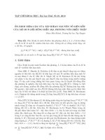
It is important to evaluate for
signs of generalized ligamentous
laxity because such signs have been
reported in 45% to 75% of patients
who have undergone surgery for
MDI.
1,5,8
These signs include
elbow hyperextension (Fig. 1),
metacarpophalangeal joint hyper-
extension, genu recurvatum, patel-
lar subluxation, and the ability of
the abducted thumb to reach the
ipsilateral forearm (thumb-to-fore-
arm test). Clinicians must recog-
nize generalized ligamentous laxity
secondary to known connective tis-
sue disorders, such as Ehlers-
Danlos syndrome and Marfan syn-
drome, because to our knowledge
patients with these conditions have
never had successful results with
soft-tissue instability repairs.
20
Patients with MDI often have an
excessive passive range of gleno-
humeral motion.
Patient confidence and relax-
ation will be gained if instability
tests are performed first on the
asymptomatic shoulder. When
performing these tests, one must
recall that laxity is not instability;
there is a wide spectrum of normal
when assessing degrees of transla-
tion, and reproduction of symp-
toms is critically important. It is
not uncommon to have to repeat
the instability tests during several
office visits because of muscle
guarding. An examination under
anesthesia at the time of a surgical
procedure can provide a more
accurate appreciation of the degree
of translation.
Inferior laxity is assessed first by
applying inferior traction with the
arm at the side (sulcus test). This
examination reflects the integrity of
the rotator interval capsule.
10
In a
positive test, an inferior translation
of at least 1 to 2 cm occurs with the
simultaneous appearance of an
anterior soft-tissue dimple just
beneath the acromion (sulcus sign).
Occasionally, this maneuver will
provoke neurologic symptoms in
the affected extremity. A similar
examination is performed with the
arm abducted to 90 degrees and an
inferior translational force being
applied to the superior proximal
humerus. A positive test in this
position reflects redundancy of the
inferior capsule.
9
Because of inade-
quate muscle relaxation, it is not
uncommon for tests of the asymp-
tomatic shoulder to appear more
positive; nevertheless, this can be a
pertinent finding supportive of a
diagnosis of MDI.
In the supine position, the pa-
tient is assessed for anterior and
posterior instability with use of the
load-and-shift test.
21
The shoulder
is placed slightly off the edge of the
examination table and is held in
approximately 20 degrees of abduc-
tion in the plane of the scapula. The
examiner gently grasps the proxi-
mal humerus and applies a slightly
compressive load to center the
humeral head on the glenoid while
the free hand supports the elbow.
Anterior and posterior translational
forces are then applied at the proxi-
mal humerus in the plane of the gle-
Multidirectional Instability of the Shoulder
Journal of the American Academy of Orthopaedic Surgeons
68
Fig. 1 The patient with MDI often has
hyperextension of the elbows.
noid surface. With maintenance of
the slightly compressive force, the
humeral head will begin to move
medially when its center has trans-
lated beyond the edge of the gle-
noid rim. This sudden change in
direction can usually be palpated by
the examiner during the dislocating
and/or relocating phases of transla-
tion. The extent of laxity (i.e.,
whether the shoulder can be sub-
luxated or dislocated) is determined
by the magnitude of the translation.
It is advantageous to perform this
examination in varying degrees of
abduction and external rotation to
effect different degrees of tension
within the capsular ligaments.
Normal degrees of posterior laxity
allow the center of the humeral
head to be translated up to half the
width of the glenoid fossa, which
patients with MDI usually sur-
pass.
17
A variation of the supine load-
and-shift test can be performed
with the patient seated and the arm
at the side. The humeral head is
centrally compressed in the glenoid
fossa with the translating hand at
the proximal humerus. The scapu-
la is stabilized at the anterior and
posterior aspects of the acromion
with the free hand to allow accu-
rate grading of the translation.
Additional tests that can demon-
strate increased translation include
the Fukuda test, the push-pull test,
and the jerk test.
17
Because the examination of
strength can provoke pain and
spasm, it should always follow the
instability assessment. The exami-
nation concludes with an assess-
ment of sensory function and the
reflexes of the peripheral nerves of
the brachial plexus.
Radiologic Evaluation
Plain radiographs should be ob-
tained to identify uncommon bone
lesions, such as Bankart and Hill-
Sachs lesions, and glenoid dyspla-
sia. Because MDI is a clinical diag-
nosis based on the findings from
the history and physical examina-
tion, we have not found any reason
to order more sophisticated imag-
ing studies.
Nonoperative Management
Nonoperative management in-
cludes patient education and a spe-
cific program of physical therapy.
Patients learn that their lax shoul-
der has become deconditioned from
its usual state and that they need to
regain both strength and neuromo-
tor coordination of the stabilizing
muscles of the rotator cuff, deltoid,
and scapula. To support this expla-
nation, the patient often can be
shown that the contralateral shoul-
der is equally loose yet functions
normally without pain. Burkhead
and Rockwood
14
reported satisfac-
tory results in 29 of 33 (88%) multi-
directionally unstable shoulders
treated with a specific program of
physical therapy.
Before the patient starts an exer-
cise program, pain can be managed
with a combination of brief immo-
bilization, nonsteroidal anti-inflam-
matory drugs, and occasionally a
mild analgesic. The exercise pro-
gram consists of two phases. Phase
I concentrates on progressive resis-
tance exercises utilizing elastic ele-
ments for strengthening the rotator
cuff and deltoid musculature. As
progress is made, strengthening
exercises for the scapula-stabilizing
muscles are added. Phase II begins
at the 10- to 12-week mark, when
additional exercises are added to
retrain humeroscapular coordina-
tion and awareness. Exercises are
continued for a minimum of 6
months. A program of mainte-
nance exercises is then given, to be
followed indefinitely.
Surgical Management
Surgery is an option for patients
who were compliant with a specific
exercise program but who remain
symptomatic. Surgery is not
offered to voluntary dislocators
with emotional problems or to
behaviorally immature teenagers.
While several surgical proce-
dures have been described, an
open inferior capsular shift, as orig-
inally described by Neer and
Foster,
1
is the standard procedure
and continues to be the most com-
monly used. Additional proce-
dures include glenoid osteotomy
22
and arthroscopic inferior capsular
shift.
23
Both procedures have
yielded satisfactory results; howev-
er, the literature to date is sparse.
Arthroscopic, laser-assisted capsu-
lar ÒshrinkageÓ procedures remain
experimental at present.
Technique for Inferior Capsular
Shift
Interscalene block anesthesia is
recommended because it allows
the patient to stand at the comple-
tion of surgery for application of a
modified shoulder spica cast. First,
an examination under anesthesia is
performed, followed by skin
preparation and draping. An ante-
rior approach has been used exclu-
sively by the senior author (J.J.B.)
because it is the only single inci-
sion that allows for a complete
shift of the capsule, closure of the
rotator interval capsule, and repair
of unexpected anterior Bankart
lesions.
The incision is made from the tip
of the coracoid process to the apex
of the axilla in line with the natural
skin creases, and the deltopectoral
interval is developed. The clavipec-
toral fascia is incised lateral to the
conjoined tendon-muscle unit up to
the coracoacromial ligament. The
subscapularis tendon is incised
sharply 1 cm medial to the lesser
Thomas J. Schenk, MD, and John J. Brems, MD
Vol 6, No 1, January/February 1998
69
tuberosity, beginning superiorly at
the rotator interval. After the
scalpel has incised through two
thirds of the anterior thickness of
the length of the tendon, it is turned
coronally, and dissection is carried
medially at the same tendon depth
(Fig. 2, A). When the subscapularis
muscle fibers are encountered, dis-
section deepens to remove the
entire subscapularis muscle belly
from the underlying capsule. Once
freed, the tendon is retracted medi-
ally with traction sutures (Fig. 2, B).
The rotator interval capsule defect
is then imbricated in 30 degrees of
external rotation with the arm at
the side.
A lateral capsular incision begins
at the rotator interval and extends
inferiorly 2 to 3 mm lateral to the
articular cartilage. Access can be
gained for posterior capsule release
by externally rotating and slightly
flexing the adducted arm. The axil-
lary nerve, which is relatively pro-
tected by this positioning, is kept
away from the incising blade by a
blunt retractor. The amount of pos-
terior release is adjusted just
enough for the shift to eliminate the
posterior pouch of redundant tis-
sue. A secondary incision is made
in the capsule, aimed at the center
of the anterior aspect of the glenoid
(Fig. 2, C). Traction sutures are
placed at the corner of each leaflet.
The humeral head is retracted pos-
teriorly with a humeral-head retrac-
tor, and the intra-articular contents
are inspected. Note is made of the
condition of the articular surfaces
and the labral complex attachment.
A dental burr is used to decorti-
cate the bone adjacent to the articu-
lar surface on the surgical neck of
the humerus. The shift is per-
formed with the arm in 30 degrees
of abduction, 40 degrees of external
rotation, and 10 degrees of flexion.
The inferior flap is shifted superior-
ly, eliminating excessive capsular
volume posteriorly and inferiorly,
and is sutured to the cuff of pre-
served lateral capsular tissue. The
superior leaflet is shifted inferiorly
and is similarly repaired (Fig. 2, D).
The subscapularis tendon is re-
paired at its anatomic length. Non-
absorbable suture material is used
throughout these reconstructive
steps.
The application of a modified
shoulder spica cast is recommended
because it is the most certain way to
immobilize the reconstructed cap-
sule during the acute healing phase,
and it eliminates the worry of com-
pliance with brace wear. The cast is
applied with the arm in neutral
rotation and in 10 to 15 degrees of
abduction. To reduce potential
strain on the rotator interval capsule
repair, an assistant pushes cephalad
on the olecranon until the cast is
firm. When the cast is applied
properly, the shoulder will be in a
mildly shrugged position.
Aftercare
A standard protocol of postoper-
ative exercises is used as a general
outline. During the healing and
Multidirectional Instability of the Shoulder
Journal of the American Academy of Orthopaedic Surgeons
70
A B
C D
Fig. 2 A, The anterior two thirds of the subscapularis tendon is dissected medially, leav-
ing the posterior portion of the tendon to reinforce the anterior capsule. B, The subscapu-
laris muscle belly and the anterior portion of the tendon are retracted medially. C, The
capsule is incised in a ÒTÓ fashion, creating superior and inferior leaflets. D, The capsule is
advanced and shifted; the superior flap overlaps the inferior flap.
Subscapularis tendon
A
B
B
A
stretching phases of postoperative
management, the standard proto-
col is adhered to rigidly for fear
that rapid gains in motion will
result in recurrent instability.
When strengthening exercises are
initiated, the program is individu-
alized depending on the patientÕs
progress.
The spica is removed at week 6,
and a sling is provided to ease the
transition from rigid immobiliza-
tion. During weeks 6 to 10, activi-
ties of daily living are allowed
below the level of the shoulder and
within 45 degrees of external rota-
tion. At week 10, a stretching pro-
gram is begun for forward eleva-
tion (limit, 160 degrees) and exter-
nal rotation (limit, 45 degrees),
emphasizing gradual restoration of
range of motion. At weeks 14 to 16,
deltoid and rotator cuff strength-
ening begins. At weeks 18 to 20,
exercises for the scapular stabiliz-
ers are added.
Contact sports are permitted
once full strength and conditioning
have been restored, usually at 10
months. Examples of activities dis-
couraged indefinitely include
wrestling, waterskiing, and certain
lifting exercises, including bench
presses and dips.
Outcomes
There have been only a few pub-
lished reports of the results of sur-
gical treatment of MDI. These
demonstrate a high degree of
patient satisfaction and subjective
stability in patients treated with an
open inferior capsular shift. In the
original article by Neer and Foster,
1
39 patients were reevaluated more
than 1 year after surgery, of whom
17 (44%) were followed up for
more than 2 years. One patient
experienced recurrent anterior sub-
luxations 7 months postopera-
tively. The remaining patients
achieved satisfactory results, as
defined by the absence of recurrent
instability events or significant
pain and by the return of normal
strength and the ability to partici-
pate in full activities, as well as the
capacity for elevation within 10
degrees of that possible in the con-
tralateral shoulder and external
rotation within 40 degrees. Three
patients had neurapraxia of the
axillary nerve.
Cooper and Brems
5
reported on
38 patients (43 shoulders) with a
minimum follow-up of 2 years
(average follow-up, 38 months).
Symptomatic MDI recurred in 4
shoulders (9%) in 4 patients within
2 years of surgery; one instance of
MDI was attributable to a defined
event of significant trauma, and
three instances presumably oc-
curred because the repair became
stretched. The remaining 34 pa-
tients were subjectively satisfied
with the status of their shoulder,
although 5 patients (15%) had per-
sistent episodes of apprehension.
Bigliani et al
24
reported on surgi-
cal treatment of 49 patients with
MDI. An anterior approach was
used when largely anteroinferior
instability was identified (34
patients) and a posterior approach
was used when instability was
greatest posteroinferiorly (15
patients). The results after an aver-
age follow-up interval of 5 years
were satisfactory for 91% of the
patients treated with an anterior
approach and for 100% of the
patients treated with a posterior
approach.
Summary
A diagnosis of MDI is arrived at on
the basis of a careful history and
physical examination. Most
patients can be successfully treated
with a well-executed exercise pro-
gram. For the minority of patients
for whom nonoperative manage-
ment is a failure, surgical recon-
struction can be reasonably recom-
mended. The most widely report-
ed surgical procedure is an open
inferior capsular shift. When com-
bined with meticulous aftercare,
this procedure has yielded favor-
able results in the relatively few
series published to date.
Thomas J. Schenk, MD, and John J. Brems, MD
Vol 6, No 1, January/February 1998
71
References
1. Neer CS II, Foster CR: Inferior capsu-
lar shift for involuntary inferior and
multidirectional instability of the
shoulder: A preliminary report. J Bone
Joint Surg Am 1980;62:897-908.
2. Neer CS II: Dislocations, in Neer CS II
(ed): Shoulder Reconstruction. Phila-
delphia: WB Saunders, 1990, pp 273-
341.
3. Thomas SC, Matsen FA: An approach
to the repair of glenohumeral ligament
avulsion in the management of trau-
matic anterior glenohumeral instability.
J Bone Joint Surg Am 1989;71:506-513.
4. Neer CS II: Involuntary inferior and
multidirectional instability of the
shoulder: Etiology, recognition, and
treatment. Instr Course Lect 1985;34:
232-238.
5. Cooper RA, Brems JJ: The inferior
capsular-shift procedure for multidi-
rectional instability of the shoulder. J
Bone Joint Surg Am 1992;74:1516-1521.
6. Hawkins RJ, Abrams JS, Schutte J:
Multidirectional instability of the
shoulder: An approach to diagnosis.
Orthop Trans 1987;11:246.
7. Bigliani LU, Kurzweil PR, Schwartz-
bach CC, Wolfe IN, Flatow EL: Infe-
rior capsular shift procedure for ante-
rior-inferior shoulder instability in ath-
letes. Am J Sports Med 1994;22:578-584.
8. Altchek DW, Warren RF, Skyhar JM,
Ortiz G: T-plasty modification of the
Bankart procedure for multidirectional
instability of the anterior and inferior
types. J Bone Joint Surg Am 1991;73:
105-112.
9. Warner JJP, Deng XH, Warren RF,
Torzilli PA: Static capsuloligamentous
restraints to superior-inferior transla-
tion of the glenohumeral joint. Am J
Sports Med 1992;20:675-685.
10. Harryman DT II, Sidles JA, Harris SL,
Matsen FA III: The role of the rotator
interval capsule in passive motion and
stability of the shoulder. J Bone Joint
Surg Am 1992;74:53-66.
11. Lippitt SB, Vanderhooft JE, Harris SL,
Sidles JA, Harryman DT II, Matsen FA
III: Glenohumeral stability from concav-
ity-compression: A quantitative analy-
sis. J Shoulder Elbow Surg 1993;2:27-35.
12. Blasier RB, Guldberg RE, Rothman
ED: Anterior shoulder stability: Con-
tributions of rotator cuff forces and the
capsular ligaments in a cadaver
model. J Shoulder Elbow Surg 1992;1:
140-150.
13. Ozaki J: Glenohumeral movements of
the involuntary inferior and multidi-
rectional instability. Clin Orthop 1989;
238:107-111.
14. Burkhead WZ Jr, Rockwood CA Jr:
Treatment of instability of the shoul-
der with an exercise program. J Bone
Joint Surg Am 1992;74:890-896.
15. Blasier RB, Carpenter JE, Huston LJ:
Shoulder proprioception: Effect of
joint laxity, joint position, and direc-
tion of motion. Orthop Rev 1994;23:
45-50.
16. Lephart SM, Warner JJP, Borsa PA, Fu
FH: Proprioception of the shoulder
joint in healthy, unstable, and surgical-
ly repaired shoulders. J Shoulder Elbow
Surg 1994;3:371-380.
17. Matsen FA III, Thomas SC, Rockwood
CA Jr: Anterior glenohumeral insta-
bility, in Rockwood CA Jr, Matsen FA
III (eds): The Shoulder. Philadelphia:
WB Saunders, 1990,vol 1, pp 526-622.
18. Kumar VP, Balasubramaniam P: The
role of atmospheric pressure in stabil-
ising the shoulder: An experimental
study. J Bone Joint Surg Br 1985;67:719-
721.
19. Rowe CR, Pierce DS, Clark JG:
Voluntary dislocation of the shoulder:
A preliminary report on a clinical,
electromyographic, and psychiatric
study of twenty-six patients. J Bone
Joint Surg Am 1973;55:445-460.
20. Jerosch J, Castro WHM: Shoulder
instability in Ehlers-Danlos syndrome:
An indication for surgical treatment?
Acta Orthop Belg 1990;56:451-453.
21. Hawkins RJ, Bokor DJ: Clinical evalu-
ation of shoulder problems, in Rock-
wood CA Jr, Matsen FA III (eds): The
Shoulder. Philadelphia: WB Saunders,
1990, vol 1, pp 149-177.
22. Nobuhara K, Ikeda H: Glenoid oste-
otomy for loose shoulder, in Bateman
JE, Welsh RP (eds): Surgery of the
Shoulder. Philadelphia: BC Decker,
1984, pp 100-103.
23. Duncan R, Savoie FH III: Arthroscopic
inferior capsular shift for multidirec-
tional instability of the shoulder: A
preliminary report. Arthroscopy 1993;
9:24-27.
24. Bigliani LU, Pollock RG, Owens JM,
McIlveen SJ, Flatow EL: The inferior
capsular shift procedure for multidi-
rectional instability of the shoulder.
Orthop Trans 1993;17:576.
Multidirectional Instability of the Shoulder
Journal of the American Academy of Orthopaedic Surgeons
72