Báo cáo y học: " Prevalence of a virus similar to human hepatitis B virus in swine" ppt
Bạn đang xem bản rút gọn của tài liệu. Xem và tải ngay bản đầy đủ của tài liệu tại đây (1.15 MB, 7 trang )
RESEARC H Open Access
Prevalence of a virus similar to human hepatitis B
virus in swine
Wengui Li
1,2
, Ruiping She
1*
, Liqiang Liu
3
, Hua You
1
, Jun Yin
1
Abstract
Background: The objective of this study is to established evidence of the existence of a novel member of the
hepadnavirus family endemic in swine. Temporarily this virus was designated as swine hepatitis B virus (SHBV). This
SHBV can be detected by using human hepatitis B virus diagnostic kits including ELISA, immunohistochemical
staining, and transmission electron microscopy (TEM). Also seroprevalence of pig farms in Beijing, China, and
pathological features of SHBV infection was determined.
Results: Screened result shows that overall prevalence of HBsAg was 24.8%, closed to that of anti-HBsAg, whereas
HBeAg and anti-HBe were barely detectable. The distribution of HBsAg and HBcAg was examined by
immunohistochemistry of liver samples. Typical hepatitis pathological change, such as spotty parenchymal cell
degeneration, necrosis of hepatocytes and proliferation of fibrous connective tissue were observed during
histopathological analysis. Analysis of HBsAg-positive serum with TEM revealed two morphologic forms, 20 nm and
40 nm sized particles, similar to small spherical and Danes particles of HBV. Observation of the ultrastructure of the
liver also found HBV-like particles in the nucleus of hepatocytes.
Conclusion: Our research result implies that SHBV cou ld be a causative agent of swine. The discovery of SHBV will
unveil novel evolutionary aspects of hepatitis and provides new information for further hepadnavirus research.
Background
Viral hepatitis B remain a serious medical challenge
worldwide [1]. A strong epidemiological relationship has
been established between persistent hepatitis B virus
(HBV) infection and hepatocellular carcinoma (HCC)
[2]. HBV is one of the smallest enveloped animal viruses
with a virion diameter of 42 nm. But pleomorphic forms
exist, including filamentou s and spherical bodies lacking
a core. As most hepadnaviruses, HBV will only replicate
in specific hosts, and this makes experiments using in
vitro methods very difficult.
Formerly, hepatitis B was called serum hepatitis.
Detection of HBV infection involves serum or blood
tests that detect either viral antigens (surface antigen
HBsAg and e antigen HBeAg) and ant ibodies (anti-HBs,
anti-HBc, anti-HBe), known as HBV serological marker.
HBsAg is most frequently used to screen for the pre-
sence of this infection, the presence of HBeAg in a
host’s serum is associated with much higher rates of
viral replication and enhanced infectivity. Nevertheless,
interpretation of these assays is complex.
HBV is the prototype member of a steadily growing
family of hepadnaviruses which can be found in both
mammals (orthohepadnaviruses) and birds (avihepadna-
viruses). Orthohepadnaviruses have been identified so
far in woodchucks (WHV), ground and arctic squirrels
(GSHV, ASHV), and primates including woolly monkeys
(WMHBV), orangutans, gorillas, and gibbons [3-8]. Avi-
hepadnavirus has b een reported in various duck species
(DHBV), grey herons (HHBV), geese (GHBV), Ross’s
goose (RGHBV), storks (STHBV), and cranes (CHBV)
[9-11]. The discovery of HBV-related viruses offers
ample opportunities for in vivo studies of various ani-
mals with naturally occurring hepadnaviruses. This has
been valuable in determin ing the mechanisms of hepad-
navirus replication, pathogenesis of hepatocellular carci-
noma (HCC), and for antiviral drug studies.
HBV-related hepadnaviruses in mammalian and avian
species has been valuable in HBV studies. Like deter-
mining the mechanisms of hepadnavirus replication,
pathogenesis of HCC, and antiviral drug studies [12].
* Correspondence:
1
College of Veterinary Medicine, China Agricultural University, Beijing 100193,
China
Li et al. Virology Journal 2010, 7:60
/>© 2010 Li et al; licensee BioMed Central Ltd. This is an Open Access article distributed under the terms of the Creative Commons
Attribution License ( which permits unrestricted use, distribution, and reproduction in
any medium, provided the original work is properly cited.
However, most of the corresponding animals are diffi-
cult to handle in captiv ity or not easily available. Since
none of the currently available animal models are ideal,
the development of additional experimental animal
models promises to provide answers for many HBV
research questions [13].
Researchers have concentrated on a group of HBV-
like viruses in domestic animals since 1985 [14]. Using
humanHBVdiagnostickits,anumberofdomesticani-
mals are positive for HBV serological marker [15,16],
electron microscope observed HBV-like virion in HBsAg
positive serum of swine, Holstein, cattle, canine and
sheep; even gene sequence highly homologous to HBV
has been amplified [17-20]. Nevertheless, Up to the pre-
sent time, none of these HBV-like viruses been systema-
ticallyidentifiedandrelatedreportsfoundonlyin
China. Here we characterize the prevalence of HBV-like
virus in swine which may provide an interesting model
for comparative studies of live r pathology and cancer
associated with chronic hepadnavirus infections.
Results
Enzyme-linked immunosorbent assay
To investigate the current prevalence of SHBV in swine
herds, 416 samples of swine serum collected from 5 ran-
domly selected farms in Beijing, China, were tested for
HBV serological markers using a comm ercial ELISA kit.
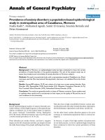
Briefly, overall prevalence of HBsAg was 24.8%, and pro-
foundly close to anti-HBs (25.0%), whil e HBe and anti-
HBe was hardly detected (0.5% and 0.7%), indicating no
common antigen existed in HBe. The ov erall prevalence
of anti-HBc was 63.9% (Fig. 1, Table 1).
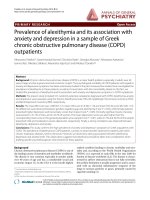
Histopathological analysis and Mallory’s trichrome stain
For swine CP74 and DX385, although obvious patholo-
gical changes were not observed at autopsy, pathological
changes were observed under light microscope. Gross
histopathological findings showed desmoplasia in hepa-
tic lobules, infiltration of lymphocytes, hyperplasy of bile
canaliculus, and fibrous tissue at the portal area (Fig. 2A
and 2B). Severe fibrous connective tissue proliferation
was observed by Mallory staining (Fig. 2C and 2D). In
contrast , no obvious changes were found in liver tissues
collected from swine CP59.
Immunohistochemistry
Immunohistochemical scanning of expression of viral
antigens found that liver tissues from both swine con-
tained HBsAg and HBcAg. Strong immunohistochemical
signal was seen within hepatitis lesions. HBsAg was
detected in the nucleus and cytoplasm of hepatocytes,
while HBcAg was mainly distributed in the nucleus of
hepatocytes. Necrosis as karyorrhexi s, pyknosis and kar-
yolisis was observed in immunohistochemically positive
hepatocytes. This indicates that SHBV was pathogenic
to swine, an d replication of SHBV caused the necrosis
of hepatocytes directly (Fig. 3A, B, C and 3D).
Detection of viral particles in swine sera and liver cells by
electron microscopy
To obtain ultrastructural evidence for the presence of
HBV-related viral particles in the swine sera containing
S antigen, HBsAg-positive serum was collected, viral
particles in the sera of infected swine were morphologi-
cally analyzed by electron microscopy and sera negative
for HBsAg served as controls. Essentially, two types of
particles closely resembled i n size (20 nm and 40 nm)
and morphology, like complete and empty viral particles
of HBV, were observed. However, it is puzzling that no
tubular particles were seen. Particles were observed only
in serum positive for HBsAg, and the number of 40 nm
particles was much more than expected (Fig. 4A). Ultra-
structurally, HBV-like particles were observed in the
nucleus of hepatocytes (Fig. 4B).
Discussion
Serological d iagnosis of hepatitis B virus infection relies
on a combination of qualitative assay results and differ-
ent patterns are representative of acute or chro nic dis-
ease in a carrier [21]. By examining the antigen-
antibody system, hepatitis B infection is diagnosed, the
course of the disease is observed and treatment is moni-
tored [22]. The screening of HBV serological markers in
swine herds showed that nearly a quarter of swine have
been infect ed. However, profiles in SH BV serology were
quite different from human HBV (data not shown).
Anti-HBc is found in all people infected with HBV,
which can persist for many years and act as a lifelong
marker of hepatitis B [23]. The high prevalence of anti-
HBc in swine (63.9%) may indicate that these swine
have a history of infection. Nevertheless, existence of
anti-HBc as the only serological marker also may be the
result of nonspecific cross-reaction with other agents
[24].
Though hepadnaviruses are host specific, HBV infec-
tions also occ ur frequently in chimpanzee, gibbon and
other ape populations in sub-Saharan Africa and South-
East Asia where the HBV infection rate in apes was
remarkably comparable to that of human population in
these areas [25,26]. Scientists are concerned about the
ability of HBV to cross species barriers. Large reservoirs
of infection in apes may hamper ongoing attempts to
permanently eradicate HBV infection from the human
population through immunization [27].
The prevalence of HBV among human and the non-
human primates maybe speed up the evolution process.
Due to high error rate of the viral reverse transcriptase,
and recombination among different genotypes or
Li et al. Virology Journal 2010, 7:60
/>Page 2 of 7
hepadnavirus strains from human and nonhuman pri-
mates, the eight genotypes of HBV have further diverged
into at least 24 subgenotypes, with certainly many more
still to be identified [28]. Interspecies recombination
events of HBV also occur among human and nonhuman
primates [29], such as gibbons of different genera, chim-
panzees, and birds of different subfamilies [25,30]. Inter-
species recombination of hepadnaviruses from cross-
species hosts would provide a large variation in virus
genomes, which would change pathogenecity and trans-
missibility, and expand thehostrange.Evidencefor
recombination of human and ape HBV variants demon-
strates that human and nonh uman-associated HBV var-
iants can in deed share hosts in natu re [30]. Compared
to nonhuman primates, domestic animals a re in more
contact with humans and the possibility of interspecies
recombination is higher. Thus the discovery of SHBV
will be beneficial to research of HBV evolution.
The lack of suitable in vitro infection systems and
appropriate animal models has hampered the progress
of HBV research, but progress has been made through
the identification of avian and mammalian HBV-related
viruses. However, none of these natural hosts are com-
monly used laboratory animals, and the expense and dif-
ficulty in handling these animals have limited their
usage [4,31,32]. In fact, chimpanzees are the only ani-
mals fully permissive and well tested for HBV infection.
Nonetheless, the limited availability and the high cost of
keeping primates severely restricts their use in research
[13]. Comparatively, pigs are widely used in medical
Figure 1 Prevalence of SHBV serological markers among 416 swine sera samples collected from five farms in Beijing, China .Scatter
graphs showed that nearly a quarter of the swine have been infected by SHBV. Prevalence rates of HBs were close to anti-HBs, while HBeAg
and anti-HBe were hardly detected.
Table 1 Prevalence of SHBV serological markers among 416 swine sera samples collected from five farms
n HBsAg, n (%) HBsAb n (%) HBeAg, n (%) Anti-HBe, n (%) Anti-HBc, n (%)
Farm A 77 17 (22.1) 8 (10.4) 0 (0) 0 (0) 23 (29.9)
Farm B 84 39 (46.4) 43 (51.2) 0 (0) 0 (0) 84 (100)
Farm C 85 12 (14.1) 10 (11.8) 2 (2.4) 3 (3.5) 60 (70.6)
Farm D 85 0 (0) 3 (3.5) 0 (0) 0 (0) 84 (98.8)
Farm E 85 35 (41.2) 40 (47.1) 0 (0) 0 (0) 15 (17.6)
Total 416 103 (24.8) 104 (25.0) 2 (0.5) 3 (0.7) 266 (63.9)
Li et al. Virology Journal 2010, 7:60
/>Page 3 of 7
research and there are abundant in supply, since the
available animal models are not ideal, the development
of additional experimental animal systems is warranted,
the finding of HBV in pigs will enhance our understand-
ing of the v irology and immunology of HBV infection
and disease pathogenesis, including major sequelae like
chronic hepatitis and hepatocellular carcinoma.
Of the 350 million to 400 million individuals world-
wide infected with the hepatitis B virus (HBV), one-
third reside in China, with 130 million carriers and
30 million chronically infected [33]. E ven though a vac-
cination program for newborn babies has been in place
since the 1990s, the incidence of hepatitis B is still
increasing, from 21.9 in 100,000 people in 1990 to 53.3
in 100,000 in 2003. The reason for this increased HBV
infection is unknown, because hepatitis B has no clear
transmission routes in many people in China [34]. The
identification of the SHBV strain confirms that a novel
class of hepadnaviridae exists in swine populations. And
thus brings about a lot of questions. Does these pigs
infected by HBV? Does swine hepadnavirus exist? Does
this virus related to the rising of h epatitis B in human
population? But before these questions could be
answered, further studies are needed to elucidate the
structure, assembly, genome organization and regulation
of gene expression of this novel hepadnavirus.
Methods
Swine and serum samples
To determine the seroprevalence of SHBV infection in
swine, 416 swine serum samples were col lected from
five randomly selected farms in Beijing, China. For
serum collection, 5 mL of blood was collected from
swine into dry tubes. After clotting and centrifugation,
Figure 2 Results of histopathological analysis(A, B) and Mallory’s trichrome stain(C, D). (A) desmoplasia between hepatic lobule (arrow),
(B) infiltration of lymphocytes (down arrow), hyperplasy of bile canaliculus and fibrous tissue at portal area(up arrow), also coagulation necrosi s
and karyopyknosis of hepatocytes could be seen. Original magnification × 400. (C, D) Showing proliferation of connective tissue between liver
lobule (arrow). Mallory staining method, Original magnifications ×200. (A, C: liver sample from CP74; B, D: liver sample from DX385).
Li et al. Virology Journal 2010, 7:60
/>Page 4 of 7
sera were separated and stored at -20°C until use. Two
swine positive for HBsAg (CP74:Boar, 7 month; DX385:
Sow, 8.5 month) were sacrificed to determine the possi-
ble relationship of SHBV infection and his topathological
changes in the liver. Another swine negative for all sero-
logical markers (CP59:Sow, 5 month) was sacrificed and
served as a negative control.
Serological analysis of hepatitis B virus markers
All serum samples were screened for hepatitis B serolo-
gical markers (anti-HBc, HBsAg, anti-HBs, HBeAg, and
anti-HBe) with a commercial enzyme-linked immuno-
sorbent assay (ELISA) kit (SIIC Kinghaw Biotech Co.
Ltd., Beijing, China) according to the manufacturer’s
recommendations. The absorbance was determined at
450 nm (Multiscan Titertek MCC). Blank, negative and
positive controls were included on each plate. Data were
analyzed with the SPSS software for Windows (SPSS
Inc., Chicago, USA) and a scatter graph was obtained by
using OriginPro 7.5 (OriginLab Corporation, Northamp-
ton, MA, USA).
Histopathology analysis and Mallory’s trichrome stain
Histopathological analysis was used to study the pathologi-
cal characteristics of SHBV infection. In consideration of
fibrosis is the pathological feature of chronic hepatitis,
Mallory trichrome stain was used to study fibrous tissue
proliferation in liver. Liver samples were collected and
fixed in 2.5% (v/v) glutaraldehyde-polyoxymethylene solu-
tion immediately after swine were sacrificed. The tissue
samples were dehyd rated and embedded in paraffin. Sec-
tions of 5-μm thickness were then prepared for
Figure 3 Immunohistochemical analysis of HBsAg and HBcAg in liver tissues.(A) Strong HBsAg immune positivity was shown in
hepatocytes (arrow). (B) Immunopositivity for HBsAg was mainly distributed in cytoplasm of hepatocytes. (C, D) HBcAg was distributed mainly
distributed nucleus of hepatocytes. Spotty parenchymal cell degeneration, with necrosis and karyopyknosis (arrow) of hepatocytes were
observed. Original magnification×400. (A, C: liver sample from CP74; B, D: liver sample from DX385).
Li et al. Virology Journal 2010, 7:60
/>Page 5 of 7
hematoxylin and eosin (H&E), and Mallory trichrome
stains. For Mallory’ s trichrome stain, paraffin sections
were washed with distilled water and immersed in 3%
dichromicum kalium for 5 min, then in solution consisting
of 0.1% acid fuchsin for 2 min, and 0.5% aniline blue for
20 min. Thereafter the slides were washed sequentially
with distilled water, 95% ethanol, and three changes of
100% xylene. After the xylene had evaporated, Cytoseal 60
mounting medium was applied, and the slides were cover-
slipped for examination under a microscope. All powdered
stains used for Mallory stain were obtained from Sigma
(Sigma Co., Beijing, China).
Immunohistochemistry
Serial paraffin sections (5 μm) were prepared and kept at
37°C for more than 12 hours. The sections were
immersed in three consecutive washings in xylol for
5 min to remove paraffin, and then hydrated through
graded alcohol. Sections were incubated for 30 min and
blocked with 3% peroxide at room temperature for endo-
genous peroxidase ablation. The following steps were
carried out in a moist chamber. Sections were incubated
with blocking buffer (Zymed Laboratories Inc., San
Diego, USA) containing 20% normal goat serum (Gibco)
and 80% PBS (0.01 M, pH 7 .4) at 37°C for 30 min. After
discarding the goat serum, sections were incubated in
primary monoclonal antibodies against HBsAg and
HBcAg (Zhongshan Golden Bridge Biotech Co. Ltd., Beij-
ing, China) diluted in PBS, for 2 hours at 37°C. After rin-
sing for 3 times in PBS-T, sections were incubated with
the goat anti-mouse IgG conjugated with HRP (Sigma) at
37°C for 1 hour and rinsed 3 times in PBS-T. The speci-
mens were incubated with 3,3-diaminobenzidin (DAB;
Zymed Laboratories Inc) at room temperature for 10 min
in the dark. Finally, sections were stained with hematoxy-
lin for 8 min after rinsing for 3 times in PBS-T, dehy-
drated, and mounted with neutral gums. Sections for the
negative control group were prepared by the same steps
as described above but with the HBsAg and HBcAg anti-
bodies replaced by PBS.
Detection of viral particles in swine sera and hepatocytes
by transmission electron microscopy
To obtain ultrastructural evidence for the presence of
HBV-related viral parti cles in s wine sera containing S
antigen, HBsAg-positive serums were collected and viral
particles i n sera of infected swine were morphologically
analyzed by electron microscopy. Sera negative for
HBsAg served as controls. Serum collected from three
swine were centrifuged at 4000 rpm for 10 min, then
0.01 M poly ethylene glycol 6000 (PEG6000) was added
into the subsequent upper aqueous phase. After
Figure 4 Viral particles in swine sera and hepatocytes revealed by electron microscopy. A: Electron micrographs of negativ ely stained
SHBV particles from HBsAg positive serum. Two types of particles were observed which are similar in size (20 nm and 40 nm) and morphology,
like complete and empty viral particles of SHBV. B: Virus-like particles in the nucleus of hepatocytes (liver sample from DX385).
Li et al. Virology Journal 2010, 7:60
/>Page 6 of 7
incubation overnight at 4°C, the serum was ce ntrifuged
at 20,000 rpm for 1 hour, resuspended in PBS and
stained for 1 min with 1% uranyl acetate. For the thin
section study, the fixative used was 2.5% paraformalde-
hyde-glutaraldehyde in 0.1 M cacodylate buffer (pH 7.4).
The sections were postfixed in 1% OsO
4
for 1 hour, and
treated with 1% uranyl acetate, dehydrated in ethanol
and embedded in Epon 812. Ultrathin sections were
obtained using a routine method and stained with ura-
nyl acetate and lead citrate. All electron micrographs
were obtained with JEV1230 transmission electron
microscope (JEOL Ltd., Tokyo, Japan) at 80 kV.
Acknowledgements
This work was supported by the National Natural Science Foundation of
China (Grant No. 30871853) and the Yunnan Provincial Program for
Introducing High-level Scientists (Grant No. 2009CI125).
The authors would like to thank Prof. Bin Wang (State Key Laboratory for
Agro-Biotechnology, China Agricultural University) for permission and help in
using the laboratory facilities.
Author details
1
College of Veterinary Medicine, China Agricultural University, Beijing 100193,
China.
2
College of Animal Science and Technology, Yunnan Agricultural
University, Kunming 650201, China.
3
College of Agriculture, Hebei University
of Engineering, Handan 056021, China.
Authors’ contributions
WGL carried out the serological analysis of hepatitis B virus markers and
drafted the manuscript. LQL carried out the Histopathology analysis and
Mallory’s trichrome stain. HY and JY carried out the immunohistochemical
staining and transmission electron microscope investigations. RPS carried out
the design of the study and revision of the manuscript. All authors read and
approved the final manuscript.
Competing interests
The authors declare that they have no competing interests.
Received: 16 December 2009 Accepted: 17 March 2010
Published: 17 March 2010
References
1. Lai CL, Ratziu V, Yuen MF, Poynard T: Viral hepatitis B. The Lancet 2003,
362:2089-2094.
2. Buendia MA: Hepatitis B viruses and cancerogenesis. Biomed
Pharmacother 1998, 52:34-43.
3. Summers J, Smolec JM, Snyder R: A virus similar to human hepatitis B
virus associated with hepatitis and hepatoma in woodchucks. Proc Natl
Acad Sci USA 1978, 75:4533-4537.
4. Testut P, Renard CA, Terradillos O, Vitvitski-Trepo L, Tekaia F, Degott C,
Blake J, Boyer B, Buendia MA: A new hepadnavirus endemic in arctic
ground squirrels in Alaska. J Virol 1996, 70:4210-4219.
5. Lanford RE, Chavez D, Brasky KM, Burns RB Iii, Rico-Hesse R: Isolation of a
hepadnavirus from the woolly monkey, a new World primate. Proc Natl
Acad Sci USA 1998, 95:5757-5761.
6. Warren KS, Heeney JL, Swan RA, Heriyanto , Verschoor EJ: A new group of
hepadnaviruses naturally infecting orangutans (Pongo pygmaeus). J Virol
1999, 73:7860-7865.
7. Linnemann CC Jr, Kramer LW, Askey PA: Familial clustering of hepatitis B
infections in gorillas. AM J Epidemiol 1984, 119:424-430.
8. Noppornpanth S, Haagmans BL, Bhattarakosol P, Ratanakorn P,
Niesters HGM, Osterhaus ADME, Poovorawan Y: Molecular epidemiology of
gibbon hepatitis B virus transmission. J Gen Virol 2003, 84:147-155.
9. Prassolov A, Hohenberg H, Kalinina T, Schneider C, Cova L, Krone O,
Frolich K, Will H, Sirma H: New Hepatitis B Virus of Cranes That Has an
Unexpected Broad Host Range. J Virol 2003, 77:1964-1976.
10. Pult I, Netter HJ, Bruns M, Prassolov A, Sirma H, Hohenberg H, Chang SF,
FrÖlich K, Krone O, Kaleta EF, Will H: Identification and analysis of a new
hepadnavirus in white storks. Virology 2001, 289:114-128.
11. Funk A, Mhamdi M, Will H, Sirma H: Avian hepatitis B viruses: Molecular
and cellular biology, phylogenesis, and host tropism. World J
Gastroenterol 2007, 13:91-103.
12. Olive DS, Konishi M, Wu GY: Cell culture and animal models for human
viral hepatitis. Hepatol Res 2004, 28:61-67.
13. Dandri M, Volz TK, Lutgehetmann M, Petersen J: Animal models for the
study of HBV replication and its variants. J Clin Virol 2005, 34:S54-S62.
14. Qifeng X: Experimental infection of chickens with hepatitis B virus.
Chinese Journal of Nature 1985,
9:238-239.
15. She RP, Li WG, Wang YH, Liu L, Hu YX, Xu JC, Bao HH, Wang DC: Viral
Hepatitis: A Dangerous Zoonosis. Sci Technol Rev 2007, 25:44-52.
16. Xu YW, Chu X: Study progress on HBV-like virus in animal. Chinese J Prev
Vet Sci Technol 1993, 23:16-20.
17. Shao XA, Xu W, Wang Y, Xiong SD: HBsAg-like protein detected in the
bovine serum. Fudan Univ J Med Sci 2004, 31:585-587, +596
18. Din Z, Wang CY, jin NY, Zou XH, Bai L, Nie Y, Liu CG: Hereditary variation
in S gene sequence of hepatitis B virus from canine. Chinese J Prev Vet
Med 2003, 25:24-28.
19. Din Z, Jin NY, Chen ZW, Zou XH, Yang HSO: S Gene Sequence
Comparison between the HBV like virus from chicken and human HBV.
Chinese J Prev Vet Sci Technol 1999, 19:18-21.
20. Din Z, Jin NY, Chen CF, Zou XH, Wang CY: Study on S Gene Sequence
Homologous Analysis between the Hepatitis B Virus from Sheep and
Human. Progress in Veterinary Medicine 2001, 22:54-58.
21. Salassa B, Daziano E, Bonino F, Lavarini C, Smedile A, Chiaberge E, Rosina F,
Rossana Brunetto M, Pessione E, Spezia C, et al: Serological diagnosis of
hepatitis B and delta virus (HBV/HDV) coinfection. J Hepatol 1991,
12:10-13.
22. Badur S, Akgun A: Diagnosis of hepatitis B infections and monitoring of
treatment. J Clin Virol 2001, 21:229-237.
23. Tseliou P, Spiliotakara A, Dimitracopoulos GO, Christofidou M: Detection of
hepatitis B virus DNA in blood units with anti-HBc as the only positive
serological marker. Haematologia 2000, 30:159-165.
24. Sanchez-Quijano A, Jauregui JI, Leal M, Pineda JA, Castilla A, Abad MA,
Civeira MP, Garcia de Pesquera F, Prieto J, Lissen E: Hepatitis B virus occult
infection in subjects with persistent isolated anti-HBc reactivity. J Hepatol
1993, 17:288-293.
25. Yang J, Xi Q, Deng R, Wang J, Hou J, Wang X: Identification of
interspecies recombination among hepadnaviruses infecting cross-
species hosts. J Med Virol 2007, 79:1741-1750.
26. Starkman SE, MacDonald DM, Lewis JCM, Holmes EC, Simmonds P:
Geographic and species association of hepatitis B virus genotypes in
non-human primates. Virology 2003, 314:381-393.
27. Makuwa M, Souquière S, Clifford SL, Mouinga-Ondeme A, Bawe-Johnson M,
Wickings EJ, Latour S, Simon F, Roques P: Identification of hepatitis B virus
genome in faecal sample from wild living chimpanzee (Pan troglodytes
troglodytes) in Gabon. J Clin Virol 2005, 34:S83-S88.
28. Schaefer S: Hepatitis B virus taxonomy and hepatitis B virus genotypes.
World J Gastroenterol 2007,
13:14-21.
29. Magiorkinis EN, Magiorkinis GN, Paraskevis DN, Hatzakis AE: Re-analysis of a
human hepatitis B virus (HBV) isolate from an East African wild born
Pan troglodytes schweinfurthii: Evidence for interspecies recombination
between HBV infecting chimpanzee and human. Gene 2005, 349:165-171.
30. Simmonds P, Midgley S: Recombination in the genesis and evolution of
hepatitis B virus genotypes. J Virol 2005, 79:15467-15476.
31. Menne S, Cote PJ: The woodchuck as an animal model for pathogenesis
and therapy of chronic hepatitis B virus infection. World J Gastroenterol
2007, 13:104-124.
32. Feitelson MA, DeTolla LJ, Zhou XD: A chronic carrierlike state is
established in nude mice injected with cloned hepatitis B virus DNA. J
Virol 1988, 62:1408-1415.
33. Liu J, Fan D: Hepatitis B in China. The Lancet 2007, 369:1582-1583.
34. Wang XJ, Zhang RZ, Hu YS, F LX: Analysis on epidemic status of viral
hepatitis in China: the report from Chinese Center for Disease Control
and Prevention. Dis Surveillance 2004, 19:290-292.
doi:10.1186/1743-422X-7-60
Cite this article as: Li et al.: Prevalence of a virus similar to human
hepatitis B virus in swine. Virology Journal 2010 7:60.
Li et al. Virology Journal 2010, 7:60
/>Page 7 of 7