Báo cáo y học: " Pathological studies on postvaccinal reactions of Rift Valley fever in goats" pptx
Bạn đang xem bản rút gọn của tài liệu. Xem và tải ngay bản đầy đủ của tài liệu tại đây (1.23 MB, 15 trang )
Virology Journal
BioMed Central
Open Access
Research
Pathological studies on postvaccinal reactions of Rift Valley fever in
goats
Samia Ahmed Kamal
Address: Animal Health Research Institute, Dokki, Giza, Egypt
Email: Samia Ahmed Kamal -
Published: 6 July 2009
Virology Journal 2009, 6:94
doi:10.1186/1743-422X-6-94
Received: 23 April 2009
Accepted: 6 July 2009
This article is available from: />© 2009 Kamal; licensee BioMed Central Ltd.
This is an Open Access article distributed under the terms of the Creative Commons Attribution License ( />which permits unrestricted use, distribution, and reproduction in any medium, provided the original work is properly cited.
Abstract
RVF live attenuated vaccine (Smithburn strain) was evaluated by using goats as experimental animal.
The results indicate that this vaccine cause severe deleterious pathological changes in liver
especially in kids and causing abortion in pregnant does. The virus was seen to be propagated inside
hepatic cells forming intranuclear inclusions which was also seen by E.M. Viral antigens were
detected in hepatic cells, gall bladder, endothelial lining of blood vessels, leukocytes, kidneys and
heart by using immunoflourescent technique. It could be concluded that the use of live attenuated
vaccine of RVF (Smithburn strain) for immunization of live stock is not safe in Egypt as it considered
an endemic area.
Introduction
Rift Valley Fever (RVF) is a febrile disease that affected
livestock and humans, transmitted by mosquitoes and
caused by a virus (genus: Phlebovirus, family: Bunyaviridae) that can persist in nature [1]. The virus was first isolated in Kenya in 1930 [2]. Until 1977, Rift Valley Fever
disease was geographically limited to Africa, and then it
was recorded for the first time in Egypt as an epizootic [3].
The Rift Valley Fever virus is a single stranded RNA with
three segments (S, M, and L), each segment is enclosed in
a separate nucleocapsid within the virion [4].
Rift Valley Fever (RVF) virus causes abortion in pregnant
animals and a high mortality in young ones and humans
especially those suffering other diseases [5]. The most
prominent and pathognomonic lesions of RVF virus infection were found in the liver [6]. RVF virus is a hepatotropic
virus in vivo and can cause liver necrosis and death in animals [7]. The intranuclear inclusion bodies inside the
hepatic cells and the necrotic foci are of diagnostic importance which characterizes RVF infection [8]. The Aim of
this Work was to: Evaluate the live attenuated RVF vaccine
which was produced locally in Egypt from pathological
point of view with discussing the picture of the disease
that might result from this vaccine by using young kids,
adult does and pregnant does.
Materials and methods
Materials
1-Experimental animals
The number of animals used in this study was fourteen
(14). They were classified into three groups. The first
group was five kids (1.5 month old), the second group
was five non pregnant adult does (1 year old) and the
third group was four pregnant does (1.5 year old and of
three months pregnancy). Meanwhile, the animals were
tested serologically against RVF virus to prove that they
were completely free from infection and did not contain
neutralizing antibodies against RVF virus. Four kids, four
non pregnant does and three pregnant does were vaccinated by RVF attenuated vaccine subcutaneously 1 ml/
animal.
Page 1 of 15
(page number not for citation purposes)
Virology Journal 2009, 6:94
2-Samples
1-Whole blood. 2-Serum samples. 3-Frozen tissues specimens. 4-Formalin fixed tissues specimens. 5-Glutaraldehyde fixed tissues specimens. 6-Frozen tissues specimens
for A.G.P.T.
Methods
1-Haematological studies
Erythrocytic count (RBCs) was performed using improved
neubauer haemocytometer and hayme's solution. The
packed cell volume (PCV) was done using the microhaematocrit centrifuge. Total leucocytic count (TLC) was performed using improved neubauer hemocytometer and
turkey's solution. Blood films were prepared and stained
using Giemsa stain for differential leucocytic count, which
was done by Battelement method. All hematological studies were done according to [9]. While haemoglobin (Hb)
was determined using test kits of Diamond according to
[10]. Whole Blood Clotting Time was done after [11]; protocols require standardization of blood volume, clean
glass tubes of a standard size and a water bath (25–37°C).
Using whole blood immediately after collection; contact
activation of coagulation is initiated by the glass tube.
2-Biochemical Analysis
Aspartate Aminoransferase (AST), Alanine Aminotransferase (ALT) and serum urea were determined according
to [12]. Alkaline phosphatase (ALP) was measured
according to [13].
3-Viral antigens
The detection of viral antigen was done by indirect
immunofluroscent technique on frozen sections and paraffin sections using hyperimmunserum of RVF which prepared in rabbit and anti-rabbit IgG conjugate with
fluorescein isothiocyanate according to[14], and also by
agar gel precipitating test (AGPT) on organ homogenates
according to [15].
4-Histopathological Examination
Formalin fixed specimens were prepared and examined
microscopically [16]. Another paraffin sections were
stained by PAS procedure (Periodic acid-Schiff) and
Phloxine-Tartarzine stain according to [14].
5-Transmission electron microscopy examination
The glutaraldehyde fixed tissues specimens were prepared
and examined microscopically by the electron microscope
[17].
Results
1-Haematological results
The RBCs count, Hb concentration and PCV% showed a
significant decrease than control in all vaccinated animals
beginning from second day postvaccination in group 1&2
and started at 3rd day P.V. in group three. Then showed a
/>
significant decrease at 3rd, 4th, 5th, 6th and 7th days P.V. in
all vaccinated goats than control. The type of anaemia was
detected after determination of MCV and MCHC. The
results indicated that the type of anaemia was normocytic
normochromic at the 2nd, 3rd and 4th days P.V. in groups
(1&2) and at 3rd and 4th days P.V. in group (3). Then the
anaemia type became macrocytic hypochromic at 5th, 6th
and 7th days P.V. in all groups.
Total leucocytic count (TLC) and differential leucocytic
count; there was a significant increase in TLC in 1st, 2nd
and 3rd day P.V. in group (1) and group (2) and up to 4th
day P.V. in group (3) than control. Significant decrease in
TLC was recorded at 6th and 7th days P.V. in group (2). Significant decrease in TLC was recorded at 2nd, 3rd and 4th
weeks P.V. in vaccinated animals of all groups. There was
a significant increase in segmented neutrophil at 1st, 2nd
and 3rd days P.V. in group (1) and group (2) followed by
a significant decrease at 6th,7th days P.V. and at and 2nd,3rd
and 4th weeks P.V. in both groups. In group (3) a significant increase in segmented neutrophil was recorded at 1st,
2nd, 3rd and 4th days P.V. then a significant decrease in segmented neutrophil was recorded at 7th day P.V. and at 2nd,
3rd and 4th weeks P.V. There was a significant increase in
lymphocyte in group (1) at 1st day P.V. and at 1st and 2nd
day P.V. in group (2) and at 1st, 2nd and 6th day P.V. in
group (3) followed by significant decrease at 2nd, 3rd and
4th weeks P.V. in groups (1) and (2) but at 3rd and 4th
weeks P.V. in group (3).
2-The biochemical results
a-Whole blood clotting time(WBCT)
There was a significant prolongation of the whole blood
clotting time than control in all vaccinated animals with
differences according to the age and physiological states
(pregnant or not). The highest level of clotting time was
observed in the pregnant does (7.2 minutes) at 7th day
P.V. Then gradually decreased in 2nd, 3rd and 4th weeks
P.V.
b-Clinicopathological results
Group (1)
a- AST and ALT in young kids showed an increase beginning from 2nd day postvaccination (P.V.) and reach its
peak at 7th day P.V. & 6th day P.V. respectively and was still
high at 2nd, 3rd & 4th weeks P.V. than control. b- ALP in
young kids showed an increase beginning from 1st day
P.V. and reaches its peak at 6th day P.V. and still was high
at 2nd, 3rd and 4th weeks P.V. than control.
Group (2)
a- AST and ALT in adult doe showed an increase beginning
from 2nd day postvaccination respectively and reach its
peak at 5th day and 6th day postvaccination respectively
and was still high at 2nd, 3rd and 4th weeks P.V., b- ALP in
adult doe showed increase beginning from 1st day P.V.
Page 2 of 15
(page number not for citation purposes)
Virology Journal 2009, 6:94
/>
and reaches its peak at 5th day P.V. and was still high at
2nd, 3rd and 4th weeks P.V.
Group (3)
a- AST and ALT in pregnant does showed increase beginning from 2nd day postvaccination and reach its peak at 6th
day and 5th day P.V. respectively and was still high at 2nd,
3rd and 4th weeks P.V., b- ALP in pregnant does showed
increase beginning from 1st day P.V. and reaches its peak
at 6th day P.V. and was still high at 2nd, 3rd and 4th weeks
P.V.
4- Histopatholoical Results
Group (1) Kids
Kid sacrificed one week postvaccination showed that, the
liver has the most prominent lesions (necrogranulomes).
These granulomes were focal areas of necrosis invaded by
macrophages, lymphocytes and plasma cells. These
necrotic foci were scattered allover the entire hepatic lobules. The necrotic foci have necrotic debris in the centre
and the border showed signs of necrosis and degenerated
hepatic cells. Councilman's-like bodies were seen inside
the cytoplasm of swollen, degenerated and necrotic hepatocytes. These bodies appeared spherical, refractile and
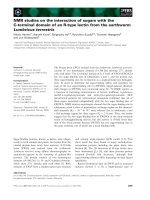
eosinophilic hyaline masses (Fig. 1). In some cases, the
necrotic foci contained extracellular and intracellular
spherical eosinophilic refractile bodies seen among the
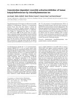
inflammatory cells (Russell's bodies) (Fig. 2). The hepatic
parenchyma also showed degeneration and necrosis in
other parts (paracentral necrosis). Nearly all the subcapsular hepatocytes appeared swollen degenerated and contained intranuclear inclusion bodies. The inclusion
bodies were confirmed by positive reaction to Phloxine-
Tartrazine stain. The detected inclusions sometimes
appeared rounded and surrounded with hallo zone in
degenerated nucleus (Fig. 3). The detected inclusion
appeared as one, two or three inclusions inside the nuclei
(Fig. 4). Some areas in the hepatic lobules showed disorganization of the hepatocytes in which the cells were not
arranged in cords. The hepatic cell plates have been
destroyed (lobular disarray) and the surviving hepatocytes were forming rounded hyperplastic nodules without
lobular arrangement. Apoptosis was observed near the
Figure
necrotic necrotic focus with Councilman-like bodies inside
showing kid in group-1, animal sacrificed
Liver of 1and degenerated hepatocytes one week P.V.,
Liver of kid in group-1, animal sacrificed one week
P.V., showing necrotic focus with Councilman-like
bodies inside necrotic and degenerated hepatocytes.
(H&E × 400).
Figure
nuclei the
and surrounded with hallo inside degenerated hepatocytes
showing kid intranuclear inclusion bodies appeared rounded
Liver of 3 in group-1, animal sacrificed one week P.V.,
Liver of kid in group-1, animal sacrificed one week
P.V., showing the intranuclear inclusion bodies
appeared rounded and surrounded with hallo inside
degenerated hepatocytes nuclei. (Phloxine-Tartrazine
stain × 1000).
Figure
bodies Councilman-like bodies (green one oesinophilic
masses kid
showing2 in group-1, animal sacrificed arrow) and Russell's
Liver ofsurrounded by hallo zoneappeared as week P.V.,
Liver of kid in group-1, animal sacrificed one week
P.V., showing Councilman-like bodies appeared as
oesinophilic masses surrounded by hallo zone (green
arrow) and Russell's bodies. (H&E × 1000).
Page 3 of 15
(page number not for citation purposes)
Virology Journal 2009, 6:94
/>
Figure the
two of kid group-1,
showing4 ininclusions animal sacrificed one week as one,
Liveror threeintranuclear inclusion bodies appearedP.V.,
Liver of kid in group-1, animal sacrificed one week
P.V., showing the intranuclear inclusion bodies
appeared as one, two or three inclusions. (PhloxineTartrazine stain × 1000).
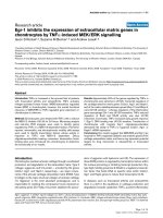
Figure megakaryocytes in
nuclei were
showing kid hyperchromaticthe liver parenchyma and their
Liver of 6 in group-1, animal sacrificed one week P.V.,
Liver of kid in group-1, animal sacrificed one week
P.V., showing megakaryocytes in the liver parenchyma and their nuclei were hyperchromatic. (H&E ×
400).
necrotic foci and nearly allover the entire hepatic lobules,
affecting the individual cells (Fig. 5). The endothelial lining of the hepatic sinusoids showing signs of necrosis and
degeneration & sinusoidal dilatation were observed. The
bile ducts showed hyperplastic proliferation of their epithelial lining. In the liver megakaryocytes were seen inside
hepatic sinusoids and their nuclei were hyperchromatic
accompanied by sinusoidal leuckocytosis (Fig. 6). The
lymph nodes showed hyperplastic activation of lymphocytes in the form of follicular and paracortical hyperplasia that manifested by numerous large lymphoblasts in
the paracortical zone with evidence of mitosis and presence of intranuclear inclusion bodies. The spleen also
exhibited lymphocytic activation of the white pulp. The
kidneys showed areas of necrosis. The proximal convoluted and distal tubules were suffering from degenerative
and necrotic changes. Intranuclear inclusion bodies were
demonstrated inside the tubular epithelium as demonstrated by phloxine-Tartrazine stain. The adrenal gland
showed hyperplastic activation in zona fasiculata (Fig. 7)
and some necrotic changes in the medulla were also seen.
Kid sacrificed two weeks postvaccination showed diffuse
vacuolar degeneration of hepatocytes (Fig. 8) with kupffer
cells activation. Minute necrotic foci were seen in the
hepatic parenchyma invaded by macrophage and lymphocytes. The endothelial lining of the hepatic sinusoids
showing signs of necrosis and degeneration & sinusoidal
dilatation were observed. Councilman's-like bodies were
Figure apoptosis affecting the individual cells
showing kid
Liver of 5 in group-1, animal sacrificed one week P.V.,
Liver of kid in group-1, animal sacrificed one week
P.V., showing apoptosis affecting the individual cells.
(H&E × 400).
Figure 7
lata
week P.V., showing kid in group-1, animal sacrificed fasicuThe adrenal gland ofhyperplastic proliferation in zona one
The adrenal gland of kid in group-1, animal sacrificed
one week P.V., showing hyperplastic proliferation in
zona fasiculata. (H&E × 400).
Page 4 of 15
(page number not for citation purposes)
Virology Journal 2009, 6:94
/>
Figure diffuse vacuolar degeneration of hepatocytes
showing kid
Liver of 8 in group-1, animal sacrificed two weeks P.V.,
Liver of kid in group-1, animal sacrificed two weeks
P.V., showing diffuse vacuolar degeneration of hepatocytes. (H&E × 400).
Figure blood vessels animal sacrificed two weeks P.V.,
showing kid
Brain of 10 in group-1,congested with perivascular oedema
Brain of kid in group-1, animal sacrificed two weeks
P.V., showing blood vessels congested with perivascular oedema. (H&E × 100).
seen in the degenerated hepatocytes. Intranuclear inclusion bodies were detected inside degenerated hepatocytes
adjacent to areas of coagulative necrosis. Hyperplasia of
the epithelial lining of the bile ducts and lymphocytic
infiltration were seen in the portal areas (Fig. 9). The
lymph nodes and spleen revealed lymphocytic depletion
and the kidneys exhibited necrotizing changes in the proximal and distal convoluted tubules and the adrenal glands
showed hyperplasia of zona fasciculata and degenerative
changes in the medulla. The brain blood vessels were congested and engorged with blood with perivascular
oedema (Fig. 10 &11). Neuronal degeneration and necrosis was seen accompanied by astrocytic oedema, microglial proliferation and neuronophagia. Kid sacrificed three
weeks postvaccination showed large necrotic foci with
destructed center surrounded by macrophages, lymphocytes and necrotic hepatocytes. Intracytoplasmic
inclusion-like bodies surrounded by hallo zone (Councilman's-like bodies) were seen in the degenerated hepatocytes. Near these necrotic foci, abnormal cellular growth
accompanied by dilated blood vessels and haemorrhages
were observed. The endothelial lining of the hepatic sinusoids showing signs of necrosis and degeneration & sinusoidal dilatation were observed. Severe hemorrhages were
seen in some areas of the hepatic lobules that suffer from
degeneration and necrosis. Sinusoidal dilatation and disconfiguration of the hepatic parenchyma was seen. Some
bile ducts showed hyperplasia of the epithelial lining.
Large number of mononuclear cells infiltration was
Figure hyperplasia of animal sacrificed two the bile duct
with of kid
showing9 lymphocytic infiltration
Liversevere in group-1, the epithelial lining of weeks P.V.,
Liver of kid in group-1, animal sacrificed two weeks
P.V., showing hyperplasia of the epithelial lining of
the bile duct with severe lymphocytic infiltration.
(H&E × 400).
Figure blood vessels animal sacrificed two weeks P.V.,
showing kid
Brain of 11 in group-1,congested with perivascular oedema
Brain of kid in group-1, animal sacrificed two weeks
P.V., showing blood vessels congested with perivascular oedema. (H&E × 400).
Page 5 of 15
(page number not for citation purposes)
Virology Journal 2009, 6:94
observed in some parts of hepatic tissue together with
necrosis and loss of cellular details that gave hepatocytes
washed out appearance (Fig. 12). Kid sacrificed four
weeks postvaccination showed severe centrolobular
hepatic necrosis (periacinar necrosis) and severe haemorrhages around the central veins with hemosiderin pigments deposition (Fig. 13). The subcapsular hepatic
parenchyma showed swollen hepatocytes with coagulative necrosis. The degenerated and swollen hepatocytes
contained numerous intracytoplasmic inclusion-like bodies that appeared as eosinophilic rounded and well-circumscribed masses of different sizes surrounded by hallo
zone (Councilman's-like bodies). The kupffer cells were
highly proliferated. Some hepatocytes undergo apoptosis
and appeared shrinked, differed in shape from the adjacent hepatocytes with condensed cytoplasm, detached
from other adjacent hepatocytes. Nuclear chromatin was
condensed, clumped and the apoptotic bodies were seen.
The endothelial lining of the hepatic sinusoids showing
signs of necrosis and degeneration & sinusoidal dilatation
were observed. Some bile ducts undergo massive destruction of epithelial lining and invaded by large numbers of
lymphocytes and macrophages. The myocardium showed
areas of hemorrhages around the dilated and necrotic
blood vessels. Zenker's necrosis and focal infiltration with
macrophages and lymphocytes were observed. The spleen
showed severe lymphocytic depletion. The kidneys
showed signs of necrosis in the renal corpuscles and the
renal tubular epithelium showed intranuclear inclusion
bodies surrounded by hallo zone. The adrenal glands
showed hyperplastic changes in zona fasciculata and
medulla. The brain blood vessels were slightly congested.
Group (2) Adults non pregnant
Doe sacrificed one week postvaccination showed diffuse
centrolobular coagulative necrosis of liver parenchyma
Figure with
out appearance)
together large group-1, animal sacrificed three weeks P.V.,
showing kid necrosis and loss of cellular details (washed
Liver of 12 in number of mononuclear cells infiltration
Liver of kid in group-1, animal sacrificed three weeks
P.V., showing large number of mononuclear cells
infiltration together with necrosis and loss of cellular
details (washed out appearance). (H&E × 200).
/>
Figure centrolobular with brownish haemosiderin pigment
deposition
around kid
showing13 in group-1, animal necrosis four weeks P.V.,
Liver ofthe central veinhepatic sacrificedand haemorrhage
Liver of kid in group-1, animal sacrificed four weeks
P.V., showing centrolobular hepatic necrosis and
haemorrhage around the central vein with brownish
haemosiderin pigment deposition. (H&E × 400).
(periacinar). Sinusoidal dilatation that causes disconfiguration of the liver parenchyma and thrombus formation
in the hepatic artery were also demonstrated. The hepatic
cells under the liver capsule were swollen and undergo
degeneration and some of them undergo necrotic changes
(Fig. 14). Necrotic foci were found in some hepatic lobules near the central veins (midzonal). The degenerated
hepatocytes that found adjacent to the necrotic areas contained eosinophilic intranuclear inclusion bodies and
their cytoplasm contained fine granules which stained
positive with Phloxine-Tartrazine stain. The swollen and
degenerated hepatocytes contained intracytoplasmic bodies surrounded by a hallo zone (Councilman's-like bodies) (Fig. 15). Haemorrhages were detected in areas
around the central veins. Some bile ducts were infiltrated
with lymphocytes and others were hyperplastic and its
Figure adult
P.V., of 14 goat in group-2, animal sacrificed
Livershowing subcapsular hepatic degeneration one week
Liver of adult goat in group-2, animal sacrificed one
week P.V., showing subcapsular hepatic degeneration. (H&E × 200).
Page 6 of 15
(page number not for citation purposes)
Virology Journal 2009, 6:94
epithelium was elongated and branched inside the lumen.
The endothelial lining of the hepatic sinusoids showing
signs of necrosis and degeneration & sinusoidal dilatation
were observed. The lymph nodes appeared to be hyperplastic and the lymphoid follicles revealed activation that
manifested by large lymphoblast suffering from mitosis
especially in paracortical area (paracortical hyperplasia).
The spleen also revealed hyperplastic white pulp. The kidneys showed nephrosis of the renal tubules. The lining
epithelium of proximal tubules showed pyknotic and
lysed nuclei beside the degenerative and necrotic changes
that observed in the adjacent tissue. Intranuclear oesinophilic inclusion bodies were demonstrated inside the
tubular epithelium as demonstrated by Phloxine-Tartrazine stain. The adrenal glands were hyperplastic particularly in zona fasciculata with some necrotic changes in the
medulla. Doe sacrificed two weeks postvaccination
showed that the hepatic lesions were centrolobular coagulative necrosis (periacinar), preceded by granularity of
the hepatocytes cytoplasm. Areas of haemorrhages
around the central veins were noticed. Kupffer cells were
seen engulfing hemosiderin pigments. Moderate number
of apoptotic cells were seen allover the hepatic lobule. The
endothelial lining of the hepatic sinusoids showing signs
of necrosis and degeneration & sinusoidal dilatation were
observed. Disconfiguration of the hepatic parenchyma
was seen in some areas together with macrophage aggregations around necrotic hepatocytes (Fig. 16). Pyknotic
nuclei of hepatic cells with karyorrhexis and karyolysis
were also seen in the necrotic areas around the central
veins. The hepatocytes that lying under the hepatic capsule were swollen and contained eosinophilic intracytoplasmic bodies of different sizes and was surrounded by
hallo zone (Councilman's-like bodies). Some bile ducts
mic inclusion-like in hepatocytes contained intracytoplasP.V.,bodies) goatbodies surrounded sacrificed (CouncilmanLivershowing swollengroup-2, animal by a hallo one week
Figure adult
like of 15
Liver of adult goat in group-2, animal sacrificed one
week P.V., showing swollen hepatocytes contained
intracytoplasmic inclusion-like bodies surrounded by
a hallo (Councilman-like bodies). (H&E × 1000).
/>
Figure adult
around 16 goat in necrosis
hepatic parenchyma group-2, and macrophages aggregation
P.V., of necrotic hepatocytes animal sacrificed two weeks
Livershowing hepatictogether with disconfiguration of the
Liver of adult goat in group-2, animal sacrificed two
weeks P.V., showing hepatic necrosis and disconfiguration of the hepatic parenchyma together with macrophages aggregation around necrotic hepatocytes.
(H&E × 1000).
showed necrotic epithelium and others showed hyperplastic overgrowth with vesicular elongated epithelium
and lymphocytic infiltration. The myocardium showed
areas of necrosis (Zenker's necrosis) accompanied by
haemorrhages and lymphocytic infiltration. Depletion of
lymphocytes from the spleen and lymph nodes associated
with subcapsular macrophages in lymph nodes. The kidneys showed necrosis in the renal corpuscles and tubules.
The adrenal glands showed hyperplasia in the cortex and
necrosis in the medulla. The brain showed perivascular
and astrocytic edema, focal gliosis and lymphocytic infiltration in the Virchow-Robin spaces (cuffing), microglial
proliferation and some neurons were necrotic and
invaded by microglia (neuronophagia). Doe sacrificed
three weeks postvaccination showed centrolobular
hepatic necrosis (periacinar necrosis). The haemorrhages
were seen near the necrotic areas with discontinued blood
vessels (central veins). Apoptotic cells were seen in the
hepatic parenchyma around and inside the necrotic areas.
The bile ducts showed macrophages and lymphocytes
around it. Some bile ducts showed severe hyperplastic
proliferation. The myocardium, spleen, lymph nodes, kidneys, adrenal glands and brain showed similar but more
severe necrotic changes than that mentioned before at two
weeks postvaccination. Doe sacrificed four weeks postvaccination showed more severe destruction of the hepatic
parenchyma and necrosis accompanied by lymphocytic
infiltrations (Fig. 17). Almost all the hepatic cells were
swollen and contained intracytoplasmic bodies (councilman's-like bodies). Some areas in the hepatic lobules were
undergoing vacoulation and necrosis (Fig. 18). The
necrotic areas at this stage appeared with large number of
macrophages and lymphocytes (Fig. 19). The bile ducts
Page 7 of 15
(page number not for citation purposes)
Virology Journal 2009, 6:94
/>
Figure
areas adult
P.V., of 17 goat in group-2, animal sacrificed four weeks
Livershowing lymphocytic aggregations around the portal
Liver of adult goat in group-2, animal sacrificed four
weeks P.V., showing lymphocytic aggregations
around the portal areas. (H&E × 100).
Figure adult
lymphocytes
P.V., of 19 goat in group-2, animal sacrificed four weeks
Livershowing necrotic area invaded with macrophages and
Liver of adult goat in group-2, animal sacrificed four
weeks P.V., showing necrotic area invaded with macrophages and lymphocytes. (H&E × 1000).
were massively destructed and some of them showed
severe hyperplasia.
the peripheral cells contained Councilman's like bodies
inside cytoplasm also intranuclear inclusion could be
seen. Extravasated RBCs aggregated around central veins
and portal areas were heavly infiltrated with macrophages
and lymphocytes (Fig. 22). Thrombus was found inside
another cenral vein and was infiltrated by lymphocytes.
Kupffer cells proliferation and vacular degeneration were
also seen (Fig. 23). The lymph nodes and spleen were
depleted from mature lymphocytes with necrosis. The
uteri showed necrotic endometrial lining with areas of
necrosis in the tunica muscularis and lymphocytic infiltration (Fig. 24). The endometrial blood vessels showed
necrotic endothelial lining accompanied by areas of
haemorrhages. The renal tubules showed degeneration
The myocardium showed Zenker's necrosis with severe
lymphocytic infiltration and its blood vessels endothelium showed necrosis and discontinuation accompanied
by areas of haemorrhages (Fig. 20). The spleen, lymph
nodes, kidneys, adrenal glands and brain showed more
exaggerated changes similar to those observed at three
weeks postvaccination.
Group (3) Pregnant Does
The hepatic lesions in this group were quite similar and
characterized by periacinar necrosis (Fig. 21). The hepatocytes around central veins were completely necrotized and
Figure adult
P.V., of 18 goat in group-2, vacuolation and necrosis
Livershowing hepatocytes withanimal sacrificed four weeks
Liver of adult goat in group-2, animal sacrificed four
weeks P.V., showing hepatocytes with vacuolation
and necrosis. (H&E × 400).
Figure 20
with few lymphocytic infiltrations
weeks P.V., of adult goat in group-2, animal sacrificed four
Myocardiumshowing congestion of myocardial blood vessels
Myocardium of adult goat in group-2, animal sacrificed four weeks P.V., showing congestion of myocardial blood vessels with few lymphocytic infiltrations.
(H&E × 200).
Page 8 of 15
(page number not for citation purposes)
Virology Journal 2009, 6:94
/>
Figure aborted doe in
area of 21
P.V., showing periportal focal necrosis with invasion of portal
Liverwith lymphocytes group-3, animal sacrificed 10 days
Liver of aborted doe in group-3, animal sacrificed 10
days P.V., showing periportal focal necrosis with invasion of portal area with lymphocytes. (H&E × 400).
Figure
eration aborted doe cells proliferation and vacuolar degenP.V., of 23
Livershowing Kupfferin group-3, animal sacrificed 28 days
Liver of aborted doe in group-3, animal sacrificed 28
days P.V., showing Kupffer cells proliferation and vacuolar degeneration. (H&E × 400).
and necrosis. The adrenal glands showed necrotic cells in
the medulla and hyperplasia in zona fasciculata. The
brain showed more severe necrotic changes in the neurons
with microgliosis and lymphocytic infiltration in the Virchow-Robin spaces. Astrocytic and perivascular oedema
were also seen. The aborted and born foeti were showed
severe hepatic necrosis (pan-necrosis) accompanied by
lymphocytic infiltration. The swollen and degenerated
hepatocytes contained intracytoplasmic bodies surrounded by a hallo zone (Councilman's-like bodies) and
also some hepatocytes contained inclusion bodies confirmed by Phloxine-Tartrazine stain. The renal tubules
were degenerated and some times appeared necrosed. The
brain showed meningoencephalitis, oedema and gliosis
(Fig. 25).
Figure aborted
haemosiderin doe in inside infiltrated macrophages
P.V., of 22 depositionnecrosis and haemorrhages days
Liver showing periacinar group-3, animal sacrificed 10with
Liver of aborted doe in group-3, animal sacrificed 10
days P.V., showing periacinar necrosis and haemorrhages with haemosiderin deposition inside infiltrated macrophages. (H&E × 200).
Figure and
necrosis24 the doe in endometrial lining with
P.V., showinglymphocytic infiltration in the tunicaeareas of
Uterus of abortednecroticgroup-3, animal sacrificed 10 days
Uterus of aborted doe in group-3, animal sacrificed
10 days P.V., showing the necrotic endometrial lining
with areas of necrosis and lymphocytic infiltration in
the tunicae. (H&E × 400).
4-Results of electron microscopic studies
The hepatic cells in young vaccinated kids (group no.1)
showed indentation of the nuclear membrane and margination and disintegration of the chromatin. The hepatic
cells of vaccinated adult goats (group no.2) showed swollen mitochondria and destructed cytoplasm, (Fig. 26).
Some hepatic cells of vaccinated pregnant does (group
no.3) revealed condensed chromatin on the nuclear
membranes and others revealed concentrated chromatin
inside the nucleus accompanied by destructed cytoplasmic organelles. In vaccinated adult goat (group no.2) the
Page 9 of 15
(page number not for citation purposes)
Virology Journal 2009, 6:94
/>
Figure
gliosis aborted foetus in group-3, 28 days P.V., showing
Brain of 25
Brain of aborted foetus in group-3, 28 days P.V.,
showing gliosis. (H&E × 200).
Figure hepatocytes with strong positive fluorescent stain
showing kid
Liver of 27 in group-1, animal sacrificed one week P.V.,
Liver of kid in group-1, animal sacrificed one week
P.V., showing hepatocytes with strong positive fluorescent stain. (IFA & Evan's blue × 400).
proximal convoluted tubules showed necrotic and
destructed nucleus with lysed nuclear membrane, fragmented chromatin and lysed cytoplasmic organelles. The
microvilli were short and necrotic.
positive fluorescing reactions were detected inside white
blood cells in central vein and in the areas of haemorrhages around this vein (Fig. 30). The proliferated kupffer
cells also gave strong and characteristic fluorescing reaction, (Fig. 31). The myocardium in control group gave
negative results by showing only the Evan's blue stain
reaction. Strong positive fluorescing reactions were
detected in the myocardium.
4-Immunofluorescent microscopic results
The liver in control group gave negative results by showing only the Evan's blue stain reaction. The liver in all vaccinated groups gave positive reaction (Fig. 27). The bile
ducts gave strong and characteristic reaction as the antigen
appears inside the cytoplasm of its epithelium in all bile
ducts (Fig. 28) and also the viral antigen was also detected
in the endothelium of the blood vessels (Fig. 29). Strong
Figure and
chondriaadult goat in group-2, swollen cytoplasm
P.V., of 26 hepatocyte with animal sacrificed one week
Livershowing viral particles inside their and destructed mitoLiver of adult goat in group-2, animal sacrificed one
week P.V., showing hepatocyte with swollen and
destructed mitochondria and viral particles inside
their cytoplasm. (E.M. × 8000).
5-Agar gel precipitation test (AGPT)
The detection of RVF virus antigen in organs of vaccinated
goats with the live attenuated RVF vaccine by agar gel pre-
Figure epithelium (intracytoplasmic)
bile duct bile
showing kid duct with animal sacrificed one week P.V.,
Liver of 28 in group-1,strong positive fluorescent stain in the
Liver of kid in group-1, animal sacrificed one week
P.V., showing bile duct with strong positive fluorescent stain in the bile duct epithelium (intracytoplasmic). (IFA & Evan's blue × 400).
Page 10 of 15
(page number not for citation purposes)
Virology Journal 2009, 6:94
/>
Figure central vein of the white blood fluorescing reaction
inside the
showing kid in group-1, animal sacrificed one
Liver of 30cytoplasm with strong positivecells week P.V.,
Liver of kid in group-1, animal sacrificed one week
P.V., showing central vein with strong positive fluorescing reaction inside the cytoplasm of the white
blood cells. (IFA & Evan's blue × 400).
Group-(3)
Adult pregnant does: Gave positive results from 3rd day
P.V. and and reached its highest level at 20th day P.V.
(0.820) and persist at a high level till the end of the experiment (55 days P.V.).
Discussion
Figure adult
with of 29 goat in group-2, reaction
P.V., showing hepatocytes and animal sacrificed two vessels
Liverstrong positive fluorescingendothelium of bloodweeks
Liver of adult goat in group-2, animal sacrificed two
weeks P.V., showing hepatocytes and endothelium of
blood vessels with strong positive fluorescing reaction. (IFA × 400).
In the present work we tested the live attenuated RVF vaccine (Smithburn strain) in kids, adult and pregnant goats
to investigate the adverse effects induced by the vaccine.
Abortions and parturition of dead foeti were additional
signs in group three and occurred after sudden and sharp
cipitation test (AGPT) on organ homogenates (liver,
spleen, kidneys, brain, myocardium and lymph nodes)
had given positive reaction in groups 1 and 2 at 1st, 2nd
and 3rd days and 4th weeks P.V. and in group 3 (pregnant
does) at abortion and parturition either from does or dead
or stillbirth foeti.
6-ELISA
Group-(1)
Kids: Gave positive results from fourth day postvaccination and reached its highest level at 15th day P.V. (0.782)
and persist at a high level till the end of the experiment (4
weeks P.V.).
Group-(2)
Adult does: Gave positive results from third day postvaccination and reached its highest level at 20th day P.V.
(1.006) and persist at a high level till the end of the experiment (4 weeks P.V.).
Figure adult
kupffer 31
P.V., of cells goat in group-2, animal sacrificed two the
Livershowing strong positive fluorescing reaction in weeks
Liver of adult goat in group-2, animal sacrificed two
weeks P.V., showing strong positive fluorescing reaction in the kupffer cells. (IFA × 400).
Page 11 of 15
(page number not for citation purposes)
Virology Journal 2009, 6:94
rise in body temperature (41°C). The abortion takes place
in two pregnant does out of three. The 1st one was aborted
after 10 days postvaccination and the other pregnant one
was aborted after 28 days postvaccination. In this concern,
[18] mentioned that during the epidemic of Rift Valley
Fever that broke out in Egypt in 1977, abortions occurred
at any stage of pregnancy after a sharp rise of body temperature accompanied by bloodstained nasal discharge. [19]
Mentioned that the inoculation of RVF virus in pregnant
ewes resulted in four abortions after 4–12 days with
retained placenta.
The results suggested that the causes of abortions and parturition of dead foeti were primarily the direct effect of
RVF virus on the genital organs of the pregnant does by
causing necrotic changes in the uteri and secondary due to
death of the foeti resulted from the infection of the foeti
by the RVF virus and propagation of the virus inside the
liver of the foeti (RVF antigens in the livers of the aborted
and dead foeti were detected by immunoflurescent technique).
The clinical pathological result revealed normocytic normochromic anaemia. [20] Reported that RVF infection in
sheep resulted in a significant decrease in RBCs at 1 to 7
days postinfection. [21] Found that the experimental
infection of RVF virus in sheep, caused fall of RBCs during
the course of the disease.
However, from histopathological point the anaemia was
haemorrhagic and this was indicated by widespread
haemorrhages in most organs (liver, kidneys, epicardium,
endocardium and brain). This haemorrhage may be
attributed to three reasons; vascular damage which confirmed by viral antigen in endothelium lining of blood
vessels by immunofluorescing staining or; consumption
of clotting factors which indicated by massive hepatic
necrosis of all vaccinated animals or; thrombocytopenia
which may be occurred due to adherence of thrombocytes
on damaged endothelium blood vessels and this was
proved by extramedullary haematopoiesis in which megakaryocytes observed in hepatic sinusoids, lymph nodes
and splenic sinusoids [22].
The clinical results also revealed a significant increase in
TLC in the 1st three days in all vaccinated groups and this
was resulted from neutrophilia and lymphocytosis and
such finding could be attributed to the ability of vaccine
to evoke the immune response of animals by stimulation
and activation of bone marrow for neutrophilia and
lymph nodes for lymphocytosis and this was confirmed
histologically by follicular and paracortical hyperplasia of
lymph nodes.
On the other hand, decrease in TLC was observed by 2nd
week P.V. and this leucopenia was resulted either from
/>
neutropenia due to bone marrow affection or from lymphopenia which may be resulted from lymphocytic depletion of lymph nodes and spleen. Our results were
concomitant with [23] who mentioned that leucopenia
and lymphopenia resulted in case of RVF virus infection
while leucocytosis and lymphocytosis resulted from vaccination. Similar findings were observed by [24] and [21].
The results revealed prolongation of the blood clotting
time in all vaccinated animals and the highest level of
delayed clotting time was observed in the pregnant does
(7.2 minutes) at 7th day postvaccination. Our findings
were in agreement with [25] who mentioned that one of
the main functions that have been deteriorated by RVF
virus infection was the coagulation factors of the blood as
demonstrated from the significant prolongation of the
clotting time. [26] Reported that clotting factors produced
by the hepatocytes include fibrinogen, prothrombin and
other factors. Consequently, decreased functional hepatic
mass may result in a diminishing of clotting factor activity
in proportion to the degree of hepatocellular damage,
leading to prolonged coagulation times and possible
bleeding tendencies. Loss of greater than 70 to 80% of the
functional hepatic mass is considered sufficient to cause a
clinical coagulopathy.
The results also revealed that there were increases in levels
of AST, ALT and ALP in all vaccinated animals. This may
resulted from hepatic damage and this was in agreement
with [27] who stated that natural infection of RVF virus in
sheep caused a significant increase in level of AST and ALT
due to liver necrosis caused by RVF virus.
Liver was the most affected organ in all vaccinated groups.
The lesions were more obvious and severe in kids than
adults. This was in accordance with [28] who stated that
the hepatic necrosis in older animals was slightly less
extensive than young kids. There were different types of
necrosis, periacinar, midzonal, paracentral or even massive necrosis (pan-necrosis) of most hepatocytes. The
necrosis could be attributed to either cytotoxic effect of
virus or ischemia that resulted from hepatic haemorrhages
due to the damage of the endothelial lining of hepatic
sinusoids. However, [29] mentioned that the frequent
paracentral location of the liver lesions is suggesting the
association of the changes with anoxia. This finding was
in agreement with [30] who mentioned that in livers of
lambs infected with Rift Valley Fever virus naturally,
showed scattered grayish-white foci of 1–2 mm in diameter and hemorrhages of varying sizes were seen throughout.
Intranuclear inclusion bodies were detected inside the
cells of liver and kidneys of all the vaccinated kids, adults
and pregnant does. These inclusions were differed in
shape and in some cells the chromatin of the affected
Page 12 of 15
(page number not for citation purposes)
Virology Journal 2009, 6:94
nuclei were marginated and disintegrated as shown by the
electron microscope examination. The intranuclear inclusions were eosinophilic by Phloxine-Tartrazine stain and
surrounded by a hallo and founded inside cells which suffering degeneration and necrosis and located inside or
near the necrotic areas in the hepatocytes and the epithelial lining of the renal tubules. Our findings were in agreement with [22]. However, [7] mentioned that viruses of
Bunyaviridae family encode their structural proteins but
moreover Phlepoviruses also encode a nonstructural protein (NSs) in the viral RNA (vRNA) of their S segment. For
RVF virus, the NSs proteins were reported to be phosphorylated and to accumulate in the nuclei of the infected
cells. Studies concerning genomic replication indicated
that continous proteins synthesis was required for replication of the RVF viral genome and these proteins were of
viral origin. For the viruses in the family Bunyaviridae the
structural protein, nucleocapsid protein (N), would function to regulate replication and would be tempered by the
presence of nonstructural proteins (NSs), where they
exist.
Recently, [31] mentioned that a better understanding of
the factors that govern RVFV virulence and pathogenicity
is required, given the urgent need for antiviral therapies
and safe vaccines. NSs with anti-IFN activity accumulated
in the nucleus. IFN synthesis is regulated by specific transcription factors, including interferon regulatory factor
(IRN-3), NF-kappaB, and AP-1. In the presence of NSs,
IRF-3 was still activated and moved to the nucleus. So,
these results suggest that NSs, unlike other viral IFN antagonists, does not inhibit IFN-specific transcription factors
but blocks IFN gene expression at a subsequent step.
The Councilman's-like bodies were seen inside the cytoplasm of swollen, degenerated and necrotic hepatocytes
in all vaccinated animals and in the aborted foeti and
dead foeti as well. The bodies appeared as intracytoplasmic eosinophilic masses surrounded by hallo zone.
However, in this concern [32] gave description to the
Councilman's bodies as small, hyalinous, round or oval
eosinophilic inclusions in the cytoplasm of hepatic cells
infected with Yellow fever virus and suggested that these
bodies represent necrosis around viral particles. He added
that these bodies might also see in other form of toxic or
viral hepatitis. [29] Mentioned that the affected liver cells
with RVF virus were swollen, eosinophilic, hyaline cytoplasm, and pyknotic or fragmented nuclei, their appearance suggesting the Councilman's bodies of yellow fever.
Recently, [22] stated that Councilman's-like bodies were
seen in some viral infections and may be due to condensation of cytoplasmic organelles and squestrated from
remaining cytoplasm by membranes that fuse with lyso-
/>
somes (autolysosomes) and may also be derived from
other hepatocytes that suffered from apoptosis (apoptotic
bodies) and engulfed by remaining hepatocytes.
In the present work, we observed large numbers of apoptotic cells in liver of vaccinated animals by RVF live attenuated vaccine. Our findings are in concurrence with [33]
who mentioned that viral infection can induce PCD in
various host target cells. Also we agree with [22] who
mentioned that RVF virus increases the apoptotic hepatic
cells. These findings are in concurrence with [34] who
mentioned that Bunyamwera virus nonstructural protein
(NSs) counteracts interferon regulatory factor 3 (IRF-3),
so leads to the induction of early cell death (PCD).
Russell's bodies were seen inside the necrotic foci in the
form of intracellular and extracellular spherical strongly
eosinophilic bodies in some cases of group-1 (kids). However, [35] described the so called Russell's bodies as small,
spherical hyaline bodies in cancerous and simple inflammatory growth and in degenerating plasma cells. Then,
[36] discussed the various theories of Russell's bodies origin. The possibilities included, origin from the lymphocytes, origin in plasma cells with later degeneration,
origin from mitochondria of cells, and even an origin
from a red blood cell swallowed up by a plasma cell. These
bodies were seen within the tissue cells (intracellular) and
outside the cells (extracellular). The size of Russell's bodies ranged from barely visible up to "half again" as large as
red blood corpuscles. The largest round forms were easily
seen microscopically. Recently, [37] mentioned that Russell bodies (RB) are dilated ER cisternae containing condensed immunoglobulins (Ig). As to their biogenesis, it
was shown that the synthesis of a mutated Ig, which is neither secreted nor degraded, is sufficient to induce RB formation in cells of different species and histotype. Many
disease-linked cases of intralumenal protein accumulation have been described including thyrocytes of congenital goiter patients and hepatocytes of individuals carrying
mutated α1 anti-trypsin alleles (PiZ). The correlation
between liver disease and PiZ lumenal depositions indicates that the latter can be harmful for cells, but the exact
mechanisms that lead to toxicity are not known.
Electron microscopic examination revealed ultrastructure
changes in liver and kidneys and viral particles inside
hepatocytes cytoplasm. The hepatic cells of vaccinated
goats with live attenuated RVF vaccine showed cell with
indentation of the nuclear membrane, margination and
disintegration of the chromatin. The mitochondria were
swollen. Some hepatocytes reveals condensed chromatin
on the borders of the nuclei and others revealed more
chromatin inside the nuclei. In kidneys the proximal convoluted tubules epithelial cells showed necrotic and fragmented brush borders with signs of necrosis and lyses in
Page 13 of 15
(page number not for citation purposes)
Virology Journal 2009, 6:94
/>
their nuclei and other organelles. The distal convoluted
tubules showed necrotic microvilli with necrotic and
destructed nuclei and lyses of other cell organelles.
cine (Smithburn's strain) for control RVF virus is not
advisable in Egypt as it considered an endemic area for
RVF virus.
Our findings agree with [38] who mentioned that in liver
of newborn lamb infected with RVF virus showed
ultrastructural changes. Hepatocytes were primarily
affected, while inflammatory and structural changes were
secondary. [39] Mentioned that the characteristic lesions
of RVF were in the liver. Margination of the chromatin in
a beaded form or its accumulation in clumps on the
nuclear membranes was also observed. In this study we
detected the RVF virus antigen in tissues by using the
immunoflourescent technique (IFAT) and by agar gel precipitation test (AGPT). The IFAT gave strong positive reactions in organs of all vaccinated animals especially in the
liver. The hepatocytes, especially those adjacent to the
necrotic foci and to the bile ducts and adjacent to the
blood vessels were the most affected and gave the strong
fluorescent stain. It was a surprise to found that antigen of
RVF virus was seen inside the cytoplasm of the epithelial
cells of the bile ducts, nearly in all the examined specimens. These findings suggesting that the RVF virus affects
the bile ducts in strong manner and propagates inside its
epithelium. However, [40] mentioned that the RVF viral
antigen was studied by immunohistochemistry in the
liver and was most prominent in the liver and detected in
the cytoplasm of hepatocytes which were sparsely scattered throughout the lobules. [41] Mentioned that RVF
virus antigen was detected by immunohistochemical
analysis in the liver, where positive staining was localized
in coalescing foci of hepatocellular necrosis.
Competing interests
The author declares that they have no competing interests.
Authors' contributions
SAK conceived of the study, and participated in its design
and coordination. The author read and approved the final
manuscript.
References
1.
2.
3.
4.
5.
6.
7.
8.
9.
10.
From this point of view, RVF virus (Smithburn's strain)
could be dessiminated from the vaccinated animals as a
result of its propagation and circulation in the blood of
the vaccinated animals and resulted in spreading of the
RVF virus in the adjacent environment and infected other
susceptible animals and even man. These side effects
became more dangerous especially in endemic areas as
mentioned by [42] who found that genetic exchange
occurred between strains from different lineages of RVF
virus and this hypothesis emphasizes the risk of generating uncontrolled chimeric viruses by using the live attenuated vaccines in areas of endemicity.
It could be concluded that the use of live attenuated vaccine of RVF (Smithburn strain) for immunization of live
stock in Egypt is danger. As the use of live attenuated vaccine of RVF (Smithburn strain) could result in the spreading of RVF virus instead of its eradication. In the present
work, the results indicate that this vaccine cause severe
deleterious changes in various organs and propagated
inside hepatic cells in a manner similar to the infection by
the virulent RVF virus. So, the use of live attenuated vac-
11.
12.
13.
14.
15.
16.
17.
18.
19.
20.
21.
Linthicum KJ, Davies FG, Kairo A: Rift Valley Fever virus (family
Bunyaviridae, genus Phlebovirus). Isolation from Diptera
collected during an inter-epizootic period in Kenya. Journal of
Hygiene 1985, 95(1):197-209.
Daubney R, Hudson JR, Graham PC: RVF or Enzootic hepatitis.
An undescribed virus disease of sheep, cattle and man from
East Africa. J Path Bact 1931, 34:545-579.
Darwish MAA, Imam ZE, Omar F, Hoogstraal M: A seroepidemiological survey in man and domestic animals in Egypt. J Egyp
Pub Health Ass 1977, 3:156-163.
Calisher C: Classification and nomenclature of viruses, fifth
Ed. Report of the International Committee on Taxonomy of
viruses. Arch Virol 1991:273-283.
Meegan JM, Khalil GM, Hoogstrall H, Adham FK: Experimental
transmission and field isolation studies implicating Culex
pipens as a vector of RVF in Egypt. Am J Trop Med Hyg 1980,
29(6):1405-1410.
Smith HA, Jones JC, Hunt RD: Veterinary Pathology. In Text Book
2nd edition. Lea & Febiger, Philadelphia, USA; 1972.
Schmaljohn CS: Fundamental Virology. 3rd edition. B.N. Fields
and lippincott. Raven publishers. Philadelphia, USA; 1996.
Coatzer JAW, Ishak KG: Sequential development of the liver
lesions in new born lambs infected with RVF virus. Onderstepoort J Vet Res 1982, 49(2):103-108.
Schalm OW, Jain NC, Carroll EJ: Veterinary Hematology. 3rd
edition. Lea & Febiger, Philadelphia, USA; 1975.
Wintrobe MM: Colorimetric Determination of Haemoglobin.
1st ed. Clinical Chemistry 1965, 5:719.
Lee RI, White PD: A clinical study of coagulation time of blood.
Amer J Med Sc 1913, 145:495.
Rej R, Horder M: Aspartate aminotransferase and Alanine
aminotransferase activities. In Methods Of Enzymatic Analysis Volume 3. 3rd edition. Edited by: Bergmeyer HU, Grassl M. Weinheim,
Verlag-Chem; 1983:416-433. 444–456
King PR, King EG: Calorimetric determination of alkaline phosphatase activity. J Clin Path 1954, 7:322.
Payment P, Trudal M: Methods and Techniques in Virology. In
Text Book Marcel Dekker, Inc. New York, USA; 1993.
Mansi W: Slide gel diffusion precipitin. Nature (London) 1958,
181:1289-1290.
Lillie RD, Fullmer HM: Histopathology Technique And Practical
Histochemistry. 4th edition. McGrew Hill Book Company, NY.,
USA; 1976.
Hayat MA: Basic Technics for Transmission Electron Microscopy. Academic Press Inc., Orlando, Florida; 1986.
Abdel-Ghaffar S, Ayoub NNK, El-Nimr M, Nafeh EK, Malik SK: Rift
Valley Fever in Egypt. Bull. De L'Office International des Epizootices
1980, 92:7-8.
Wasel MS, Ateia M, Mohsen A, El-Sayed M, Zaki K: Effect of RVF
virus on pregnant local Barki ewes after artificial infection.
Vet Bull 1990, 60(1):21-22.
Shehta MM: Investigation of blood changes in Rift Valley Fever
in Egypt. MV Sc Thesis (Microbiology) College Vet Med Cairo Univ 1982.
Olaleye OD, Tomori O, Fajimi JL, Schmitz H: Experimental infection of three Nigerian breeds of sheep with the Zinga strain
of the Rift Valley Fever virus. Rev Elev Med Vet Pays Trop 1996,
49(1):6-16.
Page 14 of 15
(page number not for citation purposes)
Virology Journal 2009, 6:94
22.
23.
24.
25.
26.
27.
28.
29.
30.
31.
32.
33.
34.
35.
36.
37.
38.
39.
40.
41.
42.
Jubb KVF, Kennedy PG, Palmer N: Pathology Of Domestic Animals. 2nd edition. Academic Press Orlando Florida; 1994.
Embert HC: Leucocytes: Veterinary Clinical Pathology. 2nd
edition. London, Academic Press; 1974:55-81.
Mouaz MA, Gihan KM, Khirat AM, Aida ED, Khodeir MH: Evaluation of immune response in Egyptian Balady sheep vaccinated with attenuated RVF and PBR vaccines. Assiut Vet Med J
1998, 38(76):329-341.
Agag AEM: Morphopathological and Clinicopathological studies on goats following experimental infection with the RVF
virus. MVSc Thesis, Microbiology, Fac Vet Med Cairo University 1983.
Prater MR: Acquired Coagulopathy II;Liver Disease. In Chalm's
Veterinary Hematology 4th edition. Lippincott Williams & Wilkins, USA;
2000.
Abou-Zaid AA, Nakashly SA, Nasr MY, Mokhabatly A: Studies on
RVF in sheep. J Egypt Vet Med Associ 1995, 55(1):339-350.
Hubbard KA, Baskerville A, Stephanson JR: Ability of a mutagenized virus variant to protect young lambs from RVF. Amer
J Vet Res 1991, 52(1):50-55.
Jones TC, Hunt RD, King NW: Veterinary Pathology. In Text
Book, Library of Congress 6th edition. Williams and Wilkins Baltimore,
Maryland, USA; 1997.
Coatzer JAW: The pathology of Rift Valley Fever 1. Lesions
occurring in natural cases in new-born lambs. Onderstepoort
Journal of Veterinary Research 1977, 44:205-212.
Billecocq A, Spiegel M, Vialat P, Kohl A, Weber F, Bouloy M, Haller
O: NSs Protein of Rift Valley Fever Virus Blocks Interferon
Production by Inhibiting Host Gene Transcription. J Virol
2004, 78(18):9798-806.
Councilman WT: Diseases and Its Causes. 1st edition. H Holt &
Co. New York; 1913.
Caillet FP, Perros M, Brandenburger A: Programmed killing of
human cells by means of an inducible clone of parvoviral
genes encoding nonstructural proteins.
Embo J 1990,
9:2989-2995.
Kohl A, Reginald FC, Freidemann W, Anne B, Richard ER, Richard ME:
Bunyamwera Virus Nonstructural Protein NSs Counteracts
Interferon Regulatory Factor 3-Mediated Induction of Early
Cell Death. Journal of Virology 2003, 77(14):7999-8008.
Russel W: An address on a characteristic organism of cancer.
British Medical Journal, London 1890, 2:1356-1360.
Schleicher EM: Giant Russell's bodies in neoplastic cells in a
case of leukemic lymphosarcomatosis. Minnesota Medicine
1965, 48:1125-1130.
Kopito RR, Sitia R: Aggresomes and Russell bodies, symptoms
of cellular indigestion. EMBO Reports 2000, 1(3):225-231.
Coatzer JAW, Ishak KG, Calvert RC: Sequential development of
the liver lesions in new born lambs infected with RVF virus.
II. Ultrastructural findings. Onderstepoort J of Vet Res 1982,
49:109-122.
Mahmoud AZ, Youssef MS, Ibrahim MK: Pathological Studies on
Some Liver Affection In Camel: II-Viral Hepatitis. Egypt J
Comp Path Clin Path 1989, 2(2):27-32.
Lugt JJ, Coatzer JAW, Smith MME: Distribution of viral antigen in
tissues of newborn lambs infected with RVF virus. Onderstepoort journal of Veterinary Research 1996, 63(4):341-347.
Rippy MK, Topper MJ, Mebus CA, Morril JC: RVF virus-induced
encephalomyelitis and hepatitis in calves. Veterinary Pathology
1992, 29(6):495-502.
Sall AA, Zanotto PM, Sene OK, Zeller HG, Digoutte JP, Thiongane Y:
Genetic reassortment of Rift Valley Fever virus in nature. J
Virol 1999, 73:8196-2000.
/>
Publish with Bio Med Central and every
scientist can read your work free of charge
"BioMed Central will be the most significant development for
disseminating the results of biomedical researc h in our lifetime."
Sir Paul Nurse, Cancer Research UK
Your research papers will be:
available free of charge to the entire biomedical community
peer reviewed and published immediately upon acceptance
cited in PubMed and archived on PubMed Central
yours — you keep the copyright
BioMedcentral
Submit your manuscript here:
/>
Page 15 of 15
(page number not for citation purposes)