Báo cáo khoa học: "A rat model of picornavirus-induced airway infection and inflammation" pdf
Bạn đang xem bản rút gọn của tài liệu. Xem và tải ngay bản đầy đủ của tài liệu tại đây (1.81 MB, 10 trang )
BioMed Central
Page 1 of 10
(page number not for citation purposes)
Virology Journal
Open Access
Research
A rat model of picornavirus-induced airway infection and
inflammation
Louis A Rosenthal*
1,2
, Svetlana P Amineva
2,3
, Renee J Szakaly
1,2
,
Robert F Lemanske Jr
1,2,3
, James E Gern
2,3
and Ronald L Sorkness
1,2,3,4
Address:
1
Department of Medicine, University of Wisconsin School of Medicine and Public Health, 600 Highland Avenue, Madison, WI 53792,
USA,
2
Morris Institute for Respiratory Research, University of Wisconsin School of Medicine and Public Health, 600 Highland Avenue, Madison,
WI 53792, USA,
3
Department of Pediatrics, University of Wisconsin School of Medicine and Public Health, 600 Highland Avenue, Madison, WI
53792, USA and
4
School of Pharmacy, University of Wisconsin-Madison, Madison, WI 53705, USA
Email: Louis A Rosenthal* - ; Svetlana P Amineva - ; Renee J Szakaly - ;
Robert F Lemanske - ; James E Gern - ; Ronald L Sorkness -
* Corresponding author
Abstract
Background: Infection of the lower airways by rhinovirus, a member of the picornavirus family,
is an important cause of wheezing illnesses in infants, and plays an important role in the
pathogenesis of rhinovirus-induced asthma exacerbations. Given the absence of natural rhinovirus
infections in rodents, we investigated whether an attenuated form of mengovirus, a picornavirus
whose wild-type form causes systemic rather than respiratory infections in its natural rodent hosts,
could induce airway infections in rats with inflammatory responses similar to those in human
rhinovirus infections.
Results: After inoculation with 10
7
plaque-forming units of attenuated mengovirus through an
inhalation route, infectious mengovirus was consistently recovered on days 1 and 3 postinoculation
from left lung homogenates (median Log
10
plaque-forming units = 6.0 and 4.8, respectively) and
right lung bronchoalveolar lavage fluid (median Log
10
plaque-forming units = 5.8 and 4.0,
respectively). Insufflation of attenuated mengovirus, but not vehicle or UV-inactivated virus, into
the lungs of BN rats caused significant increases (P < 0.05) in lower airway neutrophils and
lymphocytes in the bronchoalveolar lavage fluid and patchy peribronchiolar, perivascular, and
alveolar cellular infiltrates in lung tissue sections. In addition, infection with attenuated mengovirus
significantly increased (P < 0.05) lower airway levels of neutrophil chemoattractant CXCR2 ligands
[cytokine-induced neutrophil chemoattractant-1 (CINC-1; CXCL1) and macrophage inflammatory
protein-2 (MIP-2; CXCL2)] and monocyte chemoattractant protein-1 (MCP-1; CCL2) in
comparison to inoculation with vehicle or UV-inactivated virus.
Conclusion: Attenuated mengovirus caused a respiratory infection in rats with several days of
viral shedding accompanied by a lower airway inflammatory response consisting of neutrophils and
lymphocytes. These features suggest that mengovirus-induced airway infection in rodents could be
a useful model to define mechanisms of rhinovirus-induced airway inflammation in humans.
Published: 11 August 2009
Virology Journal 2009, 6:122 doi:10.1186/1743-422X-6-122
Received: 18 March 2009
Accepted: 11 August 2009
This article is available from: />© 2009 Rosenthal et al; licensee BioMed Central Ltd.
This is an Open Access article distributed under the terms of the Creative Commons Attribution License ( />),
which permits unrestricted use, distribution, and reproduction in any medium, provided the original work is properly cited.
Virology Journal 2009, 6:122 />Page 2 of 10
(page number not for citation purposes)
Background
Human rhinovirus (HRV) infections are the most fre-
quent cause of common colds and virus-induced asthma
exacerbations, and wheezing HRV infections in infancy
are associated with an increased risk for the development
of childhood asthma [1-3]. A central conundrum with
regard to HRV, a member of the picornavirus family, is
explaining how a virus that usually causes a self-limiting
upper airway infection, a common cold, can induce
asthma exacerbations and provoke persistent lower air-
way sequelae in susceptible children [4,5]. An important
clue in addressing this issue is the substantial evidence
that HRV can infect the lower airways [6-11]. HRV infec-
tion of lower airway epithelial cells induces the secretion
of a variety of proinflammatory cytokines, chemokines,
and mediators [4].
Neutrophils are the predominant inflammatory cell ini-
tially recruited to the airways during HRV infections
[12,13], and clinical studies have demonstrated that there
is a positive correlation between this inflammatory
response and respiratory symptoms and airway dysfunc-
tion [14-17]. Although these relationships have been
observed in a variety of clinical and experimental infec-
tion studies, the nature of this relationship is still enig-
matic. It is possible that 1) neutrophilic inflammation
causes respiratory symptoms, 2) neutrophils recruited to
the airways in response to HRV infection have antiviral
effects and contribute to resolution of the infection, or 3)
neutrophilic inflammation is an epiphenomenon that
does not significantly affect the course of the disease.
Finally, perhaps the difference between a relatively une-
ventful cold and more severe HRV-induced airway seque-
lae resides in the balance between beneficial and
detrimental effects of the neutrophilic inflammatory
response.
Progress in understanding the relationship between HRV
infection, inflammation, and respiratory symptoms has
been significantly hampered by the absence of rodent-spe-
cific rhinoviruses. Recently, murine experimental models
have been established using either minor group HRV in
wild-type mice or major group HRV in mice that are trans-
genic for human intercellular adhesion molecule-1
(ICAM-1; CD54), the receptor for major group HRV
[18,19]. While these models will be useful, a significant
drawback to these models is that HRV replication is short-
lived (≤ 24 h) in the mouse. In studying the relationship
between viral replication, inflammation, and respiratory
dysfunction, it would be advantageous to develop a
model with viral replication lasting several days, as occurs
during clinical or experimental infections with HRV.
Mengovirus is a picornavirus that naturally infects rodents
[20], and the native virus causes systemic infections that
resemble poliovirus infections, rather than HRV infec-
tions, of humans. The poly(C) tract in the distal region of
the 5' untranslated region of the mengovirus genome is a
critical virulence determinant that inhibits interferon
responses [21-25]. A panel of attenuated mengovirus
mutants with varying deletions of the poly(C) tract (wild-
type mengovirus has a poly(C) tract length of 44) has
been derived, including vMC
0
, which has no poly(C) tract
[21-25]. In contrast to the systemic and often lethal infec-
tions caused by wild type mengovirus, intracerebral or
intraperitoneal administration of vMC
0
induces self-lim-
ited infections, and vMC
0
also stimulates vigorous type I
interferon responses [21-25]. Furthermore, attenuated
mengoviruses replicate well in epithelial cells but poorly
in macrophage lineage cells [25]. These features are simi-
lar to those of HRV infection [4], and led us to hypothe-
size that inoculation of rats with vMC
0
via inhalation
could produce infection limited to the respiratory tract,
and could serve as a model for HRV infections in humans.
Results
Expression of infectious virus in the lungs after inhalation
of attenuated mengovirus
To examine whether attenuated mengovirus could induce
lower airway infections in rats, 10
7
plaque-forming units
(PFU) of attenuated mengovirus, vMC
0
, an equivalent
amount of UV-inactivated vMC
0
, or vehicle were insuf-
flated into the lungs of adult BN rats. On days 1 and 3
postinoculation, significant levels of infectious mengovi-
rus were recovered from left lung homogenates (median
Log
10
PFU = 6.0 and 4.8, respectively) and right lung bron-
choalveolar lavage (BAL) fluid (median Log
10
PFU = 5.8
and 4.0, respectively) of BN rats inoculated with the atten-
uated mengovirus, vMC
0
(Figure 1; P < 0.005). By day 5
postinoculation, viral titers in the lung homogenates and
BAL fluid of vMC
0
-inoculated rats were either low or
undetectable. Infectious mengovirus was not detected in
lung homogenates and BAL fluid from BN rats inoculated
with either UV-inactivated vMC
0
or vehicle. Examination
of brain, heart, and spleen homogenates and plasma
revealed no evidence of systemic infection with vMC
0
.
Reduction in body weight gain after inhalation of
attenuated mengovirus
A reduction in body weight or in the rate of body weight
gain is a sensitive measure of viral respiratory infections in
rodents [26]. The percent gain in body weight from the
day of the inoculation to day 3 postinoculation was signif-
icantly lower in BN rats inoculated with 10
7
PFU of vMC
0
(median = 0.8%; n = 10 rats) than in those receiving the
vehicle (median = 2.2%; n = 6 rats; P = 0.04). However,
there was no significant difference between the percent
gain in body weight in rats inoculated with UV-inacti-
vated vMC
0
(median = 1.6%; n = 5 rats) and those inocu-
lated with vehicle, indicating the requirement for
Virology Journal 2009, 6:122 />Page 3 of 10
(page number not for citation purposes)
replication-competent virus for the observed effects on
body weight.
Development of neutrophilic lower airway inflammation
after inhalation of attenuated mengovirus
Insufflation of vMC
0
(10
7
PFU) into the lungs of adult BN
rats induced the recruitment of neutrophils and lym-
phocytes into the lower airways. The total number of BAL
cells and the numbers of BAL neutrophils and lym-
phocytes were significantly elevated on days 3 and 5 posti-
noculation in BN rats inoculated with attenuated
mengovirus compared with those inoculated with an
equivalent amount of UV-inactivated vMC
0
or vehicle
(Figure 2; P < 0.05). Levels of BAL lymphocytes were also
significantly elevated on day 1 postinoculation in vMC
0
-
inoculated BN rats as compared with vehicle-inoculated
rats (Figure 2; P < 0.05). No significant differences were
observed among the vMC
0
-, UV-inactivated vMC
0
-, and
vehicle-inoculated groups with regard to the numbers of
BAL macrophages or eosinophils. Examination of
Giemsa-stained lung sections revealed patchy peribron-
chial, perivascular, and alveolar cellular infiltrates in the
lungs of BN rats inoculated with 10
7
PFU of vMC
0
but not
in those inoculated with vehicle or UV-inactivated vMC
0
(Figure 3). These data demonstrate the development of a
neutrophilic and lymphocytic lower airway inflammatory
response in rats after inhalation of attenuated mengovi-
rus, which required replication-competent virus.
Expression of CXCR2 ligands in the lower airways after
inhalation of attenuated mengovirus
Given the significant neutrophilia in the lower airways
that was induced in BN rats by inhalation of vMC
0
, we
examined the BAL fluid for the expression of the rat
CXCR2 ligands, CINC-1 and MIP-2, which are neutrophil
chemoattractants [27]. The BAL fluid levels of CINC-1 and
MIP-2 were significantly elevated on days 1, 3, and 5
postinoculation in rats inoculated with 10
7
PFU of vMC
0
as compared with those inoculated with vehicle or an
equivalent amount of UV-inactivated vMC
0
(Figure 4A
and 4B; P ≤ 0.05).
Expression of MCP-1 in the lower airways after inhalation
of attenuated mengovirus
Because HRV infection induces high levels of MCP-1
expression [28], and MCP-1 indirectly contributes to neu-
trophil recruitment to the lungs [29-32], we examined the
BAL fluid from BN rats that had been inoculated with 10
7
PFU of vMC
0
for MCP-1 expression. The levels of MCP-1
in BAL fluid were significantly increased on days 1 and 3
or day 3 postinoculation in vMC
0
-inoculated rats com-
pared with vehicle- or UV-inactivated vMC
0
-inoculated
rats, respectively (Figure 4C; P < 0.05). As shown with
regard to CXCR2 ligand expression, UV-inactivation of
vMC
0
abrogated its ability to induce a significant elevation
in BAL fluid MCP-1 levels, demonstrating the need for
replication-competent virus.
Effect of inoculation dose on inflammatory response to
inhalation of attenuated mengovirus
Inoculation with a ten-fold lower dose of vMC
0
yielded a
similar inflammatory response in the lower airways.
Insufflation of 10
6
PFU of vMC
0
into the lungs of BN rats
(n = 4) induced a significant increase (P < 0.05) in the
numbers [10
6
cells: median (interquartile range)] of neu-
trophils [0.19 (0.16, 0.21)] and lymphocytes [0.23 (0.20,
0.30)], but not total cells, eosinophils, or macrophages in
the BAL fluid on day 3 postinoculation as compared with
the values from vehicle-inoculated rats. In addition, the
levels [pg: median (interquartile range)] of CINC-1 [715
(611, 835)], MIP-2 [188 (168, 208)], and MCP-1 [385
(266, 452)] in the BAL fluid were significantly elevated (P
< 0.05) in these rats as compared with vehicle-inoculated
controls. An inoculation dose of 10
5
PFU of vMC
0
was
substantially less effective at generating an inflammatory
response in the lower airways of the rats, leading to the
recruitment of about 75% fewer BAL neutrophils and 60%
fewer BAL lymphocytes on day 3 postinoculation com-
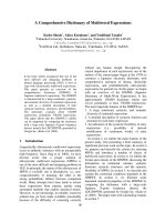
Lung viral titers after inhalation of attenuated mengovirusFigure 1
Lung viral titers after inhalation of attenuated men-
govirus. Viral titers in left lung homogenates and BAL fluid
(obtained from the right lung) from BN rats inoculated with
10
7
PFU of attenuated mengovirus, vMC
0
, an equivalent
amount of UV-inactivated vMC
0
, or vehicle were determined
by plaque assays. Data are the total amount of virus present
in the lung homogenate or BAL fluid (virus concentrations
were multiplied by the volumes of lung homogenate or BAL
fluid). Symbols represent data from individual rats. Dotted
lines indicate the limits of detection. * P < 0.005 (vMC
0
vs.
vehicle and UV-inactivated vMC
0
).
Virology Journal 2009, 6:122 />Page 4 of 10
(page number not for citation purposes)
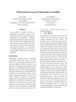
Recruitment of neutrophils and lymphocytes to the lungs after inhalation of attenuated mengovirusFigure 2
Recruitment of neutrophils and lymphocytes to the lungs after inhalation of attenuated mengovirus. Numbers
of (A) total cells, (B) neutrophils, (C) lymphocytes, (D) eosinophils, and (E) macrophages in the BAL fluid harvested on days 1,
3, and 5 postinoculation from the right lungs of BN rats inoculated with 10
7
PFU of vMC
0
(n = 4, 10, and 4 rats, respectively)
and on day 3 postinoculation from those inoculated with an equivalent amount of UV-inactivated vMC
0
(n = 5 rats) or vehicle
(n = 7 rats). Data are presented as box plots. * P < 0.05 (mengovirus vs. vehicle); † P < 0.05 (vMC
0
vs. UV-inactivated vMC
0
).
Virology Journal 2009, 6:122 />Page 5 of 10
(page number not for citation purposes)
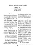
Recruitment of inflammatory cell infiltrates to the lungs after inhalation of attenuated mengovirusFigure 3
Recruitment of inflammatory cell infiltrates to the
lungs after inhalation of attenuated mengovirus.
Giemsa-stained sections of the left lungs from BN rats inocu-
lated with (A) vehicle, (B) vMC
0
(10
7
PFU) or (C) an equiva-
lent amount of UV-inactivated vMC
0
. Lungs were harvested
on day 3 postinoculation. Magnification, 20×.
B
C
A
Figure 4
Virology Journal 2009, 6:122 />Page 6 of 10
(page number not for citation purposes)
pared with that observed using inoculation doses of 10
7
or
10
6
PFU.
Effect of inhalation of attenuated mengovirus on
pulmonary physiology and airway hyperresponsiveness
(AHR)
To examine whether infection of the lower airways with
attenuated mengovirus induced changes in pulmonary
physiology, either vehicle or 10
7
PFU of vMC
0
were insuf-
flated into the lungs of adult BN rats, and pulmonary
function was measured on day 3 postinoculation. No sig-
nificant differences were observed between vehicle- and
vMC
0
-inoculated groups of rats with regard to respiratory
system resistance (Rrs) or the input impedance variables,
Newtonian resistance (Rn), tissue viscance (G), and
elastance (H), either at baseline or in response to metha-
choline challenge (Figure 5 and data not shown), indicat-
ing a lack of viral effects on pulmonary physiology and
AHR.
Discussion
The establishment of useful small animal models to study
HRV pathogenesis has been an important goal to enable
mechanistic studies and facilitate the development of new
therapies. The earliest reported effort to develop a HRV
infection model in mice required very large input doses of
virus and pretreatment of the mice with actinomycin D
[33]. Recently, more robust murine experimental models
of HRV infection have been established. These models
employ either a murine cell culture-adapted minor group
HRV in wild-type mice or a major group HRV in mice that
are transgenic for human ICAM-1 [18,19]. Although the
development of these novel tools represents a significant
advance in the study of HRV-induced airway inflamma-
tion, an important limitation is that HRV shedding is lim-
ited to ≤ 24 h postinoculation [18].
In the rat model described here, infectious mengovirus
was consistently detected in the lungs at high levels, and
persisted for at least 3 days after inoculation. The inocula-
tion dose of 10
6
–10
7
PFU of attenuated mengovirus in the
rats was similar to the dose of 5 × 10
6
TCID
50
(50% tissue-
culture infective dose) administered in the HRV models in
mice [18,19], especially considering that the body weight
of the rats is about an order of magnitude greater com-
pared to that of mice. Furthermore, inhalation of attenu-
ated mengovirus, but not vehicle or UV-inactivated virus,
into the lungs of BN rats resulted in increases in chemok-
ines (CINC-1, MIP-2, and MCP-1) and cellular inflamma-
tion (neutrophils, lymphocytes, and total BAL cells).
Compared to the HRV mouse models, infection with
vMC
0
represents a rodent model of picornavirus-induced
airway inflammation in which the roles of viral replica-
tion and persistence are more prominent.
Mengovirus-induced expression of CXCR2 ligands is con-
sistent with the increased expression of CXCR2 ligands
that is observed in response to rhinovirus infection [34-
36]. A similar induction of CXCR2 ligands was also
observed in the murine HRV infection models [18,19].
We also observed the induction of MCP-1 expression in
response to inhalation of attenuated mengovirus, which
represents another similarity between this rat model of
attenuated mengovirus-induced airway inflammation
Effect of inhalation of attenuated mengovirus on pulmonary physiologyFigure 5
Effect of inhalation of attenuated mengovirus on pul-
monary physiology. BN rats were inoculated with either
vehicle or 10
7
PFU of vMC
0
(n = 5 rats per group), and on
day 3 postinoculation, pulmonary physiology measurements
were obtained after exposure to aerosols of normal saline
followed by escalating concentrations of methacholine. Val-
ues for respiratory system resistance (Rrs) are presented as
the group means ± the standard error. There were no signif-
icant differences between the vehicle- and vMC
0
-inoculated
groups.
Inhalation of attenuated mengovirus enhanced pulmonary expression of the chemokines, CINC-1, MIP-2, and MCP-1Figure 4
Inhalation of attenuated mengovirus enhanced pul-
monary expression of the chemokines, CINC-1, MIP-
2, and MCP-1. BAL fluid was harvested on days 1, 3, and 5
postinoculation from the right lungs of BN rats inoculated
with 10
7
PFU of vMC
0
(n = 4, 10, and 4 rats, respectively) and
on day 3 postinoculation from those inoculated with an
equivalent amount of UV-inactivated vMC
0
(n = 5 rats) or
vehicle (n = 5–6 rats), and (A) CINC-1, (B) MIP-2, and (C)
MCP-1 levels were determined by ELISA. Data are the total
amount of chemokine recovered from the right lung BAL
(ELISA values, corrected for the 15× concentration, were
multiplied by the BAL fluid volume). (A, B) Data are pre-
sented as box plots. (C) Symbols represent data from individ-
ual rats; bars indicate medians. * P < 0.05, ‡ P = 0.05 (vMC
0
vs. vehicle); † P < 0.05 (vMC
0
vs. UV-inactivated vMC
0
).
Virology Journal 2009, 6:122 />Page 7 of 10
(page number not for citation purposes)
and human host responses to HRV infection [28]. There-
fore, the induction of rat CXC2 ligand and MCP-1 expres-
sion in airway fluids in response to inhalation of
attenuated mengovirus closely resembles the HRV-
induced enhancement of these chemokines.
Another similarity between this rat model and HRV infec-
tion in humans is the relative kinetics of the viral infection
vs. the lower airway neutrophilic inflammatory response.
Mengovirus titers in the lung peak earlier than the neu-
trophilic inflammatory response in the lower airways.
This parallels data from experimental HRV inoculations in
human volunteers [11,13]. In addition, the patchiness of
the mengovirus-induced airway inflammation in this rat
model is consistent with the patchy infection of airway
epithelial cells observed in HRV infections in human sub-
jects [11,37-39].
Infection of the lower airways with mengovirus did not
result in significant changes in baseline pulmonary phys-
iology measurements or in AHR to methacholine chal-
lenge in this rat model. It is important to note that
experimentally naïve adult rats without existing airway
disease were used in these studies. Similar to this rat
model, several studies involving experimental HRV inoc-
ulations of healthy, nonasthmatic, nonallergic human
subjects have demonstrated no changes in baseline pul-
monary function or AHR after HRV infection [10,40-44].
In one study showing a small change in AHR after experi-
mental HRV infection of nonasthmatic, nonallergic sub-
jects, the small difference was only detected by employing
a methacholine concentration that was a half-log higher
than the highest concentration typically used [45]. In con-
trast, experimental inoculation with HRV has been shown
to increase AHR in individuals with asthma and/or aller-
gic rhinitis in several studies [10,17,44,46,47], although
not in others [40,41,43,45,48]. Therefore, the absence of
changes in AHR in these healthy adult rats without exist-
ing airway disease is consistent with the outcomes of
experimental HRV infections in healthy humans who had
no underlying airway disease, such as asthma or allergic
rhinitis. The absence of viral effects on AHR in this men-
govirus model and in the experimental HRV inoculations
in humans is consistent with the murine experimental
model of HRV infection described by Bartlett et al. in
which there was no increase in AHR to methacholine chal-
lenge after HRV infection unless the BALB/c mice had also
been sensitized and challenged with allergen [18]. How-
ever, in the murine experimental HRV infection model
described by Newcomb et al., an increase in AHR to meth-
acholine challenge was observed after infection of C57BL/
6 mice with HRV [19], which may be related to the use of
a different mouse strain. Overall, the lack of significant
changes in pulmonary physiology during mengovirus-
induced respiratory infection in adult rats without existing
airway disease is consistent with previous observations in
experimental HRV infections in humans. In future studies,
it will be of interest to investigate the effects of mengovi-
rus-induced respiratory infection on rats with existing air-
way injury related to prior exposures to allergens or other
respiratory viruses [49] with the objective of modeling
aspects of HRV-induced asthma exacerbations.
A potential limitation of this animal model is the use of
mengovirus, which is neurotropic, to serve as a model for
HRV, which primarily causes respiratory infections. In this
regard, it is important to note that poliovirus, which is
closely related to HRV, is also neurotropic. The attenuated
mengovirus, vMC
0
, used in these studies induced a self-
limited respiratory infection when administered through
an inhalation route. This indicates that there is plasticity
in the tissue tropism of vMC
0
that makes it suitable for a
model of picornavirus-induced airway infection and
inflammation. Another consideration is that there are
both similarities and differences in CXCR2 and its ligands
between rats and humans [50]. Humans express IL-8 and
two IL-8 receptors, CXCR1 and CXCR2, whereas rats do
not express an IL-8 ortholog and only express CXCR2.
However, rats do express relevant CXCR2 ligands, such as
CINC-1 and MIP-2, which are functional analogs of IL-8
with regard to neutrophil recruitment and activation. We
believe that the rat represents an attractive, relevant, and
simplified model for examining the role of CXC chemok-
ines in neutrophil recruitment and activation in response
to picornavirus-induced respiratory infection because of
the reduced number of chemokines and chemokine
receptors to be examined.
Conclusion
Overall, our data support the feasibility of using this novel
rat model of picornavirus-induced lower airway infection
and inflammation to study, among other questions, the
role of neutrophilic inflammation in the host response to
picornavirus-induced respiratory infections. Although
this model does not fully encompass all aspects of HRV
infection in humans, it does demonstrate a remarkable
number of parallel developments that will provide novel
opportunities to study the interactions between picornavi-
ral replication and the host antiviral immune responses in
a relevant small animal model.
Methods
Animals
BN/SsN male rats were purchased from Harlan (Indiana-
polis, IN) and had a median body weight of 250 g when
used for inoculation studies. The rats were housed in
HEPA-filtered isolation cubicles (Britz and Co., Wheat-
land, WY) in an American Association for Accreditation of
Laboratory Animal Care-accredited laboratory animal
facility at the University of Wisconsin School of Medicine
Virology Journal 2009, 6:122 />Page 8 of 10
(page number not for citation purposes)
and Public Health. All procedures were approved by the
University of Wisconsin Animal Care and Use Committee
and conformed to the Guide for the Care and Use of Lab-
oratory Animals (1996).
Virus
Stock preparations of the attenuated mengovirus, vMC
0
(which has no poly(C) tract) [21-25], were prepared by
transfection of HeLa cells with viral RNA transcribed from
a plasmid encoding the vMC
0
genome followed by ampli-
fication of viral titers via passage in HeLa cell cultures as
described [51]. Supernates from uninfected HeLa cell cul-
tures were used as vehicle controls, and UV-inactivated
vMC
0
stocks were prepared by exposing vMC
0
to a germi-
cidal UV lamp at a distance of 10 cm for 1 h. Plaque assays
using HeLa cells were employed to determine the titer of
the active virus preparations and to verify UV-inactivation.
Active virus was undetectable (< 10 PFU/ml) in the UV-
inactivated preparations.
Virus inoculation
Rats were lightly anesthetized by inhalation of 5% isoflu-
rane, and vMC
0
, UV-inactivated vMC
0
, or vehicle in a total
volume of 0.1 ml were insufflated into the lungs via an
orotracheal catheter.
Measurements of pulmonary inflammation
At various times after inoculation, rats were anesthetized
with urethane and euthanized by exsanguination. The
chest was opened, and the left mainstem bronchus was
clamped to allow BAL of the right lung. The right lung was
filled with phosphate buffered saline (PBS) to total lung
capacity by gravity and drained 5 times, the BAL fluid was
centrifuged, and the cell pellet was resuspended in 1 ml
PBS. The total number of BAL leukocytes was determined
with an automated cell counter (model Z1, Beckman
Coulter, Hialeah, FL), and cytospin slides were prepared
for a differential leukocyte count based on 200 cells. BAL
fluid was concentrated 15× using a centrifugal filter device
with a molecular weight cutoff of 5,000 (Millipore, Bed-
ford, MA) and stored at -80°C until analyzed for chemok-
ine expression. Samples of unconcentrated BAL fluid were
used for viral titer determinations. The left lung was either
removed for viral titer determinations or filled to total
lung capacity by gravity with 10% buffered formalin for
histological analysis.
Measurements of pulmonary physiology
Rats were anesthetized with pentobarbital (Abbott, North
Chicago, IL), intubated via tracheostomy, paralyzed with
succinylcholine HCl (Sigma, St. Louis, MO), and venti-
lated mechanically (flexiVent, SCIREQ, Montreal, Can-
ada). Aerosol challenges were delivered by the ventilator
via an inline nebulizer (Aeroneb, SCIREQ) as 10 breaths
of aerosolized normal saline, followed by methacholine
HCl (Sigma) solutions in concentrations of 0.1, 0.3, 1, 3,
and 10 mg/ml. Each challenge was preceded by two lung
inflations to 30 cmH2O, and the challenges were deliv-
ered every 4 min. After each aerosol challenge, measure-
ments of pulmonary physiology were performed by the
flexiVent system every 15 s for 2 min, alternating measures
of Rrs with measures of input impedance variables (Rn, G,
and H). For each variable, the highest value occurring after
each aerosol challenge was recorded as the response, ref-
erenced to the value obtained after saline challenge.
Measurement of viral titers
Viral titers in left lung homogenates, prepared in PBS
(10% w/v) and clarified by centrifugation, and in uncon-
centrated BAL fluid were determined by plaque assay
using HeLa cells as described [24,51]. Briefly, HeLa cell
monolayers were inoculated with dilutions of the sam-
ples, incubated for 24–48 h at 37°C (until plaques form),
formalin fixed, stained with crystal violet, and scored for
plaques. Stock vMC
0
preparations served as the positive
control.
Histological assessment of pulmonary inflammation
Sections (5 μM) were prepared from formalin-fixed, par-
affin-embedded left lungs. Giemsa staining was per-
formed on these sections, which were evaluated for
inflammation by light microscopy.
Measurement of chemokine expression
Chemokine levels in BAL fluid were determined using
commercially available rat-specific enzyme-linked immu-
nosorbent assay (ELISA) kits for CINC-1 (R&D Systems,
Minneapolis, MN), MIP-2, and MCP-1 (Biosource,
Camarillo, CA) with sensitivities of 7.8, 7.8, and 8 pg/ml,
respectively, according to the manufacturers' instructions.
Statistical analysis
Analysis of variance (general linear model) was per-
formed on the BAL fluid CINC-1 and MIP-2 ELISA data
and on pulmonary physiology data after a log transforma-
tion, and Fischer's least significant difference test was used
for planned pairwise comparisons. A residual analysis was
employed to test the adequacy of the models. Nonpara-
metric tests were used to analyze all other data. For com-
parisons between two groups, the Mann-Whitney test was
used. The Kruskal-Wallis test was used for comparisons
among three or more groups and was followed by
planned pairwise comparisons using the Mann-Whitney
test. Because infectious virus was undetectable in the lung
homogenate and BAL fluid samples from rats inoculated
with vehicle or UV-inactivated virus, these groups were
combined for statistical analysis of viral titers. Box plots
depict the median and the interquartile range between the
25th and 75th percentile, and whiskers show the 10th and
90th percentiles. Analyses were performed using the sta-
Virology Journal 2009, 6:122 />Page 9 of 10
(page number not for citation purposes)
tistical software package SYSTAT 11.0 (Systat Software,
Chicago, IL).
Competing interests
The authors declare that they have no competing interests.
Authors' contributions
LAR co-conceived the study, designed and coordinated
the experiments, participated in the animal and immuno-
logical studies, performed the data and statistical analysis,
analyzed and interpreted the data, and drafted the manu-
script. SPA carried out the virology studies and partici-
pated in the experimental design and interpretation of the
data. RJS carried out the animal, immunological, and his-
tological studies and participated in the interpretation of
the data. RFL participated in the interpretation of the data
and revision of the manuscript. JEG co-conceived the
study and participated in the interpretation of the data
and revision of the manuscript. RLS co-conceived the
study and participated in the experimental design, the ani-
mal and immunological studies, the interpretation of the
data, and the revision of the manuscript. All authors read
and approved the final manuscript.
Acknowledgements
The authors thank Dr. Ann Palmenberg (The Institute for Molecular Virol-
ogy, University of Wisconsin-Madison) for generously providing the plas-
mid containing the attenuated mengovirus, vMC
0
, and for helpful
discussions. We also thank Maria Bulat and LaCinda Burchell for technical
assistance with the virology and histology studies, respectively. This work
was funded by National Institutes of Health grants AI070503 to LAR and
JEG and AI50500 to RFL.
References
1. Lemanske RF Jr, Jackson DJ, Gangnon RE, Evans MD, Li Z, Shult PA,
Kirk CJ, Reisdorf E, Roberg KA, Anderson EL, Carlson-Dakes KT,
Adler KJ, Gilbertson-White S, Pappas TE, DaSilva DF, Tisler CJ, Gern
JE: Rhinovirus illnesses during infancy predict subsequent
childhood wheezing. J Allergy Clin Immunol 2005, 116:571-577.
2. Jackson DJ, Gangnon RE, Evans MD, Roberg KA, Anderson EL, Pappas
TE, Printz MC, Lee WM, Shult PA, Reisdorf E, Carlson-Dakes KT,
Salazar LP, DaSilva DF, Tisler CJ, Gern JE, Lemanske RF Jr: Wheez-
ing rhinovirus illnesses in early life predict asthma develop-
ment in high-riskchildren. Am J Respir Crit Care Med 2008,
178:667-672.
3. Kusel MM, de Klerk NH, Kebadze T, Vohma V, Holt PG, Johnston SL,
Sly PD: Early-life respiratory viral infections, atopic sensitiza-
tion, and risk of subsequent development of persistent
asthma. J Allergy Clin Immunol 2007, 119:1105-1110.
4. Kelly JT, Busse WW: Host immune responses to rhinovirus:
mechanisms in asthma. J Allergy Clin Immunol 2008, 122:671-682.
5. Singh AM, Moore PE, Gern JE, Lemanske RF Jr, Hartert TV: Bronchi-
olitis to asthma: a review and call for studies of gene-virus
interactions in asthma causation. Am J Respir Crit Care Med 2007,
175:108-119.
6. Gern JE, Galagan DM, Jarjour NN, Dick EC, Busse WW: Detection
of rhinovirus RNA in lower airway cells during experimen-
tally induced infection. Am J Respir Crit Care Med 1997,
155:1159-1161.
7. Papadopoulos NG, Bates PJ, Bardin PG, Papi A, Leir SH, Fraenkel DJ,
Meyer J, Lackie PM, Sanderson G, Holgate ST, Johnston SL: Rhinovi-
ruses infect the lower airways. J Infect Dis 2000, 181:1875-1884.
8. Mosser AG, Brockman-Schneider R, Amineva S, Burchell L, Sedgwick
JB, Busse WW, Gern JE: Similar frequency of rhinovirus-infect-
ible cells in upper and lower airway epithelium. J Infect Dis
2002, 185:734-743.
9. Schroth MK, Grimm E, Frindt P, Galagan DM, Konno SI, Love R, Gern
JE: Rhinovirus replication causes RANTES production in pri-
mary bronchial epithelial cells. Am J Respir Cell Mol Biol 1999,
20:1220-1228.
10. Message SD, Laza-Stanca V, Mallia P, Parker HL, Zhu J, Kebadze T,
Contoli M, Sanderson G, Kon OM, Papi A, Jeffery PK, Stanciu LA,
Johnston SL: Rhinovirus-induced lower respiratory illness is
increased in asthma and related to virus load and Th1/2
cytokine and IL-10 production. Proc Natl Acad Sci USA 2008,
105:13562-13567.
11. Mosser AG, Vrtis R, Burchell L, Lee WM, Dick CR, Weisshaar E, Bock
D, Swenson CA, Cornwell RD, Meyer KC, Jarjour NN, Busse WW,
Gern JE: Quantitative and qualitative analysis of rhinovirus
infection in bronchial tissues. Am J Respir Crit Care Med 2005,
171:645-651.
12. Jarjour NN, Gern JE, Kelly EA, Swenson CA, Dick CR, Busse WW:
The effect of an experimental rhinovirus 16 infection on
bronchial lavageneutrophils. J Allergy Clin Immunol 2000,
105:1169-1177.
13. Gern JE, Vrtis R, Grindle KA, Swenson C, Busse WW: Relationship
of upper and lower airway cytokines to outcome of experi-
mental rhinovirusinfection. Am J Respir Crit Care Med 2000,
162:2226-2231.
14. Fahy JV, Kim KW, Liu J, Boushey HA: Prominent neutrophilic
inflammation in sputum from subjects with asthma exacer-
bation. J Allergy Clin Immunol 1995, 95:843-852.
15. Wark PA, Johnston SL, Moric I, Simpson JL, Hensley MJ, Gibson PG:
Neutrophil degranulation and cell lysis is associated with
clinical severity in virus-induced asthma. Eur Respir J 2002,
19:68-75.
16. Gern JE, Martin MS, Anklam KA, Shen K, Roberg KA, Carlson-Dakes
KT, Adler K, Gilbertson-White S, Hamilton R, Shult PA, Kirk CJ, Da
Silva DF, Sund SA, Kosorok MR, Lemanske RF Jr: Relationships
among specific viral pathogens, virus-induced interleukin-8,
and respiratory symptoms in infancy. Pediatr Allergy Immunol
2002, 13:386-393.
17. Grunberg K, Timmers MC, Smits HH, de Klerk EP, Dick EC, Spaan
WJ, Hiemstra PS, Sterk PJ: Effect of experimental rhinovirus 16
colds on airway hyperresponsiveness to histamine and inter-
leukin-8 in nasal lavage in asthmatic subjects in vivo. Clin Exp
Allergy 1997, 27:36-45.
18. Bartlett NW, Walton RP, Edwards MR, Aniscenko J, Caramori G, Zhu
J, Glanville N, Choy KJ, Jourdan P, Burnet J, Tuthill TJ, Pedrick MS,
Hurle MJ, Plumpton C, Sharp NA, Bussell JN, Swallow DM, Schwarze
J, Guy B, Almond JW, Jeffery PK, Lloyd CM, Papi A, Killington RA,
Rowlands DJ, Blair ED, Clarke NJ, Johnston SL: Mouse models of
rhinovirus-induced disease and exacerbation of allergic air-
way inflammation. Nat Med 2008, 14:
199-204.
19. Newcomb DC, Sajjan US, Nagarkar DR, Wang Q, Nanua S, Zhou Y,
McHenry CL, Hennrick KT, Tsai WC, Bentley JK, Lukacs NW, John-
ston SL, Hershenson MB: Human rhinovirus 1B exposure
induces phosphatidylinositol 3-kinase-dependent airway
inflammation in mice. Am J Respir Crit Care Med 2008,
177:1111-1121.
20. Palmenberg AC, Osorio JE: Cardioviral poly(C) tracts and viral
pathogenesis. Arch Virol Suppl 1994, 9:67-77.
21. Duke GM, Osorio JE, Palmenberg AC: Attenuation of Mengo
virus through genetic engineering of the 5' noncoding
poly(C) tract. Nature 1990, 343:474-476.
22. Martin LR, Duke GM, Osorio JE, Hall DJ, Palmenberg AC: Muta-
tional analysis of the mengovirus poly(C) tract and surround-
ing heteropolymeric sequences. J Virol 1996, 70:2027-2031.
23. Osorio JE, Grossberg SE, Palmenberg AC: Characterization of
genetically engineered mengoviruses in mice. Viral Immunol
2000, 13:27-35.
24. Osorio JE, Martin LR, Palmenberg AC: The immunogenic and
pathogenic potential of short poly(C) tract Mengo viruses.
Virology 1996, 223:344-350.
25. Martin LR, Neal ZC, McBride MS, Palmenberg AC: Mengovirus and
encephalomyocarditis virus poly(C) tract lengths can affect
virus growth in murine cell culture. J Virol 2000, 74:3074-3081.
26. Sorkness RL, Castleman WL, Kumar A, Kaplan MR, Lemanske RF Jr:
Prevention of chronic post-bronchiolitis airway sequelae
Publish with BioMed Central and every
scientist can read your work free of charge
"BioMed Central will be the most significant development for
disseminating the results of biomedical research in our lifetime."
Sir Paul Nurse, Cancer Research UK
Your research papers will be:
available free of charge to the entire biomedical community
peer reviewed and published immediately upon acceptance
cited in PubMed and archived on PubMed Central
yours — you keep the copyright
Submit your manuscript here:
/>BioMedcentral
Virology Journal 2009, 6:122 />Page 10 of 10
(page number not for citation purposes)
with interferon-γ treatment in rats. Am J Respir Crit Care Med
1999, 160:705-710.
27. Shibata F, Konishi K, Kato H, Komorita N, Al-Mokdad M, Fujioka M,
Nakagawa H: Recombinant production and biological proper-
ties of rat cytokine-induced neutrophil chemoattractants,
GRO/CINC-2 alpha, CINC-2 beta and CINC-3. Eur J Biochem
1995, 231:306-311.
28. Hall DJ, Bates ME, Guar L, Cronan M, Korpi N, Bertics PJ: The role
of p38 MAPK in rhinovirus-induced monocyte chemoattract-
ant protein-1 production by monocytic-lineage cells. J Immu-
nol 2005, 174:8056-8063.
29. Gonzalo JA, Lloyd CM, Wen D, Albar JP, Wells TN, Proudfoot A,
Martinez A, Dorf M, Bjerke T, Coyle AJ, Gutierrez-Ramos JC: The
coordinated action of CC chemokines in the lung orches-
trates allergic inflammation and airway hyperresponsive-
ness. J Exp Med 1998, 188:157-167.
30. Maus U, von GK, Kuziel WA, Mack M, Miller EJ, Cihak J, Stangassinger
M, Maus R, Schlondorff D, Seeger W, Lohmeyer J: The role of CC
chemokine receptor 2 in alveolar monocyte and neutrophil
immigration in intactmice. Am J Respir Crit Care Med 2002,
166:268-273.
31. Maus UA, Waelsch K, Kuziel WA, Delbeck T, Mack M, Blackwell TS,
Christman JW, Schlondorff D, Seeger W, Lohmeyer J: Monocytes
are potent facilitators of alveolar neutrophil emigration dur-
ing lung inflammation: role of the CCL2-CCR2 axis. J Immunol
2003, 170:3273-3278.
32. Vozzelli MA, Mason SN, Whorton MH, Auten RL Jr: Antimacro-
phage chemokine treatment prevents neutrophil and mac-
rophage influx in hyperoxia-exposed newborn rat lung. Am J
Physiol Lung Cell Mol Physiol 2004, 286:L488-L493.
33. Yin FH, Lomax NB: Establishment of a mouse model for human
rhinovirus infection. J Gen Virol 1986, 67:2335-2340.
34. Zhu Z, Tang W, Gwaltney JM Jr, Wu Y, Elias JA: Rhinovirus stimu-
lation of interleukin-8 in vivo and in vitro: role of NF-kappaB.
Am J Physiol 1997, 273:L814-L824.
35. Subauste MC, Jacoby DB, Richards S, Proud D: Infection of a
human respiratory epithelial cell line with rhinovirus. Induc-
tion of cytokine release and modulation of susceptibility to
infection by cytokineexposure.
J Clin Invest 1995, 96:549-557.
36. Donninger H, Glashoff R, Haitchi HM, Syce JA, Ghildyal R, van RE,
Bardin PG: Rhinovirus induction of the CXC chemokine epi-
thelial-neutrophil activating peptide-78 in bronchial epithe-
lium. J Infect Dis 2003, 187:1809-1817.
37. Arruda E, Boyle TR, Winther B, Pevear DC, Gwaltney JM Jr, Hayden
FG: Localization of human rhinovirus replication in the upper
respiratory tract by in situ hybridization. J Infect Dis 1995,
171:1329-1333.
38. Pitkaranta A, Puhakka T, Makela MJ, Ruuskanen O, Carpen O, Vaheri
A: Detection of rhinovirus RNA in middle turbinate of
patients with common colds by in situ hybridization. J Med
Virol 2003, 70:319-323.
39. Winther B, Gwaltney JM Jr, Mygind N, Turner RB, Hendley JO: Sites
of rhinovirus recovery after point inoculation of the upper
airway. JAMA 1986, 256:1763-1767.
40. Skoner DP, Doyle WJ, Seroky J, Vandeusen MA, Fireman P: Lower
airway responses to rhinovirus 39 in healthy allergic and non-
allergicsubjects. Eur Respir J 1996, 9:1402-1406.
41. Zambrano JC, Carper HT, Rakes GP, Patrie J, Murphy DD, Platts-Mills
TA, Hayden FG, Gwaltney JM Jr, Hatley TK, Owens AM, Heymann
PW: Experimental rhinovirus challenges in adults with mild
asthma: Response to infection in relation to IgE. J Allergy Clin
Immunol 2003, 111:1008-1016.
42. de Kluiver J, Grunberg K, Sont JK, Hoogeveen M, van Schadewijk WA,
de Klerk EP, Dick CR, van Krieken JH, Sterk PJ: Rhinovirus infec-
tion in nonasthmatic subjects: effects on intrapulmonary air-
ways. Eur Respir J 2002, 20:274-279.
43. Angelini B, Van Deusen MA, Doyle WJ, Seroky J, Cohen S, Skoner DP:
Lower airway responses to rhinovirus-Hanks in healthy sub-
jects with and without allergy. J Allergy Clin Immunol 1997,
99:618-619.
44. Gern JE, Calhoun W, Swenson C, Shen G, Busse WW: Rhinovirus
infection preferentially increases lower airway responsive-
ness in allergicsubjects.
Am J Respir Crit Care Med 1997,
155:1872-1876.
45. Fleming HE, Little FF, Schnurr D, Avila PC, Wong H, Liu J, Yagi S,
Boushey HA: Rhinovirus-16 colds in healthy and in asthmatic
subjects: similar changes in upper and lower airways. Am J
Respir Crit Care Med 1999, 160:100-108.
46. Lemanske RF Jr, Dick EC, Swenson CA, Vrtis RF, Busse WW: Rhino-
virus upper respiratory infection increases airway hyperre-
activity and late asthmatic reactions. J Clin Invest 1989, 83:1-10.
47. de Gouw HW, Grunberg K, Schot R, Kroes AC, Dick EC, Sterk PJ:
Relationship between exhaled nitric oxide and airway hyper-
responsiveness following experimental rhinovirus infection
in asthmatic subjects. Eur Respir J 1998, 11:126-132.
48. Avila PC, Abisheganaden J, Wong H, Liu J, Yagi S, Schnurr D, Kishi-
yama JL, Boushey HA: Effects of allergic inflammation of the
nasal mucosa on the severity of rhinovirus 16 cold. J Allergy Clin
Immunol 2000, 105:923-932.
49. Sorkness RL, Herricks KM, Szakaly RJ, Lemanske RF Jr, Rosenthal LA:
Altered allergen-induced eosinophil trafficking and physio-
logical dysfunction in airways with preexisting virus-induced
injury. Am J Physiol Lung Cell Mol Physiol 2007, 292:L85-L91.
50. Roth I, Hebert C: CXCR1 and CXCR2. In Cytokine Reference: a
Compendium of Cytokines and Other Mediators of Host Defense Edited
by: Oppenheim JJ, Feldman M. San Diego: Academic Press;
2001:1982-2002.
51. Amineva SP, Mosser AG, Binder JJ, Aminev AG, Palmenberg AC, Gern
JE: Synthesis of the allergen ovomucoid by a replicating
Mengo virus. Arch Virol 2006, 151:1933-1946.