OCEANOGRAPHY and MARINE BIOLOGY: AN ANNUAL REVIEW (Volume 44) - Chapter 2 potx
Bạn đang xem bản rút gọn của tài liệu. Xem và tải ngay bản đầy đủ của tài liệu tại đây (614.14 KB, 24 trang )
61
Oceanography and Marine Biology: An Annual Review,
2006,
44
, 61-83
© R. N. Gibson, R. J. A. Atkinson, and J. D. M. Gordon, Editors
Taylor & Francis
ROLE, ROUTES AND EFFECTS OF MANGANESE
IN CRUSTACEANS
SUSANNE P. BADEN* & SUSANNE P. ERIKSSON
Göteborg University, Department of Marine Ecology,
Kristineberg Marine Research Station, S-450 34 Fiskebäckskil, Sweden
*E-mail:
Abstract
This review provides an overview of the role, routes and effects of manganese in aquatic
crustaceans. Manganese is a naturally abundant metal in marine and freshwater sediments where
it is involved in a large number of chemical processes. Although sediments contain high natural
concentrations of manganese, the potential danger to benthic organisms has been neglected in
studies to date. Manganese bioavailability increases as the result of human impact and it accumulates
in biota. Manganese may occur in toxic concentrations (10–20 mg l
–1
) in the bottom water of marine
coastal areas after hypoxia, or more locally (e.g., close to industries) as well as in acidic lakes and
aquaculture shrimp ponds. Though manganese is an essential metal, it is also an unforeseen toxic
metal in the aquatic environment. Although the uptake and elimination of manganese is rapid,
manganese affects processes that decrease the fitness of organisms. As manganese bioavailability
increases, its uptake is predominately through the water. The midgut gland, nerve tissue, blood
proteins and parts of the reproductive organs have the highest accumulation factors and are the
main target tissues. The functional effects of manganese in aquatic environments are still sparsely
investigated. Recent results show that the immune system, the perception of food via chemosensory
organs and a normal muscle extension are affected at manganese concentrations observed in the field.
Geochemical role of manganese
Manganese is the 12th most common element, the fourth most abundant metal and is universally
distributed in the earth’s crust and waters (Anonymous 2005). This metal is involved in a large
number of chemical processes, due mainly to its redox sensitivity. The literature on manganese (Mn)
geochemistry in the aquatic environment is immense (Elderfield 1976), whereas literature on the
occurrence and biological effects of manganese in aquatic animals is comparatively sparse. Man-
ganese concentrations in soil vary from 0.001–7 mg g
–1
dry weight (dw), averaging 0.75 mg g
–1
dw
(Saric 1986). Ocean sediment concentrations vary from approximately 1–50 mg g
–1
dw (Elderfield
1976). Since the 1800s an intensive and ongoing debate has been centred on the origin and amount
of the manganese flux to the oceans. Three main sources have been identified: continental weather-
ing (lithogenous origin), submarine volcanism and an upward migration in porewaters as a conse-
quence of sediment diagenesis (Elderfield 1976). The anthropogenic supplies of manganese to
aquatic biotopes derive mainly from mine tailings and from steel manufacturing industries where
approximately 90% of total manganese is used as a deoxidising and desulphurising additive and
as an alloying constituent (Saric 1986). Manganese (MnO
2
) is also widely used in dry cell batteries
(Saric 1986), as a contrasting agent for nuclear magnetic resonance tomography, and as an agri-
cultural fungicide (Gerber et al. 2002). A manganese antiknock additive (methylcyclopentadienyl
manganese tricarbonyl (MMT)) was introduced to Canada in 1990 to substitute for lead in fuel,
7044_C002.fm Page 61 Monday, April 17, 2006 1:37 PM
© 2006 by Taylor & Francis Group, LLC
SUSANNE P. BADEN & SUSANNE P. ERIKSSON
62
and since 1995 MMT has also been used in several states of the USA (Shukla & Singhal 1984,
Davis 1998, Normandin et al. 2002). In the rapidly expanding shrimp farming industry of tropical
regions, manganese is added to shrimp ponds in the form of potassium permanganate (KMnO4),
as disinfectant, in concentrations causing potential hazards to life in the ponds and in the coastal
zone close to the effluent water (Gräslund & Bengtsson 2001, Visuthismajarn et al. 2005).
Manganese becomes bioavailable as Mn(II) in water when it is reduced by hypoxic/anoxic
conditions in sediment. The reduction of manganese dioxides occurs during the degradation of
sedimenting organic matter (Dehairs et al. 1989). The process is directly or indirectly microbially
mediated but is fastest when sulphide and Fe(II) are reductants (Johnson et al. 1991). In general,
the solubility (and bioavailability) of manganese increases with decreasing oxygen tension and pH,
but not with increasing temperature (Wollast et al. 1979, Faust & Aly 1983).
During oxic
conditions in bottom water, sediment porewater may contain Mn concentrations
of 0.16–24.0 mg l
–1
(Canfield et al. 1993, Aller 1994, Magnusson et al. 1996), whereas bottom
water concentrations are between 0.18–16.5
µ
g l
–1
(Laslett & Balls 1995, Hall et al. 1996). During
hypoxia (O
2
< 3 mg l
–1
)), the Mn(II) of the bottom water can increase by several orders of magnitude
to 1.5 mg l
–1
, as in the Kiel Bight (Balzer 1982), and up to 22 mg l
–1
in the anoxic bottom water
of the Orca Basin in the Mexican Gulf (Trefry et al. 1984).
This review aims to give an overview of the role of manganese in aquatic animals, its routes
of uptake, and biological effects, mainly focusing on marine crustaceans living in and on the
sediment. As the biological chemistry of manganese is poorly explored in invertebrates, the relevant
medical and biological literature on basic processes involving Mn in vertebrates is cited.
Biological role of manganese
Essentiality
Manganese is an essential trace metal for metabolism belonging to the borderline elements (Mn, Fe,
Co, Ni, Cu, Zn, Cd, Hg, Pb) of the periodic table. It lies between the oxygen-seeking elements of
class A (Na, Mg, K and Ca being the most abundant) and the sulphur- and nitrogen-seeking elements
(including heavy metals like Ag, Au and Hg) of class B, and thus exhibits aspects of both classes
(Nieboer & Richardson 1980). In its divalent form, Mn(II), manganese has a relatively high affinity
for sulphur or nitrogen in functional groups of proteins and other molecules, which enables Mn to
interfere in a wide spectrum of biological processes (Simkiss 1979, Williams 1981). The divalent
Mn(II) exchanges water and ligands rapidly and the binding constant of the metal in proteins is weak.
Manganese is important as a cofactor or activator of different enzymatic reactions (e.g., electron-
transfer reactions, antioxidant defences, and phosphorylation) (Simkiss & Taylor 1989). In the case
of enzymes containing metal ions (mainly Mg(II), Mn(II) and Zn(II)) the metal ion itself can bind
with groupings in the substrate and act as a strain-producing agent by forming a chelated interme-
diary compound. At the same time the metal ion, because of its positive charge, is an efficient
electrophilic agent that can act as an effective participant in the reaction (White et al. 1973).
Examples of enzymatic reactions having Mn as an activator are acetyl-CoA carboxylase (the first
reaction in the fatty acid formation in the endoplasmatic reticulum), pyruvate carboxylase (in the
mitochondrial formation of oxaloacetate), glycylglycine dipeptidase (in the degradation of dena-
tured intracellular proteins) and the well-known Mn-super oxide dismutase (Mn-SOD) (a redox
enzyme in the mitochondria facilitating the production of dioxygen) (Cotzias 1958, White et al.
1973, da Silva & Williams 1991).
Manganese is mainly accumulated in organelles like the mitochondria, Golgi apparatus and
vesicles, whereas concentrations in the cytoplasm are relatively low. These concentration gradients
are sustained by metal transporters over the membrane (e.g., Luk & Culotta 2001). The elimination
7044_C002.fm Page 62 Monday, April 17, 2006 1:37 PM
© 2006 by Taylor & Francis Group, LLC
ROLE, ROUTES AND EFFECTS OF MANGANESE IN CRUSTACEANS
63
of Mn(II) from the mitochondria is a slow, energy-requiring, Na-dependent efflux mechanism
(Gavin et al. 1999).
In general, those metals having an essential biochemical role, such as the metals mentioned
above, are regulated at the individual level, while for non-essential metals such as mercury (Hg),
cadmium (Cd) and silver (Ag) there is only weak evidence of controls on accumulation. Under
constant ambient conditions, the net balance between inward and outward fluxes of metals provides
the underlying control on tissue burdens and, in general, metals that exchange rapidly tend to be
accumulated less efficiently than metals that exchange slowly. Accumulation may give rise to body
concentrations in excess of four orders of magnitude above background in non-regulating organisms
(Rainbow 1992, 1997).
Toxicity
Many borderline metals are thus essential to metabolism as micronutrients but may have the
potential of being toxic in high concentrations. The toxicity of manganese has been known for over
150 years after it was recognised that mine workers inhaling dust rich in Mn developed ‘manganism’
(Couper 1837). Manganism is an irreversible brain disease with prominent psychological and
neurological disturbances. Such neurological responses have received close attention because they
resemble several clinical disorders collectively described as ‘extra pyramidal motor system dys-
function’ and in particular Parkinson’s disease. The disease is regarded as chronic and the clinical
signs of intoxication include many symptoms dominated by speech disturbance, compulsive actions
and motor dysfunction like tremor and stiff gait (Mena et al. 1967, Iregren 1990, Aschner & Aschner
1991). Recently, however, a manganese-induced epileptic syndrome was cured after treatment with
a chelating treatment of CaNa
2
EDTA (Hernandez et al. 2003). Another much debated theory
connects excess Mn exposure with the initiation of transmissible spongiform encephalopathy (TSE),
also called scrapie in sheep and Creutzfeldts Jacobs disease (CJD) in humans. Imbalance of Mn
and Cu is established when Mn- and Cu-chelating insecticides (organo-phosphates) are taken up
at the same time, giving a substitution of Cu with Mn as Mn(III) in the CNS prion protein. This
substitution conforms the prions, preventing their degradation, and TSE may develop (Purdey 2000).
As Mn(III), manganese is able to accumulate in the brain, likely carried through the blood-
brain barrier via transferrin and receptor-mediated endocytosis (Simkiss & Taylor 1989, Aschner &
Aschner 1991). Transferrin is a protein containing a Fe-cluster crucial for absorption, transport,
storage and excretion of Fe in mammals and is able to cross the otherwise relatively impermeable
blood-brain barrier. Manganese may mirror Fe and bind to transferrin, not necessarily replacing
Fe, and in this way passes the blood-brain barrier (Aschner & Aschner 1991). Within the brain the
main part of Mn(III) appears to release from transferrin and concentrate in certain parts via axonal
transport (Henriksson et al. 1999). In freshwater crayfish a structural analogue to the vertebrate
blood-brain barrier called the glial perineurium, has been identified. The glial perineurium ensures
protection of the CNS by having a high degree of ion selectivity and regulation (Butt et al. 1990).
A direct uptake from the media through the nasal chamber in rats and olfactory chamber of pike
(Esox lucius)
followed by axonal transport along primary and secondary neurones into the olfactory
bulb has been documented (Tjälve et al. 1995, 1996). A similar uptake and transport into nerve
tissue of invertebrates has not been described to date.
Hydrated Mn has an ionic ratio close to that of Ca(II), and its ability to affect various aspects
of neuronal transmission has been ascribed primarily to its mimicry of Ca (Aschner & Aschner
1991). Manganese ions are known to affect various steps in the chemical synapses of nerve-muscle
transmission in a wide range of animal groups. At low concentrations, Mn ions have been found
to pass through Ca channels in a number of different preparations, e.g., giant squid axons (Yamagishi
1973), mammalian cardiac muscle (Ochi 1970, 1975; Delahayes 1975), mouse oocytes (Okamoto et al.
7044_C002.fm Page 63 Monday, April 17, 2006 1:37 PM
© 2006 by Taylor & Francis Group, LLC
SUSANNE P. BADEN & SUSANNE P. ERIKSSON
64
1977), starfish eggs (Hagiwara & Miyazaki 1977), larval beetle skeletal muscle fibres (Fukunda &
Kawa 1977) and frog skeletal muscle fibres (Palade & Almers 1978). However, at higher concen-
trations Mn ions are potent inhibitors of synaptic transmission (Katz & Miledi 1969, Ross & Stuart
1978, Xiao & Bevan 1994) and also act as competitive inhibitors of Ca ion flow through calcium
channels in muscle membranes (Fatt & Ginsborg 1958, Hagiwara & Takahashi 1967, Takeda 1967,
Mounier & Vassort 1975). Manganese affects not only the presynaptic site of action but also the
postsynaptic site (Katz & Miledi 1969). This is consistent with earlier studies on the excitation-
contraction coupling mechanisms in crustacean muscles, which indicated that Mn ions compete
with Ca ions to pass through sarcolemmal calcium channels and thus affect muscle membrane
depolarisation (Fatt & Ginsborg 1958, Hagiwara & Nakajima 1966, Chiarandini et al. 1970,
Mounier & Vassort 1975). More recently, Hirata (2002) presented evidence that Mn(II) can induce
DNA fragmentation, a biochemical hallmark for apoptosis, in neuronal cells.
Deficiency
The theoretical requirement of manganese for crustaceans has been calculated to be 3.9
µ
g Mn g
1
dw
(White & Rainbow 1987). The calculation was based on the animals’ total content, thus including
the exoskeleton where the majority of the manganese is incorporated into the calcareous matrix.
In the literature pelagic crustaceans are reported to have an average muscle and midgut gland
concentration of less than 2
µ
g Mn g
–1
dw and a total manganese body concentration of 1.2–1.4
µ
g
Mn g
–1
dw (Table 1). Even the benthic lobster
Nephrops norvegicus
from the pristine Faeroe Islands
contains very low Mn concentrations (Table 1). When excluding the exoskeleton and the stomach
(which may contain sediment rich in Mn) in these animals, the rest of the body (the soft tissue)
contains an Mn concentration of 2.5
µ
g Mn g
–1
dw (n = 32) (S.P. Eriksson & S.P. Baden, unpublished
observations). The theoretical required concentration of manganese in the soft tissue of crustaceans
is thus likely to be somewhat overestimated. Since no data exist on crustacean manganese deficiency,
the precise Mn requirements of Crustacea remain unresolved. It is hoped that further investigations
will provide an answer. Most field-caught animals contain manganese concentrations well above
the assumed basic requirements needed and manganese deficiency does not appear to pose a general
threat to aquatic crustaceans (Table 1).
Manganese in Crustacea — Overview
Manganese is an essential metal and is thus required in at least a minimum concentration for an
animal to be able to fulfil its metabolic functions. When discussing the basic body requirements
of manganese, it is, however, also important to differentiate between metabolically active soft tissues
and relatively inert tissues. Each tissue is likely to have its own kinetics (reaction rate) of metal
uptake and loss, the determination of which can often be valuable when interpreting the biological
significance of metal burdens. The interpretation of animal kinetic data and animal metal concen-
tration is potentially complicated by a combination of factors including organism condition, growth,
food supply, moulting and reproduction cycles, and may also depend directly or indirectly on
environmental conditions like temperature, oxygen saturation and metal concentration. Some tissue
metal concentrations are maintained within a narrow range and for others there may be less tight
regulation and even storage. Clearly, under such circumstances, increased metal burdens in specific
tissues could easily be obscured when analysing whole organisms.
The literature on background manganese concentrations in different crustaceans derives from
field-collected animals from marine and freshwater environments (Table 1). Average total Mn con-
centration was 63
µ
g Mn g
–1
dw, with the lowest concentrations found in marine pelagic crustaceans
and benthic lobsters from the pristine Faroe Islands. The highest total Mn concentration was found
7044_C002.fm Page 64 Monday, April 17, 2006 1:37 PM
© 2006 by Taylor & Francis Group, LLC
65
ROLE, ROUTES AND EFFECTS OF MANGANESE IN CRUSTACEANS
Table 1
Manganese concentrations found in field-caught crustaceans from pristine areas
Habitat/Order/Species Subhabitat Total Eggs Exo. Gills Haem. Midg.gl. Muscle Ovary Testes References
Marine
Amphipoda
Talitrus saltator
S,B 25.2–97.4 Rainbow et al. 1998,
Fialkowski et al. 2003
Thoracica
Balanus crenatus
(
–
shell) S,B 53 Rainbow et al. 2002
Tetraclita squamosa
(
–
shell) S,B 6.7–10 Blackmore 1999,
Rainbow & Blackmore
2001
Stomatopoda
Squilla mantis
D,B 32 Blasco et al. 2002
Decapoda
Acantephyra eximia
D 12.3* Kress et al. 1998
Aristeus antennatus
D 27.9* 0.9–7.4 Kress et al. 1998, Drava
et al. 2004
Bythograea thermydon
D,H 0.4–1.6 Baden & Childress
unpublished
Callinectes sapidus
S,B 36–76 14–17 3.6–4.3 Weinstein et al. 1992
Cancer irroratus
S,B 36 10 10–28 7–42 0.2–0.3 7–18 2.5–5.0 6 Martin 1974, 1975, 1976,
Martin & Ceccaldi 1976
Carcinus maenas
S,B 74–206 92–286 175–282 0.36–0.38 7.5–10 10–24 3.1–19 Martin 1975, Bjerregaard
& Depledge 2002
Heterocarpus vicarius
D,B 0.4–0.6 Hendrickx et al. 1998
Nephrops norvegicus
D,B 91.7 5.5–120 150 45 1.4 11 3.1 5.5 33 Eriksson & Baden 1998,
Eriksson 2000a,b
Nephrops norvegicus
(Faroe islands)
D,B 8.0 3.5 11 5.9 0.12 4.7 1.9 5.3 25 Eriksson unpublished,
Eriksson & Baden 1998
Pandalus borealis
D,B 5.1* Heu et al. 2003
7044_C002.fm Page 65 Monday, April 17, 2006 1:37 PM
© 2006 by Taylor & Francis Group, LLC
SUSANNE P. BADEN & SUSANNE P. ERIKSSON
66
Table 1 (continued)
Manganese concentrations found in field-caught crustaceans from pristine areas
Habitat/Order/Species Subhabitat Total Eggs Exo. Gills Haem. Midg.gl. Muscle Ovary Testes References
Panulirus inflatus
D,B 11 9.4–13 5.4–11 1.1–2.0 3.1 1.9 Paez-Osuna et al. 1995
Melicertus
(as
Penaeus
)
kerathurus
B 0.8* Balkas et al. 1982
Polycheles typhlops
D 29.3* Kress et al. 1998
Portunus pelagicus
S,P 0.6 0.1 0.7–1.2* Balkas et al. 1982,
Al-Mohanna &
Subrahmanyam 2001
Trachypenaeus curvirostris
B 3.0* Heu et al. 2003
Decapoda, Mysidacea,
Euphausiacea
Larvae
P 1.2–4.0 Ridout et al. 1989
Freshwater
Decapoda
Asellus aquaticus
S,B 160 Akyuz et al. 2001
Astacus astacus
S,B 67 3.6 Jorhem et al. 1994
Austropotamobius pallipes
B6952Gherardi et al. 2002
Cambarus bartonii
S,B 52 32 11 Alikhan et al. 1990
Orconectes virilis
S,B 66–106 12–33 4–8 Young & Harvey 1991
Pacifastacus leniusculus
S,B 361 2 Jorhem et al. 1994
Potamon fluviatile
B53305 Gherardi et al. 2002
Potamonautes warreni
S,B 239 340 508 374 87 107 89 Steenkamp et al. 1994,
Sanders et al. 1998
Mean 63 22 96 100 0.63 102 9 23 37
Max/min ratio 199 34 36 847 12 3740 218 35 47
Notes:
All values are given as
µ
g Mn g
–1
dry weight tissue, except for haemolymph which is in wet weight. * Values calculated from wet weight by using ww/dw ratio stated in
the original papers. Abbreviations: S-shallow, D-deep, B-benthic, H-hydrothermal vent, P-pelagic, Exo-Exoskeleton, Haem-Haemolymph and Midg.gl Midgut gland.
7044_C002.fm Page 66 Monday, April 17, 2006 1:37 PM
© 2006 by Taylor & Francis Group, LLC
ROLE, ROUTES AND EFFECTS OF MANGANESE IN CRUSTACEANS
67
in a freshwater crayfish (
Potamonautes warreni
). Highest mean tissue Mn concentration was found in
the animal’s midgut gland (102
µ
g Mn g
–1
dw) and the lowest concentration in the muscle tissue
(average 9
µ
g Mn g
–1
dw). All haemolymph values were presented as wet weight (ww) values and
were thus compared as such, giving an average Mn concentration of 0.63
µ
g Mn g
–1
ww. The dw/ww
ratio of haemolymph equivalent to approximately 7–17% (S.P. Baden, unpublished observations).
In general, all tissue concentrations showed a high interspecies variability, with the largest
difference (almost 4000-fold) found in the midgut gland of a freshwater, benthic crayfish compared
with that of a marine, pelagic crab (Table 1). Due to the high interspecies variability, and the fact
that often only a few of the tissues are measured in each species, caution should be made when
comparing the mean tissue concentrations at the bottom of Table 1. In two cases, sufficient data
were obtained to statistically compare tissue concentrations in crustaceans of different habitats.
The results showed that freshwater decapods had a significantly higher Mn concentration in the
midgut gland than marine decapods (one-way ANOVA, df 10, F-value 7.4, p < 0.05), but that no
difference could be observed for the Mn concentration in the exoskeleton of freshwater and marine
decapods (one-way ANOVA, df 8, F-value 0.34, p > 0.05).
The variability within individuals (between tissues) was in comparison lower. By ranking the
tissue concentrations of Mn in Table 1 for species, where more than two tissues had been measured,
the following general relationship between tissues was observed: exoskeleton, gill > egg > testes >
ovary, midgut gland > muscle > haemolymph.
Even when animals are exposed to elevated Mn concentrations, as in environments that are
polluted (industrial waste), acidic (lakes and rivers) or hypoxic (mainly eutrophic marine areas),
the relative relationship between the exoskeleton, gills, midgut gland, muscle and haemolymph
holds, though concentrations are higher than in animals from pristine areas (Table 2).
The routes and effects of manganese
In the following sections an up-to-date review on the routes and effects of manganese in crustaceans
is presented. In Figure 1 the uptake of manganese from water is described as well as the accumu-
lation and effects in separate target tissues. Existing data on elimination kinetics are described
under the respective tissue section.
Uptake of manganese from water
For many organisms the key determinant that influences metal accumulation from water is the
speciation of the metal. Metals are usually considered more bioavailable as free ions than as complex
ligands with anions. In sea water as much as 58% of the total Mn concentration is free hydrated
ions whereas 37% is complexed with chloride, 4% with sulphate and 1% with carbonate (Simkiss &
Taylor 1989). Hydrated ions are clearly larger than the equivalent ions in a crystal. These hydration
properties of ions in aqueous solution are important in determining the permeability and selectivity
of ions crossing membranes (Simkiss & Taylor 1989).
Of the borderline metals, only Mn has a sufficiently low enthalpy to be able to shed its hydration
and pass through membrane channels. The uptake of divalent trace metal ions
occurs mainly at
permeable respiratory surfaces, for example gills, and is driven by passive diffusion via ligand
binding occurring through calcium channels (Rainbow 1997).
Gills
Crustaceans are relatively impermeable animals, having the main part of the body covered with a
calcareous exoskeleton. The uptake of ions, including metals, dissolved in water thus occurs largely
7044_C002.fm Page 67 Monday, April 17, 2006 1:37 PM
© 2006 by Taylor & Francis Group, LLC
SUSANNE P. BADEN & SUSANNE P. ERIKSSON
68
through the gills (Rankin et al. 1982). The diffusion over the gill membrane is dependent on the
concentration gradient of free metal ions. Crustaceans may accumulate essential as well as non-
essential metals above the concentration of the medium as the metals may bind to e.g., blood
proteins and thus maintain an inward flux (Baden & Neil 1998). The mean Mn concentration in
animals from pristine areas is 100
µ
g g
–1
dw, but varies from 0.6–508
µ
g g
–1
dw (Table 1). During
hypoxia in the SE Kattegat, Sweden, in 1995, the mean gill concentration of Mn in Norway lobster
(Nephrops norvegicus)
increased by 30 times to
1560
µ
g Mn g
–1
(Eriksson & Baden 1998; Table 2).
The fraction of absorbed and adsorbed Mn is poorly investigated. However, in the SE Kattegat, a
black layer of precipitated Mn on the gills was observed indicating that large amounts of adsorbed
Mn may occur in the field (Baden et al. 1990). The effects of the precipitated layer of Mn on
respiration is not yet investigated but it may hamper a normal function and internal hypoxia may
Table 2
Manganese concentrations in field-caught crustaceans from pristine, polluted
(industrial waste), acidic (lakes and rivers) and hypoxic (eutrophic) areas
Habitat/Order/Species Tissue Pristine Polluted Acidic Hypoxic References
Marine
Amphipoda
Talitrus saltator
Total 31 105 Rainbow et al. 1998
Thoracica
Tetraclita squamosa
Total (–shell) 6.7 64 Blackmore 1999
Decapoda
Callinectes sapidus
Gills 56 83 Weinstein et al. 1992
Midgut gland 16 29 Weinstein et al. 1992
Muscle 4.0 6.6 Weinstein et al. 1992
Portunus pelagicus
Gills 0.6 1.0 Al-Mohanna &
Subrahmanyam 2001
Midgut gland 0.1 1.6 Al-Mohanna &
Subrahmanyam 2001
Muscle 0.7 1.9 Balkas et al. 1982,
Al-Mohanna &
Subrahmanyam 2001
Nephrops norvegicus
Exoskeleton 223 304 Eriksson & Baden 1998
Gills 58 1560 Eriksson & Baden 1998
Haemolymph 3.3 4.3 Eriksson & Baden 1998
Freshwater
Decapoda
Cambarus bartonii
Total 52 68 513 Alikhan et al. 1990
Exoskeleton 32 102 248 Alikhan et al. 1990,
Young & Harvey 1991
Midgut gland 11 59 337 Alikhan et al. 1990
Orconectes virilis
Exoskeleton 86 106 Young & Harvey 1991
Gills 23 36 Young & Harvey 1991
Muscle 6.0 4.5 Young & Harvey 1991
Potamonautes warreni
Total 239 662 Sanders et al. 1998
Exoskeleton 340 1203 Steenkamp et al. 1994
Gills 508 886 Steenkamp et al. 1994
Midgut gland 374 773 Steenkamp et al. 1994
Muscle 87 168 Steenkamp et al. 1994
Notes:
All concentrations are given as mean
µ
g Mn g
–1
dry weight tissue, except haemolymph which is in wet weight.
7044_C002.fm Page 68 Monday, April 17, 2006 1:37 PM
© 2006 by Taylor & Francis Group, LLC
ROLE, ROUTES AND EFFECTS OF MANGANESE IN CRUSTACEANS
69
develop, as has been found by Spicer & Weber (1991) for crustaceans when exposed to other
essential metals like Cu and Zn.
Since the gills are part of the exoskeleton, changes in Mn concentration during the moult cycle
follow the same pattern in these two tissues (Eriksson, 2000a). This is further discussed in the
section ‘Exoskeleton’ below.
Haemolymph
Having passed the gill epithelium, Mn is transported in the haemolymph to target tissues either
dissolved in the plasma or bound to the haemolymph proteins, predominantly (80–90%) to the
respiratory protein haemocyanin (Baden & Neil 1998). Exposing
N. norvegicus
to realistic con-
centrations of dissolved Mn (5 and 10 mg Mn l
–1
for 2 weeks) the haemolymph plasma reaches
the same concentration as the ambient water, whereas the Mn concentrations of the haemocyanin
and whole haemolymph (plasma and haemocyanin) are about twelve and three times higher,
respectively (Baden & Neil 1998). However, when
N. norvegicus
were exposed to Mn concentra-
tions of 60 mg Mn l
–1
for 2 weeks
the plasma and whole haemolymph reached only 0.5 and 1.5
times the concentration of the ambient water (Selander 1997).
The biological half-life for manganese accumulation in
N. norvegicus
during exposure to 5 and
10 mg Mn l
–1
and elimination in undosed sea water is relatively fast in haemolymph (about 24 h
for both processes) (Baden et al. 1999).
As the competitive binding of metals by organic ligands (the Irving-Williams series) is stronger
for Cu
2+
than Mn
2+
(Rainbow 1997), Mn does not replace Cu as apostethic metal in the haemocyanin,
as indicated by a constant Cu concentration with increasing Mn concentration of the haemolymph
(Baden & Neil 1998).
Removal and displacement of Ca from haemocyanin may change the quaternary structure and
thus the functional properties of the haemocyanin (Van Holde & Brenowitz 1981, Brouwer et al.
1983). The binding of Cd and Zn is stronger than Ca and has been shown to replace Ca in the
haemolymph of the blue crab,
Callinectes sapidus
. Even though Mn binds slightly stronger than Ca,
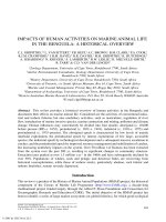
Figure 1
Routes and effects of manganese in a crustacean. Dissolved Mn II in water may enter via the gills
or antennules or get precipitated on the exoskeleton. Entrance may also occur via the food in a variety of
chemical form. Octagonal boxes indicate the route and target tissues of Mn and square boxes indicate the
effects of Mn exposure. Observed effects (
√
) and hypothetical but not yet investigated effects(?).
Mn (ll)
Gills
Midgut
gland
Reproductive
organs
Haemato-
poetic tissue
O
2
uptake/
respiration?
Immune
Suppression √
Storage √
Necrosis ?
Reduced muscle
function √
Fertility ?
No synthesis of
Hc in hypoxia √
Reduced chemo-
sensitivity √
Stomach Antennulae
Mn (ll)Mn
Haemolymph Nerve tissue
Muscle
7044_C002.fm Page 69 Monday, April 17, 2006 1:37 PM
© 2006 by Taylor & Francis Group, LLC
SUSANNE P. BADEN & SUSANNE P. ERIKSSON
70
no change in Ca concentration of whole haemolymph was found in
Nephrops
norvegicus
with
increasing exposure to Mn of 60 mg l
–1
(Selander 1997). This constancy in the whole haemolymph,
however, does not rule out the possibility that Mn has displaced Ca from the haemocyanin to the plasma.
An important source of Mn in the ocean is from hydrothermal vents. The crustaceans adapted
to live close to these vents may hypothetically contain a higher concentration of Mn than non-vent
crustaceans. Professor J.J. Childress from the University of California, Santa Barbara, kindly
provided the authors with haemolymph from a vent crab
,
Bythograea thermydon,
which was found
to have Mn concentrations between 0.44 and 1.6
µ
g g
–1
ww. These Mn concentrations are within
the range of haemolymph concentration from non-vent crustaceans as seen from Table 1. The max-
imum mean Mn concentrations of 7.35 µg g
1
ww in a field-caught crustacean (Nephrops norvegicus)
is reported from the SE Kattegat following a hypoxic period in 1995 (Eriksson & Baden 1998).
The effects of manganese on haemocyanin synthesis and adaptation to hypoxia are described
in a subsequent section discussing the midgut gland, as this is the primary organ for haemocyanin
synthesis (Taylor & Antiss 1999).
The synthesis of haemocytes takes place in the haematopoietic tissue localised as a thin sheet
on the dorsal site of the stomach in crustaceans (Chaga et al. 1995). The haemocytes of crustaceans
consist of hyaline, semigranular and granular cells playing an important role in, for example, the
innate immune defence (Ratcliffe & Rowley 1979, Söderhäll 1981, Söderhäll & Cerenius 1992).
Immunotoxicology of invertebrates is an unexplored field and as a result no early investigations
can be cited. Recently, Hernroth et al. (2004) discovered that when exposed to 20 mg l
–1
Mn for
10 days several immunological processes of N. norvegicus were affected. The number of haemocytes
decreased by 60%. Despite the great loss of haemocytes, renewal through increased proliferation
of the haematopoietic stem cells did not appear to occur. Additionally, maturation of the stem cells
to immune-active haemocytes was inhibited in Mn-exposed lobsters (N. norvegicus). To release the
prophenoloxidase system (ProPO), which is necessary for the immune defence of arthropods, the
granular haemocytes must degranulate. This degranulation activity was also significantly suppressed
after Mn treatment. Furthermore, the activation of ProPO by the non-self molecule, lipopolysac-
caride, was blocked. Probably Mn replaces Ca and thereby inhibits protein required for mobilisation
and activation of the haemocytes.
Immune suppression may explain the occurrence of shell disease caused by microbial infection
of the exoskeleton in blue crab, Callinectes sapidus, from North Carolina, U.S. (Weinstein et al.
1992). The infection is related to elevated Mn concentrations in the body tissues. Similar findings
might explain the high frequency of the parasitic dinoflagellate Hematodinium sp. that has been
found in Nephrops norvegicus from the west coast of Scotland (Field et al. 1992). In the same area
high concentrations of Mn have been recorded in the tissue of this species (Baden & Neil 1998).
Midgut gland
In contrast to other target tissues, where manganese accumulation reaches an equilibrium deter-
mined by the exposure concentration within 5 days, the midgut gland of N. norvegicus continuously
accumulates manganese at a relatively slow rate and does not reach equilibrium after a 3-week
period of exposure. This slow accumulation to the hepatopancreas has also been observed for zinc
in Carcinus maenas by Chan & Rainbow (1993). The elimination rate of manganese from the
midgut gland is, however, much faster. The biological half-lives for accumulation and elimination
of manganese are about 4 and 1.5 days, respectively (Baden et al. 1999). Insoluble granules
containing metals bound with phosphorus or sulphur have been observed in the epithelial cells of
the midgut gland (or comparable organ) in many invertebrates (for review see Ahearn et al. 2004).
The granules scavenge and detoxify surplus metals, and are later eliminated through exocytosis.
Several marine snails have been shown to eliminate manganese this way (Simkiss 1981, Nott &
7044_C002.fm Page 70 Monday, April 17, 2006 1:37 PM
© 2006 by Taylor & Francis Group, LLC
ROLE, ROUTES AND EFFECTS OF MANGANESE IN CRUSTACEANS
71
Nicolaidou 1994). Although no such granules have yet been described in manganese-rich crustaceans,
the surplus of manganese is clearly delivered to the midgut gland for net accumulation as indicated
in Rainbow (1997) and Baden et al. (1999). Accumulation is also demonstrated by the relatively
high levels of Mn in midgut glands from different species of crustaceans (Table 1). The highest
mean tissue concentrations found in the literature are from the midgut glands of a marine hermit
crab, Clibanarius erythropus (1596 µg Mn g
–1
dw) and a freshwater crayfish, Procambarus clarkii
(1677 µg Mn g
–1
dw), both collected in areas with known anthropogenic input (Gherardi et al.
2002, Nott & Nicolaidou 1994). Unfortunately, no background data are available for either of these
species which is why they have not been included in Table 2. However, unpublished data on
background Mn concentrations in another marine hermit crab, Pagurus bernhardus, varied from
15–28 µg Mn g
–1
dw in the midgut gland (Andersson 1993), and the highest overall midgut gland
background concentration published is 374 µg Mn g
–1
dw in a freshwater crayfish, Potamonautes
warreni (Steenkamp et al. 1994; Table 1).
The synthesis of haemocyanin is primarily recognised to take place in the midgut gland
(Taylor & Antiss 1999, for review). In a recent study the combined and separate effects of hypoxia
(2.5 mg l
–1
) and manganese (20 mg l
–1
) on the haemocyanin concentration were investigated after
an exposure period of 2 weeks. Crustaceans adapt to hypoxia by increasing or decreasing (depending
on the initial value) the haemocyanin concentration, presumingly to an optimal concentration
(Spicer & Baden 2001). A simultaneous exposure to manganese affects this adaptation by preventing
the synthesis of haemocyanin (Baden et al. 2003).
Muscle
The manganese concentration of the muscle tissue remains relatively constant throughout the moult
cycle and is less dependent on the exposure concentration of Mn compared with other tissues
(Bryan & Ward 1965, Baden et al. 1995, Baden & Neil 1998, Eriksson & Baden 1998, Bjerregaard &
Depledge 2002). This constancy is especially interesting since the muscle is a metabolically active
tissue with high mitochondrial content. Calculations indicate that an increase in Mn concentration
of muscle tissue after exposure to elevated Mn concentrations can, in principle, be explained by
the increase in Mn in the extracellular haemolymph of the muscle tissue (Hille 1992, Baden et al.
1995). A plausible explanation for the relatively stable concentration in the muscle cells themselves
is, thus, either that turnover rates of manganese in these cells are high enough to disguise increased
uptake (at least for the exposure concentrations that have so far been studied) or that the metal
never enters the muscle cells but remains in the extracellular haemolymph.
Normal muscle concentrations of Mn lie in the range of 0.4–8.0 µg Mn g
–1
dw with the exception
of the extremely high values of 24 µg Mn g
–1
found in small Carcinus maenas by Bjerregaard &
Depledge (2002) and 87 µg Mn g
–1
found in the freshwater crayfish, Potamonautes warreni by
Steenkamp et al. (1994). Many values in the literature are stated as wet weight concentrations with
the primary objective being risk assessment of heavy metals in human food. Taken that the daily
recommended intake for humans is 2.5–5 mg Mn day
–1
, a person would have to eat ca 1 kg of
crustacean meat just to fulfil the daily requirement. Manganese at natural levels in crustaceans is
thus not likely to pose a threat for human consumption.
When lobsters (Nephrops norvegicus) are exposed to 10 mg Mn g
–1
their muscle extension and
thus most probably (consequently) the swimming capacity is affected as will be discussed under
the section ‘Nervous system’.
Exoskeleton
Due to its chemical properties, manganese is found in highest concentrations in the calcified parts
of crustaceans, mainly in the exoskeleton, gills and the gastric mill of the stomach (Bryan & Ward
7044_C002.fm Page 71 Monday, April 17, 2006 1:37 PM
© 2006 by Taylor & Francis Group, LLC
SUSANNE P. BADEN & SUSANNE P. ERIKSSON
72
1965, Baden et al. 1990, 1995, Eriksson & Baden 1998, Eriksson 2000a). Depending on the
thickness of an animal’s exoskeleton the vast majority of manganese is found in this tissue, as it
contains more than 98% of the total Mn content of the decapod lobsters Homarus gammarus (Bryan
& Ward 1965) and Nephrops norvegicus (Baden et al. 1995). The manganese incorporated in the
matrix of the exoskeleton is believed to have little effect on the animals.
The manganese concentration of the exoskeleton changes during the moult cycle, and lobsters
(N. norvegicus) collected in the field show a step-wise increase in average Mn concentration from
postmoult, intermoult to premoult (Eriksson & Baden 1998). The crustacean moult cycle is dom-
inated temporally by the intermoult phase, with brief periods of postmoult and premoult. There is,
however, no correlation between the contemporary environmental Mn(II) concentration of ambient
sea water and that of the exoskeleton in field-caught intermoult lobsters (Eriksson & Baden 1998).
It was thus proposed that the amount of Mn found in the exoskeleton of intermoult individuals
primarily depends on the Mn concentration to which the animals are exposed during the calcification
process at postmoult, rather than the current ambient Mn concentrations (Eriksson & Baden 1998,
Eriksson 2000a). During growth, the shell of the barnacle Balanus amphitrite has been shown to
incorporate Mn in direct proportion to the concentration of the sea water (Hockett et al. 1997).
Unlike most crustaceans, the calcified shells in barnacles grow more or less continuously (Bourget &
Crisp 1975), thus having continuous calcification. In most crustaceans, however, calcification occurs
during a short postmoult period. To test the theory, newly moulted Nephrops norvegicus were
exposed to flow-through sea water with <0.06 mg Mn l
–1
(controls) or 10 mg Mn l
–1
for 20 days
(S.P. Eriksson, unpublished observations). The animals were sacrificed and the Mn concentration
was measured in the exoskeleton and in the cast exuviae (exuviae were removed immediately after
moulting, prior to Mn addition). The cast exuviae showed no difference (one factor ANOVA, F
1,8
=
0.09, P = 0.77, n = 5) between the control group and the (later) Mn-exposed group; Mn concen-
trations were 352 ± 70 and 326 ± 55 (mean ± SE) µg Mn g
–1
dw, respectively. After 20 days the
newly calcified intermoult exoskeletons showed significant differences between the two groups
(one factor ANOVA, F
1,8
= 151, P < 0.001, n = 5). The Mn-exposed animals had exoskeletal Mn
concentrations of 2524 ± 201 µg Mn g
–1
dw (mean ± SE) whereas the control animals contained
only 44 ± 8 µg Mn g
–1
dw. In comparison, an earlier study on intermoult animals also exposed to
10 mg Mn l
–1
dw for 20 days showed a modest increase from 200 µg Mn g
–1
to 290 µg Mn g
–1
dw (Baden et al. 1999). The results, though not extensive, thus appear to support the theory that
intermoult exoskeleton Mn concentrations are mainly the result of prevailing Mn concentrations
during the calcification process.
In contrast, the increase from intermoult to premoult found in N. norvegicus is thought to be
the result of exoskeletal breakdown (Eriksson 2000a). During premoult, crustaceans degrade and
resorb some of the old cuticle. Cuticle components, such as calcium, are stored for later use in
hardening of the new ‘shell’ (Aiken & Waddy 1992). The breakdown of the old cuticle results in a
decreased dry weight/wet weight ratio which in turn also leads to an apparent increase in Mn
concentration from intermoult to premoult (Eriksson 2000a).
Moulting has been suggested as one possible way for decapods (Homarus gammarus, Palaemon
elegans, Systellaspis debilis) to dispose of excess unwanted metals (Bryan & Ward 1965; Ward
1966; White & Rainbow 1984a,b, 1987; Swift 1992). Although crustaceans on occasion eat part
or all of their cast exuviae, preliminary data on Mn uptake from food suggests that Mn incorporated
in exoskeletal parts is not easily accessible when ingested, as described in the section of Mn uptake
from food. Moulting might thus serve as an important regulator of the Mn content providing there
are low Mn(II) concentrations in the water at the time of moult.
Manganese precipitations on the hard-shelled exoskeleton are visible as persistent black dots
mainly in crevices as observed after hypoxia on Nephrops norvegicus in the SE Kattegat (Baden
et al. 1990). Being insoluble, the precipitation of Mn on the exoskeleton is a potential biomarker
7044_C002.fm Page 72 Monday, April 17, 2006 1:37 PM
© 2006 by Taylor & Francis Group, LLC
ROLE, ROUTES AND EFFECTS OF MANGANESE IN CRUSTACEANS
73
of exposure to Mn either from industry or hypoxia. However, the interindividual variation is high
due to patchiness as shown in Baden et al. (1999). The uptake of Mn on mobile appendages of the
exoskeleton is higher than on the non-mobile exoskeleton and shows less variability. Baden & Neil
(2003) showed a linear time- and dose-dependent uptake of Mn and there was no elimination after
2 weeks of return to undosed sea water.
Female reproductive system and fertilized eggs
Martin (1975) showed that the manganese concentration of the ovary of the green crab (Carcinus
maenas) correlated negatively with the Rapport Gonado-Somatique (RGS) gonad index (=matura-
tion stage). Other studies on the lobster Nephrops norvegicus have shown that the Mn concentration
of the oocytes, during maturation and throughout most of the embryogenesis, remains very stable
regardless of ambient Mn concentrations. However, due to stable Mn concentrations but increase
in gonad mass over the maturation period of the oocytes, the Mn load of the whole gonad increased
over time (Eriksson, 2000b).
Egg membranes of decapod crustaceans increase their permeability to water and minerals
dramatically just before hatching (Pandian 1970a,b; Petersen & Anger 1997). This increases the
internal pressure of the egg and, in combination with the weakening of the shell membrane, is
believed to help the larvae to burst the eggshells and hatch. The Mn concentration of eggs from
N. norvegicus is stable at around 5.5 µg g
–1
dw egg
–1
during the first 6 months of development. At
the end of the embryonic development the Mn concentration increases dramatically so that at the
time of hatching (approximately 9 months after fertilization) the eggs have reached concentrations
of 120 µg g
–1
dw egg (Table 1). At this late stage the eggshell gives no protection against external
Mn, and dissolved Mn(II) passes through the eggshell where it is taken up by the embryo (Eriksson
2000b). Manganese can replace calcium at many sites (Nassrallah-Aboukais et al. 1996), and most
of the Mn in aquatic crustaceans is therefore incorporated into calcified regions such as the
exoskeleton and the ossicles and teeth of the gastric mill (Bryan & Ward 1965, Eriksson & Baden
1998, Eriksson 2000a, Steenkamp et al. 1994). Since the cuticle of the zoea larva has shown to be
poorly calcified (Spicer & Eriksson 2003) the dramatic increase in Mn concentration found in the
embryos prior to hatching would most likely not have been caused by Mn being incorporated into
the animal’s cuticle (Eriksson 2000b). The hatched larva is a carnivorous zoea with a complete
functional alimentary canal and it is therefore more likely that the sudden increase in egg Mn
concentration might be explained by the development of the gastric mill in the embryo. Since,
many crustacean embryos are brooded externally in an open clutch on the abdomen of the female,
they will be exposed to prevailing benthic conditions and the Mn concentration of mature eggs
may thus serve as a useful tool to indicate elevated Mn(II) concentrations in the field. This is, of
course, dependent on embryonic mortality not being affected, since the female carrier removes
dead eggs from the egg mass.
Male reproductive system
In astacidean crustaceans the manganese concentration in male reproductive organs (testis, vas
deferens and sperm mass) is relatively high (33.2 µg g
–1
) (as found in N. norvegicus from the field
that were not exposed to Mn) compared with the concentration found in other tissues (Eriksson
2000a). This finding may be explained by the large amount of acidic mucopolysaccaride (AMPS)
containing condroitin sulphate (cartilage precursor) in the vas deferens (Radha & Subramoniam
1985, Subramoniam 1993). The mucopolysaccaride protects the sperm and makes the main part
of the spermatophore delivered to the female spermatheca. The negative charge of this substance
attracts the positive charged metals like manganese. Besides, manganese is an important factor in
the production of chondroitin sulphate (Leach 1971). After in vitro exposure of N. norvegicus to
7044_C002.fm Page 73 Monday, April 17, 2006 1:37 PM
© 2006 by Taylor & Francis Group, LLC
SUSANNE P. BADEN & SUSANNE P. ERIKSSON
74
manganese (20 mg Mn l
–1
) for 14 days the accumulation factor was highest in the testis (× 10)
reaching 100 µg g
–1
dw, whereas the concentration in the vas deferens and sperm mass increased
from about 80 µg g
–1
dw to 210 and 140 µg g
–1
dw, respectively (Krönström 2002).
During mating, the male places a spermatophore in the spermatheca (thelycum) of a newly
moulted female. The gelatinous component of the spermatophore hardens, protecting the contained
spermatozoa. A flap of exoskeleton covers the spermatheca and hardens with the rest of the
exoskeleton following mating (Farmer 1974). After manganese exposure (20 mg Mn l
–1
for 14 days)
the spermatophore in the spermatheca showed an increase in Mn concentration from 10 to 50 µg
g
–1
dw (inner part of spermatheca) and 15 to 40 µg g
–1
dw (outer part of spermatheca). Thus
manganese may reach and hypothetically affect the sperm either from the surrounding water through
the opening of the spermatheca and/or from the body of the female (Krönström 2002).
Central nervous system
A primary target tissue for Mn is the central nervous system. The accumulation of Mn in the nerve
tissue and the effects therein are thus of great importance. The literature on Mn accumulation effects
and toxicity in vertebrate nerve systems is extensive (see above), whereas only a few papers on
this topic exist for invertebrates. The toxicity of the heavy metals Pb, Hg and Cd on synaptic
transmission is reviewed for crustaceans by Devi & Fingerman (1995) and Fingerman et al. (1996).
The biological half-life of Mn (after exposure to 5 & 10 ml Mn l
–1
) in the brain and ganglion of
N. norvegicus is about 1 day for the accumulation of Mn and 2–4 days for Mn elimination, which
is slowest from the brain (Baden et al. 1999). The brain and ganglionic chain may contain about
5 µg Mn g
–1
dw when unexposed whereas the accumulation of Mn by exposed animals resulted
in a four times higher concentration in the brain than in the ganglia during exposure and may reach
250 µg Mn g
–1
dw in the brain when exposed to 10 mg Mn l
–1
for 3 weeks (Baden & Neil 1998).
In the SE Kattegat (Sweden) a concentration of 193 µg Mn g
–1
in the brain of N. norvegicus has
been reported after hypoxic events in the autumn of 1995 (Eriksson & Baden 1998). This could
indicate a field exposure to at least 10 mg Mn l
–1
.
Accumulated manganese has an impact on neuromuscular performance. In crustacean skeletal
muscle, depolarisation involves an inflow of calcium ions rather than sodium ions across the muscle
membrane (Fatt & Ginsborg 1958). Manganese ions can suppress muscle excitation (Suarez-Kurz
1979) by acting as a competitive inhibitor to calcium ion flow through calcium channels in the
muscle membrane (Hagiwara & Takahashi 1967). The neuromuscular performance of N. norvegicus
after manganese exposure was investigated in muscle preparations (Holmes et al. 1999) and in
whole animals (Baden & Neil 1998). Low concentrations (ca 1 mg l
–1
) of manganese increased
the contractile force of the abdominal superficial flexor muscle preparations whereas concentrations
above 5 mg l
–1
Mn successively decreased the contractile force until total abolition at concentrations
above 320 mg l
–1
(Holmes et al. 1999). Exposure of N. norvegicus for 3 weeks to 10 mg l
–1
Mn
affected the free tail flip swimming by reducing the postflip extension by about 40% whereas the
flip flexion was unaffected. The explanation of this difference is probably that the extension involves
a chemical neuromuscular synapse that is known to be affected by manganese, whereas the flexion
is elicited primarily by an electrical synapse not affected by manganese (Baden & Neil 1998).
In decapod crustaceans thin-walled hairs (the aesthetascs) on the first antennae (antennules)
are the major chemoreceptor organs. They play a critical role in orientation toward an odour source,
and are therefore important in social recognition and food search (Devine & Atema 1982). Each
aesthetasc is innervated by over 300 neurones connected to the olfactory neurons of the brain as
described for Homarus americanus by Shepheard (1974). The aesthetascs are very sensitive to
amino acids and Pearson & Olla (1977) found that blue crabs, Callinectes sapidus, can detect clam
extract in concentrations of 10
–15
g l
–1
. The response of the aesthetasc receptor cells to changes in
7044_C002.fm Page 74 Monday, April 17, 2006 1:37 PM
© 2006 by Taylor & Francis Group, LLC
ROLE, ROUTES AND EFFECTS OF MANGANESE IN CRUSTACEANS
75
stimulus concentration is enhanced by movement of the antennules known as ‘flicking’ (Schmitt &
Ache 1979). Flicking decreases the boundary layer thickness, and provides increased odour access
to the receptor cells (Moore et al. 1991). The effect of Mn on chemosensitivity has been investigated
in Nephrops norvegicus exposed to combinations of manganese (0, 10, 20, 40 mg Mn l
–1
) and either
normoxia (8.9 mg O
2
l
–1
) or hypoxia (1.3 mg O
2
l
–1
) for 4 and 10 days (Engdahl 1997). Exposure
length as well as Mn concentration up to 20 mg Mn l
–1
significantly increased the mean flick
frequency by about 15–25%, whereas the frequency decreased significantly between exposures of
20–40 mg Mn l
–1
. When exposed to a combination of hypoxia and increasing Mn concentrations,
the flick frequency decreased significantly. It thus seems that manganese affects the perception of
odour at the aesthetascs. This could be the result of either physical precipitation of Mn on the
aesthetascs, or by chemical action as a neurotoxin in such a way that increasing the flick frequency
may compensate for a reduced perception of the stimulus. Hypoxia or unrealistically high Mn
concentration seemed to hamper this compensation of increased flicking (Engdahl 1997).
Uptake of manganese from food
The ingestion of manganese could potentially be quite significant as Mn can occur in sediment
concentrations of up to 80 mg g
–1
(Elderfield 1976) and large amounts of sediment are frequently
found in the stomachs of N. norvegicus (S.P. Baden, unpublished observations). The oral intake of
manganese via food is sparsely investigated. Most metals including manganese are bound electro-
statically to phosphate or covalently to sulphur and are thus unavailable for digestion. When feeding
hermit crabs with the digestive gland of marine snails the metals of these glands were found to go
straight through the gut of the hermit crab without being absorbed (Nott & Nicolaidou 1994).
In a feeding experiment N. norvegicus, starved for 4 weeks, were individually fed three different
diets ad libitum for 2 weeks (Norstedt 2004). The diets (shrimp muscle, shrimp muscle + exo-
skeleton, shrimp muscle + sediment containing 1.4, 5.4 and 145 µg Mn g
–1
dw, respectively) were
composed to mirror the natural food selection following Baden et al. (1990). No significant differ-
ence in Mn concentration was found in the lobster soft tissue despite the large difference in Mn
concentration of the diets offered. The Mn concentrations obtained were normal for lobsters from
reference areas and were much lower than the concentrations found in N. norvegicus following
hypoxia (Table 2) (Eriksson & Baden 1998, S.P. Baden & S.P. Eriksson, unpublished observations).
Uptake from water via the gills thus seems to be the most important path of Mn into aquatic
crustaceans during hypoxic situations when bioavailable dissolved Mn is at high concentrations.
Excretion of manganese
In aquatic environments the excretion of toxins, including metals, from organisms to the surrounding
media is faster than in the terrestrial environment due in part to the large surface of the gills where
an exchange occurs between the internal liquid of the haemolymph and the external water of
different salinities (Rand et al. 1995).
The excretion of metals including Mn from different tissues of aquatic invertebrates is reviewed
by Viarengo & Nott (1993). From crustaceans, the excretion of metals to the medium may occur
through antennary glands (via the urine), gills, gut and during moulting (e.g., Marsden & Rainbow
2004, for review). The rate and dominant route of excreting excess Mn depends on physical and
chemical factors. Excretion of
54
Mn from the lobsters Homarus gammarus (Bryan & Ward 1965)
and Nephrops norvegicus (Baden et al. 1995) revealed that part of the
54
Mn was excreted through
the antennary glands and also in the faeces. These routes, however, only accounted for a small
portion of the
54
Mn lost. Bryan & Ward (1965) estimated the loss by urinary excretion to be 20–40%
7044_C002.fm Page 75 Monday, April 17, 2006 1:37 PM
© 2006 by Taylor & Francis Group, LLC
SUSANNE P. BADEN & SUSANNE P. ERIKSSON
76
of the total
54
Mn loss, compared with a maximum of 3% in N. norvegicus. The major portion of
loss is suggested to take place via the body surface.
Conclusions
Evaluating the literature on the role, routes and effects of manganese in crustaceans shows that
manganese, though an essential metal, is also an unforeseen toxic metal in the marine environment.
Manganese may occur in toxic concentrations in the bottom water of larger coastal areas after
hypoxia or more locally close to industrial sources. Although the uptake and elimination is rapid
with a half-life of a couple of days, Mn adversely affects physiological processes and can decrease
fitness. Uptake from water seems to be the most important mechanism giving body concentrations
above basic Mn requirements. The main target tissues and accumulation levels in different parts
of the body have been investigated for many marine and freshwater species indicating that the
midgut gland, nerve tissue, blood proteins and parts of the reproductive organs have the highest
accumulation factors. The functional effects of manganese are, however, sparsely investigated.
Recent results show that several steps in a well-functioning immune defence, the perception of
food via chemosensory organs, and normal muscle extension are affected by commonly occurring
concentrations of manganese. To get a more complete understanding of this metal in biological
systems it is necessary to explore why Mn gets accumulated more in brain tissue than other nerve
tissues, why high concentrations of Mn accumulate into the vas deferens wall and sperm mass, and
how sperm viability is affected. Is respiration affected by a precipitation of MnO
2
covering the
gills and by elevated Mn concentrations in the oxygen-carrying protein (haemocyanin) and does
Mn induce hemocytopenia, etc.?
Human concern about metals has mainly focused on highly toxic, rare and unessential heavy
metals, like Pb, Hg and Cd. Due to its common occurrence and possibly also because it is essential,
the potential danger of manganese has been neglected. One has to remember that any metal has
the potential to cause biological damage, it is just a matter of reaching a high enough concentration.
Manganese is widespread and found in very high concentrations, in particular in soft aquatic
sediments. Its bioavailability increases as the result of human impact, and it can become accumu-
lated in biota where it has the potential to cause damage. As more about the mechanisms underlying
metal handling by animals is understood, and the details of human impact on the environment are
further elucidated, more attention is likely to be given to previously overlooked metals, like manganese.
Acknowledgements
We are sincerely grateful to Prof. Robert C. Aller, Prof. Helge H. Baden, Dr. Bodil Hernroth and
Prof. Philip S. Rainbow for inspiration and encouragement during our work and for valuable
comments on the manuscript of this review.
Financial support was received from The Swedish Research Council for Environment, Agri-
cultural Sciences and Spatial Planning (FORMAS no. 22.3/2001-1077) to SPB and from The
Natural Swedish Research Council (VR no. 621-2001-3670) to SPE.
References
Ahearn, G.A., Mandal, P.K. & Mandal, A. 2004. Mechanisms of heavy-metal sequestration and detoxification
in crustaceans: a review. Journal of Comparative Physiology B 174, 439–452.
Aiken, D.E. & Waddy, S.L. 1992. The growth process in crayfish. Reviews in Aquatic Sciences 6, 335–381.
7044_C002.fm Page 76 Monday, April 17, 2006 1:37 PM
© 2006 by Taylor & Francis Group, LLC
ROLE, ROUTES AND EFFECTS OF MANGANESE IN CRUSTACEANS
77
Akyuz, T., Erkan, B.M. & Bassari, A. 2001. Radioisotope excited X-ray flurescence analysis of Asellus
aquaticus Crustacea: Isopoda from Istanbul as an indicator of environmental metal pollution. Journal
of Radioanalytical and Nuclear Chemistry 249, 649–651.
Alikhan, M.A., Bagatto, G. & Zia, S. 1990. The crayfish as a “biological indicator” of aquatic contamination
by heavy metals. Water Research 24, 1069–1076.
Aller, R.C. 1994. The sedimentary Mn cycle in Long Island Sound: its role as intermediate oxidant and the
influence of bioturbation, O
2
, and C
org
flux on diagenetic reaction balances. Journal of Marine Research
52, 259–295.
Al-Mohanna, S.Y. & Subrahmanyam, M.N.V. 2001. Flux of heavy metal accumulation in various organs of
the intertidal marine blue crab, Portunus pelagicus L. from the Kuwait coast after the Gulf war.
Environment International 27, 321–326.
Andersson, I. 1993. Fördelning av koppar och mangan i eremitkräftan Pagurus bernhardus exponerad för
syrebrist. (Distribution of copper and manganese in the hermit crab Pagurus bernhardus exposed to
hypoxia). MSc Thesis, Department of Zooecology, Göteborg University, Sweden, In Swedish.
Anonymous 2005. The International Manganese Institute (IMnI). Paris, France. Online. Available at www.man-
ganese.org/reserves.php and www.manganese.org/intro.php (accessed 4 September 2005).
Aschner, M. & Aschner, J.L. 1991. Manganese neurotoxicity: cellular effects and blood-brain barrier transport.
Neuroscience and Biobehavioral Revues 15, 333–340.
Baden, S.P., Depledge, M.H. & Hagerman, L. 1994. Glycogen depletion and altered copper and manganese
handling in Nephrops norvegicus following starvation and exposure to hypoxia. Marine Ecology
Progress Series 103, 65–72.
Baden, S.P., Eriksson, S.P. & Gerhardt, L. 1999. Accumulation and elimination kinetics of manganese from
different tissues of the Norway lobster Nephrops norvegicus (L.). Aquatic Toxicology 46, 127–137.
Baden, S.P., Eriksson, S.P. & Weeks, J.M. 1995. Uptake, accumulation and regulation of manganese during
experimental hypoxia and anoxia in the decapod Nephrops norvegicus (L.). Marine Pollution Bulletin
31, 93–102.
Baden, S.P., Håkansson, C.L.J. & Spicer, J.I. 2003. Between-individual variation in haemocyanin concentra-
tions in the Norway lobster Nephrops norvegicus following exposure to hypoxia and manganese.
Marine Biology 143, 267–273.
Baden, S.P. & Neil, D.M. 1998. Accumulation of manganese in the haemolymph, nerve and muscle tissue of
Nephrops norvegicus (L.) and its effect on neuromuscular performance. Comparative Biochemistry
and Physiology 119A, 351–359.
Baden, S.P. & Neil, D.M. 2003. Manganese accumulation by the antennule of the Norway lobster Nephrops
norvegicus (L.) as a biomarker of hypoxic events. Marine Environmental Research 55, 59–71.
Baden, S.P., Pihl, L. & Rosenberg, R. 1990. Effects of oxygen depletion on the ecology, blood physiology
and fishery of the Norway lobster, Nephrops norvegicus. Marine Ecology Progress Series 67, 141–155.
Balkas, T.I., Tugrul, S. & Salihoglu, I. 1982. Trace metal levels in fish and Crustacea from northeastern
Mediterranean coastal waters. Marine Environmental Research 6, 281–289.
Balzer, W. 1982. On the distribution of iron and manganese at the sediment/water interface: thermodynamic
versus kinetic control. Geochimica et Cosmochimica Acta 46, 1153–1161.
Bjerregaard, P. & Depledge, M.H. 2002. Trace metal concentrations and contents in the tissue of the shore
crab Carcinus maenas: effects of size and tissue hydration. Marine Biology 141, 741–752.
Blackmore, G. 1999. Temporal and spatial biomonitoring of heavy metals in Hong Kong coastal waters using
Tetraclita squamosa. Environmental Pollution 106, 273–283.
Blasco, J., Arais, A.M. & Saenz, V. 2002. Heavy metal concentrations in Squilla mantis (L.) (Crustacea,
Stomatopoda) from the Gulf of Cádiz: evaluation of the impact of the Aznalcollar mining spill.
Environmental International 28, 111–116.
Bourget, E. & Crisp, D.J. 1975. Factors affecting deposition of the shell in Balanus balanoides. Journal of
the Marine Biological Association of the United Kingdom 55, 231–249.
Brouwer, M., Bonaventura, C. & Bonaventura, J. 1983. Metal ion interactions with Limulus polyphemus and
Callinectes sapidus haemocyanins: stoichiometry and structural and functional consequences of cal-
cium (II), cadmium (II), zinc (II) and mercury (II). Biochemistry 22, 4713–4723.
7044_C002.fm Page 77 Monday, April 17, 2006 1:37 PM
© 2006 by Taylor & Francis Group, LLC
SUSANNE P. BADEN & SUSANNE P. ERIKSSON
78
Bryan, G.W. & Ward, E. 1965. The absorption and loss of radioactive and non-radioactive manganese by the
lobster, Homarus vulgaris. Journal of the Marine Biological Association of the United Kingdom 45,
65–95.
Butt, A.M., Hargittai, P.T. & Lieberman, E.M. 1990. Calcium-dependent regulation of potassium permeability
in the glial perineurum Blood-Brain-Barrier of the crayfish. Neuroscience 38, 175–185.
Canfield, D.E., Thamdrup, B. & Hansen, J.W. 1993. The anaerobic degradation of organic matter in Danish
coastal sediments: Fe reduction, Mn reduction, and sulfate reduction. Geochimica et Cosmochimica
Acta 57, 3867–3884.
Chaga, O., Lignell, M. & Söderhäll, K. 1995. The haemopoietic cells of the freshwater crayfish, Pacifastacus
leniusculus. Animal Biology 4, 59–70.
Chan, H.M. & Rainbow, P.S. 1993. The accumulation of dissolved zinc by the shore crab Carcinus maenas
(L.) Ophelia 38, 13–30.
Chiarandini, D.J., Reuben, J.P., Brandt, P.W. & Grundfest, H. 1970. Effects of caffeine on crayfish muscle
fibres. I. Activation of contraction and induction of Ca spike electrogenesis. Journal of General
Physiology
55, 640–664.
Cotzias, J.C. 1958. Manganese in health and disease. Physiological Revue 38, 503–532.
Couper, J. 1837. On the effects of black oxide of manganese when inhaled into the lungs. British Annals of
Medicine, Pharmacy, Vital Statistics, and General Science (London) 1, 41–42.
da Silva, J.J.R.F. & Williams, J.P. 1991. The Biological Chemistry of the Elements – The Inorganic Chemistry
of the Elements. Oxford, England: Clarendon Press.
Davis, J.M. 1998. Methylcyclopentadienylmanganese tricarbonyl: health risk uncertainties and research direc-
tions. Environmental Health Perpectives 106, suppl 1, 191–201.
Dehairs, F., Baeyens, W. & Van Gansbeke, D. 1989. Tight coupling between enrichment of iron and manganese
in North Sea suspended matter and sedimentary redox processes: evidence for seasonal variability.
Estuarine Coastal and Shelf Science 29, 457–471.
Delahayes, G.F. 1975. Depolarisation-induced movement of Mn
2+
across the cell membrane in the guinea-pig
myocardium. Circulation Research 36, 713–718.
Devi, M. & Fingerman, M. 1995. Inhibition of acetylcholinesterase activity in the central nervous system of
the red swamp crayfish, Procambarus clarkii, by mercury, cadmium, and lead. Bulletin of Environ-
mental Contamination and Toxicology 55, 746–750.
Devine, D.V. & Atema, J. 1982. Function of chemoreceptor organs in spatial orientation of the lobster, Homarus
americanus: differences and overlap. Biological Bulletin (Woods Hole) 163, 144–153.
Drava, G., Capelli, R., Mingati, V., De Pellegrini, R., Relini, L.O. & Ivaldi, M. 2004. Trace elements in the
muscle of red shrimp Aristeus antennatus, Risso, 1816 Crustacea, Decapoda from Ligurian sea NW
Mediterranean: variations related to the reproductive cycle. Science of the Total Environment 321, 87–92.
Elderfield, H. 1976. Manganese fluxes to the oceans. Marine Chemistry 4, 103–132.
Engdahl, S. 1997. Effects of manganese and hypoxia on the chemoreception behaviour of the Norway lobster,
Nephrops norvegicus. MSc Thesis in Marine Zoology, Department of Marine Ecology, Göteborg
University, Sweden.
Eriksson, S.P. 2000a. Temporal variations of manganese in the haemolymph and tissues of the Norway lobster,
Nephrops norvegicus (L.). Aquatic Toxicology 48, 297–307.
Eriksson, S.P. 2000b. Variations of manganese in the eggs of the Norway lobster, Nephrops norvegicus. Aquatic
Toxicology 48, 291–295.
Eriksson, S.P. & Baden, S.P. 1998. Manganese in the haemolymph and tissues of the Norway lobster, Nephrops
norvegicus (L.), along the Swedish west coast, 1993–95. Hydrobiologia 375–376, 255–264.
Farmer, A.S.D. 1974. Reproduction in Nephrops norvegicus (Decapoda: Nephropidae). Journal of Zoology
174, 161–183.
Fatt, P. & Ginsborg, B.L. 1958. The ionic requirements for the production of action potentials in crustacean
muscle fibers. The Journal of Physiology, London 142, 516–543.
Faust, S.D. & Aly, O.M. 1983. Chemistry of Water Treatment. Boston, MA: Butterworths.
Fialkowski, W., Rainbow, P.S., Smith, B.D. & Zmudzinski, L. 2003. Seasonal variation in trace metal con-
centrations in three talitrid amphipods from the Gulf of Gdansk, Poland. Journal of Experimental
Marine Biology and Ecology 288, 81–93.
7044_C002.fm Page 78 Monday, April 17, 2006 1:37 PM
© 2006 by Taylor & Francis Group, LLC
ROLE, ROUTES AND EFFECTS OF MANGANESE IN CRUSTACEANS
79
Field, R.H., Chapman, C.J., Taylor, A.C., Neil, D.M. & Vickerman, K. 1992. Infection of the Norway lobster
Nephrops norvegicus by a Hematodinium-like species of dinoflagellate on the west coast of Scotland.
Diseases of Aquatic Organisms 13, 1–15.
Fingerman, M., Devi, M., Reddy, P.S. & Katyayani, R. 1996. Impact of heavy metal exposure on the nervous
system and endocrine-mediated processes in crustaceans. Zoological Studies 35, 1–8.
Fukunda, J. & Kawa, K. 1977. Permeation of manganese, cadmium, zinc, beryllium through calcium channels
of insect muscle membrane. Science 196, 309–311.
Gavin, C.E., Gunter, K.K. & Gunter, T.E. 1999. Manganese and calcium transport in mitochondria implications
for manganese toxicity. Neurotoxicology 20, 445–453.
Gerber, G.B., Léonard, A. & Hantson, P. 2002. Carcinogenicity, mutagenicity and teratogenicity of manganese
compounds. Critical Reviews in Oncology and Haematology 42, 25–34.
Gherardi, F., Barbaresi, S., Vaselli, O. & Bencini, A. 2002. A comparison of trace metal accumulation in
indigenous and alien freshwater macro-decapods.
Marine and Freshwater Behaviour and Physiology
35, 179–188.
Gräslund, S. & Bengtsson B E. 2001. Chemicals and biological products used in southeast Asian shrimp
farming, and their potential impact on the environment — a review. The Science of the Total Envi-
ronment 280, 93–131.
Hagiwara, S. & Miyazaki, S. 1977. Ca and Na spikes in egg cell membrane. Progress in Clinical Biological
Research 15, 147–158.
Hagiwara, S. & Nakajima, S. 1966. Differences in Na
2+
and Ca
2+
spikes as examined by application of
tetrodotoxin, procaine and manganese ions. Journal of General Physiology 49, 793–806.
Hagiwara, S. & Takahashi, K. 1967. Surface density of calcium ions and calcium spikes in the barnacle muscle
fibre membrane. Journal of General Physiology 50, 583–601.
Hall, I.R., Hydes, D.J., Statham, P.J. & Overnell, J. 1996. Dissolved and particulate trace metals in a Scottish
Sea Loch: an example of a pristine environment? Marine Pollution Bulletin 32, 846–854.
Hendrickx, M.E., Paez-Osuna, F. & Zazueta-Padilla, H.M. 1998. Biology and biochemical composition of the
deep-water shrimp Heterocarpus vicarius Faxon, Crustacea: Decapoda: Caridea: Pandalidae from the
Southeastern gulf of California, Mexico. Bulletin of Marine Science 63, 265–275.
Henriksson, J., Tallkvist, J. & Tjälve, H. 1999. Transport of manganese via the olfactory pathway in rats:
dosage dependency of the uptake and subcellular distribution of the metal in the olfactory epithelium
and the brain. Toxicology and Applied Pharmacology 156, 119–128.
Hernandez, E.H., Discalzi, G., Dassi, P., Jarre, L. & Pira, E. 2003. Manganese intoxication: the cause of an
inexplicable epileptic syndrome in a 3 year old child. Neurotoxicology 24, 633–639.
Hernroth, B., Baden, S.P., Holm, K., Andrén, T. & Söderhäll, I. 2004. Manganese induced immunosupression
of the lobster Nephrops norvegicus (L.). Aquatic Toxicology 70, 223–231.
Heu, M S., Kim, J S. & Shahidi, F. 2003. Components and nutritional quality of shrimp processing by-
products. Food Chemistry 82, 235–242.
Hille, B. 1992. Ionic Channels of Excitable Membranes. Sunderland, MA: Sinauer Associates Incorporated.
Hirata, Y. 2002. Manganese-induced apoptosis in PC12 cells. Neurotoxicology and Teratology 24, 639–653.
Hockett, D., Ingram, P. & LeFurgey, A. 1997. Strontium and manganese uptake in the barnacle shell: electron
probe microanalysis imaging to attain fine temporal resolution of biomineralization activity. Marine
Environmental Research 43, 131–143.
Holmes, J.M., Gräns, A S., Neil, D.M., & Baden, S.P. 1999. Effects of the metal ions Mn
2+
and Co
2+
on
muscle contraction in the Norway lobster, Nephrops norvegicus. Journal of Comparative Physiology B
169, 402–410.
Iregren, A. 1990. Physiological test performance in foundry workers exposed to low levels of manganese.
Neurotoxicology and Teratology 12, 673–675.
Johnson, C.A., Ulrich, M., Sigg, L. & Imboden, D.M. 1991. A mathematical model of the manganese cycle
in a seasonally anoxic lake. Limnology and Oceanography 7, 1415–1426.
Jorhem, L., Engman, J., Sundstrom, B. & Thim, A.M. 1994. Trace elements in crayfish: regional differences
and changes induced by cooking. Archives of Environmental Contamination and Toxicology 26, 137–142.
Katz, B. & Miledi, R. 1969. The effect of divalent cations on transmission in the squid giant synapse.
Pubblicazioni della Stazione Zoologica di Napoli 37, 303–310.
7044_C002.fm Page 79 Monday, April 17, 2006 1:37 PM
© 2006 by Taylor & Francis Group, LLC
SUSANNE P. BADEN & SUSANNE P. ERIKSSON
80
Kress, N., Hornung, H. & Herut, B. 1998. Concentrations of Hg, Cd, Cu, Zn, Fe and Mn in deep-sea benthic
fauna from the southeastern Mediterranean sea: a comparison study between fauna collected at a
pristine area and at two waste disposal sites. Marine Pollution Bulletin 36, 911–921.
Krönström, J. 2002. A study of the reproductive system in the Norway lobster, Nephrops norvegicus —
Accumulation of manganese and acrosome reaction. MSc Thesis, Department of Marine Ecology,
Göteborg University, Sweden.
Laslett, R.E. & Balls, P.W. 1995. The behaviour of dissolved Mn, Ni and Zn in the Forth, an industrialised,
partially mixed estuary. Marine Chemistry 48, 311–328.
Leach, R.M. 1971. Role of manganese in mucopolysaccaride metabolism. Federation Proceedings 30,
991–994.
Luk, E.E. & Culotta, V.C. 2001. Manganese superoxide dismutase in Saccaromyces cerevisiae acquires its
metal co-factor through a pathway involving the Nramp metal transporter, Smf2p. Journal of Biological
Chemistry 276, 47,556–47,562.
Magnusson, K., Ekelund, R., Dave, G., Granmo, Å., Förlin, L., Wennberg, L., Samuelsson, M O., Berggren,
M. & Brorström-Lundén, E. 1996. Contamination and correlation with toxicity of sediment samples
from the Skagerrak and Kattegat. Journal of Sea Research 35, 223–234.
Marsden, I.D. & Rainbow P.S. 2004. Does the accumulation of trace metals in crustaceans affect their
ecology — the amphipod example? Journal of Experimental Marine Biology and Ecology 300,
373–408.
Martin, J L.M. 1974. Metals in Cancer irroratus (Crustacea: Decapoda): concentrations, concentration factors,
discrimination factors, correlations. Marine Biology 28, 245–251.
Martin, J L.M. 1975. Étude comparée du métabolisme et des facteurs de concentration de Fe, Cu, Zn, Mn,
Co et Mg dans l’ovaire de Carcinus maenas et Cancer irroratus durant l’ovogenése. Cahiers de
Biologie Marine 16, 149–157.
Martin, J L.M. 1976. Le métabolisme du manganése chez Cancer irroratus Say, 1817, Crustacé, Décapode.
Cahiers de Biologie Marine 17, 359–373.
Martin, J L.M. & Ceccaldi, H.J. 1976. Métabolisme de Fe, Cu, Zn, Mg, Mn et Co dans les œufs de Cancer
irroratus Crustacé Décapode. Competes Rendus des Seances de la Societe Biologie et de ses Filiales
170, 157–162.
Mena, I., Marin, O., Fuenzalida, S. & Cotzias, G.C. 1967. Chronic manganese poisoning-clinical picture and
manganese turnover. Neurology 17, 128–136.
Moore, P., Atema, J. & Gerhardt, G.A. 1991. Fluid dynamics and microscale chemical movement in the
chemosensory appendages of lobster Homarus americanus. Chemical Senses 16, 663–674.
Mounier, Y. & Vassort, G. 1975. Initial and delayed membrane currents in crab muscle fibre under voltage-
clamp conditions.
The Journal of Physiology, London 251, 589–608.
Nassrallah-Aboukais, N., Boughriet, A., Fischer, J.C., Wartel, M., Langelin, H.R. & Aboukais, A. 1996.
Electron paramagnetic resonance EPR study of Cu
2+
and Mn
2+
ions interacting as probes with calcium
carbonate during the transformation of vaterite into cubic calcite. Journal of the American Chemical
Society — Faraday Transactions 92, 3211–3216.
Nieboer, E. & Richardson, D.H.S. 1980. The replacement of the nondescript “heavy metals” by a biologically
and chemically significant classification of metal ions. Environmental Pollution
1B, 3–26.
Normandin, L., Carrier, G., Gardiner, P.F., Kennedy, G., Hazell, A.S., Mergler, D., Butterworth, R.F.,
Philippe, S. & Zayed, J. 2002. Assessment of bioaccumulation, neuropathology, and neurobehaviour
following subchronic 90 days inhalation in Sprague-Dawley rats exposed to manganese phosphate.
Toxicology and Applied Pharmacology 183, 135–145.
Norstedt, M. 2004. Uptake of manganese through experimental feeding in Norway lobster, Nephrops norveg-
icus. MSc Thesis, Department of Marine Ecology, Göteborg University, Sweden.
Nott, J.A. & Nicolaidou, A. 1994. Variable transfer of detoxified metals from snails to hermit crabs in marine
food chains. Marine Biology 120, 369–377.
Ochi, R. 1970. The slow inward current and the action of manganese ions in guinea-pig’s myocardium. Pflügers
Archives 316, 81–94.
Ochi, R. 1975. Manganese action potentials in mammalian cardiac muscle. Experientia 31, 1048–1049.
7044_C002.fm Page 80 Monday, April 17, 2006 1:37 PM
© 2006 by Taylor & Francis Group, LLC
ROLE, ROUTES AND EFFECTS OF MANGANESE IN CRUSTACEANS
81
Okamoto, H., Takahashi, K. & Yamashita, N. 1977. Ionic currents through the membrane of the mammalian
oocyte and their comparison with those in the tunicate and sea urchin. The Journal of Physiology,
London 267, 465–495.
Paez-Osuna, F., Perez-Gonzalez, R., Izaguirre-Fierro, H., Zazueta-Padilla, M. & Flores-Campana, L.M. 1995.
Trace metal concentrations and their distribution in the lobster Panulirus inflatus Bouvier, 1895, from
the Mexican Pacific coast. Environmental Pollution 90, 163–170.
Palade, P.T. & Almers, W. 1978. Slow Na and Ca currents across the membrane of frog skeletal muscle fibres.
Biophysical Journal 21, 168a.
Pandian, T.J. 1970a. Ecophysiological studies on the developing eggs and embryos of the European lobster
Homarus gammarus. Marine Biology 5, 154–167.
Pandian, T.J. 1970b. Yolk utilization and hatching time in the Canadian lobster Homarus americanus. Marine
Biology 7, 249–254.
Pearson, W.H. & Olla, B.L. 1977. Chemoreception in the blue crab, Callinectes sapidus. Biological Bulletin
(Woods Hole) 153, 346–354.
Petersen, S. & Anger, K. 1997. Chemical and physiological changes during the embryonic development of
the spider crab, Hyas araneus L. Decapoda: Majidae. Comparative Biochemistry and Physiology B
117, 299–306.
Purdey, M. 2000. Ecosystems supporting clusters of sporadic TSEs demonstrate excesses of theradical-
generating divalent cation manganese and deficiencies of antioxidant cofactors Cu, Se, Fe, Zn. Medical
Hypothesis 54, 278–306.
Radha, T. & Subramoniam, T. 1985. Origin and nature of spermatophoric mass of the spiny lobster Panulirus
homarus. Marine Biology 86, 13–19.
Rainbow, P.S. 1992. The significance of accumulated heavy metal concentration in marine organisms. In
Proceedings of a Bioaccumulation Workshop. A.G. Miskiewicz (ed.), Australia, Sydney: Water Board
and Australian Marine Sciences Association Incorporated, 1–13.
Rainbow, P.S. 1997. Trace metal accumulation in marine invertebrates: marine biology or marine chemistry?
Journal of the Marine Biological Association of the United Kingdom 77, 195–210.
Rainbow, P.S. & Blackmore, G. 2001. Barnacles as biomonitors of trace metal availabilities in Hong Kong
coastal waters: changes in space and time. Marine Environmental Research 51, 441–463.
Rainbow, P.S., Fialkowski, W. & Smith, B.D. 1998. The sandhopper Talitrus saltator as a trace metal
biomonitor in the Gulf of Gdansk, Poland. Marine Pollution Bulletin 36, 193–200.
Rainbow, P.S., Smith, B.D. & Lau, S.S.S. 2002. Biomonitoring of trace metal availabilities in the Thames
estuary using a suite of littoral biomonitors. Journal of the Marine Biological Association of the
United Kingdom 82, 793–799.
Rand, G.M., Wells, P.G., & McCarthy, L.S. 1995. Introduction to aquatic toxicology. In Fundamentals of
Aquatic Toxicology, 2nd edition, G.M. Rand & S.R.C Petrocelli (eds), Bristol (Philadelphia), PA:
Taylor and Francis.
Rankin, J.C., Stagg, R.M. & Bolis, L. 1982. Effects of pollutants on gills. In Gills, D.F. Houlihan et al. (eds),
Cambridge (Cambridgeshire); New York: Press syndicate of the University of Cambridge, 207–219.
Ratcliffe, N.A. & Rowley, A.R. 1979. A comparative synopsis of the structure and function of the blood cells
of insects and other invertebrates. Developmental and Comparative Immunology 3, 189.
Ridout, P.S., Rainbow, P.S., Roe, H.S.J. & Jones, H.R. 1989. Concentrations of V, Cr, Mn, Fe, Ni, Co, Cu,
Zn, As and Cd in mesopelagic crustaceans from the North East Atlantic Ocean. Marine Biology 100,
465–471.
Ross, W.N. & Stuart, A.E. 1978. Voltage sensitive calcium channels in the presynaptic terminals of a detri-
mentally conducting photoreceptor. The Journal of Physiology, London 274, 173–191.
Sanders, M.J., DuPreez, H.H. & VanVuren, J.H.J. 1998. The freshwater river crab, Potamonautes warreni, as
a bioaccumulative indicator of iron and manganese pollution in two aquatic systems. Ecotoxicology
and Environmental Safety 41, 203–214.
Saric, M. 1986. Manganese. In Handbook on the Toxicology of Metals, Vol. 2 — Specified Metals. L. Friberg
et al. (eds), Amsterdam: Elsevier, 354–386.
Schmitt, B.C. & Ache, B.W. 1979. Olfactory: response of a decapod crustacean are enhanced by flicking.
Science 205, 204–206.
7044_C002.fm Page 81 Monday, April 17, 2006 1:37 PM
© 2006 by Taylor & Francis Group, LLC
SUSANNE P. BADEN & SUSANNE P. ERIKSSON
82
Selander, E. 1997. Effects of hypoxia and manganese on mortality, glycogen utilisation, and calcium homeo-
stasis of Norway lobster, Nephrops norvegicus (L.) MSc Thesis, Department of Marine Ecology,
Göteborg University, Sweden.
Shepheard, P. 1974. Chemoreception in the antennule of the lobster, Homarus americanus.
Marine and
Freshwater Behaviour and Physiology 2, 262–273.
Shukla, G.S. & Singhal, R.L. 1984. The present status of biological effects of toxic metals in the environment:
lead, cadmium and manganese. Canadian Journal of Physiology and Pharmacology 62, 1015–1031.
Simkiss, K. 1979. Metal ions in cells. Endeavour, New Series 3, 2–6.
Simkiss, K. 1981. Cellular discrimination processes in metal accumulation cells. Journal of Experimental
Biology 94, 317–327.
Simkiss, K. & Taylor, M.G. 1989. Metal fluxes across the membranes of aquatic organisms. Aquatic Sciences
1, 173–188.
Söderhäll, K. 1981. Fungal cell wall beta-1,3-glucans induce clotting and phenoloxidase attachment to foreign
surfaces of crayfish haemocyte lysate. Developmental and Comparative Immunology 5, 565–573.
Söderhäll, K. & Cerenius, L. 1992. Crustacean immunity. Annual Review of Fish Diseases 2, 3–23.
Spicer, J.I. & Baden, S.P. 2001. Environmental hypoxia and haemocyanin variability in Norway lobsters
Nephrops norvegicus L. Marine Biology 139, 727–734.
Spicer, J.I. & Eriksson, S.P. 2003. Does the development of respiratory regulation always accompany the
transition from pelagic larvae to benthic fossorial postlarvae in the Norway lobster Nephrops norveg-
icus L. Journal of Experimental Marine Biology and Ecology 295, 219–243.
Spicer, J.I. & Weber, R.E. 1991. Respiratory impairment in crustaceans and molluscs due to exposure to
heavy-metals. Comparative Biochemistry and Physiology 100C, 339–342.
Steenkamp, V.E., du Preez, H.H., Schoonbee, H.J. & van Eeden, P.H. 1994. Bioaccumulation of manganese
in selected tissues of the freshwater crab, Potamonautes warreni (Calman), from industrial and mine-
polluted freshwater ecosystems. Hydrobiologia 288, 137–150.
Suarez-Kurz, G. 1979. The role of calcium in exitation-contraction sampling in crustacean muscle fibres.
Canadian Journal of Physiology and Pharmacology 60, 446–458.
Subramoniam, T. 1993. Spermatophores and sperm transfer in marine crustaceans. Advances in Marine Biology
29, 129–214.
Swift, D.J. 1992. The accumulation of plutonium by the European lobster Homarus gammarus (L.). Journal
of Environmental Radioactivity 16, 1–24.
Takeda, K. 1967. Permeability changes associated with the action potential in procaine-treated crayfish
abdominal muscle fibres. Journal of General Physiology 50, 1049–1074.
Taylor, H.H. & Antiss, J.M. 1999. Copper and haemocyanin dynamics in aquatic invertebrates. Marine and
Freshwater Research 50, 907–931.
Tjälve, H., Henriksson, J., Tallkvist, J., Larsson, B.S. & Lindquist, N.G. 1996. Uptake of manganese and
cadmium from the nasal mucosa into the central nervous systemvia olfactory pathways in rats.
Pharmacology and Toxicology 79, 347–356.
Tjälve, H., Mejàre, C. & Borg-Neczak, K. 1995. Uptake and transport of manganese in primary and secondary
olfactory neurones in Pike. Pharmacology and Toxicology 77, 23–31.
Trefry, J.H., Presley, B.J., Keeney-Kennicutt, W.L. & Trocine, R.P. 1984. Distribution and chemistry of
manganese, iron, and suspended particulates in Orca Basin. Geo-Marine Letters 4, 125–130.
Van Holde, K.E. & Brenowitz, M. 1981. Subunit structure and physical properties of the haemocyanin of the
giant isopod Bathynomus giganteus. Biochemistry 20, 5232–5239.
Viarengo, A. & Nott, J.A. 1993. Mechanisms of heavy metal cation homeostasis in marine invertebrates.
Comparative Biochemistry and Physiology 104C, 355–372.
Visuthismajarn, P., Vitayavirasuk, B., Leeraphante, N. & Kietpawpan, M. 2005. Ecological risk assessment
of abandoned shrimp ponds in southern Thailand. Environmental Monitoring and Assessment 104,
409–418.
Ward, E.E. 1966. Uptake of plutonium by the lobster Homarus vulgaris. Nature 209, 625–626.
Weinstein, J.E., West, T.L. & Bray, J.T. 1992. Shell disease and metal content of blue crabs, Callinectes
sapidus, from the Albemarle-Pamlico estuarine system, North Carolina. Archives of Environmental
Contamination and Toxicology 23, 355–362.
7044_C002.fm Page 82 Monday, April 17, 2006 1:37 PM
© 2006 by Taylor & Francis Group, LLC
ROLE, ROUTES AND EFFECTS OF MANGANESE IN CRUSTACEANS
83
White, A., Handler, P. & Smith, E.L. 1973. Principles of Biochemistry. Tokyo: McGraw-Hill Kogakusha, Ltd.
Fifth edition.
White, S.L. & Rainbow, P.S. 1984a. Regulation of zinc concentration by Palaemon elegans Crustacea:
Decapoda: zinc flux and effects of temperature, zinc concentration and moulting. Marine Ecology
Progress Series 16, 135–147.
White, S.L. & Rainbow, P.S. 1984b. Zinc flux in Palaemon elegans (Crustacea: Decapoda): moulting, individual
variation and tissue distribution. Marine Ecology Progress Series 19, 153–166.
White, S.L. & Rainbow, P.S. 1987. Heavy metal concentrations and size effects in the mesopelagic decapod
Systellaspis debilis. Marine Ecology Progress Series 37, 147–151.
Williams, R.J.P. 1981. Natural selection of the chemical elements. Proceedings of the Royal Society of London.
Biological Sciences 213, 361–397.
Wollast, R., Billen, G. & Duinker, J.C. 1979. Behaviour of manganese in the Rhine and Scheldt estuaries. I.
Physico-chemical aspects. Estuarine and Coastal Marine Science 9, 161–169.
Xiao, X.H. & Bevan, J.A. 1994. Pharmacological evidence that flow-induced and potassium-induced contrac-
tion of rabbit facial vein may involve the same calcium-entry pathway. Journal of Pharmacology and
Experimental Therapeutics 268, 25–31.
Yamagishi, S. 1973. Manganese-dependant action potentials in intracellularly perfused squid giant axons.
Proceedings of the Japan Academy. Ser. B. Physical and Biological Sciences 49, 218–222.
Young, L.B. & Harvey, H.H. 1991. Metal concentrations in crayfish tissues in relation to lake pH and metal
concentrations in water and sediments. Canadian Journal of Zoology 69, 1076–1082.
7044_C002.fm Page 83 Monday, April 17, 2006 1:37 PM
© 2006 by Taylor & Francis Group, LLC
7044_C002.fm Page 84 Monday, April 17, 2006 1:37 PM
© 2006 by Taylor & Francis Group, LLC