Access: Acid-Base, Fluids, and Electrolytes - part 3 pot
Bạn đang xem bản rút gọn của tài liệu. Xem và tải ngay bản đầy đủ của tài liệu tại đây (320.26 KB, 50 trang )
DISORDERS OF WATER BALANCE 85
TABLE 3–28: Treatment of Hypovolemic Hyponatremia
• Discontinue diuretics, correct GI losses, and expand ECF
volume with normal saline
• ECF volume deficit is replaced to eliminate nonosmotic
AVP release and promote maximally dilute urine
• Replace one-third of the Na
+
deficit over the first 6–12 h
and the remainder over the ensuing 24–48 h
Na
؉
deficit = (total body water)
× (140 – current serum [Na
+
])
• K
+
deficits must be corrected in the setting of hypokalemia
Abbreviations: GI, gastrointestinal; ECF, extracellular fluid; AVP,
arginine vasopressin
TABLE 3–29: Treatment of Euvolemic Hyponatremia
Water restriction is used in the asymptomatic patient
Fluid restriction rarely increases serum [Na
+
] by more than
1.5 mEq/L per day
Demeclocycline (600–1200 mg/day) is used for incurable
SIADH providing that the patient has normal liver function
Conivaptan hydrochloride injection (20 mg load, followed
by 20 mg IV over 24 h) is a V1a/V2 receptor antagonist that
was recently approved for SIADH
Oral vasopressin receptor antagonists are in clinical trials
and may be useful for therapy of SIADH in the future
Abbreviations: SIADH, syndrome of inappropriate antidiuretic
hormone
86 DISORDERS OF WATER BALANCE
TABLE 3–30: Treatment of Hypervolemic Hyponatremia
Hypervolemia is managed with salt and water restriction
An increase in cardiac output will suppress AVP release
in CHF
Large volume paracentesis, albumin infusion, and water
restriction reduces hyponatremia in cirrhotics
Abbreviations: AVP, arginine vasopressin; CHF, congestive
heart failure
TABLE 3–31: Example of Change in TBW to Correct
Hypervolemic Hyponatremia
A 75-kg man has a total body water of 45 L and a serum
[Na
+
] of 115 mEq/L
Desired TBW = (actual serum [Na
+
]/normal serum [Na
+
]) ×
current TBW
Desired TBW = (115/140) × 45 L = 36.9 L
45 L − 36.9 L = 8.1 L must be lost to restore serum [Na
+
]
to 140 mEq/L
Abbreviation: TBW, total body water
DISORDERS OF WATER BALANCE 87
TABLE 3–32: Important Concepts in Therapy
of Hyponatremia
A fear of CPM delays appropriate correction of severe
hyponatremia
• Neurologic sequellae are more commonly related to a slow
correction rate rather than rapid correction
• Hypertonic saline should be employed in hyponatremic
encephalopathy, even in the absence of seizures
• Prevention of seizures and respiratory arrest are critical to
avoid permanent neurologic injury triggered by hypoxia
Rapid correction may occur in patients with the abrupt
withdrawal or correction of a stimulus that inhibits free
water excretion such as liver transplantation, and elderly
women on thiazides in whom the drug is held, and steroid
replacement in the patient with panhypopituitarism
• Magnetic resonance imaging best diagnoses CPM
(changes are seen 1–2 weeks after onset of signs and
symptoms, not immediately)
Patients at high risk for hyponatremic encephalopathy
include premenopausal women in the postoperative setting
• Postoperative patients should never receive hypotonic
solutions
• Normal saline or Ringers lactate are appropriate
SIADH should never be treated with normal saline alone,
as it will result in a further fall in serum Na
؉
concentration
• Monitor the patient closely; a falling serum Na
+
concentration with normal saline administration is highly
suggestive of SIADH
Abbreviations: CPM, central pontine myelinolysis; SIADH,
syndrome of inappropriate antidiuretic hormone
88 DISORDERS OF WATER BALANCE
HYPERNATREMIA
TABLE 3–33: Example of Saline Therapy in SIADH
A patient with SIADH and U
osm
of 600 mOsm/kg is
administered 1 L of normal saline (300 mOsms)
The osmolar load is excreted in 500 mL of urine
300 mOsms/ 600 mOsm/kg (U
osm
) = 500 mL final urine
volume
This results in the generation of 500 mL of free water (rest
of the liter) and a fall in serum Na
+
concentration occurs
Abbreviation: U
osm
, urine osmolality
TABLE 3–34: Pathophysiologic Mechanisms
of Hypernatremia
Hypernatremia is defined as a serum Na
+
concentration
greater than 145 mEq/L
Normally, water loss leads to an increase in osmolality
(hypernatremia), which stimulates both AVP and thirst to
return osmolality back to normal (see Figure 3–3)
A disturbance in either of these homeostatic mechanisms
leads to hypernatremia
Abbreviation: AVP, arginine vasopressin
DISORDERS OF WATER BALANCE 89
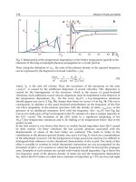
FIGURE 3–3: Net water loss increases serum osmolality
and serum Na
؉
concentration, thereby stimulating both
thirst and AVP production to return water balance
to baseline
90 DISORDERS OF WATER BALANCE
TABLE 3–35: Hypernatremia
Develops in two major settings
• AVP concentration or effect is decreased
• Water intake is less than insensible, GI or renal water losses
■
Inadequate free water intake (access to water or thirst
sensation is impaired) in either the presence or absence
of a urinary concentrating defect
Hypernatremia can result from salt ingestion or
administration of hypertonic saline solutions
The body’s major protective mechanisms include thirst and
the ability of the kidney to reabsorb water from the urine
Serum osmolality and [Na
+
] increase with free water loss
• The rise in serum osmolality has two effects
■
Stimulates thirst
■
Increases AVP release
Normal renal concentration allows for excretion of urine that is
four times as concentrated as plasma (1200 mOsm/kg H
2
O)
Components of the renal concentrating mechanism include
• Generation of a hypertonic interstitium— Henle’s loop acts
as a countercurrent multiplier, which dilutes tubular fluid
and renders the interstitium hypertonic from cortex to papilla
• AVP secretion—The collecting duct is made permeable to
water and allows fluid equilibration with the interstitium
Abbreviations: AVP, arginine vasopressin, GI, gastrointestinal
DISORDERS OF WATER BALANCE 91
ETIOLOGY
Hypernatermia due to renal water loss is broadly categorized as
either central or nephrogenic diabetes insipidus.
TABLE 3–36: Central DI
• Requires destruction of greater than 80% of vasopressin-
producing neurons
• Polyuria (urine volume ranges from 3–15 L/day) is the
most common symptom
• Occurs in young patients with nocturia and is associated
with a preference for cold water
• Complete central DI is associated with inability to
concentrate urine above 200 mOsm/kg with dehydration
• Exogenous AVP increases urine osmolality 100 mOsm/kg
above the value achieved following water deprivation
• Partial DI is associated with a smaller concentrating defect
• Increased P
osm
effectively stimulates thirst, thus serum
Na
+
concentration is only slightly elevated
• Central DI is idiopathic or secondary to head trauma,
surgery, or neoplasm
■
One-third to one-half are idiopathic with a lymphocytic
infiltrate in the posterior pituitary and pituitary stalk
(± circulating antibodies against vasopressin-producing
neurons)
• Familial central DI is rare and inherited in three ways
■
Autosomal dominant disorder (most common)
■
X-linked recessive inheritance
■
Autosomal recessive disorder (very rare)
Abbreviations: DI, diabetes insipidus; AVP, arginine vasopressin
92 DISORDERS OF WATER BALANCE
TABLE 3–37: Nephrogenic DI
Collecting duct does not respond appropriately to AVP
• Inherited forms of nephrogenic DI
• Sex-linked disorder (most common)
■
Caused by mutations in the V2 receptor
• Autosomal dominant and recessive forms
■
Aquaporin-2 gene mutations
■
Results in complete resistance to AVP
•
Acquired nephrogenic DI is more common
but less severe
■
Chronic kidney disease, hypercalcemia, lithium
treatment, obstruction, and hypokalemia are causes
■
Both hypokalemia and hypercalcemia are associated
with a significant downregulation of aquaporin-2
■
Drugs may cause a renal concentrating defect
■
Lithium and demeclocycline cause tubular resistance
to AVP
■
Amphotericin B and methoxyflurane injure the renal
medulla
Abbreviations: DI, diabetes insipidus; AVP, arginine vasopressin
DISORDERS OF WATER BALANCE 93
TABLE 3–38: DI Induced by Degradation of AVP
by Vasopressinase
Develops in women during the peripartum period
Vasopressinase is produced by the placenta and degrades
AVP and oxytocin
It is expressed early in pregnancy and increases in activity
throughout gestation
Desmopressin (dD-AVP), which is not degraded by
vasopressinase, is effective therapy
After delivery vasopressinase becomes undetectable
Abbreviations: AVP, arginine vasopressin; dD-AVP, 1-deamino-
8-D-arginine vasopressin
94 DISORDERS OF WATER BALANCE
SIGNS AND SYMPTOMS
Signs and symptoms of hypernatremia are related to cell swelling
and shrinking.
TABLE 3–39: Signs and Symptoms of Hypernatremia
Neuromuscular irritability with twitches, hyperreflexia,
seizures, coma, and death result from cellular dehydration
The underlying cause of hypernatremia may be the primary
symptom early in hypernatremia
• Polyuria and thirst from DI
• Nausea and vomiting or diarrhea with inadequate
water access
• Hypodipsia or adipsia (central defect in thirst)
Cellular dehydration in the brain is defended by an increase
in brain osmolality
• This is due in part to increases in free amino acids
• The mechanism is unclear, but the phenomenon is referred
to as the generation of idiogenic osmoles
In children, severe acute hypernatremia (serum Na
+
concentration >160 mEq/L) has a mortality rate of 45%
• Two-thirds of survivors have permanent neurological
injury
In adults, acute hypernatremia has a mortality of 75%;
chronic hypernatremia has a mortality of 60%
Hypernatremia is often a marker of serious underlying
disease
Abbreviation: DI, diabetes insipidus
DISORDERS OF WATER BALANCE 95
DIAGNOSIS
TABLE 3–40: Diagnosis of Hypernatremia
Hypernatremia occurs most commonly with hypovolemia,
but can occur in association with hypervolemia and
euvolemia (see Figure 3–4)
A stepwise approach allows appropriate diagnosis of
hypernatremia by assessing thirst, access to water, and the
central production of AVP or effect of AVP on the kidney
Step 1 Is thirst intact?
• If the serum Na
+
concentration >147 mEq/L the patient
should be thirsty
Step 2 If thirsty, can patient get to water?
• This assesses if the thirst center is intact and if the patient
has access to water or other hypotonic solutions
Step 3 Evaluate the hypothalamic-pituitary-renal axis
• This involves an examination of urine osmolality
Abbreviation: AVP, arginine vasopressin
96 DISORDERS OF WATER BALANCE
FIGURE 3–4: Hypernatremia is classified initially based on
ECF volume (Total body Na
؉
content)
DISORDERS OF WATER BALANCE 97
TABLE 3–41: Hypothalamic-Pituitary Axis
An intact axis maximally stimulates AVP release and results
in U
osm
> 700 mOsm/kg when serum Na
+
concentration
>147 mEq/L
Free water losses are often extrarenal if urine osmolality
>700 mOsm/kg
U
osm
less than plasma indicates that there is renal source of
free water loss (central or nephrogenic DI)
Differentiate by the response to exogenous AVP
[subcutaneous aqueous vasopressin (5 units) or intranasal
dD-AVP (10 mcg)]
• Increases urine osmolality by ≥50% in central DI
• No effect on urine osmolality in nephrogenic DI
U
osm
in the intermediate range (300–600 mOsm/kg) may be
secondary to psychogenic polydipsia, osmotic diuresis, and
partial central or nephrogenic DI
Psychogenic polydipsia is associated with a mildly
decreased rather than increased serum Na
+
concentration
Partial central and nephrogenic DI may require a water
deprivation test to distinguish
Abbreviations: AVP, arginine vasopressin; U
osm
, urine osmolality;
DI, diabetes insipidus; dD-AVP, 1-deamino-8-D-arginine vasopressin
98 DISORDERS OF WATER BALANCE
TREATMENT
TABLE 3–42: Water Deprivation Test
Water is prohibited, urine volume and osmolality is
measured hourly, and serum Na
+
concentration and
osmolality is measured every 2h
The test is stopped if any of the following occur
• U
osm
reaches normal levels
• P
osm
reaches 300 mOsm/kg
• U
osm
is stable on two successive readings despite a rising
serum osmolality
• In the last two circumstances exogenous AVP is
administered and the U
osm
and volume measured
■
Partial central DI has urine osmolality increase >50
mOsm/kg
■
Partial nephrogenic DI has no or minimal increase in
urine osmolality
Abbreviations: U
osm
, urine osmolality; P
osm
, plasma osmolality;
AVP, arginine vasopressin; DI, diabetes insipidus
Table 3–43: General Treatment of Hypernatremia
Treatment of hypernatremia is divided into two parts
• Restore plasma tonicity to normal and correct Na
+
imbalances by correcting the water deficit
• Provide treatment directed at the underlying disorder
DISORDERS OF WATER BALANCE 99
Table 3–44: Therapy of Hypernatremia: Correcting
the Water Deficit
Water deficits are restored slowly to avoid sudden shifts in
brain cell volume
•
Increased oral water intake
•
Intravenous administration of hypotonic solution
Serum Na
+
concentration should not be lowered faster than
8–10 mEq/day
The formula below calculates the initial amount of free water
replacement needed (not ongoing losses)
Ongoing renal free water losses should be added to the
replacement calculation
Renal free water losses are calculated as the electrolyte-free
water clearance, dividing urine into two components
• Isotonic component (the volume needed to excrete Na
+
and K
+
at their concentration in serum)
• Electrolyte-free water
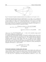
Formula for electrolyte-free water clearance
• Urine volume = C
Electrolytes
+ C
H
2
O
• C
Electrolytes
= (Urine [Na
+
] + [K
+
])/serum [Na
+
])
× urine volume
• C
H
2
O
= the volume of urine from which the
electrolytes were removed during elaboration
of a hypotonic urine
100 DISORDERS OF WATER BALANCE
TABLE 3–45: Example of Treatment of Hypernatremia
A 70-kg male with a history of central DI is found
unconscious; serum [Na
+
] = 160 mEq/L and urine output is
500 mL/h
Urine electrolytes reveal the following values: [Na
+
] = 60
mEq/L, [K
+
] = 20 mEq/L and U
osm
= 180 mOsm/kg
How much water is required to correct the serum [Na
+
]
to 140 mEq/L?
Water needed (L) = (0.6 body weight in kg) ((actual
[Na
+
]/desired [Na
+
]) – 1)
= (0.6 ϫ 70)((160/140) – 1)
= 42 ϫ 0.14 or 6L
If serum [Na
+
] were decreased by 8 mEq/L in the first 24 h,
then 2.4 L of water (100 mL/h) would be required for the
deficit
The serum [Na
+
] increases with this solution because the
calculation did not include the large ongoing free water loss
in urine
To include renal free water losses one must calculate the
electrolyte-free water clearance as illustrated
C
Electrolytes
= ((Urine [Na
+
] + [K
+
])/serum [Na
+
])
ϫ urine volume
C
H
2
O
= Urine volume – C
Electrolytes
Ongoing renal free water losses of 250 mL/h are added to the
replacement solution (100 mL/h), giving a total of 350
mL/h required to correct the serum Na
+
concentration
Abbreviation: DI, diabetes insipidus
DISORDERS OF WATER BALANCE 101
TABLE 3–46: Therapy of Hypernatremia: Based on the
Underlying Disorder
Nephrogenic diabetes insipidus
• Reduce urine volume and renal free water excretion
• Urine volume can be reduced by
■
Decreasing osmolar intake (protein or salt restriction)
■
Increasing U
osm
• Urine volume = solute intake or excretion (the same in the
steady state)/ U
osm
• Thiazide diuretics inhibit urinary dilution and increase
urine osmolality
• Nonsteroidal anti-inflammatory drugs (NSAIDs) inhibit
synthesis of renal prostaglandins (which normally antagonize
AVP effect) and increase concentrating ability
Electrolyte disturbances
• Both hypokalemia and hypercalcemia reduce urinary
concentration and should be corrected
Lithium-induced nephrogenic diabetes insipidus
• Stop lithium and/or use amiloride to ameliorate DI by
preventing entry of lithium into the CCD
Central diabetes insipidus
• Intranasal dD-AVP (5 µg at bedtime) is initiated and
titrated up (5–20 µg once or twice daily)
• Oral desmopressin is an alternative (0.1 mg tablet =
2.5–5.0 µg of nasal spray)
• Drugs that increase AVP release (clofibrate) or enhance its
effect (chlorpropamide, carbamazepine) can be added
Abbreviations: U
osm
, urine osmolality; NSAIDs, nonsteroidal
anti-inflammatory drugs; AVP, arginine vasopressin; DI,
diabetes insipidus; CCD, cortical collecting duct, dD-AVP,
1-deamino-8-D-arginine vasopressin
102 DISORDERS OF WATER BALANCE
TABLE 3–47: Treatment of Central DI
Condition Drug Dose
Complete DI
dD-AVP 5–20 µg intranasal
q 12–24 h
0.1–0.4 mg orally
q12–24 h
Incomplete DI
Chlorpropamide 125–500 mg/day
Carbamazepine 100–300 mg BID
Clofibrate 500 mg QID
Abbreviations: DI, diabetes insipidus; BID, twice a day; QID, four
times a day; dD-AVP, 1-deamino-8-D-arginine vasopressin
103
4
Diuretics
OUTLINE
Introduction 105
4–1. Basics of Diuretics 105
4–2. Renal Regulation of NaCl and Water Excretion 105
Figure 4–1. Sites of Diuretic Action 106
4–3. General Characteristics of Diuretics 107
Sites of Diuretic Action in Kidney 108
4–4. Proximal Tubule 108
4–5. Proximal Tubule Diuretics 109
4–6. Thick Ascending Limb of the Loop of Henle 110
4–7. Loop of Henle Diuretics 111
4–8. Ceiling Doses of IV and Oral Loop Diuretics 112
in Various Clinical Conditions
4–9. Adverse Effects of Loop Diuretics 113
4–10. Distal Convoluted Tubule 113
4–11. DC Tubule Diuretics 114
4–12. Adverse Effects of DCT Diuretics 115
4–13. Cortical Collecting Duct 115
Copyright © 2007 by The McGraw-Hill Companies, Inc.
Click here for terms of use.
104 DIURETICS
4–14. CCD Diuretics 116
4–15. Adverse Effects of CCD Diuretics 117
Diuretic Resistance 118
4–16. Approach to the Patient with Diuretic Resistance 118
Clinical Conditions Associated 120
with Diuretic Resistance
4–17. Congestive Heart Failure and Na
+
Retention 120
4–18. Diuretic Resistance Associated with Nephrotic 121
Syndrome
4–19. Edema Formation in Cirrhosis 122
4–20. Na
+
Contribution to Hypertension 122
4–21. Diminished Diuretic Effect in Kidney Disease 123
Treatment of Diuretic Resistance 123
4–22. Oral versus IV Diuretic Therapy 123
4–23. Advantages of Continuous Diuretic Infusions 124
4–24. Dosing Guidelines for Continuous Infusions 124
of Loop Diuretics
4–25. Synergistic Effect of Combined Loop 125
and DCT Diuretic
4–26. Dosing Guidelines for Diuretics Added to 126
Loop Diuretics
4–27. Monitoring Combination Diuretic Therapy 127
4–28. Cardiovascular Drugs Employed to Enhance 128
Diuresis
DIURETICS 105
INTRODUCTION
TABLE 4–1: Basics of Diuretics
Kidneys regulate ECF volume by modulating NaCl
and water excretion
Diuretics increase the amount of urine formed, due primarily
to inhibition of Na
+
and water reabsorption along the nephron
Diuretics are used to treat a variety of clinical disease states:
• Hypertension, edema, congestive heart failure,
hyperkalemia, and hypercalcemia
Abbreviation: ECF, extracellular fluid volume
TABLE 4–2: Renal Regulation of NaCl and Water Excretion
Na
+
absorption is regulated by several factors:
• Hormones (renin, AII, aldosterone, atrial natriuretic
peptide, prostaglandins, and endothelin)
• Physical properties (mean arterial pressure, peritubular
capillary pressure, and renal interstitial pressure) affect
handling of Na
+
and water
Na
+
reabsorption is driven by Na
+
-K
+
ATPase located on
basolateral membrane
• It provides energy for transporters located on the apical
membrane that reabsorb Na
+
from glomerular filtrate
Cell-specific transporters are present on these tubular cells
• Diuretics enhance renal Na
+
and water excretion by
inhibiting these transporters at different nephron sites
(see Figure 4–1)
Abbreviation: AII, angiotensin II
106 DIURETICS
FIGURE 4–1: Sites of Diuretic Action in the Nephron
DIURETICS 107
TABLE 4–3: General Characteristics of Diuretics
Act on the luminal surface (except spironolactone or
eplerenone) and must enter tubular fluid to be effective
Secretion across the proximal tubule via organic acid or base
transporters is the primary mode of entry (except mannitol,
which undergoes glomerular filtration)
Potency depends on the following:
• Drug delivery to the nephron site of action
• Glomerular filtration rate
• State of the effective arterial blood volume (congestive
heart failure, cirrhosis, and nephrosis)
• Treatment with medications such as NSAIDs and
probenecid (reduce potency)
Diuretics have adverse effects, some that are common to all
diuretics and others that are unique
Abbreviation: NSAIDs, nonsteroidal anti-inflammatory drugs
108 DIURETICS
SITES OF DIURETIC ACTION IN KIDNEY
TABLE 4–4: Proximal Tubule
Na
+
delivered via glomerular filtration
Na
+
transport in the proximal tubular cell is driven by
Na
+
-K
+
ATPase activity
• Energy derived from ATP moves three Na
+
ions out of the
cell in exchange for two K
+
ions
• A reduction of intracellular Na
+
concentration results
• Na
+
moves down its electrochemical gradient from tubular
lumen into the cell via the Na
+
-H
+
exchanger in exchange
for H
+
that moves out
• H
+
secretion is associated with reclamation of filtered
bicarbonate
Abbreviation: ATP, adenosine triphosphate
DIURETICS 109
TABLE 4–5: Proximal Tubule Diuretics
Mannitol
Employed for prophylaxis to prevent ischemic or
nephrotoxic renal injury and to reduce cerebral edema
Nonmetabolizable osmotic agent that is freely filtered, raises
intratubular osmolality and drags water and Na
+
into the
tubule
Active only when given intravenously
Acts within 10 min and has a t
1/2
of approximately 1.2 h in
patients with normal renal function
Toxicity develops when filtration of mannitol is impaired, as
in renal dysfunction
• Retained mannitol increases P
osm
■
Exacerbates CHF, induces hyponatremia, and causes a
hyperoncotic syndrome
■
Contraindicated in patients with CHF and moderate to
severe kidney disease
Nausea and vomiting, and headache are adverse effects
Acetazolamide (primarily proximal tubular)
A CA inhibitor that alkalinizes the urine, prevents and treats
altitude sickness, and decreases intraocular pressure in
glaucoma
Disrupts bicarbonate reabsorption by impairing the
conversion of carbonic acid (H
2
CO
3
) into CO
2
and H
2
O in
tubular fluid and within renal tubular epithelial cells
• Excess bicarbonate in the tubular lumen associates with
Na
+
and exits the proximal tubule
(continued)