Access: Acid-Base, Fluids, and Electrolytes - part 9 potx
Bạn đang xem bản rút gọn của tài liệu. Xem và tải ngay bản đầy đủ của tài liệu tại đây (367.54 KB, 50 trang )
DISORDERS OF SERUM PHOSPHORUS 385
TABLE 10–13 (Continued)
Treatment
Oral sodium phosphate solution should be used with caution
in those above age 55, those with decreased gastrointestinal
motility, patients with decreased GFR, and in the presence
of volume depletion
Renal dysfunction is often irreversible
• Vitamin D intoxication
• High dose liposomal amphotericin (phosphatidylcholine,
phosphatidylserine)
• Solvent detergent treated fresh frozen plasma
■
Contained improper amounts of dihydrogen phosphate
used as a buffer in the purification process
Abbreviations: ECF, extracellular fluid; GFR, glomerular
filtration rate
386 DISORDERS OF SERUM PHOSPHORUS
TABLE 10–14: Signs and Symptoms
Signs and symptoms of hyperphosphatemia are primarily the
result of hypocalcemia (see previous chapter)
Pathophysiology of hypocalcemia induced by
hyperphosphatemia
The most common explanation offered for hypocalcemia
is that the calcium phosphorus product exceeds a certain
level and Ca
2+
deposits in soft tissues and serum Ca
2+
concentration falls
Calcium phosphorus product of > 72 mg/dL is commonly
believed to result in “metastatic” calcification
Short-term infusions of phosphorus increase bone Ca
2+
deposition and reduce bone resorption
Hypocalcemia can also result from decreased calcitriol
concentration from suppression of 1-
α
-hydroxylase by
increased serum phosphorus; these effects may be more
important than physicochemical precipitation
The hypothesis that hypocalcemia results from soft tissue
deposition is inconsistent with the observation in
experimental animals that serum Ca
2+
concentration
continues to decline for up to 5 days after phosphorus
infusions are discontinued and long beyond the time period
when serum phosphorus concentration normalizes
387
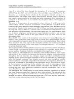
FIGURE 10–2: An Algorithm for the Approach to the Patient with Hyperphosphatemia. The
cause is generally acute kidney injury or chronic kidney disease. Unexplained persistent
hyperphosphatemia should raise the suspicion of pseudohyperphosphatemia, the most
common cause is paraproteinemia secondary to multiple myeloma. No consistent
relationship of immunoglobulin type or subclass was identified. This is a method-dependent
artifact. The assay must be rerun with sulfosalycylic acid deproteinized serum
388 DISORDERS OF SERUM PHOSPHORUS
TABLE 10–15: Treatment
The cornerstone of treatment is reduction of intestinal
phosphorus absorption
Dietary phosphorus restriction
Early in chronic kidney disease hyperphosphatemia can be
controlled with dietary phosphorus restriction
Dietary phosphorus absorption is linear over a wide range of
intakes (4–30 mg/kg/day) and absorption depends on the
amount of dietary phosphorus and its bioavailability
The majority of dietary phosphorus is contained in three
food groups: (1) milk and related dairy products such as
cheese; (2) meat, poultry, and fish; and (3) grains
Processed foods may contain large amounts of phosphorus;
in one study an additional 1154 mg/day of phosphorus was
ingested secondary to phosphorus-containing additives in
fast food with no change in dietary protein intake
Phosphorus contained in plants is largely in the form of
phytate and has low bioavailability since humans do not
express intestinal phytase that is necessary to degrade
phytate and release phosphorus
Phosphorus in meats and dairy products is well absorbed
Inorganic phosphorus salts in processed foods are virtually
completely absorbed and patients with hyperphosphatemia
should avoid these foods including hot dogs, cheese
spreads, colas, processed meats, and instant puddings
Dietary estimates of phosphorus ingestion commonly
underestimate phosphorus intake
DISORDERS OF SERUM PHOSPHORUS 389
TABLE 10–15 (Continued)
Phosphate binders
As chronic kidney disease worsens phosphate binders must
be added
The optimal choice of a phosphate binder remains
controversial
The ideal binder should efficiently bind phosphate, have
minimal effects on comorbid conditions, have a favorable
side-effect profile, and be low in cost; none of the currently
available binders fulfill all of these criteria
Ca
2+
-containing binders are low in cost but may contribute
to net positive Ca
2+
balance and vascular Ca
2+
deposition
Aluminum-containing binders can be employed in the short
term but should be avoided chronically because of
aluminum toxicity (osteomalacia and dementia)
Sevelamer HCl, a synthetic Ca
2+
-free polymer, has a
favorable side-effect profile but is costly
Lanthanum carbonate was recently approved by the FDA;
it is costly and associated with significant GI toxicity
The hyperphosphatemic patient with coexistent
hypocalcemia
It is preferable to first lower serum phosphorus concentration
below 6 mg/dL, if possible, before treating the
hypocalcemia
This is not always possible and clinical judgment must
be used
Abbreviations: GI, gastrointestinal; FDA, Food and Drug
Administration
390 DISORDERS OF SERUM PHOSPHORUS
HYPOPHOSPHATEMIA
TABLE 10–16: Etiologies of Hypophosphatemia
Decreased intestinal absorption
Decreased dietary intake
Phosphate-binding agents
Alcoholism
Redistribution from extracellular to intracellular fluid
Respiratory alkalosis
Refeeding
Diabetic ketoacidosis
Hungry bone syndrome
Sepsis
Increased renal excretion
Primary hyperparathyroidism
Secondary hyperparathyroidism from vitamin D deficiency
with intact renal function
X-linked hypophophatemic rickets
Autosomal dominant hypophosphatemic rickets
Oncogenic osteomalacia
Fibrous dysplasia of bone
DISORDERS OF SERUM PHOSPHORUS 391
TABLE 10–16 (Continued)
Hereditary hypophosphatemic rickets with hypercalciuria
Imatinib mesylate
Fanconi’s syndrome
Osmotic diuresis
Hepatic resection
Pseudohypophosphatemia
392 DISORDERS OF SERUM PHOSPHORUS
TABLE 10–17: Hypophosphatemia-Extrarenal Causes
(Cell Shift)
Shift of phosphorus from ECF to intracellular fluid
Respiratory Alkalosis
Pathophysiology
The rise in intracellular pH that occurs with respiratory
alkalosis stimulates phosphofructokinase, the rate-limiting
step in glycolysis, and phosphorus moves intracellularly
and is incorporated into ATP
Presentation
Severe hypophosphatemia with phosphorus concentrations
less than 0.5–1.0 mg/dL is common
The most common cause of hypophosphatemia in
hospitalized patients
Hypophosphatemia was reported with a rise in pH even
within the normal range in ventilated chronic obstructive
pulmonary disease patients; in concert with the rise in pH
that occurs after intubation serum phosphorus
concentration falls over the span of several hours
Refeeding Syndrome
Pathophysiology
Carbohydrate repletion and insulin release enhance
intracellular uptake of phosphorus, glucose, and K
+
The combination of total body phosphorus depletion from
decreased intake and increased cellular uptake during
refeeding leads to profound hypophosphatemia
DISORDERS OF SERUM PHOSPHORUS 393
TABLE 10–17 (Continued)
Presentation
With refeeding the time of onset of hypophosphatemia depends
on the degree of malnutrition, caloric load, and amount of
phosphorus in the formulation; in undernourished patients it
develops in 2–5 days
Hypophosphatemia can occur with both enteral and parenteral
refeeding
The fall in serum phosphorus concentration is more marked
with liver disease
In adolescents with anorexia nervosa the fall in serum
phosphorus concentration is directly proportional to the
percent loss of ideal body weight
Serum phosphorus concentration rarely declines below
0.5 mg/dL with glucose infusion alone
Treatment of Diabetic Ketoacidosis
Insulin administration results in phosphorus movement into
cells
Renal phosphate loss from osmotic diuresis also contributes
Post Partial Parathyroidectomy for Secondary Hyperpar-
athyroidism—“Hungry Bone Syndrome”
Serum Ca
2+
and phosphorus concentration often fall
abruptly in the immediate postoperative period
From a clinical standpoint hypocalcemia is the more
important management issue
Patients should be observed carefully for hyperkalemia with
Ca
2+
replacement in the postoperative period
(continued)
394 DISORDERS OF SERUM PHOSPHORUS
TABLE 10–17 (Continued)
Sepsis
Catecholamines and cytokines may also cause a phosphorus
shift into cells and this may be the mechanism whereby
sepsis results in hypophosphatemia
Abbreviations: ECF, extracellular fluid; ATP, adenosine
triphosphate
TABLE 10–18: Hypophosphatemia—Extrarenal Causes (GI )
Decreased intestinal absorption
Decreased GI absorption alone is an uncommon cause of
hypophosphatemia since dietary phosphorus intake
invariably exceeds GI losses and the kidney is
extraordinarily effective at conserving phosphorus
decreased dietary intake must be combined with the use of
phosphate binders or increased GI losses as with diarrhea
• Decreased dietary intake
• Phosphate-binding agents
• Alcoholism
Abbreviation: GI, gastrointestinal
DISORDERS OF SERUM PHOSPHORUS 395
TABLE 10–19: Hypophosphatemia—Increased Renal
Phosphate Excretion
(Selective Lesion—PTH Related)
Secondary to an increased concentration of parathyroid
hormone
Primary Hyperparathyroidism
Pathophysiology
Parathyroid hormone stimulates endocytic retrieval of Na
+
-
phosphate cotransporters from the luminal membrane of
the proximal tubular cell
Presentation
Although PTH increases renal phosphate excretion, this is
partially offset by PTH action to increase calcitriol that in
turn increases GI phosphorus absorption, and PTH effect in
bone that results in phosphorus release
Serum phosphorus concentration is rarely below 1.5 mg/dL
Secondary Hyperparathyroidism from Disorders
of Vitamin D Metabolism
Pathophysiology
Secondary hyperparathyroidism from calcitriol deficiency
may be associated with severe hypophosphatemia if the
patient has normal renal function
Presentation
Can present with severe hypophosphatemia
Abbreviations: PTH, parathyroid hormone; GI, gastrointestinal
396 DISORDERS OF SERUM PHOSPHORUS
TABLE 10–20: Hypophosphatemia—Increased Renal
Phosphate Excretion
(Selective Lesion-Phosphatonin Related)
XLH
Pathophysiology
X-linked dominant disorder with a prevalence of 1:20,000
XLH is caused by mutations in the PHEX gene
PHEX is expressed in bone, teeth, and parathyroid gland but
not in kidney
In bone, PHEX is expressed in the osteoblast cell membrane
and plays a role in mineralization
The mutated protein is not expressed in the cell membrane
and is degraded in endoplasmic reticulum
PHEX may play a role in the activation or inactivation of
peptide factors involved in skeletal mineralization, renal
phosphate transport, and vitamin D metabolism
Elevated concentrations of FGF-23 and MEPE were
described
Presentation
Growth retardation, rickets, hypophosphatemia, renal
phosphate wasting, and low serum calcitriol concentration
DISORDERS OF SERUM PHOSPHORUS 397
TABLE 10–20 (Continued)
ADHR
Pathophysiology
Mutations in FGF-23 cause ADHR
FGF-23, a 251-amino acid protein, is secreted and processed at
a cleavage site into inactive N- and C-terminal fragments;
mutations in ADHR occur at the proteolytic site and prevent
cleavage
Presentation
ADHR has a similar phenotype to XLH but is inherited in an
autosomal dominant fashion with variable penetrance
OOM
Pathophysiology
OOM is caused by overproduction of FGF-23, MEPE and
possibly other phosphatonins produced by mesenchymal
tumors
Presentation
Hypophosphatemia, renal phosphate wasting, suppression of
1-
α
-hydroxylase and osteomalacia
The tumor is often difficult to localize
Tumor resection is curative; immunohistochemical staining
shows an overabundance of FGF-23
Fibrous Dysplasia of Bone—Rare
Pathophysiology
In the subset of patients with hypophosphatemia FGF-23
levels are elevated
(continued)
398 DISORDERS OF SERUM PHOSPHORUS
TABLE 10–20 (Continued)
The result of somatic activating missense mutations of
GNAS1 which encodes the alpha subunit of the stimulatory
G protein, G
s
Presentation
McCune-Albright Syndrome—triad of precocious puberty,
café au lait spots, and fibrous dysplasia of bone
Can involve oversecretion of other hormones—thyroid
hormone, parathyroid hormone, pituitary hormones
Congenital disorder presenting with bone pain, deformity,
and fracture involving one (monostotic) or multiple
(polyostotic) bones
Abbreviations: XLH, X-linked hypophosphatemic rickets; PHEX,
phosphate regulating gene with homology to endopeptidases;
FGF, fibroblast growth factor; ADHR, autosomal dominant
hypophosphatemic rickets; OOM, oncogenic osteomalacia; MEPE,
matrix extracellular phosphoglycoprotein
DISORDERS OF SERUM PHOSPHORUS 399
TABLE 10–21: Hypophosphatemia—Increased Renal
Phosphate Excretion (Selective Lesion—Miscellaneous)
HHRH
Autosomal recessive inheritance
Secondary to a loss of function mutation in the sodium-
phosphate cotransporter gene SLC34A3
Presents with hypophosphatemia, rickets, and reduced renal
phosphate reabsorption
Calcitriol levels are increased
Imatinib mesylate
Tyrosine kinase inhibitor
Hypophosphatemia due to increased renal phosphate
excretion in patients treated for CML and gastrointestinal
stromal tumors
Imatinib through its inhibiton of tyrosine kinases may
interfere with osteoclast and osteoblast function
Abbreviation: HHRH, hereditary hypophosphatemic rickets with
hypercalciuria; CML, chronic myelogenous leukemia
400 DISORDERS OF SERUM PHOSPHORUS
TABLE 10–22: Hypophosphatemia—Increased Renal
Phosphate Excretion (Nonselective Lesion)
Fanconi’s Syndrome
Pathophysiology
Caused by a variety of disorders that result in a generalized
proximal tubular transport defect
Inherited—Cystinosis, Wilson’s disease, hereditary fructose
intolerance, and Lowe’s syndrome
Acquired—Multiple myeloma, renal transplantation,
and drugs
Drugs—Ifosfamide, streptozocin, tetracyclines, valproic
acid, ddI, cidofovir, adefovir, tenofovir, and ranitidine
Presentation
Renal phosphate wasting, glycosuria in the face of a normal
serum glucose concentration, and aminoaciduria
Less commonly patients may also have proximal renal
tubular acidosis and hypokalemia
Diagnosis
A urinalysis for glycosuria should be performed
The diagnosis is established by measuring serum and urinary
amino acids and glucose and calculating the fractional
excretion of each
Fanconi’s Syndrome Secondary to Tenofovir
Pathophysiology
Tenofovir is an acyclic nucleoside phosphonate that is
excreted by glomerular filtration and tubular secretion
DISORDERS OF SERUM PHOSPHORUS 401
TABLE 10–22 (Continued)
It enters the tubular cell across the basolateral membrane on
the hOAT1 and exits into the urine on the Mrp2
Since ritonavir inhibits Mrp2, its use with tenofovir could
result in increased toxicity
Presentation
Injury occurs weeks to months after starting treatment
Decreases in creatinine clearance and nephrogenic diabetes
insipidus were also reported
Dent’s Disease
Pathophysiology
Caused by a mutation in the Cl
−
channel CLCN 5
Presentation
Hypophosphatemia and renal phosphate wasting associated
with low molecular weight proteinuria, hypercalciuria,
nephrolithiasis, nephrocalcinosis, and chronic kidney
disease
Chinese Herb Boui-ougi-tou
Used for the treatment of obesity
Renal damage may be related to aristocholic acid
Abbreviations: hOAT1, human organic anion transporter 1; Mrp2,
multi resistant-associated protein 2; CLCN5, chloride channel 5;
PTH, parathyroid hormone; GI, gastrointestinal; MEPE, matrix
extracellular phosphoglycoprotein
402 DISORDERS OF SERUM PHOSPHORUS
TABLE 10–23: Signs and Symptoms
Hypophosphatemia causes a variety of signs and symptoms;
their severity varies with the degree of phosphorus
lowering
Moderate hypophosphatemia—(serum phosphorus
concentration 1.0–2.5 mg/dL)
With the exception of the respiratory system there is little
evidence that moderate hypophosphatemia (phosphorus
concentration 1.0–2.5 mg/dL) results in any clinically
significant morbidity
Correction improved diaphragmatic function in patients with
acute respiratory failure
In two studies patients with moderate hypophosphatemia
had an increase in ventricular arrhythmias; there was no
increase in mortality; more studies are needed to address
this issue
Moderate hypophosphatemia does not impair cardiac
contractility
Moderate hypophosphatemia increases insulin resistance but
the clinical significance of this is unclear
Severe hypophosphatemia (serum phosphorus concen-
tration <1.0 mg/dL) is associated with morbidity
Failure to wean from mechanical ventilation without
correction of severe hypophosphatemia was demonstrated
Severe hypophosphatemia produces reversible myocardial
dysfunction and an impaired response to pressors
DISORDERS OF SERUM PHOSPHORUS 403
TABLE 10–23 (Continued)
Hematologic disturbances include increases in red cell
fragility that lead to clinically significant hemolysis;
associated with reduced red cell ATP levels and large
declines in hemoglobin concentration and hematocrit;
serum phosphorus concentration is often very low
(≤ 0.2 mg/dL)
Hypophosphatemia causes a leftward shift in the oxygen
dissociation curve of unclear clinical significance
A variety of neuromuscular symptoms were reported
including paresthesias, tremor, and muscle weakness
Severe hypophosphatemia causes rhabdomyolysis in dogs
only if there is a preexisting subclinical myopathy; there are
case reports associated with severe hypophosphatemia in
alcoholics
Abbreviation: ATP, adenosine triphosphate
(10-1)
Formula for the fractional excretion (FE) of phosphorus
UP
UP
PCr
Cr P
×
×
× 100
404 DISORDERS OF SERUM PHOSPHORUS
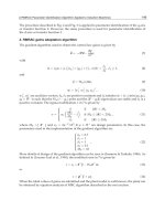
FIGURE 10–3: Approach to the Patient with
Hypophosphatemia
DISORDERS OF SERUM PHOSPHORUS 405
TABLE 10–24: Approach to the Patient with a Low Serum
Phosphorus Concentration
The most common cause of hypophosphatemia in
hospitalized patients is the result of phosphorus shift into
cells secondary to respiratory alkalosis
Primary and secondary hyperparathyroidism are the most
common causes of renal phosphate wasting
Step 1 Evaluate renal phosphorus handling
One can use the FE of phosphorus, 24-h urinary phosphorus,
or calculated renal threshold phosphate concentration
(TmPO
4
/GFR) to determine the kidneys response to
hypophosphatemia
A FE of phosphorus below 5% or a 24-h urine phosphorus
less than 100 mg/day indicates that the kidney is
responding properly to decreased intestinal absorption or
shift of phosphorus into cells
If renal phosphorus wasting is the pathophysiologic reason
for hypophosphatemia, then the FE of phosphorus
exceeds 5% and 24-h urine phosphate excretion is greater
than 100 mg
Step 2 In the patient with increased renal phosphate ex-
cretion one next evaluates the serum Ca
2+
concentration
• Serum Ca
2+
concentration low
• Secondary hyperparathyroidism from disorders of vitamin D
metabolism (normal renal function)
■
Calcidiol and calcitriol concentrations help identify the
defect
406 DISORDERS OF SERUM PHOSPHORUS
TABLE 10–24 (Continued)
• Serum Ca
2+
concentration normal or high
■
Isolated renal phosphate wasting—no glycosuria or
aminoaciduria
■
Primary hyperparathyroidism is by far the most
common diagnosis
■
Associated with high serum Ca
2+
concentration and
low serum phosphorus concentration
■
Diagnosis established by measuring PTH concentration
■
Rare inherited and acquired disorders related to
phosphatonins
■
X-linked hypophosphatemic rickets
■
Autosomal dominant hypophosphatemic rickets
■
Oncogenic osteomalacia
■
Fibrous dysplasia of bone
■
Generalized proximal tubular disorder—associated
with aminoaciduria and glycosuria
■
Fanconi’s syndrome
■
Dent’s disease
Pseudohypophosphatemia
Suspect if severe hypophosphatemia is noted without
symptoms or serum phosphorus concentration remains low
despite repletion
As in the case with pseudohyperphosphatemia paraproteins
can also result in a spuriously low serum phosphorus
concentration
Can be avoided if deproteinized serum is analyzed
Abbreviations: FE, fractional excretion; PTH, parathyroid hormone
DISORDERS OF SERUM PHOSPHORUS 407
TABLE 10–25: Treatment
There is little evidence that treatment of moderate
hypophosphatemia (serum phosphorus concentration
1.0–2.5 mg/dL) is necessary except perhaps in the
mechanically ventilated patient
Severe hypophosphatemia (≤1 mg/dL) or its symptoms are
indications for treatment
In the severely malnourished patient, such as an adolescent
with anorexia nervosa, refeeding must be accomplished
slowly; serum phosphorus concentration should be
monitored closely and the patient placed on telemetry since
sudden death and ventricular arrhythmias were reported
with refeeding
General principles
Hypophosphatemia is commonly associated with other
electrolyte disturbances (hypokalemia and
hypomagnesemia)
One must cautiously replete phosphorus in patients that have
impaired ability to excrete phosphorus loads (those with
decreased GFR) and in patients that are hypocalcemic
One must keep in mind that serum phosphorus concentration
may not be a reliable indicator of total body phosphorus
stores since the majority of phosphorus is contained
within cells
Oral repletion
Most hypophosphatemic patients can be corrected with up to
1 g of supplemental phosphorus per day orally; several forms
of oral phosphorus replacement are listed in Table 10–26
Oral repletion is most commonly limited by diarrhea
(continued)
408 DISORDERS OF SERUM PHOSPHORUS
TABLE 10–25 (Continued)
IV phosphorus administration
IV phosphate administration may be complicated by
hypocalcemia and hyperphosphatemia and is only justified
in those with severe symptomatic phosphorus depletion
Sodium phosphate should be employed except in patients
that require concomitant K
+
supplementation
During IV replacement blood chemistries including serum
phosphorus, Ca
2+
, Mg
2+
, and K
+
should be monitored
closely
Once serum phosphorus concentration has risen above
1 mg/dL, an oral preparation is begun and IV phosphorus
discontinued
Abbreviations: IV, intravenous; GFR, glomerular filtration rate
TABLE 10–26: Phosphorus Replacement (Oral)
Preparation Phosphorus Sodium Potassium
KPhos
neutral
250 mg/tab 13 mEq/tab 1.1 mEq/tab
KPhos
original
114 mg/tab None 3.7 mEq/tab
Fleets
phospho-soda
129 mg/mL 4.8 mEq/mL None
Neutra-phos-K 250 mg/cap None 13.6 mEq/cap
Neutra-phos 250 mg/cap 7.1 mEq/cap 6.8 mEq/cap
Abbreviation: IV, intravenous
DISORDERS OF SERUM PHOSPHORUS 409
TABLE 10–27: Phosphorus Replacement (IV)
Preparation
Phosphorus
(mg/mL)
Phosphorus
(mmol/mL) Sodium Potassium
IV Na
+
phosphate
93 3 4.0
mEq/
mL
None
IV K
+
phosphate
93 3 None 4.4
mEq/mL
Abbreviation: IV, intravenous