Inborn Metabolic Diseases Diagnosis and Treatment - part 3 ppt
Bạn đang xem bản rút gọn của tài liệu. Xem và tải ngay bản đầy đủ của tài liệu tại đây (1.04 MB, 55 trang )
Chapter 6 · The Glycogen Storage Diseases and Related Disorders102
II
Glycogen Metabolism
Glycogen is a macromolecule composed of glucose
units. It is found in all tissues but is most abundant in
liver and muscle where it serves as an energy store, pro-
viding glucose and glycolytic intermediates (
. Fig. 6.1).
Numerous enzymes intervene in the synthesis and deg-
radation of glycogen which is regulated by hormones.
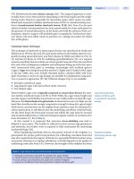
. Fig. 6.1. Scheme of glycogen metabolism and glycolysis. PGK,
phosphoglycerate kinase; P, phosphate; PLD, phosphorylase limit
dextrin; UDPG, uridine diphosphate glucose. Roman numerals
indicate enzymes whose deficiencies cause liver (italics) and/or
muscle glycogenoses: 0 glycogen synthase, I, glucose-6-phos-
phatase;
II, acid maltase (D-glucosidase); III, debranching enzyme;
IV, branching enzyme; V, myophosphorylase; VI, liver phosphory-
lase; VII, phosphofructokinase; IX, phosphorylase-b-kinase;
X, phosphoglycerate mutase; XI, lactate dehydrogenase; XII, fruc-
tose-1,6-bisphosphate aldolase A; XIII, E-enolase
6
103
The liver glycogen storage disorders (GSDs) comprise
GSD I, the hepatic presentations of GSD III, GSD IV, GSD VI,
the liver forms of GSD IX, and GSD 0. GSD I, III, VI, and IX
present similarly with hypoglycemia, marked hepato-
megaly, and growth retardation. GSD I
is the most
severe affecting both glycogen breakdown and gluco-
neogenesis. In GSD Ib there is additionally a disorder of
neutrophil function. Most patients with GSD III have a
syndrome that includes hepatopathy, myopathy, and
often cardiomyo pathy. GSD VI and GSD IX are the least
severe: there is only a mild tendency to fasting hypo-
glycemia, liver size normalises with age, and patients
reach normal adult height. GSD IV manifests in most
patients in infancy or childhood as hepatic failure with
cirrhosis leading to end-stage liver disease. GSD 0
presents in infancy or early childhood with fasting
hypoglycemia and ketosis and, in contrast, with post-
prandial hyperglycemia and hyperlactatemia. Treat-
ment is primarily dietary and aims to prevent hypoglyc-
emia and suppress secondary metabolic decompensa-
tion. This usually requires frequent feeds by day, and in
GSD I and in some patients with GSD III, continuous
nocturnal gastric feeding.
The muscle glycogenoses fall into two clinical
groups. The first comprises GSD V, GSD VII, the muscle
forms of GSD IX (VIII according to McKusick), phospho-
glycerate kinase deficiency (IX
according to McKusick),
GSD X, GSD XI, GSD XII and GSD XIII, and is character-
ised by exercise intolerance with exercise-induced
myalgia and cramps, which are often followed by rhab-
domyolysis and myoglobinuria; all symptoms are
reversible with rest. Disorders in the second group,
consisting of the myopathic form of GSD III, and rare
neuromuscular forms of GSD IV, manifest as sub-acute
or chronic myopathies, with weakness of trunk, limb,
and respiratory muscles. Involvement of other organs
(erythrocytes, central or peripheral nervous system,
heart, liver) is possible, as most of these enzymes de-
fects are not confined to skeletal muscle.
Generalized glycogenoses comprise GSD II, caused
by the deficiency of a lysosomal enzyme, and Danon
disease due to the deficiency of a lysosomal membrane
protein. Recent work on myoclonus epilepsy with
Lafora bodies (
Lafora disease) suggests that this is
a glycogenosis, probably due to abnormal glycogen
synthesis. GSD II can be treated by enzyme replace-
ment therapy, but there is no specific treatment for
Danon and Lafora disease.
The glycogen storage diseases (GSDs) and related disorders
are caused by defects of glycogen degradation, glycolysis
and, paradoxically, glycogen synthesis. They are all called
glycogenoses, although not all affect glycogen breakdown.
Glycogen, an important energy source, is found in most
tissues, but is especially abundant in liver and muscle. In
the liver, glycogen serves as a glucose reserve for the main-
tenance of normoglycemia. In muscle, glycogen provides
energy for muscle contraction.
Despite some overlap, the GSDs can be divi
ded in three
main groups: those affecting liver, those affecting muscle,
and those which are generalized (
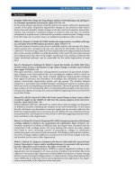
. Table 6.1). GSDs are
d enoted by a Roman numeral that reflects the historical
sequence of their discovery, by the deficient enzyme, or by
the name of the author of the first description. The Fanconi-
Bickel syndrome is discussed in Chap. 11.
6.1 The Liver Glycogenoses
The liver GSDs comprise GSD I, the hepatic presentations
of GSD III, GSD IV, GSD VI, the liver forms of GSD IX, and
GSD 0. GSD I, III, VI, and IX present with similar symp-
toms d uring infancy, with hypoglycemia, marked hepatome-
galy, and retarded growth. GSD I is the most severe of these
four conditions because not only is glycogen breakdown
impaired, but also gluconeogenesis. Most patients with
GSD III have a syndrome that includes hepatopathy, myo-
pathy, and often cardiomyopathy. GSD IV manifests in
most patients in infancy or childhood as hepatic failure with
cirrhosis leading to end-stage liver disease.
GSD VI and
the hepatic forms of GSD IX are the mildest forms: there is
only a mild tendency to fasting hypoglycemia, liver size
normalises with age, and patients reach normal adult height.
GSD 0 presents in infancy or early childhood with fasting
hypoglycemia and ketosis contrasting with postprandial
hyperglycemia and hyperlactatemia. The muscle forms of
GSD III and IX are also discussed in this section.
6.1.1 Glycogen Storage Disease Type I
(Glucose-6-Phosphatase of Translo-
case Deficiency)
GSD I, first described by von Gierke, comprises GSD Ia
caused by deficiency of the catalytic subunit of glucose-6-
phosphatase (G6Pase), and GSD Ib, due to deficiency of the
endoplasmic reticulum (ER) glucose-6-phosphate (G6P)
translocase. There is controversy about the existence of ER
phosphate translocase deficiency (GSD Ic)
and ER glucose
transporter deficiency (GSD Id) as distinct entities. In this
chapter, the term GSD Ib includes all GSD I non-a forms.
Clinical Presentation
A protruded abdomen, truncal obesity, rounded doll face,
hypotrophic muscles, and growth delay are conspicuous
clinical findings. Profound hypoglycemia and lactic acidosis
occur readily and can be elicited by trivial events, such as
delayed meals or reduced food intake associated with inter-
6.1 · The Liver Glycogenoses
Chapter 6 · The Glycogen Storage Diseases and Related Disorders104
II
current illnesses. The liver functions are normal and cir-
rhosis does not develop. In the second or third decade, the
liver’s surface may become uneven and its consistency much
firmer because of the development of adenomas. The kid-
neys are moderately enlarged. The spleen remains normal
in
GSD Ia but is enlarged in most patients with GSD Ib.
Patients bruise easily due to impaired platelet function, and
nosebleeds may be especially troublesome. Skin xanthomas
are seen in patients with severe hypertriglyceridemia, and
gouty arthritis in patients with hyperuricemia. Patients may
also suffer from episodes
of diarrhoea or loose stools.
About one in five GSD I patients has type Ib [1]. Most
patients with GSD Ib develop neutropenia before the age of
1 year, a few at an older age, and even fewer are totally
. Table 6.1. Main features of glycogen storage diseases and related disorders
Type or
synonym
Defective enzyme or transporter Main tissue involved Main clinical symptoms
Liver
Ia
Von
Gierke
Glucose-6-phosphatase Liver, kidney Hepatomegaly, short stature, hypoglycemia, lactatemia,
hyperlipidemia
Ib (non-a) Glucose-6-phosphate translocase Liver, kidney, leucocytes Same as
Ia, neutropenia, infections
III
Cori,
Forbes
Debranching enzyme and sub-
types
Liver, muscle Hepatomegaly, (cardio)myopathy, short stature, hypo-
glycemia
IV
Andersen
Branching enzyme Liver Hepato(spleno)megaly, liver cirrhosis, rare neuromuscu-
lar forms
VI
Hers
Liver phosphorylase Liver Hepatomegaly, short stature,
hypoglycemia
IX Phosphorylase kinase
and subtypes
Liver and/or muscle Hepatomegaly, short stature (myopathy),
hypoglycemia
0G
lycogen synthase Liver Hypoglycemia
Muscle
V
Mc Ardle
Myophosphorylase MuscleMyalgia, exercise intolerance, weakness
VII
Tarui
Phosphofructokinase
and variants
Muscle, erythrocytes Myopathy, hemolytic anemia,
multisystem involvement (seizures, cardiopathy)
– Phosphoglycerate kinase Muscle, erythrocytes,
central nervous system
Exercise intolerance, hemolytic anemia
convulsions
X Phosphoglycerate mutase Muscle Exercise intolerance, cramps
XI Lactate dehydrogenase Muscle Exercise intolerance, cramps, skin lesions
XII Aldolase A Muscle Exercise intolerance, cramps
XIII E-Enolase Muscle Exercise intolerance, cramps
Generalized
II
Pompe
Lysosomal
D-glucosidase
Generalized
in lysosomes
Hypotonia, cardio-myopathy
Infantile, juvenile, adult forms
IIb
Pseudo
Pompe
Danon
Lysosomal-associated
membrane protein 2
Heart, muscle Cardio-myopathy
Lafora Enzyme defect not known Polyglucosan bodies
in all organs
Myoclonic epilepsy, dementia,
convulsions
6
105
spared. Patients with neutropenia show neutrophil dys-
function, including impaired motility and migration and
impaired metabolic burst [2], and suffer with frequent
and severe infections, which can affect the upper and lower
respiratory tract, the skin, the urinary tract, or result in deep
abscesses [3]. More than 75% of the
GSD Ib patients show
symptoms of inflammatory bowel disease (IBD), including
peri-oral and peri-anal infections and protracted diar-
rhoea.
Metabolic Derangement
Among the enzymes involved in hepatic glycogen metabo-
lism, G6Pase is unique since its catalytic site is situated
inside the lumen of the ER. This means that its substrate,
G6P, must cross the ER membrane and requires a trans-
porter. There is still debate over different
proposed models
of G6Pase, over the existence of additional transporters for
its products,
phosphate and glucose [4, 5], and over the
existence of GSD Ic (putative ER phosphate/pyrophosphate
transporter deficiency), and GSD Id (putative ER glucose
transporter deficiency). In particular, patients diagnosed by
enzyme studies as GSD
Ic have been found to have the same
mutations in the G6P translocase gene as in GSD Ib (see
Genetics) [6]. The description of a GSD Id patient has been
withdrawn [7], and no other patient with GSD Ic has been
reported [8].
Hypoglycemia occurs during fasting as soon as exoge-
nous sources of glucose are exhausted, since the final steps
in both glycogenolysis and gluconeogenesis are blocked.
However, there is evidence that GSD I patients are capable
of some endogenous hepatic glucose production [9], al-
though the mechanism is still unclear. Residual G6Pase
activity or the activity
of non specific phosphatases may
result in hydrolysis of G6P to glucose; glycogen may be de-
graded into glucose by amylo-1,6-glucosidase, or autophagy
combined with lysosomal acid maltase activity.
Hyperlactatemia is a consequence of excess G6P that
cannot be hydrolysed to glucose and is further metabolised
in the glycolytic pathway, ultimately generating pyruvate
and lactate. This process is intensified under hormonal
stimulation as soon as the exogenous provision of glucose
fails. Substrates such as galactose, fructose and glycerol
need liver G6Pase to be metabolised to glucose. Conse-
quently ingestion of sucrose and lactose results in hyperlac-
tatemia, with only a small rise in blood glucose [10].
The serum of untreated patients has a milky appearance
due to hyperlipidemia, primarily from increased triglycer-
ides with cholesterol and phospholipids less elevated. The
hyperlipidemia only partially responds to intensive dietary
treatment [11, 12]. The increased concentrations of trigly-
cerides and cholesterol are reflected in increased numbers
of VLDL and LDL particles, whereas the HDL particles are
decreased [13]. VLDL particles are also increased in size
due to the accumulation of triglycerides. Hyperlipidemia is
a result of both increased synthesis from excess of acetyl-
coenzyme A (CoA) via malonyl-CoA, and decreased serum
lipid clearance [14]. Elevated hepatic G6P levels may also
play a role via activation of transcription of lipogenic genes.
Decreased plasma clearance is a result of impaired uptake
and impaired lipolysis of circulating lipoproteins. Reduced
ketone production during
fasting is a consequence of the
increased malonyl-CoA levels, which inhibit mitochon-
drial E-oxidation [15].
Hyperuricemia is a result of both increased production
and decreased renal clearance. Increased production is
caused by increased degradation of adenine nucleotides to
uric acid, associated with d
ecreased intra-hepatic phos-
phate concentration and ATP depletion [16]. Decreased
renal clearance is caused by competitive inhibition of uric
acid excretion by lactate [17].
Genetics
Both GSD Ia and Ib are autosomal recessive disorders. In
1993, the gene encoding G6Pase (G6PC) was identified on
chromosome 17q21. Today more than 75 different muta-
tions have been reported [18, 19]. Subsequently, the gene
encoding the G6P transporter (G6PT) was identified on
chromosome 11q23. More than 65 different mutations have
been reported [20]. Patients formerly diagnosed by enzyme
studies as GSD Ib, Ic and the putative Id shared the same
mutations in G6PT [6]. Recently however, a GSD Ic patient
without mutations in G
6PT was described suggesting the
existence of a distinct GSD Ic locus [21].
Diagnosis
GSD Ia is characterized by deficient G6Pase activity in intact
and disrupted liver microsomes, whereas deficient G6Pase
activity in intact microsomes, and (sub)normal G6Pase
activity in disrupted microsomes, indicates a defect in the
G6P transporter [22]. However, enzyme studies in liver
tissue obtained by biopsy are now usually un-necessary since
the diagnosis can be based on clinical and biochemical find-
ings combined with DNA investigations in leukocytes. If
patients suffer from neutropenia, recurrent infections and/
or IBD, mutation analyses of G6PT should be performed
first [18, 19], although in younger GSD Ib patients these
findings are not always present [3]. If no mutations in G6PC
or in G6PT are identified, a glucose tolerance test should be
performed. A marked decrease in blood lactate concentra-
tion from an elevated level at zero time indicates a gluconeo-
genesis disorder, including GSD I, whereas an increase in
blood lactate concentration suggests one of the other hepatic
GSDs. If the suspicion of GSD I remains, enzyme assays in
fresh liver tissue should be performed.
Identification of mutations in either G6PC or G6PT
alleles of a GSD I index case allows reliable prenatal DNA-
based diagnosis in chorionic villus samples. Carrier detec-
tion in the partners of individuals carrying a known muta-
tion is a reliable option, since a high detection rate is ob-
served for both G6PC and G6PT.
6.1 · The Liver Glycogenoses
Chapter 6 · The Glycogen Storage Diseases and Related Disorders106
II
Treatment
Dietary Treatment
The goal of treatment is, as far as possible, to prevent
hypoglycemia, thus limiting secondary metabolic derange-
ments. Initially, treatment consisted of frequent carbo-
hydrate-enriched meals during day and night. In 1974,
continuous nocturnal gastric drip feeding (CNGDF) via a
nasogastric tube was introduce
d, allowing both patients
and parents to sleep during the night [23].
CNGDF can be used in very young infants. Both a glu-
cose/glucose polymer solution and a formula (sucrose and
lactose-free/low in GSD I) enriched with maltodextrin
are suitable. There are no studies comparing these two
metho
ds. CNGDF should be started within 1 h after the
last meal. Otherwise, a small oral or bolus feed should be
given. Within 15 min after the discontinuation of the
CNGDF, a feed should be given. CNGDF can be given using
a naso gastric tube or by gastrostomy. Gastrostomy is con-
traindicated in GSD Ib patients because of the risk of IBD
and local infections. It is advisable to use a feeding pump
that accurately controls flow rate and has alarms alerting of
flaws in the system. Parents need thorough teaching with
meticulous explanation of technical and medical details and
should feel completely confident with the feeding pump
system.
In 1984 uncooked cornstarch (UCCS), from which
glucose is more slowly released than from cooked starch,
was introduced [24]. During the day, this prolongs the
period between meals, thus improving
metabolic control.
Overnight, it may be used as an alternative for CNGDF.
Theoretically, pancreatic amylase activity is insufficiently
mature in children less than 1 year of age and therefore
UCCS should not be started in these patients. Nevertheless,
it may be effective and useful even in these younger children
[25]. The starting dose of 0.25 g/kg bodyweight should be
increased slowly to prevent side-effects, such as bowel dis-
tension, flatulence, and loose stools, although these side-
effects are usually transient. Precaution is needed in GSD Ib
patients since UCCS may exaggerate IBD. UCCS can be
mixed in water in a starch/water ratio of 1:2. If UCCS is used
overnight, no glucose should be added to avoid insulin re-
lease and an UCCS tolerance test should be performed to
investigate the permissible duration of the fasting period.
It
has been documented that both
CNGDF and UCCS can
maintain normoglycemia during the night with equally
favourable (short-term) results [26, 27]. UCCS is also used
in daytime to prolong the fasting period.
Glucose requirements decrease with age and are calcu-
lated from the theoretical glucose production rate, which va-
ries between 8–9 mg/kg/min
in neonates and 2–3 mg/kg/min
in adults. Only the required amount of glucose should be
given since larger quantities of exogenous glucose may cause
undesired swings in glycemia which make patients more sen-
sitive to rebound hypoglycemia and increases peripheral
body fat storage.
During infections, a freq
uent supply of exogenous glu-
cose must be maintained, even though anorexia, vomiting,
and diarrhoea may make this difficult. Furthermore, glu-
cose metabolism is increased with fever. Replacement of
meals and snacks by glucose polymer drinks is often need-
ed. Nasogastric drip feeding 24 h a day may be
necessary. If
this is not tolerated, a hospital admission is indicated for
intravenous therapy.
There is no consensus as to the extent to which lactate
production from galactose and fructose should be avoided.
Lactate may serve as an alternate fuel for the brain, thereby
protecting
patients against cerebral symptoms from re-
duced glucose levels [28]. Furthermore, milk products,
fruits and vegetables are important sources of vitamins
and minerals. On the other hand, stringent maintenance
of normolactatemia by complete avoidance of lactose
and fructose ingestion may lead to a more favourable out-
come [29].
The dietary plan should be carefully designed and fol-
lowed to provide enough essential nutrients as recommend-
ed by the WHO. Otherwise, supplementation should be
started. Special attention should be directed to calcium
(limited milk intake) and vitamin D. Furthermore, increas-
ed carbohydrate metabolism needs an adequate
supply of
vitamin B
1
.
Prior to elective surgery, bleeding time (platelet aggrega-
tion) should be normalised by continuous gastric drip
feeding for several days or by intravenous glucose infusion
over 24–48 hours. Close peri-operative monitoring of blood
glucose and lactate concentration is essential.
Pharmacological Treatment
Until recently, (sodium)bicarbonate was recommended to
reduce hyperlactatemia. Bicarbonate also induces alkalisa-
tion of the urine, thereby diminishing the risk of urolithiasis
and nephrocalcinosis. However, it was found that a progres-
sive worsening of hypocitraturia occurs [30] so that alkali-
sation with citrate may be even more beneficial in prevent-
ing or ameliorating urolithiasis and nephrocalcinosis.
Uric acid is a potent radical scavenger and it may be a
protective factor against the development of atherosclerosis
[31]. Consequently, it is recommended to accept a serum uric
acid concentration within the high normal range. To prevent
gout and urate nephropathy, however, a xanthine-oxidase
inhibitor (allopurinol) should be started if it exceeds this.
If persistent microalbuminuria is present, a (long-act-
ing) angiotensin converting enzyme (ACE) inhibitor should
be started to reduce or prevent further deterioration of renal
function. Addi tiona l blood pressure lowering drugs should
be used if blood pressure remains above the 95th percentile
for age.
To reduce the risk of cholelithiasis and pancreatitis, tri-
glyceride-lowering drugs (nicotinic acid, fibrates) are indi-
cated only if severe hypertriglyceridemia persists. Choles-
terol-lowering drugs are not indicated in younger patients.
6
107
In adult patients however, progressive renal insufficiency
may worsen the hyperlipidemia and atherogenecity, and in
such cases statins may be indicated, although there is at present
no evidence of their efficacy. Fish-oil is not indicated since its
effect on reducing serum triglycerid
e and cholesterol is not
long lasting and it may even lead to increased lipoprotein
oxidation, thereby increasing atherogenecity [32].
There is no place for growth hormone the r apy since,
although it may enhance growth, it does not improve final
height. Similarly, neither are oest rogen and testosterone in-
dicated to
enhance pubertal development as they do not
improve final height scores. Ethinyloestradiol should be
avoided both because of its association with liver adenomas
and its incompatibility with hyperlipidemia. Oral contr acep-
tion is possible with high doses of progestagen from the
5th to the 25th day
of the cycle or with daily administration
of low doses of progestagen [33].
The benefits of prophylaxis with oral antibiotics have
not been studied in neutropenic GSD Ib patients. However,
prophylaxis with cotrimoxazol may be of benefit in sympto-
matic patients or in those with a neutrophil count < 500 u
10
6
/l [34].
Although patients with GSD Ib and neutropenia have
been treated with granulocyte colony-stimulating factor
(GCSF) from 1989 and it is widely thought that the severity
of infections decreases and IBD regresses, no unequivocal
improvement in outcome has been established [35]. It is
advised to limit the use of GCSF to one or more of the fol-
lowing indications: (1) a persistent neutrophil count below
200 u 10
6
/1; (2) a single life threatening infection requiring
antibiotics intravenously; (3) serious IBD documented by
abnormal colonoscopy and through biopsies; or (4) severe
diarrhoea requiring hospitalisation or disrupting normal
life [36]. Patients generally respond to low doses (starting
dose 2.5 µg/kg every other day). Data on the safety and
efficacy of long-term GCSF administration are limited. The
most serious frequent complication is splenomegaly includ-
ing hypersplenism. Reports of acute myelogenous leukemia
[37] and renal carcinoma [38] arising during long-term use
of GCSF make stringent follow-up necessary. Bone marrow
aspiration with cytogenetic studies before treatment and
once yearly during GCSF treatment are advised, along with
twice yearly abdominal ultrasound.
Follow-up, Complications, Prognosis, Pregnancy
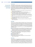
The biomedical targets are summarised in . Table 6.2 and
are based on what level of abnormality constitutes an added
health risk [39]. One should attempt to approach these
targets as far as possible, but without reducing the quality of
life. A single blood glucose assay in the clinical setting is not
useful; it is preferable to make
serial glucose measurements
at home preprandially and in the night over 48–72 h. Lactate
excretion in urine should be estimated in samples collected
at home and delivered frozen [40, 41]. Serum uric acid,
cholesterol and triglyceride concentrations, and venous
blood gases should be estimated during each
outpatient visit.
A good marker for the degree of apparent asymptomatic IBD
activity in GSD Ib is faecal alpha-1-antitrypsine [42].
Intensive dietary treatment with improved metabolic
and endocrine control has led to reduced morbidity and
mortality, and improved quality of life [29]. Long-term
cerebral function is normal if hypoglycemic damage is pre-
vented. Most patients are able to lead fairly normal lives.
With ageing, however, patients may develop complications
of different organ systems [1, 25, 43].
Proximal and distal renal tubular as well as glomerular
functions are at risk [44, 45]. Proximal renal tubular dys-
function is observed in patients with poor metabolic con-
trol and improves after starting intensive dietary treatment
[46]. However, distal renal tubular dysfunction can occur
even in patients with optimal metabolic control and may
lead to hypercalciuria and hypocitraturia [47, 48]. Regular
ultrasonography of the kidneys is recommended. Progres-
sive glomerular renal disease starts with a silent period of
hyperfiltration that begins in the first years of life [49].
Microalbuminuria may develop at the end of the first or in
the second decade of life and is an early manifestation of the
progression of renal disease [50]. Subsequently,
proteinuria
and hypertension may develop, with deterioration of renal
function leading to end-stage renal disease in the 3rd–5th
decade of life. The similarities in the natural history of renal
disease in GSD I and of nephropathy in insulin dependent
diabetes mellitus is striking. The pathogenesis however, is
still unclear. As in diabetic nephropathy, ACE inhibitors
should be started if microalbuminuria persists over a period
of 3 months with a moderate dietary restriction of protein
and sodium. Hemodialysis, continuous ambulatory peri-
toneal dialysis and renal transplantation are therapeutic
options for end-stage renal disease in GSD I.
Single or multiple liver adenomas may develop in the
second or third decade [51, 52] but remain unchanged
. Table 6.2. Biomedical targets in GSD I
1. Preprandial blood glucose >3.5–4.0 mmol/l (adjusted
to target 2)
2. Urine lactate/creatinine ratio <0.06 mmol/mmol
(or urine lactate <0.4–0.6 mmol/l)
3. Serum uric acid concentration in high normal range for
age and laboratory
4. Venous blood base excess >–5 mmol/l and venous
blood bicarbonate >20 mmol/l
5. Serum triglyceride concentration <6.0 (<10.0 mmol/l in
adult patients)
6. Normal faecal alpha-1-antitrypsine for GSD Ib patients
7. Body mass index <+2.0 SDS (in growing children
between 0 and +2.0 SDS)
6.1 · The Liver Glycogenoses
Chapter 6 · The Glycogen Storage Diseases and Related Disorders108
II
during many years of intensive dietary treatment; a reduc-
tion in size and/or number has been observed in some pa-
tients following optimal metabolic control. Liver adenomas
may cause mechanical problems and acute haemorrhage;
further more, they may develop into carcinomas. To screen
for adenomas and to follow
their size and number, ultra-
sonography should be performed regularly. Increase in size
of nodules or loss of definition of their margins necessitate
further investigations, such as CT scans or MRI. In addi-
tion, serum D-fetoprotein and carcino-embryonal antigen
can be used to screen for malignant transformation. How-
ever,
neither CT nor MRI are highly predictive of malignant
transformation, and false negative results for both tumour
markers have been reported [53]. The management of liver
adenomas is either conservative or surgical. In severe cases
of adenomas, enucleation or partial liver resection are ther-
apeutic options. Where there is
a recurrence of adenomas
or suspected malignant transformation, liver transplanta-
tion is a therapeutic option provided there are no metas-
tases [54]. Liver transplantation also corrects glucose home-
ostasis, but in GSD 1b does not correct neutropenia and
neutrophil dysfunction, nor does it prevent the develop-
ment of renal failure [55]. Immunosuppression
may worsen
renal function.
Osteopenia appears to be a result of both decreased bone
matrix formation and decreased mineralisation [56, 57].
Limited peak bone mass formation increases the risk of
fractures later in life. It is important for normal bone forma-
tion to suppress secondary metabolic and hormonal dis-
turbances, especially chronic lactatemia.
Anemia is observed at all ages, but especially in adoles-
cent and adult patients [1, 43]. The anemia may be refrac-
tory to iron because of inappropriate production, by hepatic
adenomas, of hepcidin, a peptide hormone that controls the
release of iron from intestinal cells and macrophages [58].
Polycystic ovaries (PCOs) have been observed in adoles-
cent and adult female patients [59]. Their pathophysiology
is unresolved and their effects on reproductive function are
unclear. PCOs may cause acute abdominal pain as a result
of vascular disturbances. This should be differentiated from
pancreatitis and haemorrhage into liver adenoma.
Despite severe hyperlipidemia, car d io vascular morbidity
and mortality is infrequent and, when present, may be re-
lated to secondary metabolic changes caused by the pro-
gressive renal disease. The preservation of normal endothe-
lial function [1, 43, 60] may
result from diminished platelet
aggregation [61], increased levels of apolipoprotein E [62],
decreased susceptibility of LDL to oxidation – possibly re-
lated to the altered lipoprotein fatty acid profile in GSD Ia
[32] – and increased antioxidative defences in plasma pro-
tecting against lipid peroxidation [31].
A vascular complication that may cause more morbid-
ity and mortality in the ageing patient is pulmonary hyper-
tension followed by progressive heart failure [63]. It may
develop in the second decade or later. No specific treatment
is available. Monitoring by ECG and cardiac ultrasonogra-
phy is recommended after the first decade.
Depressive illness needing therapy is observed rather
frequently in adult patients [1, 43]. Lifelong intensive
dietary treatment 24 hours a day, together with the threat
of serious medical problems,
is a major burden for both
patients and their parents.
Successful pregnancies have been reported [1, 33]. Close
supervision and reintroduction of intensive dietary treat-
ment is necessary.
6.1.2 Glycogen Storage Disease Type III
(Debranching Enzyme Deficiency)
The release of glucose from glycogen requires the activity of
both phosphorylase and glycogen debranching enzyme
(GDE). GSD III, also known as Cori or Forbes disease, is an
autosomal recessive disorder due to deficiency of GDE
which causes storage of glycogen with an abnormally com-
pact structure, known as
phosphorylase limit dextrin. Dif-
ferences in tissue expression of the deficient GDE explain
the existence of various subtypes of GDS III. Most patients
with GSD III have a generalized defect in which enzyme
activity is deficient in liver, muscle, heart, leukocytes and
cultured fibroblasts, and have a syndrome that includes
both hepatic and myopathic symptoms, and often cardio-
myopathy (GSD IIIa). About 15% of patients only have
symptoms of liver disease and are classified as GSD IIIb.
Subgroups due to the selective deficiency of either the glu-
cosidase activity (GSD IIIc) or of the transferase activity
(GSD IIId) are very rare.
Clinical Presentation
Hepatic Presentation
Hepatomegaly, short stature, hypoglycemia, and hyper-
lipidemia predominate in children, and this presentation
may be indistinguishable from GSD I. Splenomegaly can be
present, but the kidneys are not enlarged and renal function
is normal. With increasing age, these symptoms improve in
most GSD III patients and may disappear around puberty.
Myopathic Presentation
Clinical myopathy may not be apparent in infants or chil-
dren, although some show hypotonia and delayed motor
milestones. Myopathy often appears in adult life, long after
liver symptoms have subsided. Adult-onset myopathies
may be distal or generalised. Patients with distal myopathy
develop atrophy of leg and
intrinsic hand muscles, often
leading to the diagnosis of motor neurone disease or peri-
pheral neuropathy [64]. The course is slowly progressive
and the myopathy is rarely crippling. The generalised myo-
pathy tends to be more severe, often affecting respiratory
muscles. In the EMG, myopathic features are mixed with
irritative features (fibrillations, positive sharp waves,
6
109
myotonic discharges), a pattern that may reinforce the diag-
nosis of motor neurone disease in patients with distal
muscle atrophy. Nerve conduction velocities are often
d ecreased [65]. Although GDE works hand-in-hand with
myophosphorylase and one would therefore expect GDE
deficiency to cause symptoms similar to those of
McArdle
disease, cramps and myoglobinuria are exceedingly rare.
Muscle biopsy typically shows a vacuolar myopathy. The
vacuoles contain PAS-positive material and corresponds to
large pools of glycogen, most of which is free in the cyto-
plasm. However, some of the glycogen is present within
lysosomes. Biochemical analysis shows
greatly increased
concentration of phosphorylase-limit dextrin, as expected.
In agreement with the notion that the enzyme defect is
generalised, peripheral neuropathy has been documented
both electrophysiologically and by nerve biopsy and may
contribute to the weakness and the neurogenic features of
some patients. Similarly, while symptomatic cardiopathy is
uncommon, cardiomyopathy (similar to idiopathic hyper-
trophic cardiomyopathy) is detectable in virtually all pa-
tients with myopathy [66].
Metabolic Derangement
GDE is a bifunctional enzyme, with two catalytic activities,
oligo-1,4o1,4-glucantransferase and amylo-1,6-gluco-
sidase. After phosphorylase has shortened the peripheral
chains of glycogen to about four glycosyl units, these re-
sidual stubs are removed by GDE in two steps. A maltotriosyl
unit is transferred from a donor to an acceptor chain (trans-
ferase activity), leaving behind a single glucosyl unit, which
is hydrolysed.
During infancy and childhood patients suffer from
fasting hypoglycemia, associated with ketosis and hyper-
lipidemia. Serum transaminases are also increased in child-
hood but decrease to (almost) normal values with increas-
ing age. In contrast to GSD I, blood lactate concentration is
normal. Elevated levels of serum creatine kinase (CK) and
aldolase suggest muscle involvement, but normal values do
not exclude the future development of myopathy.
Genetics
The gene for GDE (GDE) is located on chromosome 1p21.
At present, at least 48 different mutations in the GDE gene
have been associated with GSD III. GSD IIIb is associated
with mutations in exon 3, while mutations beyond exon 3
are associated with GSD IIIa. When all known GSD III
mutations
are taken into consideration, there is no clear
correlation between the type of mutation and the severity of
the disease. This makes prognostic counselling based on
mutations difficult [67].
Diagnosis
Diagnosis is based on enzyme studies in leukocytes, erythro-
cytes and/or fibroblasts, combined with DNA investigations
in leukocytes. Prenatal diagnosis is possible by identifying
mutations in the GDE gene if these are already known. If
not, polymorphic markers may be helpful in informative
families. Prenatal diagnosis based on GDE activity in cul-
tured amniocytes or chorionic villi is technically difficult
and does not always discriminate between the carrier state
and the affected fetus.
Treatment
The main goal of dietary treatment is prevention of hypo-
glycemia and correction of hyperlipidemia. Dietary man-
agement is similar to GSD Ia but, since the tendency to
develop hypoglycemia is less marked, only some younger
patients will need continued nocturnal gastric drip feeding,
and a late evening
meal and/or uncooked corn starch
will
be sufficient to maintain normoglycemia during the night.
In GSD III (as opposed to GSD I), restriction in fructose
and galactose is unnecessary and dietary protein intake can
be increased since no renal dysfunction exists. The latter
would not only have a beneficial effect on
glucose homeos-
tasis, but also on atrophic myopathic muscles.
Complications, Prognosis, Pregnancy
With increasing age, both clinical and biochemical abnor-
malities gradually disappear in most patients; parameters of
growth normalise, and hepatomegaly usually disappears
after puberty [43]. In older patients, however, liver fibrosis
may develop into cirrhosis. In about 25% of these patients,
liver adenoma may occur, and transformation into hepato-
cellular carcinoma has been described, although this risk is
apparently small. Liver transplantation has been performed
in patients with end-stage cirrhosis and/or hepatocellular
carcinoma [55, 66].
Generally, prognosis is favourable for the hepatic form
(GSD IIIb), whereas it is less favourable for GSD IIIa, be-
cause severe progressive myopathy and cardiomyopathy
may develop even after a long period of latency. Currently
there is no satisfactory treatment for either the myopathy or
cardiomyopathy.
Successful pregnancy has been reported; regular dietary
management with respect to the increasing needs for energy
(carbohydrates) and nutrients is warranted [68].
6.1.3 Glycogen Storage Disease Type IV
(Branching Enzyme Deficiency)
GSD IV, or Andersen Disease, is an autosomal recessive
disorder due to a deficiency of glycogen branching enzyme
(GBE). Deficiency of GBE results in the formation of an
amylopectin-like compact glycogen molecule with fewer
branching points and longer outer chains. The pathophysi-
ological consequences of this abnormal glycogen for the
liver still need to be elucidated. Patients with the classical
form of GSD IV develop progressive liver disease early in
life. The non-progressive hepatic variant of GSD IV is less
6.1 · The Liver Glycogenoses
Chapter 6 · The Glycogen Storage Diseases and Related Disorders110
II
frequent and these patients usually survive into adulthood.
Besides these liver related presentations, there are rare
neuro muscular forms of GSD IV.
Clinical Presentation
Hepatic Forms
Patients are normal at birth and present generally in early
childhood with hepatomegaly, failure to thrive, and liver
cirrhosis. The cirrhosis is progressive and causes portal
hypertension, ascites, and oesophageal varices. Some pa-
tients may also develop hepatocellular carcinoma [69]. Life
expectancy is limited due to severe progressive liver
failure
and – without liver transplantation – death generally occurs
when patients are 4 to 5 years of age [70, 71].
Patients with the non-progressive form present with
hepatomegaly and sometimes elevated transaminases.
Although fibrosis can be detected in liver biopsies, this is
apparently non-progressive. No cardiac or skeletal muscle
involvement is
seen. These patients have normal parameters
for growth.
Neuromuscular Forms
Neuromuscular forms can be divided into four clinical
presentations according to the age of onset. A neonatal
form, which is extremely rare, presents as fetal akinesia
d eformation sequence (FADS), consisting of arthrogryposis
multiplex congenita, hydrops fetalis, and perinatal death.
A congenital form presents with hypotonia, cardiomyo-
pathy, and death in early infancy. A third form manifests in
childhood with either myopathy or cardiomyopathy. Lastly,
the adult form may present as a myopathy or as a multi-
systemic disease also called adult polyglucosan body dis-
ease (APBD) [72]. APBD is characterised by progressive
upper and lower motor neurone dysfunction (sometimes
simulating amyotrophic lateral sclerosis), sensory loss,
sphincter problems and, inconsistently, dementia. In APBD,
polyglucosan bodies have been described in processes (not
perikarya) of neurones and astrocytes in both grey and
white matter.
Muscle biopsy in the neuromuscular forms shows the
typical foci of polyglucosan accumulation, intensely PAS-
positive and diastase-resistant. Similar deposits are seen in
the cardiomyocytes of children with cardiomyopathy and in
motor neurones of infants with Werdnig-Hoffmann-like
presentation [73].
Metabolic Derangement
Hypoglycemia is rarely seen, and only in the classical hepatic
form, when liver cirrhosis is advanced, and detoxification
and synthesis functions become impaired. The clinical
and biochemical findings under these circumstances are
identical to those typical of other causes of cirrhosis, with
elevated liver transaminases and abnormal values for blood
clotting factors, including prothrombin and thromboplas-
tin generation time.
Genetics
The GBE gene has been mapped to chromosome 3p14.
Three important point mutations, R515C, F257L and R524X
were found in patients with the classical progressive liver
cirrhosis form [74]. In patients with the non-progressive
liver form, the Y329S mutation has been reported. This
mutation results in a
significant preservation of GBE
activity, thereby explaining the milder course of the disease
[70]. Interestingly, the mutation found in patients with
APBD [72] also appears to be relatively mild [74] which
may explain the late onset of this disorder.
Diagnosis
The diagnosis is usually only suspected at the histological
examination of a liver or muscle biopsy which shows large
deposits that are periodic-acid-Schiff-staining but partially
resistant to diastase digestion. Electron microscopy shows
accumulation of fibrillar aggregations that are typical for
amylopectin. The enzymatic diagnosis is based on the
d emonstration of GBE deficiency in liver, muscle, fibro-
blasts, or leukocytes. Prenatal diagnosis is possible using
DNA mutation analysis in informative families, but difficult
by measuring the enzyme activity in cultured amniocytes or
chorionic villi because of high residual enzyme activity.
Treatment
There is no specific dietary treatment for GSD IV. Dietary
treatment focuses on the maintenance of normoglycemia
by frequent feedings and a late evening meal.Liver trans-
plantation is the only effective therapeutic approach at
present for GSD IV patients with the classic progressive
liver disease [55, 71].
Complications, Prognosis, Pregnancy
The ultimate prognosis depends on the results of liver trans-
plantation which was favourable in 13 GSD IV patients [55].
The prognosis also depends on the occurrence of amylo-
pectin storage in extra-hepatic tissues. This risk seems to
be especially high for cardiac tissue. Of 13 patients with
GSD IV who underwent liver transplantation, two died
from heart failure due to amylopectin storage in the myo-
cardium [55]. A positive result of liver transplantation may
be the development of systemic microchimerism, with
d onor cells present in various tissues. This would lead to a
transfer of enzyme activity from normal to deficient cells
outside the liver [70]. No pregnancies are reported in clas-
sical GSD IV.
Patients with the non-progressive liver variant have
been reported to survive into their mid forties. With in-
creasing age, liver size tends to decrease and elevated trans-
aminases return to (nearly) normal values.
6
111
6.1.4 Glycogen Storage Disease Type VI
( Glycogen Phosphorylase Deficiency)
GSD VI or Hers disease is an autosomal recessive disorder
due to a deficiency of the liver isoform of glycogen phos-
phorylase. Phosphorylase breaks the straight chains of gly-
cogen down to glucose-1-phosphate in a concerted action
with debranching enzyme. Glucose-1-phosphate in turn
is converted into glucose-6-phosphate and then into free
glucose.
Clinical Presentation
GSD VI is a rare disorder with a generally benign course.
Patients are clinically indistinguishable from those with
liver GSD type IX caused by phosphorylase kinase (PHK)
deficiency and present with hepatomegaly and growth
retardation in early childhood. Cardiac and skeletal muscles
are not
involved. Hepatomegaly decreases with age and
usually disappears around puberty. Growth usually nor-
malises after puberty [66].
Metabolic Derangement
The tendency towards hypoglycemia is not as severe as seen
in GSD I or GSD III and usually appears only after pro-
longed fasting in infancy. Hyperlipidemia and hyperketosis
are usually mild. Lactic acid and uric acid are within normal
limits.
Genetics
Three isoforms of phosphorylase are known, encoded by
three different genes. The gene encoding the liver isoform,
PYGL, is on chromosome 14q21-q22, and mutations have
been described [75].
Diagnosis
Deficient phosphorylase activity can be documented in
liver tissue.
Treatment
Treatment of liver phosphorylase deficiency is symptomatic,
and consists of preventing hypoglycemia using a high-carbo-
hydrate diet and frequent feedings; a late evening meal is
unnecessary in most patients.
6.1.5 Glycogen Storage Disease Type IX
(Phosphorylase Kinase Deficiency)
GSD IX, or phosphorylase kinase (PHK) deficiency, is the
most frequent glycogen storage disease. According to the
mode of inheritance and clinical presentation six different
subtypes are distinguished: (1) X-linked liver glycogenosis
(XLG or GSD IXa), by far the most frequent subtype;
(
2) combined liver and muscle PHK deficiency (GSD IXb);
(3) autosomal liver PHK deficiency (GSD IXc); (4) X-linked
muscle glycogenosis (GSD IXd); (5) autosomal muscle
PHK deficiency (GSD IXe); and (6) heart PHK deficiency
(GSD IXf) with the mode of inheritance not clear yet
[75, 76
], but probably due to AMP kinase mutations [76a].
Clinical Presentation
Hepatic Presentation
The main clinical symptoms are hepatomegaly due to glyco-
gen storage, growth retardation, elevated liver transami-
nases, and hypercholesterolemia and hypertriglyceridemia.
Symptomatic hypoglycemia and hyperketosis are only seen
after long periods of fasting in young patients. The clinical
course is generally benign. Clinical and biochemical abnor-
malities disappear with
increasing age and after puberty
most patients are asymptomatic [77, 78].
Myopathic Presentation
Not surprisingly, the myopathic variants present clinically
similar to a mild form of McArdle disease (
7 below), with
exercise intolerance, cramps, and recurrent myoglobinuria
in young adults. Less frequent presentations include infan-
tile weakness and respiratory insufficiency or late-onset
weakness. Muscle morphology shows subsarcolemmal de-
posits of normal-looking glycogen.
Metabolic Derangement
The degradation of glycogen is controlled both in liver and
in muscle by a cascade of reactions resulting in the activa-
tion of phosphorylase. This cascade involves the enzymes
adenylate cyclase and PHK. PHK is a decahexameric pro-
tein composed of four subunits, D, E, J, and G: the Dand
E subunits are regulatory, the J subunit is catalytic, and the
Gsubunit is a calmodulin and confers calcium sensitivity to
the enzyme. The hormonal activating signals
for glycoge-
nolysis are glucagon for the liver and adrenaline for muscle.
Glucagon and adrenaline activate the membrane-bound
adenylate cyclase, which transforms ATP into cyclic AMP
(cAMP) and interacts with the regulatory subunit of the
cAMP-dependent protein kinase, resulting in phosphoryla-
tion of PHK. Ultimately, this activated PHK transforms
glycogen phosphorylase into its active conformation, a
process which is defective in GSD type IX.
Genetics
Two different isoforms of the Dsubunit (D
L
for liver and D
M
for muscle) are encoded by two different genes on the
X chromosome (PHKA2 and PHKA1 respectively), while
the Esubunit (encoded by PHKB), two different isoforms
of the J subunit (J
T
for testis/liver and J
M
for muscle,
encoded by PKHG2 and PKHG1, respectively), and three
isoforms of calmodulin (CALM1, CALM2, CALM3) are
encoded by autosomal genes. The PHKA2 gene has been
mapped to chromosome Xp22.2-p22.1, the PHKB gene to
chromosome 16q12-q13, and the PKHG2
gene to chromo-
some 16p12-p11 [75, 79, 80].
6.1 · The Liver Glycogenoses
Chapter 6 · The Glycogen Storage Diseases and Related Disorders112
II
The most common hepatic variant, XLG or GSD IXa
(resulting from PHKA2 mutations), comprises two differ-
ent entities: XLG 1, the classical type, and XLG 2, the less
common variant. In XLG 1 the PHK activity is deficient in
liver and decreased in blood cells. In XLG 2, PHK activity is
normal in liver, erythrocytes and leukocytes. Therefore,
normal PHK activity in erythrocytes or even liver tissue
does not exclude XLG. This phenomenon may be explained
by the fact that XLG 2 is due to minor mutations with regu-
latory effects on PHK activity, which is not decreased in
vitro [75,
79, 80].
The predominance of affected men with the myopathic
presentation suggested that the X-linked D
M
isoform may
be involved predominantly, a concept bolstered by reports
of mutations in the PHKA1 gene in two patients [81, 82].
However, a thorough molecular study of six myopathic pa-
tients, five men and one woman, revealed only one novel
mutation in PHKA1, whereas no pathogenic mutations
were
found in any of the six genes (PHKA1, PHKB, PHKG1,
CALM1, CALM2, CALM3) encoding muscle subunits of
PHK in the other five patients [83]. This surprising result
suggested that most myopathic patients with low PHK
activity either harbor elusive mutations in PHK genes or
mutations in other unidentified genes [76a].
Diagnosis
As stated above, assays of PHK in various tissues may not
allow for a definitive diagnosis. Where possible, this should
be based on the identification of mutations within the dif-
ferent PHK genes.
Treatment and Prognosis
Treatment of the hepatic form is symptomatic, and consists
of preventing hypoglycemia using a high-carbohydrate diet
and frequent feedings; a late evening meal is unnecessary
except for young patients.
Growth improves without specific treatment with age.
XLG patients have a specific growth pattern characterised
by initial growth retardation, a late growth spurt, and com-
plete catch-up in final height occurring after puberty [84,
78]. Prognosis is generally favourable for the hepatic types,
and more uncertain for the myopathic variants.
6.1.6 Glycogen Storage Disease Type 0
( Glycogen Synthase Deficiency)
Although this rarely diagnosed enzyme defect leads to de-
creased rather than increased liver glycogen, it causes symp-
toms that resemble hepatic glycogenosis.
Clinical Presentation
The first symptom of GSD 0 is fasting hypoglycemia which
appears in infancy or early childhood. Nevertheless, patients
can remain asymptomatic. Recurrent hypoglycemia often
leads to neurological symptoms. Developmental delay is seen
in a number of GSD 0 patients and is probably associated
with these periods of hypoglycemia typically occurring in
the morning before breakfast. The size of the liver is normal,
although steatosis is frequent. Some patients display stunted
growth, which
improves after dietary measures to protect
them from hypoglycemia. The small number of patients re-
ported in the literature may reflect underdiagnosis, since the
symptomatology is usually mild and the altered metabolic
profile is not always interpreted correctly [85–87].
Metabolic Derangement
GSD 0 is caused by a deficiency of glycogen synthase (GS),
a key-enzyme of glycogen synthesis. Consequently, patients
with GS deficiency have decreased liver glycogen concen-
tration, resulting in fasting hypoglycemia. This is associated
with ketonemia, low blood lactate concentrations, and mild
hyperlipidemia. Post-prandially, there is often a character-
istic
reversed metabolic profile, with hyperglycemia and
elevated blood lactate.
Genetics
The gene that encodes GS, GYS2, is located on chromosome
12p12.2, and several mutations are known [86, 87].
Diagnosis
Patients with GSD 0 may be misdiagnosed as having
d iabetes mellitus, especially when glucosuria and ketonuria
are also present. Diagnosis of GSD 0 is based on the dem-
onstration of decreased hepatic glycogen content and defi-
ciency of the GS enzyme in a liver biopsy or by DNA ana-
lysis. Demonstration of pathological mutations in DNA
material from extra-hepatic sources makes the diagnosis
possible even without a liver biopsy.
Treatment and Prognosis
Treatment is symptomatic, and consists of preventing hypo-
glycemia with a high-carbohydrate diet, frequent feedings
and, in young patients, late evening meals. Although most
patients have normal intellect, developmental delay may
follow repeated periods of hypoglycemia. Tolerance to
fasting improves with age. Increased energy consumption
during pregnancy with reoccurrence of hypoglycemia has
been reported [86].
6.2 The Muscle Glycogenoses
At rest, muscle utilizes predominantly fatty acids. During
submaximal exercise, it additionally uses energy from blood
glucose, mostly derived from liver glycogen. In contrast,
during very intense exercise, the main source of energy is
anaerobic glycolysis following breakdown of muscle glyco-
gen. When the latter is exhauste
d, fatigue ensues. Enzyme
defects within the pathway affect muscle function.
6
113
6.2.1 Glycogen Storage Disease Type V
(Myophosphorylase Deficiency)
Clinical Presentation
GSD V, decribed in 1951 by McArdle is characterised by
exercise intolerance, with myalgia and stiffness or weakness
of exercising muscles, which is relieved by rest. Two types
of exertion are more likely to cause symptoms: brief intense
isometric exercise, such as pushing a stalled car, or less
intense but sustained dynamic exercise, such as walking in
the snow. Moderate exercise, for example walking on level
ground, is usually well tolerated. Strenuous exercise often
results in painful cramps, which are true contractures as the
shortened muscles are electrically silent. An interesting
constant phenomenon is the second win
d that affected
individuals experience when they rest briefly at the first
appearance of exercise-induced myalgia. Myoglobinuria
(with the attendant risk of acute renal failure) occurs in
about half of the patients. Electromyography (EMG) can
be normal or show non-specific myopathic features at rest,
but documents electrical silence in contracted muscles. As
in most muscle glycogenoses, resting serum CK is consis-
tently elevated in McArdle patients. After carnitine palmi-
toyl transferase II (CPT II) deficiency, McArdle disease is
the second most common cause of recurrent myoglobinuria
in adults [88].
Clinical variants of McArdle disease include the fatal
infantile myopathy described in a few cases, and fixed weak-
ness in older patients [65]. However, some degree of fixed
weakness does develop in patients with typical McArdle
disease as they grow older and is associated with chroni-
cally elevated serum CK levels.
Metabolic Derangement
There are three isoforms of glycogen phosphorylase:
brain/heart, liver, and muscle, all encoded by different
genes. GSD V is caused by deficient myophosphorylase
activity.
Genetics
GSD V is an autosomal recessive disorder. The gene for the
muscle isoform (PYGM) has been mapped to chromosome
11q13. The number of known pathogenic mutations has
rapidly increased to over 40 [89]. By far the most common
mutation in Caucasians is the R49X mutation, which
accounts
for 81% of the alleles in British patients [90], and
63% of alleles in U.S. patients [91]. This mutation, however,
has never been described in Japan, where a single codon
deletion 708/709 seems to prevail [92].
No genotype:phenotype correlations have been detect-
ed. Patients with the same genotype may have very different
clinical manifestations, not entirely explained by different
lifestyles. A study of 47 patients with GSD V for associated
insertion/deletion polymorphism in the angiotensin-con-
verting enzyme (ACE) revealed a good correlation between
clinical severity and number of ACE genes harbouring a
deletion [93].
Diagnosis
The forearm ischemic exercise (FIE) test is informative but
is being abandoned as it is neither reliable, reproducible,
nor specific, and is painful. Alternative diagnostic tests
include a non-ischemic version of the FIE [94], and a cycle
test based on the unique decrease
in heart rate shown by
McArdle patients between the 7th and the 15th minute of
moderate exercise, a reflection of the second wind pheno-
menon [95]. Muscle histochemistry shows subsarcolemmal
accumulation of glycogen that is normally digested by dias-
tase. A specific histochemical stain for phosphorylase can
be diagnostic except
when the muscle specimen is taken too
soon after an episode of myoglobinuria. Myophosphorylase
analysis of muscle provides the definitive answer, but muscle
biopsy may be avoided altogether in Caucasian patients by
looking for the common mutation (R49X) in genomic
DNA. The presence of the mutation – even only in one allele
– establishes the diagnosis.
Treatment
There is no specific therapy. Probably, the most important
therapy is aerobic exercise [96], although oral sucrose
improved exercise tolerance, and may have a prophylactic
effect when taken before planned activity. This effect is
explained by the fact that sucrose is rapidly split into glucose
and fructose; both bypass the metabolic block in GSD V
and hence contribute to glycolysis [97].
6.2.2 Glycogen Storage Disease Type VII
(Phosphofructokinase Deficiency)
Clinical Presentation
Clinically, GSD VII, first described by Tarui, is indistin-
guishable from McArdle disease, except for the absence
of the second wind phenomenon [98]. Some laboratory re-
sults are useful in the differential diagnosis, including an
increased bilirubin concentration and reticulocyte count,
reflecting a compensated hemolysis. Thus, the diagnosis of
PFK deficiency is based on the combination of muscle
symptoms and compensated hemolytic anemia: the only
other muscle glycogenosis with these features is phos-
phoglycerate kinase deficiency (
7 below).
There are two clinical variants, one manifesting as fixed
weakness in adult life (although most patients recognise
having suffered from exercise intolerance in their youth),
the other affecting infants or young children, who have both
generalised weakness and symptoms of multisystem involve-
ment (seizures, cortical blindness, corneal
opacifications, or
cardiomyopathy) [65]. The infantile variant, in which no
mutation in the PFK-M gene has been documented is prob-
ably genetically different from the typical adult myopathy.
6.2 · The Muscle Glycogenoses
Chapter 6 · The Glycogen Storage Diseases and Related Disorders114
II
Metabolic Derangement and Genetics
PFK is a tetrameric enzyme under the control of three
autosomal genes. A gene (PFK-M) on chromosome 12
encodes the muscle subunit; a gene (PFK-L) on chromo-
some 21 encodes the liver subunit; and a gene (PFK-P) on
chromosome 10 enco
des the platelet subunit. Mature
human muscle expresses only the M subunit and contains
exclusively the M homotetramer (M4), whereas erythro-
cytes, which contain both the M and the L subunits, contain
five isozymes: the two homotetramers (M4 and L4) plus
three hybrid forms (M1L3; M2L2; M3L1). In
patients with
typical PFK deficiency, mutations in PFK-M cause total lack
of activity in muscle but only partial PFK deficiency in red
blood cells, where the residual activity approximates 50%
and is accounted for by the L4 isozyme. At least 15 muta-
tions have been reported in the PFK-
M gene of patients with
typical PFK deficiency [65].
Diagnosis
Muscle histochemistry shows predominantly subsarcolem-
mal deposits of normal glycogen, most of which stains nor-
mally with the PAS and is normally digested by diastase.
Patients with PFK deficiency also accumulate increasing
amounts of polyglucosan, which stains intensely with the
PAS reaction but is resistant to diastase digestion and – in
the electron microscope – appears composed of finely
granular and filamentous material, similar to the storage
material in branching enzyme deficiency and in Lafora dis-
ease (
7 below).
The lack of the histochemical reaction for PFK is sug-
gestive, but conclusive evidence comes from the bioche-
mical documentation of PFK deficiency (provided that
the muscle specimen has been snap-frozen at the time of
biopsy: PFK is notoriously labile!). Muscle biopsy can be
avoided if the clinical
presentation is typical and a known
pathogenic mutation can be documented in blood DNA;
however, there is no common mutation.
Treatment
There is no specific therapy. Contrary to McArdle disease,
sucrose should be avoided, but aerobic exercise might be
useful. The astute observation that patients with PFK defi-
ciency noticed worsening of their exercise intolerance after
high-carbohydrate meals was explained by the fact that glu-
cose lowers the bloo
d concentration of free fatty acids and
ketone bodies, alternative muscle fuels.
6.2.3 Phosphoglycerate Kinase Deficiency
Phosphoglycerate kinase (PGK) is a single polypeptide
encoded by a gene (PGK1) on Xq13 for all tissues except
spermatogenic cells. Although this enzyme is virtually
ubiquitous, clinical presentations depend on the isolated or
associated involvement of three tissues, erythrocytes
(hemolytic anemia), central nervous system (CNS, with
seizures, mental retardation, stroke), and skeletal muscle
(exercise intolerance, cramps, myoglobinuria). The most
common association, seen in 8 of 27 reported patients, is
nonspherocytic hemolytic anemia and CNS dysfunction,
followed by isolated myopathy (7 patients), isolated blood
dyscrasia (6 patients), an
d myopathy plus CNS dysfunction
(3 patients) [99]. There was only one patient with myopathy
and hemolytic anemia, while two patients showed involve-
ment of all three tissues.
The seven myopathic cases were clinically indistin-
guishable from McArdle disease, but muscle biopsies
showed less severe glycogen accumulation [100]. Mutations
in PGK1
were identified in 4 of the 7 myopathic patients.
The different involvement of single or multiple tissues re-
mains unexplained but it may have to do with leaky muta-
tions allowing for some residual PGK activity in some
tissues.
6.2.4 Glycogen Storage Disease Type X
(Phosphoglycerate Mutase Deficiency)
GSD X or phosphoglycerate mutase (PGAM) deficiency is
an autosomal recessive disorder. Phosphoglycerate mutase
is a dimeric enzyme: different tissues contain various pro-
portions of a muscle (MM) isozyme, a brain (BB) isozyme,
and the hybrid (MB) isoform. Normal adult human muscle
has a marked predominance of the MM isozyme, whereas
in most other tissues PGAM-BB is the only isozyme de-
monstrable by electrophoresis [65]. A gene (PGAMM) on
chromosome 7 encodes the M subunit.
About a dozen patients with muscle PGAM deficiency
have been described. The clinical picture is stereotypical:
exercise intolerance and cramps after vigorous exercise,
often followed by myoglobinuria. Manifesting heterozy-
gotes have been identified in several families. The muscle
biopsy shows inconsistent and mild glycogen accumula-
tion, accompanied in one case by tubular aggregates [101].
Four different mutations in the PGAMM gene have been
identified [65].
6.2.5 Glycogen Storage Disease Type XII
(Aldolase A Deficiency)
GSD XII or aldolase A deficiency is an autosomal recessive
disorder. Aldolase exists in three isoforms (A, B, and C):
skeletal muscle and erythrocytes contain predominantly the
A isoform, which is encoded by a gene (ALDOA) on chromo-
some 16. The only reported patient with aldolase A deficien-
cy was a 4 1/2-year-old boy, who had episodes of exercise
intolerance and weakness following febrile illnesses [102].
6
115
6.2.6 Glycogen Storage Disease Type XIII
(β-Enolase Deficiency)
GSD XIII or E-enolase deficiency is an autosomal recessive
disorder. E-Enolase is a dimeric enzyme and exists in differ-
ent isoforms resulting from various combinations of three
subunits, D, E, and J. The Esubunit is encoded by a gene
(ENO3) on chromosome 17. GSD XIII is still represente
d
by a single patient, a 47-year-old Italian man with adult-
onset but rapidly progressive exercise intolerance and
myalgia, and chronically elevated serum CK [103].
6.2.7 Glycogen Storage Disease Type XI
(Lactate Dehydrogenase Deficiency)
GSD XI or lactate dehydrogenase (LDH) deficiency is an
autosomal recessive disorder. Lactate dehydrogenase is a
tetrameric enzyme composed of two subunits, M (or A) and
H (or B) resulting in five isozymes. The gene for LDH-M
(LDHM) is on chromosome 11.
The first case was i
dentified on the basis of an appar-
ently paradoxical laboratory finding: during an episode of
myoglobinuria, the patient had the expected high levels of
serum CK, but extremely low level of LDH. Several Japanese
patients and two Caucasian patients with LDH-M deficien-
cy have been reported. All
have had exercise intolerance,
cramps, with or without myoglobinuria. Skin lesions and
dystocia have been described in Japanese patients [104].
Several mutations in LD HM have been reported.
6.2.8 Muscle Glycogen Storage Disease
Type 0 (Glycogen Synthase Deficiency)
Very recently, a new muscular glycogen storage disease type
0 has been described in a child with hypertrophic cardio-
myopathy and myopathy due to a homozygous stop muta-
tion in the muscular glycogen synthase gene GYS1 [104a].
6.3 The Generalized Glycogenoses
and Related Disorders
6.3.1 Glycogen Storage Disease Type II (Acid
Maltase Deficiency)
In contrast with the diseases discussed hitherto in this
chapter, GSD II is a lysosomal storage disorder, caused by
the generalized deficiency of the lysosomal enzyme, acid
maltase or D-glucosidase.
Clinical Presentation
Although the defect involves a single ubiquitous enzyme, it
manifests as three different clinical phenotypes: infantile,
juvenile, and adult. The infantile form is generalised, and
usually fatal by 1 year of age. The diagnosis is suggested by
the association of profound hypotonia from muscle weak-
ness, (floppy infant
syndrome), hyporeflexia and an en-
larged tongue. The heart is extremely enlarged, and the
electrocardiogram is characterised by huge QRS complexes
and shortened PR intervals. The liver has a normal size
unless enlarged by cardiac decompensation. The cerebral
development is normal. The clinical course is rapid
ly down-
ward, and the child dies from cardiopulmonary failure or
aspiration pneumonia [105].
The juvenile form starts either in infancy or in child-
hood, presents with retarded motor milestones and causes
severe proximal, truncal, and respiratory muscle weakness
(sometimes with calf hypertrophy, which, in boys, can
raise
the suspicion of Duchenne muscular dystrophy), but shows
no overt cardiac disease. Myopathy deteriorates gradually
leading to death from respiratory failure in the second or
third decade.
The adult form is also confined to muscle and mimics
other myopathies with a long latency. Decreased muscle
strength and weakness develop in the third or fourth decade
of life. Cardiac involvement is minimal or absent. The slow,
progressive weakness of the pelvic girdle, paraspinal mus-
cles and diaphragm simulates limb-girdle muscular dystro-
phy or polymyositis and results in walking difficulty and
respiratory insufficiency, but old age can be attained. The
early and preferential involvement of truncal and respira-
tory muscles is an important clinical characteristic. Experi-
ence with the adult form has increased during the past few
years, leading to the detection of hitherto unknown compli-
cations, such as rupture of aneurysms of cerebral arteries
(due to accumulation of glycogen in vascular smooth mus-
cle) with fatal outcome [106]. A study on the quality of life
of a large cohort of adult-onset Pompe’s patients confirmed
that this disorder causes severe physical limitations while
not impairing mental health [107].
Metabolic Derangement
The enzyme defect results in the accumulation of glycogen
within the lysosomes of all tissues, but particularly in muscle
and heart, resulting in muscle weakness. Serum levels of
transaminases (ASAT, ALAT), CK and CK-myocardial
band (in the infantile form) are elevated [105]. Intermedi-
ary metabolism is unaffected.
Genetics
Acid maltase is encoded by a gene (GAA) on chromosome
17q25. Over 80 pathogenic mutations in GAA are known.
Some degree of genotype:phenotype correlation is becoming
apparent, with severe mutations associated with the infantile
form and leaky mutations associated with the adult variant.
However, the biochemical bases for the different phenotypes
remain largely unclear. Prenatal diagnosis is possible by
enzyme assay or DNA analysis of chorionic villi.
6.3 · T he Generalized Glycogenoses and Related Disorders
Chapter 6 · The Glycogen Storage Diseases and Related Disorders116
II
Diagnosis
In the infantile form, a tentative diagnosis can be based on
the typical abnormalities in the electrocardiogram. Muscle
biopsy shows a severe vacuolar myopathy with accumula-
tion of both intralysosomal and free glycogen in both the
infantile and childhood variants. Another clue to the correct
diagnosis in myopathic P
ompe disease is the EMG, which
shows, – besides myopathic features – fibrillation poten-
tials, positive waves, and myotonic discharges, more easily
seen in paraspinal muscles. Glycogen deposition may be
unimpressive in adult cases, with variable involvement of
different muscles. A useful histochemical stain is that for
acid phosphatase, another lysosomal
enzyme, which is vir-
tually absent in normal muscle but very prominent in the
lysosome-rich muscle of Pompe patients.
For confirmation, acid maltase should be determined in
tissues containing lysosomes. The preferred tissues are
fibroblasts or muscle, but lymphocytes may be usable. The
activity of this acid maltase must be d
ifferentiated from
contamination with a non-specific cytosolic neutral maltase.
Residual enzyme activity is found in the adult form, where-
as the enzyme is absent in the infantile form.
Treatment
Palliative therapy includes respiratory support, dietary reg-
imens (e.g. high-protein diet), and aerobic exercise. Enzyme
replacement therapy using recombinant human D-glucosi-
dase, obtained in large quantities from rabbit milk has been
used successfully. Four infants with Pompe disease were
treated with spectacular results: although one patient died
of an intercurrent infection at 4 years of age, all four patients
showed remarkable clinical improvement in motor and
cardiac function and parallel improvement in muscle mor-
phology [108]. The same therapeutic approach was applied
with success in three children with the muscular variant
[109]. Before starting enzyme replacement, all three were
wheelchair-bound and two were respirator-dependent.
After 3 years of treatment, their pulmonary function had
stabilised and their exercise tolerance had improved, and
the youngest patient resumed walking independently. Al-
glucosidase alfa (Myozyme), a recombinant analog of hu-
man D-glucosidase manufactured in CHO cell lines, has
now been approved by the EMEA for use in both the infan-
tile and later onset forms. It appears to be important to start
enzyme replacement therapy as early as possible.
6.3.2 Danon Disease
Danon Disease or GSD IIb, or pseudo-Pompe disease, is
an X-linked dominant lysosomal storage disease due to
deficiency of LAMP-2 (lysosomal-associated membrane
protein 2). The disease starts after the first decade, is
extremely rare and affects cardiac and skeletal muscle. Acid
maltase activity is normal,
muscle biopsy shows vacuolar
myopathy with vacuoles containing glycogen and cytoplas-
matic degradation products [110, 111]. Some patients are
mentally retarded. As expected, hemizygous females are
also affected, but generally show the first symptoms at a
later age. No specific therapy is available, but cardiac
trans-
plantation should be considered [112]. The gene encoding
LAMP2 was mapped to Xq28 [111].
6.3.3 Lafora Disease
Clinically, Lafora disease (myoclonus epilepsy with Lafora
bodies) is characterised by seizures, myoclonus, and de-
mentia. Onset is in adolescence and the course is rapidly
progressive, with death occurring almost always before
25 years of age.
The pathologic hallmark of the disease are the Lafora
bodies, round
, basophilic, strongly PAS-positive intracellu-
lar inclusions seen only in neuronal perikarya, especially in
the cerebral cortex, substantia nigra, thalamus, globus pal-
lidus, and dentate nucleus. Polyglucosan bodies are also seen
in muscle, liver, heart, skin, and retina, showing that Lafora
disease is a generalised glycogenosis. However, the obvious
biochemical suspect, branching enzyme, is normal.
Linkage analysis localised the gene responsible for
L afora disease (EPM2A) to chromosome 6q24 and about
30 pathogenic mutation have been identified [113]. The
protein encoded by EPM2A, dubbed laforin, may play a role
in the cascade of phosphorylation/dephosphorylation reac-
tions controlling glycogen synthesis and degradation.
References
1. Rake JP, Visser G, Labrune P et al (2002) Glycogen storage disease
type I: diagnosis, management, clinical course and outcome. Re-
sults of the European Study on Glycogen Storage Disease Type I
(ESGSD I). Eur J Pediatr 161[Suppl 1]:S20-S34
2. Kuijpers TW, Maianski NA, Too
l AT et al (2003) Apoptotic neu-
trophils in the circulation of patients with glycogen storage dis-
ease type 1b (GSD1b). Blood 101:5021-5024
3. Visser G, Rake JP, Fernandes J et al (2000) Neutropenia, neutrophil
dysfunction, and inflammatory bowel disease in glycogen storage
disease type Ib: results of the European Study on Glycogen Storage
Disease type I. J Pediatr 137:187-191
4. Foster JD, Nordlie RC (2002) The biochemistry and molecular
biology of the glucose-6-phosphatase system. Exp Biol Med (May-
wood) 227:601-608
5. Waddell
ID, Burchell A (1993) Identification, purification and
genetic deficiencies of the glucose-6-phosphatase system trans-
port proteins. Eur J Pediatr 152[Suppl 1]: S14-S17
6. Veiga-da-Cunha M, Gerin I, Chen YT et al (1999) The putative glu-
cose 6-phosphate translocase gene is mutated in essentially all
cases of glycogen storage disease type I non-a. Eur J Hum Genet
7: 717-723
7. Burchell A (1998) A reevaluation of GLUT 7. Biochem J 331:973
8. Melis D, Havelaar AC, Verbeek E et al (2004) NPT4, a new micro-
somal
phosphate
transporter: mutation analysis in glycogen stor-
age disease type Ic. J Inherit Metab Dis 27: 725-733
6
117
9. Collins JE, Bartlett K, Leonard JV, Ayynsley-Green A (1990) Glucose
production rates in type 1 glycogen storage disease. J Inherit Me-
tab Dis 13:195-206
10. Fernandes J (1974) The effect of disaccharides on the hyperlactaci-
daemia of glucose-6-phosphatase-deficient children. Acta Paediatr
Scand 63: 695-698
11. Fernandes J, Alaupovic P, Wit JM (1989) Gastric drip feeding
in patients with glycogen storage disease type I: its effects on
growth and plasma lipids and apolipoproteins. Pediatr Res 25: 327-
331
12. Greene HL, Swift LL, Knapp HR (199
1) Hyperlipidemia and fatty
acid composition in patients treated for type IA glycogen storage
disease. J Pediatr 119:398-403
13. Alaupovic P, Fernandes J (1985) The serum apolipoprotein profile
of patients with glucose-6-phosphatase deficiency. Pediatr Res
19:380-384
14. Bandsma RH, Smit GP, Kuipers F (2002) Disturbed lipid metabolism
in glycogen storage disease type 1. Eur J Pediatr 161[Suppl 1]:S65-
S69
15. Fernandes J, Pikaar NA (1972) Ketosis in hepatic glycogenosis.
Arch DisChild 47: 41-46
16. Greene HL, Wilson FA, Hefferan P et al (1978) ATP
depletion, a pos-
sible role in the pathogenesis of hyperuricemia in glycogen stor-
age disease type I. J Clin Invest 62:321-328
17. Cohen JL, Vinik A, Faller J, Fox IH (1985) Hyperuricemia in glycogen
storage disease type I. Contributions by hypoglycemia and hyper-
glucagonemia to increased urate production. J Clin Invest 75: 251-
257
18. Matern D, Seydewitz HH, Bali D, Lang C, Chen YT (2002) Glycogen
storage disease type I: diagnosis and phenotype/genotype cor-
relation. Eur J Pediatr 161[Suppl 1]: S10-S19
19. Rake JP, ten Berge AM, Visser G et al (2000) Glycogen storage dis-
ease type Ia: recent experience with mutation analysis, a summary
of mutations reported in the literature and a newly developed
diagnostic flow chart. Eur J Pediatr 159:322-330
20. Chou JY, Matern D, Mansfield
BC, Chen YT (2002) Type I glycogen
storage diseases: disorders of the glucose-6-phosphatase com-
plex. Curr Mol Med 2:121-143
21. Lin B, Hiraiwa H, Pan CJ, Nordlie RC, Chou JY (1999) Type-1c glyco-
gen storage disease is not caused by mutations in the glucose-6-
phosphate transporter gene. Hum Genet 105:515-517
22. Narisawa K, Otomo H, Igarashi Y et al (1983) Glycogen storage
disease type 1b: microsomal glucose-6-phosphatase system in
two patients with different clinical findings. Pediatr Res 17:545-
549
23. Burr IM, O'Neill JA, Karzon DT, Howard LJ, Greene HL (1974) Com-
parison of the effects of total parenteral nutrition, continuous in-
tragastric feeding, and portacaval shunt on a patient with type I
glycogen storage disease. J Pediatr 85:792-795
24. Chen YT, Cornblath M, Sidbury JB (1984) Cornstarch therapy in
type I glycogen-storage disease. N Engl J Med 310:171-175
25. Wolfsdorf JI, Crigler JF, Jr (1999) Effect of continuous glucose
therapy begun in infancy on the long-term clinical course of pa-
tients with type I glycogen storage disease. J Pediatr Gastroen-
terol Nutr 29:136-143
26. Smit GP, Ververs MT, Belderok B, Van Rijn M, Berger R, Fernandes J
(1988) Complex carbohydrates in the dietary management of pa-
tients with glycogenosis caused by glucose-6-phosphatase defi-
ciency. Am J Clin Nutr 48:95-97
27. Wolfsdorf JI, Keller RJ, Landy H, Crigler JF, Jr (1990) Glucose therapy
for glycogenosis type 1 in infants: comparison of intermittent un-
cooked cornstarch and continuous overnight glucose feedings. J
Pediatr 117:384-391
28. Fernandes J, Berger R, Smit GP (1984) Lactate as a cerebral meta-
bo
lic fuel for glucose-6-phosphatase deficient children. Pediatr
Res 18:335-339
29. Daublin G, Schwahn B, Wendel U (2002) Type I glycogen storage
disease: favourable outcome on a strict management regimen
avoiding increased lactate production during childhood and ado-
lescence. Eur J Pediatr 161[Suppl 1]:S40-S45
30. Weinstein DA., Somers MJ, Wolfsdorf JI (2001) Decreased urinary
citrate excretion in type 1a glycogen storage disease. J Pediatr
138:378-382
31. Wittenstein B, Klein M, Finckh B, Ullrich K, Kohlschutter A (2002)
Radical trapping in glycogen storage disease 1a. Eur J Pediatr
161[Suppl 1]:S70-S74
32. Bandsma RH, Rake JP, Visser G et al (2002) Increased lipogenesis
and resistance of lipoproteins to oxidative modification in two
patients with glycogen storage disease type 1a. J Pediatr 140:256-
260
33. Mairovitz V, Labrune P, Fernandez H, Audibert F, Frydman R (2002)
Contraception and
pregnancy in women affected by glycogen
storage diseases. Eur J Pediatr 161[Suppl 1]:S97-101
34. Kerr KG (1999) The prophylaxis of bacterial infections in neutro-
penic patients. J Antimicrob Chemother 44:587-591
35. Visser G, Rake JP, Labrune P et al (2002) Granulocyte colony-stimu-
lating
factor in glycogen storage disease type 1b. Results of the
European Study on Glycogen Storage Disease Type 1. Eur J Pediatr
161[Suppl 1]:S83-S87
36. Visser G, Rake JP, Labrune P et al (2002) Consensus guidelines for
management of glycogen storage disease type 1b - European
Study on Glycogen Storage Disease Type 1. Eur J Pediatr 161
[Suppl 1]:S120-S123
37. Simmons PS, Smithson WA, Gronert GA, Haymond MW (1984) Acute
myelogenous leukemia and malignant hyperthermia in a patient
with type 1b glycogen storage disease. J Pediatr 105: 428-43
1
38. Donadieu J, Barkaoui M, Bezard F, Bertrand Y, Pondarre C,
Guiband P (2000) Renal carcinoma in a patient with glycogen
storage disease Ib receiving long-term granulocyte colony-stimu-
lating factor therapy. J Pediatr Hematol Oncol 22:188-189
39. Rake JP, Visser G, Labrune P, Leonard
JV, Ullrich K, Smit GP (2002)
Guidelines for management of glycogen storage disease type I –
European Study on Glycogen Storage Disease Type I (ESGSD I).
Eur J Pediatr 161[Suppl 1]:S112-S119
40. Hagen T, Korson MS, Wolfsdorf JI (2000) Urinary lactate excretion
to monitor
the efficacy of treatment of type I glycogen storage
disease. Mol Genet Metab 70:189-195
41. Lee PJ, Chatterton C, Leonard JV (1996) Urinary lactate excretion
in type 1 glycogenosis a marker of metabolic control or renal
tubular dysfunction? J Inherit Metab Dis 19:201-204
42. Visser G, Rake JP, Kokke FT, Nikkels PG, Sauer PJ, Smit GP (2002)
Intestinal function in glycogen storage disease type I. J Inherit
Metab Dis 25:261-267
43. Talente GM, Coleman RA, Alter C et al (1994) Glycogen storage
disease in adults. Ann Intern Med 120:218-226
44. Chen YT (1991) Type I glycogen storage disease: kidney involve-
ment, pathogenesis and its treatment. Pediatr Nephrol 5:71-76
45. Lee PJ, Dalton RN, Shah V, Hindmarsh PC, Leonard JV (1995)
Glomerular and tubular function in glycogen storage disease.
Pediatr Nephrol 9:705-710
46. Chen YT, Scheinman JI, Park HK, Coleman RA, Roe CR (1990) Amel-
ioration of proximal renal tubular dysfunction in type I glycogen
storage disease with dietary therapy. N Engl J Med 323:590-593
47. Iida
S, Matsuoka K, Inouse M, Tomiyasu K, Noda S (2003) Calcium
nephrolithiasis and distal tubular acidosis in type 1 glycogen stor-
age disease. Int J Urol 10:56-58
48. Restaino I, Kaplan BS, Stanley C, Baker L (1993) Nephrolithiasis,
hypocitraturia, and a distal renal tubular acidification defect in
type 1 glycogen storage disease. J Pediatr 122:392-396
49. Baker L, Dahlem S, Goldfarb S et al (1989) Hyperfiltration and renal
disease in glycogen storage disease, type I. Kidney Int 35:1345-
1350
References
Chapter 6 · The Glycogen Storage Diseases and Related Disorders118
II
50. Chen YT, Coleman RA, Scheinman JI, Kolbeck PC, Sidbury JB (1988)
Renal disease in type I glycogen storage disease. N Engl J Med
318:7-11
51. Labrune P, Trioche P, Duvaltier I, Chevalier P, Odievre M (1997)
Hepatocellular adenomas in glycogen storage disease type I and
III: a series of 43 patients and review of the literature. J Pediatr
Gastroenterol Nutr 24:276-279
52. Lee PJ (2002) Glycogen storage disease type I: pathophysiology of
liver adenomas. Eur J Pediatr 161[Suppl 1]: S46-S49
53. Bianchi L
(1993) Glycogen storage disease I and hepatocellular
tumours. Eur J Pediatr 152[Suppl 1]:S63-S70
54. Labrune P (2002) Glycogen storage disease type I: indications for liver
and/or kidney transplantation. Eur J Pediatr 161[Suppl 1]:S53-S55
55. Matern D, Starzl TE, Arnaout W et al (1999) Liver transplantation for
glycogen storage disease types I, III, and IV. Eur J Pediatr 158[Sup-
pl 2]:S43-S48
56. Lee PJ, Patel JS, Fewtrell M, Leonard JV, Bishop NJ (1995) Bone
mineralisation in type 1 glycogen storage disease. Eur J Pediatr
154:483-487
57. Rake JP, Visser G, Huismans D et al (2003) Bone mineral density in
children, adolescents and adults with glycogen storage disease
type Ia: a cross-
sectional and longitudinal study. J Inherit.Metab
Dis 26:371-384
58. Weinstein DA, Roy
CN, Fleming MD, Loda MF, Wolfsdorf JI, Andrews
NC (2002) Inappropriate expression of hepcidin is associated with
iron refractory anemia: implications for the anemia of chronic
disease. Blood 100:3776-3781
59. Lee PJ, Patel A, Hindmarsh PC, Mowat AP, Leonard JV (1995) The
prevalence of
polycystic ovaries in the hepatic glycogen storage
diseases: its association with hyper insulinism. Clin Endocrinol
(Oxf) 42:601-606
60. Lee PJ, Celermajer DS, Robinson J, McCarthy SN, Betteridge DJ,
Leonard JV (1994) Hyperlipidaemia does not impair vascular
endothelial function in
glycogen storage disease type 1a. Athero-
sclerosis 110:95-100
61. Corby DG, Putnam CW, Greene HL (1974) Impaired platelet func-
tion in glucose-6-phosphatase deficiency. J Pediatr 85:71-76
62. Trioche P, Francoual J, Capel L, Odievre M, Lindenbaum A,
Labrune P (2000) Apolipoprotein E
polymorphism and serum con-
centrations in patients with glycogen storage disease type Ia.
J Inherit Metab Dis 23:107-112
63. Humbert M, Labrune P, Simonneau G (2002) Severe pulmonary
arterial hypertension in type 1 glycogen storage disease. Eur J
Pediatr 161[Suppl 1]: S93-S96
64. DiMauro S, Hartwig GB, Hays A et al (1979) Debrancher deficiency:
neuromuscular disorder in 5 adults. Ann Neurol 5:422-436
65. DiMauro S, Hays AP, Tsujino S (2004) Nonlysosomal glycogenosis.
In: Engel AG, Franzini-Amstrong C (eds) Myology: basic and clini-
cal. McGraw-Hill, New York, pp 1535-1558
66. Wolfsdorf JI, Weinstein DA (2003) Glycogen storage diseases. Rev
Endocrinol Metab Disord 4:95-102
67. Lucchiari S, Fogh I, Prelle A et al (2002) Clinical and genetic variabil-
ity in glycogen storage disease type IIIa: Seven novel AGL gene mu-
tations in
the Mediterranean area. Am J Med Genet 109:183-190
68. Lee P (1999) Successful pregnancy in a patient with type III glyco-
gen storage disease managed with cornstarch supplements. Br J
Obstet Gyneacol 106:181-182
69. de Moor RA, Schweizer JJ, van Hoek B, Wasser
M, Vink R, Maaswin-
kel-Mooy PD (2000) Hepatocellular carcinoma in glycogen storage
disease type IV. Arch Dis Child 82:479-480
70. Moses SW, Parvari R (2002) The variable presentations of glycogen
storage disease type IV: a review of clinical, enzymatic and
molecul
ar studies. Curr Mol Med 2:177-188
71. Selby R, Starzl TE, Yunis E et al (1993) Liver transplantation for type
I and type IV glycogen storage disease. Eur J Pediatr 152[Suppl 1]:
S71-S76
72. Lossos A, Meiner Z, Barash V et al (1998) Adult polyglucosan body
disease in
Ashkenazi Jewish patients carrying the T yr329Ser muta-
tion in the glycogen-branching enzyme gene. Ann Neurol 44:867-
872
73. Tay SK, Akman HO, Chung WK et al (2004) Fatal infantile neu-
romuscular presentation of glycogen storage disease type IV.
Neuromuscul Disord 14: 253-260
74. Bao Y, Kishnani P, Wu JY, Chen HT (1996) Hepatic and neuromuscu-
lar forms of glycogen storage disease type IV caused by mutations
in the same glycogen-branching enzyme gene. J Clin Invest
97:941-948
75. Hendrickx J, Willems PJ (1
996) Genetic deficiencies of the glyco-
gen phosphorylase system. Hum Genet 97:551-556
76. Huijing F, Fernandes J (1969) X-chromosomal inheritance of liver
glycogenosis with phosphorylase kinase deficiency. Am J Hum
Genet 21:275-284
76a. Arad M, Maron BJ, Gorham JM et al (2005) Glycogen
storage
disease presenting as hypertrophic cardiomyopathy. N Engl J Med
352:362-372
77. Fernandes J, Koster JF, Grose WF, Sorgedrager N (1974) Hepatic
phosphorylase deficiency. Its differentiation from other hepatic
glycogenoses. Arch Dis Child 49:186-191
78. Willems PJ, Gerver WJ, Berger R, Fernandes J
(1990) The natural
history of liver glycogenosis due to phosphorylase kinase defi-
ciency: a longitudinal study of 41 patients. Eur J Pediatr 149:268-
271
79. Hendrickx J, Dams E, Coucke P, Lee P, Fernandes J, Willems PJ (1996)
X-linked liver glycogenosis type II (XLG
II) is caused by mutations in
PHKA2, the gene encoding the liver alpha subunit of phosphory-
lase kinase. Hum Mol Genet 5:649-652
80. Hendrickx J, Lee P, Keating JP et al (1999) Complete genomic struc-
ture and mutational spectrum of PHKA2 in patients with x-linked
liver
glycogenosis type I and II. Am J Hum Genet 64:1541-1549
81. Bruno C, Manfredi G, Andreu AL et al (1998) A splice junction mu-
tation in the alpha(M) gene of phosphorylase kinase in a patient
with myopathy. Biochem Biophys Res Commun 249:648-651
82. Wehner M, Clemens PR, Engel AG, Kilimann MW (1994) Human
muscle glycogenosis due to phosphorylase kinase deficiency as-
sociated with a nonsense mutation in the muscle isoform of the
alpha subunit. Hum Mol Genet 3:1983-1987
83. Burwinkel B, Hu B, Schroers A et al (2003) Muscle glycogenosis
with low phosphorylase kinase activity: mutations in PHKA1,
PHKG1 or six other candidate genes explain only a minority of
cases. Eur J Hum Genet 11:516-526
84. Schippers HM, Smit GP, Rake JP, Visser G (2003) Characteristic
growth pattern in male X-linked phosphorylase-b kinase defi-
ciency (GSD IX). J Inherit Metab Dis 26:43-47
85. Aynsley-Green A, Williamson DH, Gitzelmann R (1977) Hepatic
glycogen synthetase deficiency. Definition of syndrome from
metabolic and enzyme studies on a 9-year-old girl.
Arch Dis Child
52:573-579
86. Laberge AM, Mitchell GA, van de Werve G, Lambert M (2003) Long-
term follow-up of a new case of liver glycogen synthase deficiency.
Am J Med Genet A 120:19-22
87. Orho M, Bosshard NU, Buist NR et al (1998) Mutations in
the liver
glycogen synthase gene in children with hypoglycemia due to
glycogen storage disease type 0. J Clin Invest 102:507-515
88. Tonin P, Lewis PJ, Servidei S, DiMauro S (1990) Metabolic causes of
myoglobinuria. Ann Neurol 27:181-185
89. Martin
MA, Rubio JC, Wevers RA et al (2004) Molecular analysis of
myophosphorylase deficiency in Dutch patients with McArdle‘s
disease. Ann Hum Genet 68:17-22
90. Bartram C, Edwards RH, Clague J, Beynon RJ (1993) McArdle‘s dis-
ease: a nonsense mutation in
exon 1 of the muscle glycogen phos-
phorylase gene explains some but not all cases. Hum Mol Genet
2:1291-1293
6
119
112. Dworzak F, Casazza F, Mora M et al (1994) Lysosomal glycogen
storage with normal acid maltase: a familial study with successful
heart transplant. Neuromuscul Disord 4:243-247
113. Minassian BA, Ianzano L, Meloche M et al (2000) Mutation spec-
trum and predicted function of laforin in Lafora‘s progressive
myoclonus epilepsy. Neurology 55:341-346
91. el Schahawi M, Tsujino S, Shanske S, DiMauro S (1996) Diagnosis
of McArdle‘s disease by
molecular genetic analysis of blood. Neu-
rology 47:579-580
92. Tsujino S, Shanske S, Carro
ll JE, Sabina RL, DiMauro S (1994) Two
mutations, one novel and one frequently observed, in Japanese
patients with McArdle‘s disease. Hum Mol Genet 3:1005-1006
93. Martinuzzi A, Sartori E, Fanin M et al (2003) Phenotype modulators
in myophosphorylase deficiency. Ann Neurol 53:497-502
94. Kazemi-Esfarjani P, Skomorowska E, Jensen TD, Haller RG, Vissing
A (2002) A nonischemic forearm exercise test for McArdle disease.
Ann Neurol 52:153-159
95. Vissin J, Haller RG (2003) A diagnostic cycle test for McArdle‘s
disease. Ann Neurol 54:539-542
96. Haller RG
(2000) Treatment of McArdle disease. Arch Neurol
57:923-924
97. Vissing J, Haller RG (2003) The effect of oral sucrose on exercise
tolerance in patients with McArdle‘s disease. N Engl J Med 349:
2503-2509
98. Haller RG, Vissing J (2004) No spontaneous second wind in
muscle
phosphofructokinase deficiency. Neurology 62:82-86
99. Morimoto A, Ueda I, Hirashima Y et al (2003) A novel missense
mutation (1060G o C) in the phosphoglycerate kinase gene in a
Japanese boy with chronic haemolytic anaemia, developmental
delay and rhabdomyolysis. Br J Haematol 122:
1009-1013
100. Schroder JM, Dodel R, Weis J, Stefanidis I, Reichmann H (1996)
Mitochondrial changes in muscle phosphoglycerate kinase defi-
ciency. Clin Neuropathol 15:34-40
101. Vissing J, Schmalbruch H, Haller RG, Clausen T (1999) Muscle phos-
phoglycerate mutase deficiency
with tubular aggregates: effect of
dantrolene. Ann Neurol 46: 274-277
102. Kreuder J, Borkhardt A, Repp R et al (1996) Brief report: inherited
metabolic myopathy and hemolysis due to a mutation in aldolase
A. N Engl J Med 334:1100-1104
103. Comi GP,
Fortunato F, Lucchiari S et al (2001) Beta-enolase defi-
ciency, a new metabolic myopathy of distal glycolysis. Ann Neurol
50:202-207
104. Kanno T, Maekawa M (1995) Lactate dehydrogenase M-subunit
deficiencies: clinical features, metabolic background, and genetic
heterogeneities. Muscle Nerve 3:S54-S60
104a. Holme E, Kollberg G, Oldfors A et al (2005) Muscular glycogen stor-
age disease 0 – A new disease entity in a child with hypertrophic
cardiomyopathy and myopathy due to a homozygous stop muta-
tion in the muscular glycogen synthase gene (GYS1). J Inherit
Metab Dis 28[Suppl 1]:214
105. Van den Hout HM, Hop W, van Diggelen OP et al (2003) T he natural
course of infantile Pompe‘s disease: 20 original cases compared
with 133 cases from the literature. Pediatrics 112:332-340
106. Makos MM, McComb RD, Hart MN, Bennett DR
(1987) Alpha-glu-
cosidase deficiency and basilar artery aneurysm: report of a sib-
ship. Ann Neurol 22:629-633
107. Hagemans ML, Janssens AC, Winkel LP et al (2004) Late-onset
Pompe disease primarily affects quality of life in physical health
domains. Neurology 63:1688-1692
108. Van den Hout JM, Kamphoven JH, Winkel LP et al (2004) Long-term
intravenous treatment of Pompe disease with recombinant
human alpha-glucosidase from milk. Pediatrics 113:e448-e457
109. Winkel LP, Van den Hout JM, Kamphoven JH et al (2004) Enzyme
replacement therapy in late-onset Pompe‘s disease: a three-year
follow-up. Ann Neurol 55:495-502
110. Danon MJ, Oh SJ, DiMauro S et al (1981) Lysosomal glycogen
storage disease with normal acid maltase. Neurology 31:51-57
111. Nishino I, Fu J, Tanji K et al (2000) Primary LAMP-2 deficiency caus-
es X-linked vacuol
ar cardiomyopathy and myopathy (Danon dis-
ease). Nature 406:906-910
References
7 Disorders of Galactose Metabolism
Gerard T. Berry, Stanton Segal, Richard Gitzelmann
»Whenever you consider a galactose disorder, stop milk feeding first
and only then seek a diagnosis!«
7.1 Deficiency of Galactose-1-Phosphate Uridyltransferase – 123
7.1.1 Clinical Presentation – 123
7.1.2 Metabolic Derangement – 123
7.1.3 Genetics – 123
7.1.4 Diagnostic Tests – 124
7.1.5 Treatment and Prognosis – 124
7.2 Uridine Diphosphate-Galactose 4'-Epimerase Deficiency – 126
7.2.1 Clinical Presentation – 126
7.2.2 Metabolic Derangement – 126
7.2.3 Genetics – 126
7.2.4 Diagnostic Tests – 127
7.2.5 Treatment and Prognosis – 127
7.3 Galactokinase Deficiency – 127
7.3.1 Clinical Presentation – 127
7.3.2 Metabolic Derangement – 127
7.3.3 Genetics – 127
7.3.4 Diagnostic Tests – 127
7.3.5 Treatment and Prognosis – 128
7.4 Fanconi-Bickel Syndrome – 128
7.5 Portosystemic Venous Shunting and Hepatic Arterio-Venous
Malformations – 128
References – 128
Chapter 7 · Disorders of Galactose Metabolism122
II
Galactose Metabolism
Together with its 4’-epimer, glucose, galactose forms
the disaccharide lactose, which is the principal carbo-
hydrate in milk, providing 40 % of its total energy.
Ingested, exogenous lactose is hydrolyzed in the small
intestine to galactose (
. Fig. 7.1a), and glucose by lac-
tase. Galactose is mainly metabolized into galactose-1-
phosphate (galac tose-1-P) by galactokinase (GALK).
Galactose-1-P uridyltransferase (GALT) converts uri-
dine diphosphoglucose (UDPglucose) and galactose-
1-P into uridine diphosphogalactose (UDPgalactose)
and glucose-1-P. The latter is metabolized into glu-
cose-6-P from which glucose, pyruvate and lactate
are formed (not illustrated). Galactose can also be
converted into galactitol by aldose reductase, and into
galactonate by galactose dehydro genase. UDPglucose
(or UDP-N-acetylglucosamine) can be converted into
UDPgalactose (or UDP-N-acetylgalactosomine) by
UDPgalactose 4’-epimerase (GALE). The utilization of
UDPgalactose in the synthesis of glycoconjugates in-
cluding glyco proteins, glycolipids and glycosamino-
glycans, and their subsequent degradation (
. Fig. 7.1a)
may constitute the pathways of endogenous, de novo
synthesis of galactose. All four of these uridine sugar
nucleotides are used for glycoconjugate synthesis.
UDPglucose is also the key element in glycogen pro-
duction while UDPgalactose is used for lactose syn-
thesis. The UDPglucose pyro phosphorylase enzyme
(
. Fig. 7.1b) that is primarily responsible for inter-
conversion of UDPglucose and glucose-1-P can cata-
lyze, albeit in a limited way, the interconversion of
UDPgalactose and galactose-1-P, and also contribute
to endogenous synthesis of galactose.
. Fig. 7.1a,b. Galactose metabolism (simplified). GALE, UDP
galactose 4’-epimerase; GALK, galactokinase; GALT, galactose-1-P
uridyltransferase; P, phosphate; PP
i,
pyrophosphate; UDP, uridine
diphosphate; UTP, uridine triphosphate. The pathways with multi-
ple enzymatic steps are shown by broken lines
a
b
7
123
7.1 · Deficiency of Galactose-1-Phosphate Uridyltransferase
Three inborn errors of galactose metabolism are known.
The most important is classic galactosemia due to
galactose-1-phosphate uridyltransferase (GALT) defi-
ciency. A complete or near-complete deficiency is life
threatening with multiorgan involvement and long-
term complications [1]. Partial deficiency is usually,
but not always, benign. Uridine diphosphate galactose
4-epimerase (GALE) deficiency exists in at least two
forms. The very rare profound deficiency clinically re-
sembles classical galactosemia. The more frequent par-
tial deficiency is usually benign. Galactokinase (GALK)
deficiency is extremely rare and the most insidious,
since it results in the formation of nuclear cataracts
without provoking symptoms of intolerance. The Fan-
coni-Bickel syndrome (Chap. 11) is a congenital disor-
der of galactose transport due to GLUT2 deficiency
leading to hypergalactosemia. Other secondary causes
of impaired liver handling of galactose in the neonatal
period are congenital portosystemic shunting and mul-
tiple hepatic arteriovenous malformations.
7.1 Deficiency of Galactose-1-Phos-
phate Uridyltransferase
7.1.2 Clinical Presentation
As over 167 mutations in the GALT gene have been identi-
fied [2–4], different forms of the deficiency exist. Infants
with complete or near-complete deficiency of the enzyme
(classical galactosemia) have normal weight at birth but, as
they start drinking milk, lose more weight than their healthy
peers and fail to regain birth weight. Symptoms appear in
the second half of the first week and include refusal to feed,
vomiting, jaundice and lethargy. Hepatomegaly, edema and
ascites may follow. Death from sepsis, usually due to E.coli,
may follow within days but it has been noted as early as
3 days of age. Symptoms are milder and the course is less
precipitous when milk is temporarily withdrawn and re-
placed by intravenous nutrition. Nuclear cataracts appear
within days or weeks and become irreversible within weeks
of their appearance. Congenital cataracts and vitreous he-
morrhages [5] may also be present.
In many countries, newborns with galactosemia are dis-
covered through mass screening for blood galactose, the
transferase enzyme or both; this screening is performed us-
ing dried blood spots usually collected between the second
and seventh days. At the time of discovery, the first symp-
toms may already have appeared, and the infant may al-
ready have been admitted to a hospital, usually for jaundice.
Where newborns are not screened for galactosemia or when
the results of screening are not yet available, diagnosis rests
on clinical awareness. It is crucial that milk feeding be
stopped as soon as galactosemia is considered, and resumed
only when a galactose disorder has been excluded. The
presence of a reducing substance in a routine urine speci-
men may be the first diagnostic lead. Galactosuria is present
provided the last milk feed does not date back more than a
few hours and vomiting has not been excessive. However,
owing to the early development of a proximal renal tubular
syndrome, the acutely ill galactosemic infant may also
excrete some glucose, together with an excess of amino
acids. While hyperaminoaciduria may aid in the diagnosis,
glucosuria often complicates it. When both reducing sugars
(galactose and glucose) are present and reduction and
glucose tests are done, and when the former test is strongly
positive and the latter is weakly positive, the discrepancy is
easily overlooked. Glucosuria is recognized, and galactos-
uria is missed. On withholding milk, galactosuria ceases,
but amino acids in excess continue to be excreted for a few
days. However, galactitol and galactonate continue to be
excreted in large amounts. Albuminuria may also be an
early finding that disappears with dietary lactose restric-
tion.
Partial transferase deficiency associated with 25% re-
sidual GALT activity is usually asymptomatic. It is more
frequent than classical galactosemia and is most often dis-
covered in mass newborn screening because of moder-
ately elevated blood galactose (free and/or total) and/or
low transferase activity. In partial deficiency with only
10% residual GALT activity, there may be liver disease and
mental retardation in patients left untreated during early
infancy.
7.1.2 Metabolic Derangement
Individuals with a profound deficiency of GALT can phos-
phorylate ingested galactose but fail to metabolize galac-
tose-1-phosphate. As a consequence, galactose-1-phosphate
and galactose accumulate, and the alternate pathway me-
tabolites, galactitol and galactonate, are formed. Cataract
formation can be explained by galactitol accumulation. The
pathogenesis of the hepatic, renal and cerebral disturbances
is less clear but is probably related to the accumulation of
galactose-1-phosphate and (perhaps) of galactitol.
7.1.3 Genetics
The mode of inheritance is autosomal recessive. The birth
incidence of classical galactosemia is one in 40,000–60,000.
In Ireland it is one in 10,000–20,000. The gene is situated on
chromosome 9, and over 167 mutations or polymorphisms
have been described [2–4; see the following website:
http://
www.alspac.bris.ac.uk/galtdb/genomic_seq.htm]. Some
genotype-phenotype matching is available [6-13]. For in-
stance, homozygosity for the Q188R mutation, unfortu-
Chapter 7 · Disorders of Galactose Metabolism124
II
nately prevalent, has been associated with unfavorable
clinical outcome [11–13]. Because transferase polymor-
phism abounds [2–4], partial transferase deficiency is more
frequent than classical galactosemia. Many allelic variants
associated with a partial enzyme defect have been reported,
but the best known is the Duarte variant due to a N314D
GALT gene mutation that exists in cis with a small deletion
in the 5´ flanking region [2]. Variants such as the Q188R/
N314D compound heterozygote can be distinguished by
enzyme electrophoresis or DNA analysis. The N314D Du-
arte variant when combined with the severe Q188R muta-
tion is almost always benign.
7.1.4 Diagnostic Tests
Diagnosis is made by assaying transferase in heparinized
whole blood or erythrocyte lysates, and/or by measuring
abnormally high levels of galactose-1-phosphate in red
cells. Where rapid shipment of whole blood is difficult,
blood dried on filter paper can also be used for a semiquan-
titative assay. In patients with classical galactosemia, defi-
ciency of GALT is complete or nearly complete. It should be
noted that, when an infant has received an exchange trans-
fusion, as is often the case, assays in blood must be post-
poned for three to four months. In this situation, an assay
of urinary galactitol will be extremely helpful. Mutation
ana lysis of the GALT gene in genomic DNA isolated from
leukocytes may indicate a diagnosis of GALT deficiency. In
some hospitals, a blood specimen, liquid or dried on filter
paper, is collected prior to every exchange transfusion. The
finding of reduced transferase activity in parental blood
may provide additional helpful information since, in hetero-
zygotes, the enzyme activity in erythrocytes is approximately
50% of normal. Cultured skin fibroblasts can also be used
for the enzyme assay. If taken post-mortem, liver or kidney
cortex may provide diagnostic enzyme information but
these specimens must be adequately collected and frozen,
since in vivo cell damage and/or autolysis may result in de-
creased enzyme activity. Antenatal diagnosis is possible by
measuring transferase activity in cultured amniotic fluid
cells, biopsied chorionic villi, or amniotic fluid galactitol
[14]. Restricting maternal lactose intake does not interfere
with a diagnosis based on galactitol measurements in am-
niotic fluid.
In partial transferase deficiency, there is a spectrum of
residual enzyme activities in the erythrocyte with the most
common partial deficiency, the compound heterozygote
Duarte/Galactosemia (D/G) defect, having approximately
25% of the normal mean activity. As a rule, erythrocyte
galactose-1-phosphate is also elevated. Each newborn with
partial GALT deficiency must nevertheless be observed
closely, because allelic variants other than Duarte may be
operative, and they may be true clinically relevant variants
such as individuals of African descent with a S135L/S135L
genotype [10]. Assessment involves quantitation of plasma
galactose and galactitol, of erythrocyte galactose-1-phos-
phate, galactitol, galactonate and GALT activity/enzyme
electrophoresis (isoelectric focusing) and investigation
of the parents. Galactose-tolerance tests are notoriously
noxious to the child with classical galactosemia and have
no place in evaluating the need for treatment of partial
deficiencies.
7.1.5 Treatment and Prognosis
Treatment of the newborn with classical galactosemia con-
sists of the exclusion of all lactose from the diet. This must
be started immediately after the disorder is suspected clini-
cally or following a positive newborn screening results even
before the results of diagnostic tests are available. When a
lactose-free diet is instituted early enough, symptoms dis-
appear promptly, jaundice resolves within days, cataracts
may clear, liver and kidney functions return to normal and
liver cirrhosis may be prevented.
For dietary treatment, the following facts are worthy of
consideration:
4 From early embryonic life on, man is capable of syn-
thesizing UDPgalactose from glucose through the epi-
merase reaction, which converts UDPglucose to UDP-
galactose. Therefore, man does not depend on exo-
genous galactose. Raising a child with galactosemia on
a diet completely devoid of galactose is a lofty goal of
many zealous caregivers; yet, such a diet does not exist!
In fact, and this is a point of contention in long-term
care, an ultra-strict diet has never been shown to be safe
for a patient with GALT deficiency. Utilizing an »evi-
dence-based medicine« approach, the only therapy that
is convincingly beneficial is the exclusion of lactose
from the diet of a neonate and young infant with galac-
tosemia. Single reports and anecdotal information sug-
gest that children and/or adults may suffer cataracts
[15], liver disease [16] and organic brain disease [17]
with ingestion of lactose. However, there is no evidence
that galactose contained in fruits and vegetables had
played a role in these rare patients that may harbor non-
GALT modifier genes, which render them more suscep-
tible to complications.
4 Nonetheless, milligram amounts of galactose cause an
appreciable rise of galactose-1-phosphate in erythro-
cytes (e.g. ~500 mg of galactose in a 70 kg adult with
Q188R/Q188R genotype will increase galactose-1-phos-
phate by 30% in 8 hours); it is possible that the same
happens in sensitive tissues, such as brain, liver and
kidney. However, at this time, it is impossible to define
toxic tissue levels of galactose-1-phosphate and, there-
fore, safe amounts of dietary galactose cannot be de-
fined. Patients with relatively increased alternate meta-
bolic pathway activities should have greater tolerance
7
125
for galactose. More and more cases are being described,
albeit most are anecdotal in nature, in which a child or
adult with classic galactosemia is able to ingest a normal
diet without any obvious side-effects [18].
4 Patients with galactosemia certainly synthesize galactose
from glucose. This is also true for the fetal-placental
unit. Healthy pregnant women on a lactose-restricted
diet may give birth to healthy newborns whose tissues
are laden with galactose-containing macromolecules.
In newborns first exposed to milk, then diagnosed and
treated properly, erythrocyte galactose-1-phosphate
stays high for several weeks. These facts and other
observations [19–25] are evidence for continuous self-
intoxication [26] by the patient, a matter of concern
because of some late complications such as premature
ovarian failure [27–29] and central nervous system dys-
function [11, 30–32]. In adults on a strict lactose-exclu-
sion diet, galactose intake was estimated at 20–40 mg/
day; at the same time, they produced more galactose
endogenously than they consumed in their diets [21,
33]. Minimal amounts of galactose from food and hid-
den sources may contribute to erythrocyte galactose-1-
phosphate, but only real breaks in the diet, such as with
dairy products, are likely to cause a rise above 6 mg/dl.
Such breaks do not cause any discomfort to the patient
who, therefore, never develops aversion to galactose-
containing food. The measurement of urinary galactitol
for monitoring treatment has not been successful when
used to identify acute effects, but may be beneficial
when the ingestion is on a daily basis [33].
Treatment of the Newborn Infant
Treating newborns is comparatively easy, as adequate lac-
tose-free soy-based formulas are available. However, there
has been concern about the safety of soy-based infant for-
mulas containing isoflavones. At present, there is no con-
clusive evidence of adverse effects [34]. Elimination of milk
and milk products is the mainstay of treatment.
Spoon-Feeding
When spoon-feeding is started, parents must learn to know
all other sources of lactose and need assistance from the
pediatrician and dietitian, who must have recourse to pub-
lished recommendations [35]. Parents are advised to do the
following:
4 Prepare meals from basic foodstuffs
4 Avoid canned food, byproducts and preserves unless
they are certified not to contain lactose or dairy prod-
ucts
4 Read and reread labels and declarations of ingredients,
which may change without notification
4 Look out for hidden sources of galactose and lactose
from milk powder, milk solids, hydrolyzed whey (a
sweetener labeled as such), drugs in tablet form, tooth-
paste, baking additives, fillers, sausages etc.
4 Support campaigns for complete food and drug label-
ing
Vegetables and Fruits
Parents must be trained to understand that eliminating all
galactose from the diet can never be reached. The reason for
this is that galactose is present in a great number of vege-
tables and fruits [36], as a component of galactolipids and
glycoproteins, in the disaccharide melibiose and in the
oligosaccharides raffinose and stachyose [37, 38]. The latter
two contain galactose in alpha-galactosidic linkage not
hydrolyzable by human small intestinal mucosa in vitro or
in vivo [38]. They are often considered safe for consump-
tion by patients. However, this may not be the case when
the small intestine is colonized by bacteria capable of
releasing galactose. Theoretically, ingestion of raffinose-
and stachyose-rich vegetables (beans, peas, lentils etc.) by a
patient who has diarrhea may lead to enhanced intestinal
absorption of galactose. However, gastroenterologists have
stated that the small intestine may be colonized even in the
absence of diarrhea; obviously, the issue is not closed. In
addition, the normal inhabitants of the large colon may
facilitate the release of galactose from macromolecules that
pass through.
Cheese
It is not generally known that Swiss cheeses of the Emmen-
taler, Gruyère, and Tilsiter types are galactose- and lactose-
free, as these sugars are cleared by the fermenting micro-
organisms [39]. Other hardened cheeses may prove equally
safe for patients. Calcium supplements should be prescribed
before cheese is introduced to the child's diet; supplements
may also be needed by older children and young adults [40].
Calcium prescriptions containing lactobionate [30] may
also be a source of galactose because the beta-galactosidase
of human intestinal mucosa hydrolyses lactobionate, free-
ing galactose [41].
Breaks of Discipline
Whether single or repeated breaks of discipline (such as
occasional ice cream by a school-age child or adult with
galactosemia) will cause any damage is unknown. Dietary
treatment of female patients is continued during pregnancy
[42].
Complications of Treated Galactosemia
Mild growth retardation, delayed speech development, ver-
bal dyspraxia, difficulties in spatial orientation and visual
perception, and mild intellectual deficit have been variably
described as complications of treated galactosemia. The
complete set of sequelae is not necessarily present in every
patient, and the degree of handicap appears to vary widely.
Ovarian dysfunction, an almost inescapable consequence
of galactosemia is not prevented even by strict diet and
is often signaled early in infancy or childhood by hyper-
7.1 · Deficiency of Galactose-1-Phosphate Uridyltransferase
Chapter 7 · Disorders of Galactose Metabolism126
II
gonadotropism. Less than five women with the Q188R/
Q188R genotype have experienced one or more successful
pregnancies and deliveries; some of them subsequently
developed secondary amenorrhea. Since in female patients,
the number of expected ovulatory cycles is limited, it may
be wise to temporarily suppress cycles by birth-control
medication, which is lifted when the young woman wishes
to become pregnant. This is not an established form of
therapy, in contrast with chronic estrogen and progesterone
supplementation. Prescription is hampered by the fact that
seemingly all drug tablets contain lactose, providing 100 mg
or more of the noxious sugar per treatment day [33]. How-
ever, some female patients have received the birth-control
medication containing galactose for many years without
any obvious side effects [17].
Long-Term Results
Several reports have indicated the lack of effectiveness of
dietary treatment on long-term complications [1, 11, 28–32,
43, 44]. It must be stressed here that said studies were retro-
spective, not prospective, and not multicentered using the
same instruments and endpoints, and were probably marred
by negative selection of patients. There has never been an
adequate prospective study of patients with galactosemia to
document the natural history and done in conjunction with
proper dietary monitoring. More recently, the quality of
life in treated patients has also been called into question
[45]. Also, some patients, males in particular, manifest an
introverted personality and/or depression [17].
Treatment of Partial Transferase Deficiency
due to D/G genotype
Because it is impossible to decide whether partial trans-
ferase deficiency needs to be treated, some centers have
adopted a pragmatic approach, prescribing a lactose-free
formula to all infants discovered by newborn screening for
1-4 months after birth until erythrocyte galactose-1-phos-
phate levels normalize on a regular diet with lactose. Some
centers will initiate this transition with a galactose challenge.
For example, if at the end of a 1-week trial with a daily
supplement of formula containing lactose the erythrocyte
galactose-1-phosphate level is below 1 mg/dl the infant
will be returned to normal nutrition. Other centers opt for
1 year of treatment and utilize a 1-month challenge with
cow’s milk. The utility of such treatment during early in-
fancy is unknown, and, in fact, some centers will employ no
treatment at all.
Dietary Treatment in Pregnant Woman at Risk
Based on the presumption that toxic metabolites deriving
from galactose ingested by the heterozygous mother accu-
mulate in the galactosemic fetus, mothers are often coun-
seled to refrain from drinking milk for the duration of preg-
nancy. However, despite dietary restriction by the mother,
galactose-1-phosphate and galactitol accumulate in the
fetus [26, 30, 46-48] and in the amniotic fluid [14]. It was
hypothesized [26] that the affected fetus produces galac-
tose-1-phosphate endogenously from glucose-1-phosphate
via the pyrophosphorylase/epimerase pathway (
. Fig. 7.1),
which also provides UDPgalactose and, thus, secures the bio-
synthesis of galactolipids and galactoproteins indispens able
for cell differentiation and growth. Since the affected fetus
does not depend on (but may suffer from) the galactose he
receives from his mother via the placenta, galactose restric-
tion is the prudent stance for pregnant mothers. Affected
newborns of treated mothers appear healthy at birth.
7.2 Uridine Diphosphate-Galactose
4’-Epimerase Deficiency
7.2.1 Clinical Presentation
This disorder exists in at least two forms, both of which are
discovered through newborn screening using suitable tests
sensitive to both galactose and galactose-1-phosphate in
dried blood. In the 5 patients from 3 families with the severe
form of the disorder, the enzyme defect was subtotal [49].
The newborns presented with vomiting, jaundice and
hepatomegaly reminiscent of untreated classical galactos-
emia; one was found to have elevated blood methionine on
newborn screening. All had galactosuria and hyperamino-
aciduria; one had cataracts, and one had sepsis. In some,
there was evidence for sensorineural deafness and/or dys-
morphic features, but it is unclear whether this is related
to GALE deficiency per se, as there was a high degree of
consanguinity in the families of Pakistani/Asian ancestry
with homozygosity for the V94M GALE gene mutation.
Infants with the mild form appear healthy [50]. The
enzyme defect is incomplete; reduced stability and greater
than normal requirement for the coenzyme nicotinamide
adenine dinucleotide have been described [51]. Milk-fed
newborns with the mild form detected in newborn screen-
ing are healthy and have neither hypergalactosemia, galac-
tosuria nor hyperaminoaciduria.
7.2.2 Metabolic Derangement
The enzyme deficiency provokes an accumulation of UDP-
galactose after milk feeding. This build-up also results in
the accumulation of galactose-1-phosphate (
. Fig. 7.1).
7.2.3 Genetics
Epimerase deficiency is inherited as an autosomal-recessive
trait. The epimerase gene resides on chromosome 1 [52].
Several mutations have been identified [53–57] and charac-
terized including the V94M mutation that was present in a
7
127
homozygous form in all of the patients tested with a severe
phenotype [51, 57, 58]. It is also well established that this
enzyme catalyzes the conversion of UDP-N-acetylglucos-
amine to UDP-N-acetylgalactosamine [57]. A compound
heterozygous patient (L183P/N34S) of mixed Pakistani/
Caucasian ancestry with a mild form and mental retarda-
tion, that may or may not be related to the underlying GALE
deficiency, has been reported [54]. As in GALT deficiency,
abnormal glycosylation of proteins, that appears to be de-
pendent, at least in part, on lactose consumption, has been
reported in severe GALE deficiency [49] and is thought to
be a secondary biochemical complication, not primarily
related to the genetic defect.
7.2.4 Diagnostic Tests
The deficiency should be suspected when red cell galac-
tose-1-phosphate is measurable while GALT is normal.
Diagnosis is confirmed by the assay of epimerase in erythro-
cytes. Heterozygous parents have reduced epimerase activ-
ity, a finding that usually helps in the evaluation. Diagnosis
of the severe form is based on the clinical symptoms, chem-
ical signs and more marked deficiency of epimerase in red
cells. The utility of studying the enzyme deficiency in whole
white cell pellets, isolated lymphocytes and EBV-trans-
formed lymphoblasts in potentially clinically relevant
variant cases is under scrutiny [54].
7.2.5 Treatment and Prognosis
The child with the severe form of epimerase deficiency is
unable to synthesize galactose from glucose and is, therefore,
galactose-dependent. Dietary galactose in excess of actual
biosynthetic needs will cause accumulation of UDPgalac-
tose and galactose-1-phosphate, the latter being one pre-
sumptive toxic metabolite. When the amount of ingested
galactose does not meet biosynthetic needs, synthesis of ga-
lactosylated compounds, such as galactoproteins and galac-
tolipids, is impaired. As there is no easily available chemical
parameter on which to base the daily galactose allowance
(such as, e.g., blood phenylalanine in phenyl ketonuria) treat-
ment is extremely difficult. Children known to suffer from
the disorder have impaired psychomotor development.
Infants with the mild form of epimerase deficiency
described thus far have not required treatment, but it is
advisable that the family physician or pediatrician examine
one or two urine specimens for reducing substances and
exclude aminoaciduria within a couple of weeks after diag-
nosis, while the infant is still being fed milk. He should also
watch the infant's psychomotor progress without, however,
causing concern to the parents.
7.3 Galactokinase Deficiency
7.3.1 Clinical Presentation
Cataracts are the only consistent manifestation of the
untreated disorder [58], though pseudotumor cerebri has
been described [59]. Liver, kidney and brain damage, as
seen in transferase deficiency, are not features of untreat-
ed galactokinase deficiency, and hypergalactosemia and
galactose/galactitol/glucose diabetes are the only chemical
signs.
7.3.2 Metabolic Derangement
Persons with GALK deficiency lack the ability to phos-
phorylate galactose (
. Fig. 7.1). Consequently, nearly all of
the ingested galactose is excreted, either as such or as its
reduced metabolite, galactitol, formed by aldose reductase.
As in GALT deficiency, cataracts result from the accumula-
tion of galactitol in the lens [60], causing osmotic swelling
of lens fibers and denaturation of proteins.
7.3.3 Genetics
The mode of inheritance is autosomal recessive. In most
parts of Europe, in the USA and in Japan, birth incidence is
in the order of one in 150,000 to one million. It is higher in
the Balkan countries [61], the former Yugoslavia, Rumania
and Bulgaria, where it favors Gypsies (below). In Gypsies,
birth incidence was calculated as one in 2,500.
Two genes have been reported to encode galactokinase:
GK1 on chromosome 17q24 [62] and GK2 on chromosome
15 [63]. Many GK1 mutations have now been described
[62, 64–71]. The GK1 P28T mutation was identified as the
founder mutation responsible for galactokinase deficiency
in Gypsies [64, 69] and in immigrants from Bosnia in
Berlin [61].
7.3.4 Diagnostic Tests
Provided they have been fed mother's milk or a lactose-
containing formula prior to the test, newborns with the
defect are discovered by mass screening methods for detect-
ing elevated blood galactose. If they have been fed glucose-
containing fluid, the screening test could be false-negative.
Any chance finding of a reducing substance in urine, espe-
cially in children or adults with nuclear cataracts, calls for
the identification of the excreted substance. In addition to
galactose, galactitol and glucose may be found. Every per-
son with nuclear cataracts ought to be examined for GALK
deficiency. Final diagnosis is made by assaying GALK activ-
ity in heparinized whole blood, red cell lysates, liver or
7.3 · Galactokinase Deficiency