Báo cáo y học: " Deregulation of apoptosis mediators'''' p53 and bcl2 in lung tissue of COPD patients" ppt
Bạn đang xem bản rút gọn của tài liệu. Xem và tải ngay bản đầy đủ của tài liệu tại đây (981.59 KB, 8 trang )
Siganaki et al. Respiratory Research 2010, 11:46
/>Open Access
RESEARCH
BioMed Central
© 2010 Siganaki et al; licensee BioMed Central Ltd. This is an Open Access article distributed under the terms of the Creative Commons
Attribution License ( which permits unrestricted use, distribution, and reproduction in
any medium, provided the original work is properly cited.
Research
Deregulation of apoptosis mediators' p53 and bcl2
in lung tissue of COPD patients
Marianna Siganaki
1
, Anastasios V Koutsopoulos
†2
, Eirini Neofytou
†1
, Eleni Vlachaki
3
, Maria Psarrou
1
,
Nikolaos Soulitzis
1,4
, Nikolaos Pentilas
5
, Sophia Schiza
3
, Nikolaos M Siafakas
1,3
and Eleni G Tzortzaki*
1,3
Abstract
Abnormal apoptotic events in chronic obstructive pulmonary disease (COPD) subvert cellular homeostasis and may
play a primary role in its pathogenesis. However, studies in human subjects are limited.
p53 and bcl2 protein expression was measured by western blot on lung tissue specimens from 43 subjects (23 COPD
smokers and 20 non-COPD smokers), using beta-actin as internal control. Additionally, p53 and bcl2 expression
patterns were evaluated by immunohistochemistry in formalin-fixed, paraffin-embedded lung tissue sections from the
same individuals.
Western blot analysis showed statistically significant increased p53 protein levels in COPD smokers in comparison with
non-COPD smokers (p = 0.038), while bcl2 protein levels were not statistically different between the two groups. Lung
immunohistochemistry showed increased ratio of positive p53-stained type II pneumocytes/total type II pneumocytes
in COPD smokers compared to non-COPD smokers (p = 0.01), whereas the p53 staining ratio in alveolar macrophages
and in lymphocyte-like cells did not differ statistically between the two groups. On the other hand, bcl2 expression did
not differ between the two groups in all three cell types.
The increased expression of pro-apoptotic p53 in type II pneumocytes of COPD patients not counterbalanced by the
anti-apoptotic bcl2 could reflect increased apoptosis in the alveolar epithelium of COPD patients. Our results confirm
previous experiments and support the hypothesis of a disturbance in the balance between the pro- and anti-apoptotic
mediators in COPD.
Introduction
COPD is a leading cause of morbidity and mortality
among the adult population [1]. It is a cigarette smoking-
related disorder characterized by chronic inflammation
of the airways and progressive destruction of lung paren-
chyma leading to airway remodeling and pulmonary
emphysema [1]. Several mechanisms contribute to the
pathogenesis of COPD, including influx of inflammatory
cells into the lung, disruption of the balance between pro-
teolytic and anti-proteolytic activity and oxidative stress
[1]. Recent data described abnormal apoptotic events as
the fourth important mechanism involved in the destruc-
tion of pulmonary tissue in COPD [2-7]. There are two
main apoptotic pathways the extrinsic (receptor-medi-
ated) and the intrinsic (mitochondria-mediated) pathway
[2-7].
The intrinsic pathway of apoptosis may be triggered by
both internal and external stimuli and includes many
mediators, which either promote or inhibit the process
[6,7]. The most representative regulators of the mito-
chondria-mediated pathway are p53, an inducer of apop-
tosis, and bcl2, a molecule with the opposite function [8-
10].
P53 is a tumor suppressor protein that maintains
genomic integrity during cellular stress and protects from
DNA damage either by stimulating DNA repair or by ini-
tiating apoptosis when DNA damage is beyond a certain
threshold [8,9,11].
Bcl2 family of proteins is situated upstream of the
apoptotic pathway defending from irreversible cellular
damage providing a pivotal decisional checkpoint for
cells after a death stimulus [10,11]. Both pro- and anti-
* Correspondence:
1
Laboratory of Molecular and Cellular Pulmonology, Medical School University
of Crete, Greece
†
Contributed equally
Full list of author information is available at the end of the article
Siganaki et al. Respiratory Research 2010, 11:46
/>Page 2 of 8
apoptotic bcl2-family members have been identified. Bcl2
is a mitochondrial outer membrane permeabilization
protein which functions by extending cellular survival via
inhibition of a variety of apoptotic deaths, whether these
are p53 dependent or independent [6-11].
Inhaled oxidants from cigarette smoking and increased
amount of reactive oxygen species (ROS) generated by
various inflammatory cells in the airways of COPD
patients, leads to oxidative DNA damage of host cells [12]
and subsequently triggers the intrinsic apoptotic cascade
mediated by an atypical immune response with the pre-
dominance of CD8+ cytotoxic cells [7,12,13]. Further-
more, recent studies suggested that a disruption of the
balance between apoptosis and replenishment of lung
structural cells might be involved in the pathogenesis of
COPD [7,14-16].
To the best of our knowledge, no previous reports have
examined the expression pattern of pro-apoptotic p53
and anti-apoptotic bcl2 mediators, both implicated in the
intrinsic pathway of apoptosis, in lung specimens of
smokers with and without COPD. The results of this
study revealed an imbalance between pro- and anti-apop-
totic mediators in COPD.
Materials and methods
Study Subjects
The study was performed on lung tissue specimens from
43 male subjects who underwent open lung surgery for
the excision of solitary pulmonary nodule. Subjects were
divided in two groups:
A) 23 COPD smokers, according to GOLD criteria [1].
B) 20 non-COPD smokers
Smokers were defined as subjects who had a history of
at least 20 pack-years of cigarette smoking [17]. All sub-
jects underwent routine pulmonary function testing,
measurements of arterial blood gases, and chest radiogra-
phy. The GOLD spirometric classification of COPD
severity, based on post-bronchodilator FEV
1
was used for
the diagnosis of COPD [1]. All COPD patients partici-
pated in this study were GOLD stage II (FEV
1
/FVC<0.70,
with 50% ≤ FEV
1
≤ 80% predicted), (Table 1). COPD
patients were treated with a long (tiotropium) or short-
acting (ipratropium) inhaled anticholinergic [1]. In order
to achieve the best possible baseline function peri-opera-
tive and to decrease risk of postoperative complications,
COPD group received twice-daily low dose inhaled corti-
costeroids for 10 days in total (2-3 days before surgery
and continued until hospital discharge). The data on drug
regimen of the patients are shown in the table 1.
Informed consent was obtained from all subjects par-
ticipating in the study, and the study was approved by the
Medical Research Ethics Committee of the University
Hospital of Heraklion, Crete.
Tissue preparation
Human lung tissue samples were collected from all sub-
jects from an uninvolved segment of the subpleural
parenchyma at least 5 cm away from the solitary nodule.
Samples were immediately frozen in liquid nitrogen and
stored at -80°C until use. For immunostaining, additional
tissue blocks were fixed in 10% formalin for at least 24
hours. After fixation, each tissue block was embedded in
paraffin and sections 5 μm thick were cut following rou-
tine procedures.
Western blot
Western blot detection of p53, bcl2 and b-actin, which
was used as internal control, was performed using stan-
dard protocols. In detail, lung tissue specimens from all
subjects were homogenised in order to obtain the corre-
sponding protein extracts. The protein lysate was added
to 1/3 volume of SDS-preparation buffer (NuPAGE LDS
4× LDS Sample Buffer, Invitrogen Corp., USA). Sample
preparations of each lung protein sample (50 ng) were
separated by 12.5% SDS-polyacrylamide gel electropho-
resis. The proteins were then transferred electrophoreti-
cally from the gels to a nitrocellulose membrane.
Membranes were incubated with either mouse anti-p53
monoclonal antibody (X77 Santa Cruz Biotechology Inc,
USA) or rabbit anti-bcl2 polyclonal antibody (C21 Santa
Cruz Biotechology Inc, USA). After applying a secondary
antibody, immunodetection was performed with
enhanced chemiluminescence, detected on X-ray films
(Fuji films). The mouse anti-actin antibody (MAB 1501,
Chemicon, Temecula, CA) was used in order to normal-
ize p53 and bcl2 expression. Films were scanned and the
protein lanes were quantified using the Photoshop CS2
image analysis software (Adobe Systems Inc., CA).
Immunohistochemistry
Immunostaining for p53 and bcl2 was carried out using
standardized protocols. Tissue samples were fixed in 10%
formalin and embedded in paraffin. 5 μm thick serial tis-
sue sections, were obtained and mounted in Superfrost/
Plus glass slides (Fischer Scientific). Deparaffinization
was performed by heating the sections for 1 h at 60°C fol-
lowed by washing three times for 5 minutes in xylene,
then washing in 100%, 95%, 80%, 70% ethanol three times
for 5 minutes, and finally rinsing with distilled water.
Incubation of the primary antibody was followed by
detection with a labelled streptavidin-biotin peroxidase
kit (DAKO LSAB kit). Sections were counterstained blue
with haematoxylin. Positive (breast carcinoma with
known positivity) and negative (omission of primary anti-
body) controls were used for each antibody. Given that
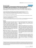
alveolar macrophages may resemble type II pneumo-
cytes, we used TTF-1 staining against type II pneumo-
cytes, as positive control (Figure 1), using the monoclonal
Siganaki et al. Respiratory Research 2010, 11:46
/>Page 3 of 8
mouse anti-TTF-1 antibody (Santa Cruz Biotechnology,
Inc) on adjacent serial sections [18]. Likewise, for the
identification of lymphocytes we have used LCA stain
(lymphocyte common antigen; DAKO Carpinteria, CA,
USA), (Figure 1). Yet, five μm sections were sufficiently
thin to guaranty that each cell was present in adjacent
sections since the diameter of the type II pneumocytes
and alveolar macrophages is much higher than 15-25 μm
[18-20].
The evaluation of total PN II (columnar alveolar lining
cells), AM (irregularly distributed in the alveoli with
foamy cytoplasm and indented nuclei) and LYM (scat-
tered spherical ovoid cells with dense nuclear chromatin
and high nuclear/cytoplasmic ratio) in the stained sec-
tions was performed using a digital camera (Sony) in a
multiread light microscope (Olympus), at 40× magnifica-
tion by two scientists experienced in lung pathology
(AVK and MS). The inter-observer variability of measure-
ments was expressed as the % coefficient of variation. The
inter-observer coefficient of variation was less than 10%.
Twenty microscopic fields under a semitransparent grid
of horizontal lines spaced at 1-mm intervals were used
for cell counting. Results were expressed as cells per mm
2
.
Statistical analysis
Statistical differences between COPD patients and non-
COPD subjects, their smoking status, anthropometric
and spirometric values, and the expression levels of each
apoptotic marker were evaluated with Mann-Whitney
and Spearman test using the SPSS 17.0 statistical soft-
ware package (SPSS Inc; Chicago, IL). A p-value of < 0.05
was considered to be significant.
Results
Clinical characteristics of the subjects
The anthropometric characteristics and spirometric val-
ues of smokers with or without COPD are shown in Table
1. As expected from the selection criteria, smokers with
COPD had a significant lower value of FEV1 (pred %) and
FEV1/FVC ratio (%) than non-COPD smokers.
Table 1: Anthropometric characteristics, spirometric values and drug regimen of the subjects.
COPD smokers Non-COPD smokers *P value
Number 23 20
Sex (M/F) 23/0 20/0
Age (years) 64 ± 7* 57 ± 10* 0.03
Smoking (P-Y) 60 ± 21* 50 ± 27* NS
FEV1 (% pred.) 64 ± 16* 95 ± 13* 0.0001
FVC (% pred) 80 ± 18* 91 ± 13* 0.02
FEV1/FVC (%) 63 ± 6.5* 82 ± 5* 0.0001
IPRATOPIUM
or
TIOTROPIUM
20 mcg, ×3/day
Once daily
NA
ICS 100 mcg ×2/day NA
M/F: Male/Female
P-Y: pack years of smoking (mean ± SD)
FEV1: forced expiratory volume in 1 second (mean ± SD)
FVC: forced vital capacity (mean ± SD)
ICS: inhaled corticosteroids (BUDESONIDE or BECLOMETHAZONE or FLUTICAZONE)
NS: non-significant
NA: not applicable
Siganaki et al. Respiratory Research 2010, 11:46
/>Page 4 of 8
Western blot
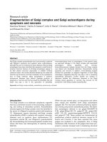
Western blot analysis revealed statistically significant
increased p53 protein levels in COPD patients compared
with non-COPD smokers (0.51 ± 0.29 versus 0.25 ± 0.07,
p = 0.03), (Figure 2). On the contrary, bcl2 protein levels
did not differ statistically between the study groups (0.08
± 0.06 versus 0.10 ± 0.02, p = 0.52), (Figure 2).
Immunohistochemistry
p53 immunostaining
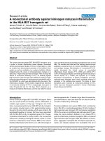
Figure 3A shows p53 immunostaining of PN II, AM and
LYM in a lung tissue section from a COPD smoker and
figure 3B in a non-COPD smoker. The ratio of p53 posi-
tive PN II cells (p53 positive PN II/total PN II) was statis-
tically significant higher in COPD patients compared to
non-COPD smokers (36% versus 10%, p = 0.01), (figure
3C). On the contrary, the ratio of p53 positive AM cells
(p53 positive AM/total AM) and the ratio of p53 positive
LYM (p53 positive LYM/total LYM) was not statistically
significant different between the two groups (25% versus
10%, p = 0.07 and 6% versus 8%, p = 0.5, respectively),
(figure 3C).
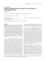
Bcl2 immunostaining
Bcl2 was faintly expressed in PN II in COPD patients
while no expression was detected in AM in both study
groups (Figure 4A, 4B). Bcl2 was expressed in LYM of
COPD and non-COPD smokers, but the ratio of bcl2 pos-
itive LYM (bcl2 positive LYM/total LYM) did not differ
significantly between smokers with or without COPD
(0.6 ± 0.1 versus 0.5 ± 0.1, p = 0.5), (Figure 4C).
Discussion
The present study demonstrated an over-expression of
the pro-apoptotic protein p53 in lung tissue of patients
with COPD compared with non-COPD smokers, not
counterbalanced by the anti-apoptotic protein bcl2. To
the best of our knowledge this is the first study to evalu-
ate, at the same time, p53 and bcl2 expression in lung tis-
sue from smokers with or without COPD, by two
different techniques. Our results validate and extend
observations made by others [3,16,21-26] of an apoptotic
imbalance in COPD investigating two apoptosis-related
proteins.
Our data as revealed by western blot analysis, showed
statistically significant increased p53 protein levels in
COPD patients compared to non-COPD smokers (Figure
2), while the immunohistochemistry revealed increased
p53 ratio in PN II in COPD patients (Figure 3) compared
to non-COPD smokers. Our results are in agreement
with those by Hodge et al [3], reporting increased levels
of p53 in airway epithelial cells and T lymphocytes gath-
ered from bronchial brushing and bronchoalveolar lavage
from ex and current COPD smokers [3]. On the contrary,
protein levels, of the anti-apoptotic mediator bcl2 in
COPD patients were faintly expressed in PN II while no
expression was detected in AM in both study groups (Fig-
ure 4A, 4B). Although Bcl2 was expressed in LYM of both
study groups did not reach statistical significance
between smokers with or without COPD (Figure 4C),
reflecting disequilibrium among pro- and anti-apoptotic
mediators in favour of apoptosis in COPD patients.
A recent study by Weaver and Liu [24] in rats after
exposure to benzene, a ubiquitous environmental pollut-
ant and a cigarette smoking by-product, showed signifi-
cant up-regulation of pro-apoptotic p53 in lung epithelia
of benzene-exposed rats compared to controls, whereas
no statistical difference was found in the expression of
bcl2 in airway epithelial cells in both study groups [24].
Other groups describe similar findings with an increase
in apoptosis of alveolar epithelial cells in patients with
emphysema compared to smokers without COPD [25,26]
while the anti-apoptotic protein bcl2 was not detected in
either normal or emphysematous lung tissue [25].
Furthermore, our data showed increased but not statis-
tically significant p53 levels in AM of COPD patients as
compared to non-COPD smokers (Figure 3). Given that
macrophages act as scavengers of apoptotic cells, we
would expect higher p53 levels in AM of COPD patients,
as a result of increased apoptosis of PN II. However, as
several groups previously demonstrated [21,22], AM
from patients with COPD are less effective in phagocyto-
sing apoptotic epithelial cells compared to controls
[21,22]. It has also been shown that neutrophil elastase
cleaves the phosphatidylserine receptor on macrophages,
resulting in impaired clearance of apoptotic cells [21].
The altered phagocytic capacity of AM in COPD could
further result in defective efferocytosis and accumulation
of apoptotic cells. Persistence of apoptotic bodies and
Figure 1 Positive TTF-1 and p53 immunostaining in type II pneu-
mocytes in serial sections from a COPD patient. Positive LCA and
bcl2 immunostaining in lymphocyte-like cells in serial sections from a
COPD patient (400× magnification).
Siganaki et al. Respiratory Research 2010, 11:46
/>Page 5 of 8
subsequent release of their toxic contents can result in
tissue damage and chronic inflammation leading to
COPD progression [23].
On the other hand, the antiapoptotic molecule bcl2 was
not expressed in AM of COPD and non-COPD smokers
(Figure 4), which could be related with AM homeostasis
implicated in lung defence [21].
p53 ratio was decreased in LYM subpopulation of both
study groups (Figure 3C), compared to PN II and AM,
while bcl2 ratio was increased only in LYM subpopula-
tion in both study groups, although not statistically sig-
nificant (Figure 4C). The imbalance between pro-
apoptotic p53 and anti-apoptotic bcl2 in LYM in favour of
bcl2, could possibly explain the persistence of lympho-
cyte survival into the lung, leading to chronic release of
inflammatory mediators.
Yet, there are limitations in this study that have to be
taken into account. First, although the study subjects
were well characterized, for feasibility reasons the lung
tissue specimens were obtained only from subjects
undergoing resection for lung cancer. Although it is
known that pulmonary malignancy could affect p53 and
bcl2 expression, all subjects included in this study had the
same comorbidity (e.g. lung cancer). Second, the surgical
lung biopsy was not performed in patients with more
advanced COPD. This could lead to the underestimation
of our results, since our data suggest that such a group
would exhibit a higher degree of apoptotic deregulation.
Third, a confounding factor could be the differences in
treatment between subjects, mainly in regard to corticos-
teroids. Inhaled corticosteroids generally enhance innate
immunity while suppress adaptive immunity, thus
enhance the survival of neutrophils and AM, but induce
the apoptosis of airway dendritic cells [27,28]. It has been
demonstrated that corticosteroids induce apoptosis of
airway epithelial cells and eosinophils in asthma [27],
while no such data are available in COPD [7,28]. Like-
wise, most of the studies discussed previously do not dis-
Figure 2 (A): Representative western blots of p53, bcl2 and b-actin in human lung tissues from two COPD and two non-COPD smokers. (B):
Quantitative analysis (mean ± SD) of p53 and bcl2 protein levels in COPD smokers in comparison to non-COPD smokers. **Statistically significant (p
< 0.05).
Siganaki et al. Respiratory Research 2010, 11:46
/>Page 6 of 8
criminate between COPD patients that are treated with
inhaled corticosteroids and those who are not [7]. In
regard to this study, all COPD patients, were stage II
GOLD and were treated accordingly, with an inhaled
anticholinergic long or short-acting [1]. Only periopera-
tive (2-3 days before surgery and continued until hospital
discharge; 10 days in total) and in order to achieve the
best possible baseline function and to prevent postopera-
tive disease-exacerbation, COPD patients received twice-
daily low dose of inhaled corticosteroids (Table 1),
[29,30]. It is still unclear whether inhaled corticosteroids,
in such low doses, are able to play a role in the control of
apoptosis and remodelling [31]. There is only one refer-
ence [32] mentioning the effect of inhaled corticosteroids
on airway inflammation in sputum of healthy volunteers,
using as a minimum dose 0.5 mg of the drug [32]. Since,
our patients received a much lower dose of inhaled corti-
costeroids (200 mcg total/day), we assume that our
results are not subjective to this limitation. However,
more studies are needed to clarify that issue.
Moreover, no data are available for the effects of
inhaled steroids on the expression of p53 and bcl2 apop-
tosis mediators [1,13,15,16,33].
Finally, the two groups were not exact matched for age
and were all male (Table 1). Although, studies in experi-
mental animals reported increased apoptosis in periph-
eral blood T-cells with increasing age [33] studies in
humans, investigating this possibility reported no signifi-
cant changes in apoptosis of airway epithelial cells or
BAL-derived T-cells, or sputum neutrophils with aging
[34,35]. Yet, to the best of our knowledge there are no
reports so far, specifically on the effect of age in p53 and
bcl2 in COPD patients, or control smokers. Furthermore,
studies report no significant differences in the levels of
apoptosis or cytokine production between males and
females [36].
Figure 3 Immunohistochemical staining of p53 protein in human lung tissue. Positive p53 PN II and AM and negative p53 LYM in (A) represen-
tative COPD smoker, and (B) non-COPD smoker. (C): Quantitative analysis (mean ± SD) of p53 expression ratio (positive cells/total cells) in three differ-
ent cell types (PN II, AM, LYM). (** p < 0.05)
Siganaki et al. Respiratory Research 2010, 11:46
/>Page 7 of 8
In conclusion, increased p53 expression in PN II of
COPD smokers may contribute to reduced integrity of
alveolar septa, resulting in cellular homeostasis defects.
In contrast, elevation of anti-apoptotic bcl2 in LYM of
COPD smokers could explain the auto-maintenance of
the "abnormal" inflammation in COPD. Nonetheless,
more studies need to be carried out in order to delineate
the above conclusions.
Abbreviations
COPD: chronic obstructive pulmonary disease; PN II: type II pneumocytes; AM:
alveolar macrophages; LYM: lymphocyte-like cells.
Competing interests
The authors declare that they have no competing interests.
Authors' contributions
MS has contributed to the acquisition of data and carried out the immunoas-
says. AVK carried out the immunoassays. EN carried out the molecular genetic
studies. EV has contributed to the acquisition of data. MP and NS performed
the statistical analysis and contributed to the interpretation of data. NP and SS
have contributed to the acquisition of data and subject recruitment. NMS has
contributed to interpretation of data and has revised critically the article. EGT
has contributed to conception and design of the study, analysis and interpreta-
tion of data and has drafted the submitted article. All authors read and
approved the final manuscript.
Acknowledgements
This study was funded by a research grant from the Hellenic Thoracic Society.
Author Details
1
Laboratory of Molecular and Cellular Pulmonology, Medical School University
of Crete, Greece,
2
Department of Pathology, Medical School, Democritus
University of Thrace, Alexandroupolis, Greece,
3
Department of Thoracic
Medicine, University Hospital of Heraklion Crete, Greece,
4
Laboratory of Clinical
Virology, Medical School University of Crete, Greece and
5
Department of
Anesthesiology, "G. Gennimatas" Hospital Athens, Greece
References
1. Rabe KF, Hurd S, Anzueto A, Barnes PJ, Buist SA, Calverley P, Fukuchi Y,
Jenkins C, Rodriguez-Roisin R, van Weel C, Zielinski J, Global Initiative for
Chronic Obstructive Lung Disease: Global Strategy for the Diagnosis,
Management, and Prevention of Chronic Obstructive Pulmonary
Disease. GOLD executive summary. Am J Respir Crit Care Med 2007,
176(6):532-55.
Received: 20 October 2009 Accepted: 27 April 2010
Published: 27 April 2010
This article is available from: 2010 Siganaki et al; licensee BioMed Central Ltd. This is an Open Access article distributed under the terms of the Creative Commons Attribution License ( which permits unrestricted use, distribution, and reproduction in any medium, provided the original work is properly cited.Respiratory Research 2010, 11:46
Figure 4 Immunohistochemical staining of bcl2 protein in human lung tissue. Positive bcl2 LYM and negative bcl2 PN II and AM (black arrows)
in: (A) representative COPD smoker and (B): representative non-COPD smoker. (C): Quantitative analysis (mean ± SD) of bcl2 expression ratio (positive
cells/total cells) in three different cell types (PN II, AM, LYM).
Siganaki et al. Respiratory Research 2010, 11:46
/>Page 8 of 8
2. Hodge S, Hodge G, Holmes M, Reynolds P: Increased peripheral blood T-
cell apoptosis and decreased Bcl-2 in chronic obstructive pulmonary
disease. Immunology and Cell Biology 2005, 83:160.
3. Hodge S, Hodge G, Holmes M, Reynolds PN: Increased airway epithelial
and T-cell apoptosis in COPD remains despite smoking cessation. Eur
Respir J 2005, 25(3):447-54.
4. Plataki M, Tzortzaki E, Rytila P, Makris D, Koutsopoulos A, Siafakas NM:
Apoptotic mechanisms in the pathogenesis of COPD. Internat J COPD
2006, 1(2):161-171.
5. Demedts IK, Demoor T, Bracke KR, Joos GF, Brusselle GG: Role of
apoptosis in the pathogenesis of COPD and pulmonary emphysema.
Respir Res 2006, 7:53.
6. Hodge S, Hodge G, Holmes M, et al.: Apoptosis in COPD. Curr Respir Med
Reviews 2005, 1:33-41.
7. Park JW, Ryter SW, Choi AM: Functional significance of apoptosis in
chronic obstructive pulmonary disease. COPD 2007, 4(4):347-53.
8. Schuler M, Green DR: Mechanisms of p53-dependent apoptosis.
Biochem Soc Trans 2001, 29:684-8.
9. Haupt S, Berger M, Goldberg Z, Haupt Y: Apoptosis - the p53 network. J
Cell Sci 2003, 116:4077-85.
10. Martin DA, Elkon KB: Mechanisms of apoptosis. Rheum Dis Clin North Am
2004, 30(3):441-54.
11. Weaver CV, Liu S-P: Differentially expressed pro- and anti-apoptogenic
genes in response to benzene exposure: Immunohistochemical
localization of p53, Bag, Bad, Bax, Bcl-2 and Bcl-w in lung epithelia.
Exper Toxicol Pathol 2008, 59:265-272.
12. Tzortzaki E, Siafakas N: A new hypothesis for the initiation of COPD. Eur
Resp J 2009, 34(2):310-5.
13. Agusti A, MacNee W, Donaldson K, Cosio M: Hypothesis: does COPD
have an autoimmune component? Thorax 2003, 58:832-834.
14. Kasahara Y, Tuder RM, Cool CD, et al.: Endothelial cell death and
decreased expression of vascular endothelial growth factor and
vascular endothelial growth factor receptor 2 in emphysema. Am J
Respir Crit Care Med 2001, 163:737-744.
15. Tuder RM, Zhen L, Cho CY, et al.: Oxidative stress and apoptosis interact
and cause emphysema due to vascular endothelial growth factor
receptor blockade. Am J Respir Cell Mol Biol 2003, 29:88-97.
16. Hodge S, Hodge G, Holmes M, et al.: Apoptosis in COPD. Current
Respiratory Medicine Reviews 2005, 1:33-41.
17. Jiménez-Ruiz C, Miravitlles M, Sobradillo V, Gabriel R, Viejo JL, Masa JF,
Fernández-Fau L, Villasante C: Can cumulative tobacco consumption,
FTND score, and carbon monoxide concentration in expired air be
predictors of chronic obstructive pulmonary disease? Nicotine Tob Res
2004, 6(4):649-53.
18. Vlachaki EM, Koutsopoulos AV, Tzanakis N, Neofytou E, Siganaki M, Drositis
I, Moniakis A, Schiza S, Siafakas NM, Tzortzaki EG: Altered surfactant
protein-A (SP-A) expression in type II pneumocytes in COPD. Chest
2010, 137(1):37-45.
19. Mascaretti RS, Mataloun MM, Dolhnikoff M, Rebello CM: Lung
morphometry, collagen and elastin content: changes after hyperoxic
exposure in preterm rabbits. Clinics 2009, 64(11):1099-104.
20. Ikeda K, Monden T, Kanoh T, Tsujie M, Izawa H, Haba A, Ohnishi T,
Sekimoto M, Tomita N, Shiozaki H, Monden M: Extraction and Analysis of
Diagnostically Useful Proteins from Formalin-fixed, Paraffin-embedded
Tissue Sections. J Histochem Cytochem 1998, 46:397-403.
21. Hodge S, Hodge G, Scicchitano R, et al.: Alveolar macrophages from
subjects with chronic obstructive pulmonary disease are deficient in
their ability to phagocytose apoptotic airway epithelial cells. Immunol
Cell Biol 2003, 81:289-296.
22. Bratton DL, Henson PM: Autoimmunity and apoptosis: refusing to go
quietly. Nat Med 2005, 11:26-27.
23. Henson PM, Cosgrove GP, Vandivier RW: Apoptosis and Cell Homeostasis
in Chronic Obstructive Pulmonary Disease. Proc Am Thorac Soc 2006,
3:512-518.
24. Weaver CV, Liu SP, Lu JF, Lin BS: The effects of benzene exposure on
apoptosis in epithelial lung cells: localization by terminal
deoxynucleotidyl transferase-mediated dUTP-biotin nick end labeling
(TUNEL) and the immunocytochemical localization of apoptosis-
related gene products. Cell Biol Toxicol 2007, 23(3):201-20.
25. Imai K, Mercer BA, Schulman LL, Sonett JR, D'Armiento JM: Correlation of
lung surface area to apoptosis and proliferation in human
emphysema. Eur Respir J 2005, 25:250-258.
26. Yokohori N, Aoshiba K, Nagai A: Increased levels of cell death and
proliferation in alveolar wall cells in patients with pulmonary
emphysema. Chest 2004, 125:626-632.
27. de Souza PM, Lindsay MA: Apoptosis as a therapeutic target for the
treatment of lung diseases. Curr Opin Pharmacol 2005, 5:232-237.
28. Schleimer RP: Glucocorticoids Suppress Inflammation but Spare Innate
Immune Responses in Airway Epithelium. Proc Am Thorac Soc 2004,
1:222-230.
29. Jenkins CR, Jones PW, Calverley PM, Celli B, Anderson JA, Ferguson GT,
Yates JC, Willits LR, Vestbo J: Efficacy of salmeterol/fluticasone
propionate by GOLD stage of chronic obstructive pulmonary disease:
analysis from the randomised, placebo-controlled TORCH study. Respir
Res 2009, 10:59.
30. Tashkin DP, Celli B, Senn S, Burkhart D, Kesten S, Menjoge S, Decramer M,
UPLIFT Study Investigators: A 4-year trial of tiotropium in chronic
obstructive pulmonary disease. N Engl J Med 2008, 359(15):1543-54.
31. Vignola AM, Riccobono L, Profita M, Foresi A, Di Giorgi R, Guerrera D,
Gjomarkaj M, Di Blasi P, Paggiaro PL: Effects of low doses of inhaled
fluticasone propionate on inflammation and remodelling in persistent-
mild asthma. Allergy 2005, 60(12):1511-7.
32. Alexis NE, Lay JC, Haczku A, Gong H, Linn W, Hazucha MJ, Harris B, Tal-
Singer R, Peden DB: Fluticasone propionate protects against ozone-
induced airway inflammation and modified immune cell activation
markers in healthy volunteers. Environ Health Perspect 2008,
116(6):799-805.
33. Pahlavani MA, Vargas DA: Aging but not dietary restriction alters the
activation-induced apoptosis in rat T cells. FEBS Lett 2001, 491:114-118.
34. Hodge S, Hodge G, Holmes M, Reynolds PN: Increased airway epithelial
and T-cell apoptosis in COPD remains despite smoking cessation. Eur
Respir J 2005, 25:447-454.
35. Makris D, Vrekoussis T, Izoldi M, Alexandra K, Katerina D, Dimitris T,
Michalis A, Tzortzaki E, Siafakas NM, Tzanakis N: Increased apoptosis of
neutrophils in induced sputum of COPD patients. Respir Med 2009,
103(8):1130-5.
36. Hodge SJ, Hodge GL, Reynolds PN, Scicchitano R, Holmes M: Increased
production of TGF-beta and apoptosis of T lymphocytes isolated from
peripheral blood in COPD. Am J Physiol Lung Cell Mol Physiol 2003,
285(2):L492-9.
doi: 10.1186/1465-9921-11-46
Cite this article as: Siganaki et al., Deregulation of apoptosis mediators' p53
and bcl2 in lung tissue of COPD patients Respiratory Research 2010, 11:46