Báo cáo y học: " Pro-inflammatory mechanisms of muscarinic receptor stimulation in airway smooth muscle" doc
Bạn đang xem bản rút gọn của tài liệu. Xem và tải ngay bản đầy đủ của tài liệu tại đây (959.22 KB, 10 trang )
RESEA R C H Open Access
Pro-inflammatory mechanisms of muscarinic
receptor stimulation in airway smooth muscle
Tjitske A Oenema
1*†
, Saeed Kolahian
1,2†
, Janke E Nanninga
1
, Daniëlle Rieks
1
, Pieter S Hiemstra
3
,
Suzanne Zuyderduyn
3
, Andrew J Halayko
4
, Herman Meurs
1
, Reinoud Gosens
1
Abstract
Background: Acetylcholine, the primary parasympathetic neurotransmitter in the airways, plays an important role
in bronchoconstriction and mucus production. Recently, it has been shown that acetylcholine, by acting on
muscarinic receptors, is also involved in airway inflammation and remodelling. The mechanism(s) by which
muscarinic receptors regulate inflammatory responses are, however, still unknown.
Methods: The present study was aimed at characterizing the effect of muscarinic receptor stimulation on
cytokine secretion by human airway smooth muscle cells (hASMc) and to dissect the intracellular signalling
mechanisms involved. hASMc expressing functional muscarinic M
2
and M
3
receptors were stimulated with the
muscarinic receptor agonist methacholine, alone, and in combination with cigarette smoke extract (CSE), TNF-a,
PDGF-AB or IL-1b.
Results: Muscarinic receptor stimulation induced modest IL-8 secretion by itself, yet augmented IL-8 secretion in
combination with CSE, TNF-a or PDGF-AB, but not with IL-1b. Pretreatment with GF109203X, a protein kinase C
(PKC) inhibitor, completely normalized the effect of methacholine on CSE-induced IL-8 secretion, whereas PMA, a
PKC activator, mimicked the effects of methacholine, inducing IL-8 secretion and augmenting the effects of CSE.
Similar inhibition was observed using inhibitors of IB-kinase-2 (SC514) and MEK1/2 (U0126), both downstream
effectors of PKC. Accordingly, western blot analysis revealed that methacholine augmented the degradation of
IBa and the phosphorylation of ERK1/2 in combination with CSE, but not with IL-1b in hASMc.
Conclusions: We conclude that muscarinic receptors facilitate CSE-induced IL-8 secretion by hASM c via PKC
dependent activation of IBa and ERK1/2. This mechanism could be of importance for COPD patients using
anticholinergics.
Background
Chronic obstructive pulmonary disease (COPD) is an
inflammatory lung disease characterized by airflow
limitation that is not fully reversible [1]. The pathophy-
siology of COPD is mainly caused by cigarette smoke.
COPD is associated with an increase in local and
systemic inflammatory cytokines including TNF-a and
IL-1b [2]. Furthermore, clinical studies reported that the
levels of IL-8 [3] and leukotriene B
4
[4] are correlated to
the proportion of neutrophils present and are increased
in induced sputum of COPD patients. Additionally,
during exacerbations periods, IL-8 levels are increased
[3]. Attracted by IL-8, neutrophil s play a significant role
in the pathogenesis of COPD. Neutrophils promote tis-
sue inflammation and injury by inducing t he release of
mediators including elastase, metalloproteases and reac-
tive oxygen species [4].
Acetylcholine, the primary parasympathetic neuro-
transmitter in the airw ays plays an important role in
COPD, by regulating bronchoconstriction and mucus
production [5]. Parasympathetic tone may b e increased
in COPD [5]. Therefore, anticholinergics -including tio-
tropium bromide, a long-acting bronchodilator- are
often used as a mainstay therapy for COPD [6].
Recently, however, it has been established that activation
of the cholinergic system may also contribute to inflam-
matory responses in the lung. For example, the release
* Correspondence:
† Contributed equally
1
Department of Molecular Pharmacology, University of Groningen, The
Netherlands
Full list of author information is available at the end of the article
Oenema et al. Respiratory Research 2010, 11:130
/>© 2010 Oenema et al; licensee BioMed Central Ltd. This is an O pen Access article distributed under the terms of the Creative
Commons Attribution License ( which permits unrestricted use, distribution, and
reproduction in any medium, provided the original work is properly cited.
of IL-8 and leukotriene B
4
by bronchial epithelial cells
[7,8] and alveolar macrophages [9]in vitro appears to be
induced by acetylcholine, resulting in increased neutro-
phil, monocyte, and eosinophil chemotactic activities, an
effect that may be enhanced in COPD. Also, animal stu-
dies showed that anticholinergics are capable of redu-
cing neutrophilic and eosinophilic inflammation induced
by inhaled diesel-soot [10], in haled allergen [11], or LPS
[12]. Furthermore, it has been reported that airway vas-
cular leakage is mediated by muscarinic receptors [13].
Collectively, these findings suggest a role in p ro-inflam-
matory responses for muscarinic receptors. Nonetheless,
it is still undefined what the potential anti-inflammatory
effects of muscarinic antagonists are in the lungs of
patients with COPD [14], which is in part due to the
unknown mechanisms behind the regulation of inflam-
matory responses by muscarinic receptors.
Human airway smooth muscle (ASM) has been attrib-
uted an important role in pro-inflammatory responses
in COPD [5]. These cells are capable of expre ssing and
releasing cytokines and growth factors, including IL-6
and IL-8 [15]. Furthermore, it has been repo rted that
ASM cells express cell surface molecules, which can
directly interact with immune cells, suggesting an
immunomodulatory role of these cells in COPD [16].
Increased pro-inflammatory cytokine release is induced
by stimulating human ASM cells (hASMc) with G- pro-
tein-coupled receptors, growth factors and extracellular
matrix proteins [15,16]. Additionaly, cigarette smoke
can evoke inflammatory responses in human hASMc,
such as IL-8 secretion [17]. Muscarinic M
2
and
M
3
receptors, both G-protein-coupled receptors, are
expressed in abundance in hASMc, suggesting that acet-
ylcholine regulates inflammatory responses by ASM
[18]. Indeed, we recently reported that muscarinic
receptor stimulation augments cigarette smoke extract
(CSE)-induced IL-8 secretion by hASMc, which was
mediated by the muscarinic M
3
receptor subtype [19].
Although these observations illustrate the potential
role for acetylcholine in regulating airway inflammation,
the mechanism(s) by which muscarinic recepto rs regu-
late inflammatory responsesarestillunknown.Inthe
present study, we investigated the regulation of cytokine
secretion from h ASMc by musc arinic receptors, alone
and in concerted action with various pro-inflammatory
stimuli involved in the pathogenesis of COPD. In addi-
tion, we investigated the intracellular signalling mechan-
isms inv olved, in particular the role of pr otein kinase C
(PKC) and downstream pathways.
Methods
Antibodies and reagents
Methacholine chloride (MCh) was p urchased from ICN
Biomedicals (Zoetermeer, the Netherlands). GF109203X
and U0126 were both from To cris Cookso n Inc. (B ristol,
UK). SC514 was obtained from Calbiochem (Amsterdam,
The Netherlands). PMA, mouse anti-ß-actin antib ody,
horseradish peroxidase (HR P)-conjugated rabbit anti-
mouse antibody, HRP-conjugated goat anti-rabbit,
recombinant human TNF-a,andIL-1b were purchased
from Sigma-Aldrich (Zwijndrecht, The Netherlands).
Human recombinant platelet-derived growth factor-AB
(PDGF-AB) was from Bachem (Weil am Rhein, Ger-
many). Phospho-p44/42 MAPK (ERK1/2) (Th r202/
Tyr204) antibody and p44/42 MAPK (ERK1/2) antibody
were obtained from Cell Signalling Technology (Beverly,
CA, USA). Rabbit anti-IBa (clone-15) was purchased
from Santa Cruz Biotechnology, INC (Santa Cruz CA,
USA). All other chemicals were of analytical grade.
Cell culture
Human bronchial smooth muscle cell lines immortalized
by stable expression of human telomerase reverse tran-
scriptase (hTERT) were prepared as described previously
[20]. The primary cultured human bronchial smooth
muscle cells used to generate these cell lines were
prepared from macroscopi cally healthy segments of
2nd-to-4th generation main bronchus obtained after
lung resection surgery from patients with a diagnosis of
adenocarcinoma. All procedures were approved by the
Human Research Ethics Board of the University of Man-
itoba. Cells were grown to confluence using DMEM
supplemented with 10% FBS, 100 μg/mL streptomycin,
100 U/mL penicil lin and 1.5 μg/mL amphotericin B.
Cultures were maintained in a humidified incubator at
37°C-5% CO2, and media was changed every 2-3 days.
Cytokine release
Cells were cultured in 24 well plates and grown until con-
fluence followed by serum-deprivation for 1 day
in DMEM supplemented with antibiotics (100 μg/mL
streptomycin, 100 U/mL penicillin and 1.5 μg/mL ampho-
tericin B) and ITS (5 μg/mL insulin, 5 μg/mL transferrin,
and 5 ng/mL selenium) before each experiment. The cells
were stimulated with the muscarinic receptor agonist
methacholine chloride (MCh, 10 μM), alone and in combi-
nation with either CSE (5%), TNF-a (1 ng/mL), PDGF-AB
(30 ng/mL) or IL-1b (1 ng/mL) for 24 hrs to determ ine
cytokine secretion in cell-free supernatant. 100% strength
CSE was prepared by combusting two 3R4F r esearch
cigarettes (without filter) (University of Kentucky,
Kentucky , USA) using a peristaltic pump and passing the
smoke through 25 mL of FBS-free medium at the rate of
one ciga rette per 5 m in. CSE was freshly prepa red before
every experiment and was used within 15 min after pre-
paration. Addit ionally, where appropriate, hASMc were
pre-incubated with either the PKC inhibitor GF109203X
(3 μM), the IKK-2-inhibitor SC514 (50 μM) or the MEK
Oenema et al. Respiratory Research 2010, 11:130
/>Page 2 of 10
inhibitor U0126 (3 μM) for 30 min. Cells were also treated
with the PKC activator PMA (0.1 μM). Cytokine levels
were quantified using enzyme-linked immunosorbent
assays (ELISA), according to the manufacturer’sinstruc-
tions (Sanquin Pharmaceutical services, Amsterdam, The
Netherlands). The detection limit was 1 pg/ml for IL-8
and 0.2 pg/ml for IL-6. We d iluted samples were needed
to remain in the range of the standard curve.
Preparation of whole cell lysates
HASMc were cultured in 6 well plates and grown until
confluence followed by serum-deprivation for 1 day in
DMEM supplemented with antibiotics (100 μg/mL
streptomycin, 100 U/mL penicillin and 1.5 μg/mL
amphotericin B) and ITS before each experiment. The
cells were stimulated with the muscarinic receptor ago-
nist MCh (10 μM), alone and in combination with either
CSE (5%) or IL-1b (1 ng/mL) for 60 or 120 min. To
obtain whole cell lysates, cells were washed once with
ice-cold PBS (NaCl 140 mM, KCl 2.6 mM, KH
2
PO
4
1.4
mM, Na
2
HPO
4
.2H
2
O 8.1 mM, pH 7.4), followed by lysis
using ice-cold RIPA buffer (Tris 40 mM, NaCl 150 mM,
Igepal 1% , deoxycholic acid 1%, NaF 1 mM, Na
3
VO
4
1
mM, aprotinin 10 μg/mL, leupeptin 10 μg/mL, pepstatin
A7μg/mL, b-glycerophosphate 1.08 mg/mL, pH 8.0).
Sonicated lysates were assayed for protein content
according to Bradford and stored at -20°C until further
use.
Western Blotting
Equal amounts of protein were separated on 10% polya-
crylamide-SDS gels and transferred to nitrocellulose
membranes. To avoid non-specific binding, membranes
were blocked with blocking buffer (Tris-HCl 50 mM,
NaCl 150 mM, TWEEN-20 0.1%, non-fat dried milk
powder 5%) for 1 hour at room temperature. The me m-
branes were then incubated with specific primary anti-
bodies, all diluted in blocking buffer, for one hour at
room temperature. After washing the membranes three
times with TBS-T 0.1% (Tris-HCl 50 mM, NaCl 150
mM,TWEEN-200.1%)for10min,incubationwiththe
secondary antibody conjugated to HRP was performed
during 1 h at room temperature, follo wed by additional
three washes with TBS-T 0.1%. Bands were subsequently
visualized on film using enhanced chemilumine scence
reagents and analyzed by densitometry (Totallab™,Non-
linear dynamics, Newcastle, UK). All bands were nor-
malized to either b-actin for IBa o r to to tal ERK1/2
for phospho ERK1/2.
Data analysis
Data are presented as mean values ± SE. Statistical sig-
nificance of differences between means was determined
by a Student’ s t-test or by one-way ANOVA, where
appropriate. Data were considered statistically significant
when p < 0.05.
Results
Muscarinic receptor stimulation facilitates cytokine
secretion induced by CSE, TNF-a and PDGF-AB
Recently, it has been reported that stimulation of mus-
carinic receptors induces the release of IL-8 from
human bronchial epithelial cells and facilitates the
releaseofIL-8fromhASMcinducedbyCSE[8,19].We
evaluated the pro-inflammatory properties of muscarinic
receptor stimulation in hASMc, alone and in concerted
action with CSE (5%), PDGF-AB (30 ng/mL), TNF-a
(1 ng/mL) or IL-1b (1 ng/mL) (Figure 1). Previous find-
ings indicated that the effects of muscarinic receptor sti-
mulation on ASM cytokine secretion were most
profound for IL-6 and IL-8 [19], with maximal effects
seen at a c oncentration of 10 μMMCh.Therefore,we
used 10 μM MCh and focused on IL-6 and IL-8 cy to-
kines for our measurements. We observed a minor
increase in IL-8 induced by MCh alone. CSE alone
induced a significant increase of both IL-8 and IL-6
secretion, which was significantly and synergistically
amplified by co-stimulation with MCh. In addition,
MCh induced a synergistic increase in both IL-8 and IL-
6 secretion in combination with TNF- a. Furthermore, a
synergistic e ffect was also observed with the combina-
tion of MCh and PDGF-AB for IL-8 secretion. However,
the effect of IL-1b, which induced a very high IL-8 and
IL-6 production by its own, was not significantly aug-
mented by MCh (Figure 1). IL-8 release in response to
IL-1b was found concentration dependent, but treat-
ment with MCh had no additional effects regardless of
the concentration IL-1b used (data not shown).
PKC is involved in the synergistic effect of muscarinic
receptor stimulation with CSE
PKC plays an important role as a signalling intermedi-
ate in pro-inflammatory cytokine secretion by indu-
cing the activation of several downstream pathways,
including the IKK-2/IBa/NF-BandRaf-1/MEK/
ERK1/2 pathways [21]. The stimulation of muscarinic
receptors induces the activation of PKC in ASM
[22,23]. We hypothesized therefore, that PKC could
play a central role in the synergism between C SE and
MCh in IL-8 secretion. HASMc were pretreated with
GF109203X (3 μM), a specific PKC inhibitor, and sub-
sequently stimulated with MCh, CSE and their combi-
nation (Figure 2). GF109203X significantly inhibited
the synergistic effect of MCh on CSE-induced IL-8
secretion, demonstrating a requirement for PKC in
this synergism. Remarkably, in the absence of the
muscarinic agonist, GF109203X tended to increase the
CSE-induced IL-8 secretion.
Oenema et al. Respiratory Research 2010, 11:130
/>Page 3 of 10
To investigate whether PKC activation was sufficient
for a synergistic IL-8 secretion in combination with
CSE, we used PMA (0.1 μM) as a PKC activator. Indeed,
CSE-induced IL-8 secretion was highly augmented in
thepresenceofPMA,whichcouldbeabolishedtothe
level of CSE-induced IL-8 secretion when pre-treated
with GF109203X ( Figure 3A). These data indicate that
PKC activation is sufficient for a synergistic interaction
with CSE, which is i n support of a central role for PKC
in regulating the synergy between MCh and CSE. In
contrast to MCh, however, PMA induced a considerable
IL-8 secretion by itself, which was abolished when the
cells were pre-treated with GF109203X.
PKC has been shown to induce activation of the
NF-B and ERK1/2 pathways in different cells [21].
Moreover, it has been reported that the stimulation of
muscarinic receptors through acetylcholine mediat es the
release of IL-8 in human bronchial epithelial cells by
NF-B- and ERK1/2-dependent mechanisms [ 8]. To test
the i nvolvement of the NF-B and ERK1/2 pathways as
a result of PKC activation, hASMc were stimulated with
PMA after pre-treatment with either an IKK-2 inhibitor,
SC514, or a MEK1/2 inhibitor, U0126. IL-8 secretion
induced by PMA was significantly decreased in presence
of these pharmacological inhibitors (Figure 3B for
SC514 and figure 3C for U0126, respectively). Moreover,
western blot analysis indicated that the activation of
PKC by PMA induced the pho sphorylation of ERK1/2
and the degradation of IBa in hASMc. Collectively,
these data indicate that PKC is able to activate the
IBa/NF-B and MEK/ERK1/2 pathways, leading to
IL-8 secretion from hASMc (Figure 3D).
Involvement of the IBa/NF-B pathway in the synergistic
effect of muscarinic receptor stimulation with CSE
HASMc were pretreated with the IKK-2 inhibitor
SC514 to test the involvement of this pathway in the
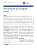
Figure 1 A-B, Muscarinic receptor stimul ation augments cytokine se cretion induced by CSE, PDGF -AB and TNF-a, but not by IL-1b.
hASMc were stimulated with CSE (5%, n = 22 and n = 6 for IL-8 and IL-6, respectively), TNF-a (1 ng/mL, n = 17 and n = 5 for IL-8 and IL-6,
respectively), IL-1b (1 ng/mL, n = 17 and n = 6 for IL-8 and IL-6, respectively) or PDGF-AB (30 ng/mL, n = 6 for IL-8), in the absence or presence
of MCh (10 μM) for 24 hours. Supernatants were analyzed for the presence of IL-8 (A) or IL-6 (B). Data shown are the means ± SE of n
independent experiments. *p < 0.05, **p < 0.01 and ***p < 0.001 compared to basal;
†
p < 0.05 and
††
p < 0.01 compared to the absence of MCh
(Student’s t-test for paired observations).
Figure 2 Involvement of PKC in the potentiation of CSE-
induced IL-8 release by muscarinic receptor stimulation. hASMc
were stimulated with CSE (5%) in the absence or presence of MCh
(10 μM) and/or GF109203X (3 μM) for 24 hours. Supernatants were
analyzed for the presence of IL-8. Data represent means ± SE of 7
independent experiments each performed in duplicate. ***p < 0.001
compared to basal;
$$$
p < 0.001 compared to CSE;
†
p < 0.05
compared to the absence of GF109203X (One-way ANOVA followed
by Newman-Keuls multiple comparisons test).
Oenema et al. Respiratory Research 2010, 11:130
/>Page 4 of 10
synergistic IL-8 secretion by MCh and CSE (Figure
4A). SC514 completely inhibited the MCh- and CSE-
induced IL-8 secretion. Furthermore, the synergistic
effect of the combination of MCh and CSE was abol-
ished (Figure 4A). These results confirm the involve-
ment of the IBa/NF-B pathway in the observed IL-8
secretion. Therefore, we next investigated the effects of
muscarinic receptor stimulation on IBa degradation,
alone and in combination with CSE at different time
points (60 and 120 min of treat ment). IBa degrada-
tion was measured by western blot analysis. Although
MCh did not induce significant IBa degradation by
itself, it augmented the response induced by CSE, par-
ticularly after 120 min of incubation (Figure 4D).
Overall, these results indicate that muscarinic receptor
stimulation promotes the activation of the IBa/
NF-B pathway induced by CSE, which likely contri-
butes to the synergistic IL-8 secretion. Interestingly,
and in line with the lack of effect of MCh on IL-1b-
induced cytokine secretion, MCh did not augment
maximal IL-1b-induced I Ba degradation at t = 60
and 120 min (Figure 4E). However, IL-1b-induced IL-8
secretion in presence or absen ce of MCh, was signif i-
cantly i nhibited by SC514 (Figure 4B).
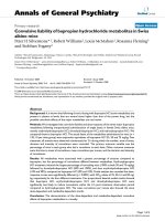
Figure 3 PKC activatio n is sufficie nt to facilitate CSE-induced IL-8 secretion in hASMc. hASMc were stimulated with PMA (0.1 μM), in the
absence or presence of CSE (5%) and GF109203X (3 μM) (Figure 3A), SC514 (50 μM) (Figure 3B) or U0126 (3 μM) (Figure 3C) for 24 hours.
Supernatants were analyzed for the presence of IL-8. Data represent means ± SE of 4-6 independent experiments each performed in duplicate.
*p < 0.05, **p < 0.01 and ***p < 0.001 compared to basal;
$$$
p < 0.001 compared to PMA;
†
p < 0.05,
††
p < 0.01 and
†††
p < 0.001 compared to
the absence of inhibitor (One-way ANOVA followed by Newman-Keuls multiple comparisons test). (Figure 3D) hASMc were stimulated with PMA
(0.1 μM) for 1 hour. Cell lysates were analyzed for IBa breakdown and phosphorylation of ERK1/2 by western blot. b-actin and total ERK1/2
were used as loading controls. Western blots shown are representative of 4 experiments.
Oenema et al. Respiratory Research 2010, 11:130
/>Page 5 of 10
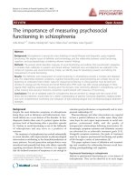
Figure 4 Involvement of the IBa/NF-B pathway in IL-8 secretion induced by CSE, IL-1b and MCh. hASMc were stimulated with CSE
(5%) (Figure 4A) or IL-1b (1 ng/mL) (Figure 4B) in the absence or presence of MCh (10 μM) and/or SC514 (50 μM) for 24 hours. Supernatants
were analyzed for the presence of IL-8. Data represent means ± SE of 5 independent experiments each performed in duplicate. **p < 0.01 and
***p < 0.001 compared to basal;
†
p < 0.05,
††
p < 0.01 and
†††
p < 0.001 compared to the absence of SC514;
$$
p < 0.01 compared to CSE (One-
way ANOVA followed by Newman-Keuls multiple comparisons test). (Figure 4C, 4 D and 4E) hASMc were stimulated with CSE (5%) (Figure 4C
and 4D) or IL-1b (1 ng/mL) (Figure 4C and 4E) in the absence or presence of MCh for 60 min and 120 min (representative blots shown in Figure
4C) as indicated. IBa degradation was determined by western blot and corrected for the expression of b-actin, which was used as a loading
control. Data represent means ± SE of 9-10 experiments. *p < 0.05 and ***p < 0.001 compared to basal and
††
p < 0.01 compared the absence of
MCh (Student’s t-test for paired observations).
Oenema et al. Respiratory Research 2010, 11:130
/>Page 6 of 10
Involvement of the MEK/ERK1/2 pathway in the
synergistic effect of muscarinic receptor stimulation with
CSE
To test the involvement of the MEK/ERK1/2 pathway in
IL-8 secretion induced by MCh and CSE, we pretreated
the cells with the MEK1/2 inhibitor, U0126 (3 μM) (Fig-
ure 5A). In the presence of U0126, IL-8 secretion
induced by co-stimulation of CSE with MCh was signifi-
cantly decreased (Figure 5A). These results confirm the
involvement of the MEK/ERK1/2 pathway in the
observed IL-8 secretion. Therefore, we next assessed
phosphorylation of ERK1/2 induced by MCh and CSE
(Figure 5C and 5D). Although, ERK1/2 phosphorylation
was not significantly i ncreased when cells were stimu-
lated with MCh alone after one hour of incubation, 15
min incubation is sufficient to induce significant ERK1/2
phosphorylation [23]. In combination with CSE, MCh
induce d a significant incr ease in the phosphorylation of
ERK1/2 at this time point (one hour). These results sup-
port the involvement of the ERK1/2 pathway in the
synergism between CSE a nd MCh a t the level o f IL-8
secretion. In contrast, IL-1b induced ERK1/2 phosphor-
ylation was not increased by MCh and also pre-
treatment with U0126 had no effect (Figure 5B and 5D).
These results are in agreement with the results of
Orsini,etal.,demonstratingthatIL-1b can induce a
transient phosphorylation of ERK1/2 in human airway
smooth muscle cells [24].
Discussion
In the present study, we demonstrate that muscarinic
receptors stimulate the secretion of the pro-inflamma-
tory cytokine IL-8 from hASMc, and augment the
response induced by TNF-a, CSE and PDGF-AB.
Furthermore, we dissected the underlying mechanism of
the synergistic IL-8 production. To permit the release of
Figure 5 Involvement of the MEK/ERK1/ 2 pathway in IL-8 release induced by MCh. hASMc were stimulated with CSE (5%) (Figure 5A) or
IL-1b (1 ng/mL) (Figure 5B), in the absence or presence of MCh (10 μM) and/or U0126 (3 μM) for 24 hours. Supernatants were analyzed for the
presence of IL-8. *p < 0.05 and **p < 0.01 compared to basal;
$$$
p < 0.001 compared to CSE alone,
†††
p < 0.001 compared to the absence of
U0126. (One-way ANOVA followed by Newman-Keuls multiple comparisons test). (Figure 5C and 5D) hASMc were stimulated with CSE (5%) or
IL-1b (1 ng/mL) in the absence or presence of MCh (10 μM) for 60 min. Cell lysates were analyzed for phospho-ERK1/2 by western blot and
corrected for the expression of total ERK1/2, which was used as a loading control. Data represent means ± SE of 5 independent experiments
each performed in duplicate.
$
p < 0.05 compared to CSE alone (Student’s t-test for paired observations).
Oenema et al. Respiratory Research 2010, 11:130
/>Page 7 of 10
the pro-inflammatory cytokine IL-8 after activation of
the muscarinic receptors and CSE, activation of PKC is
required, w hich is followed by the breakdown of IBa.
In parallel, the activation of PKC leads to the stimula-
tion of MEK1/2 inducing the phosphorylation of ERK1/
2. Both pathways regulate IL-8 secretion, which, as pre-
viously described, is dependent on NF-BandAP-1
IL-8 promoter activation [25].
Our current and previously published data [19] indi-
cate that the activation of muscarinic receptors in
hASM c facil itates the secretion of the pro-inflammatory
cytokines IL-6 and IL-8 in combina tion with CSE and
pro-inflammatory cytokines. Muscarinic receptor stimu-
lation also promoted IL-8 secretion by itself, though
only to a relatively minor extent. This suggests that the
effects of muscarinic receptor stimulation are relevant
primarily in a pro-inflammatory microenvironment. In
support, functional muscarinic receptors are expressed
on the majority of inflammatory cells [5]. Also, the
endogenous muscarinic receptor ligand acetylcholine
and its synthesizing enzyme choline acetyltra nsferase
(ChAT) are present in several extraneuronal cell types,
including airway epithelial cells, lymphocytes, eosino-
phils, neutrophils, macrophages, and mast cells [5,26].
Furthermore, animal models showed that atropine
reduces lung inflammation induced by diesel-soot in
rats [10], and that tiotropium bromide inhibits several
aspects of airway inflammation and remodeling in oval-
bumin-sensitized guinea pigs, but has little effect on
inflammatory cell counts in saline challenged controls
[11,27]. Additionally, it has been reported that carba-
chol, by activation of muscarinic receptors, is able to
increase infl ammatory gene expression in ASM, includ-
ing IL-6, IL-8 and cyclooxygenase-2 (COX-2) [28].
Furthermore, acetylcholine (ACh) can induce leuko-
triene B
4
(LTB
4
) release from sputum COPD cells [4],
also indicating a regulatory role for ACh in inflamma-
tory cells. Taken together, this indicates that acetylcho-
line is importantly involved in the regulation of pro-
inflammatory responses. Our current results provide
new insights as we demonstrate that the activation of
muscarinic receptors interacts with several cytokines
and growth factors, in particular with TNF-a, PDGF-AB
and CSE to enhance their inflammatory response in
hASMc.
HASMc produce a variety inflammatory mediators
[15,16,29]. This suggests an important role for ASM in
inflammatory responses in COPD. Indeed, hASMc are a
source of chemokines and cytokines that play a role in
chronic p ulmonary diseases like COPD and asthma,
including IL-8 and IL-6. The levels of IL-8 are correlated
with the degree of neutrophilic inflammation and are
increased in sputum in COPD patients [3,30 ]. Several
pro-inflammatory stimuli, including IL-17 [31-33], gram-
positive and gram-negative bacteria [34], b-tryptase [35],
IL-1b [32] and TNF-a [17] are able to induce IL-8 secre-
tion from human ASM. M oreover, CSE synergizes with
TNF-a to enhance IL-8 secretion by ASM [17]. We pre-
viously demonstrated that CSE and muscarinic M
3
recep-
tor stimulation leads to a synergistic increase in IL-8
secretion by hASMc [19], which as demonstrated in this
study, is dependent on downstream signalling to PKC
and the I Ba/NF-B and MEK/ERK1/2 pathways. Nicoti-
nic receptors and muscarinic M
2
receptors are not
involved in this synergism, as gallamine had no effect on
IL-8 release induced by either CSE or MCh [19]. This
indicates that acetylcholine may also play an important
role in the inflammatory/immunomodulatory processes
driven by human ASM.
Using the PKC inhibitor GF109203X, we demonstrate
that the synergism of MCh and CSE-induced IL-8 secre-
tion is mediated by PKC in hASMc. In fact, activation
of PKC was sufficient to induce synergistic IL-8 secre-
tion in combination with CSE, which was confirmed by
the use of the PKC activator, PMA. These observations
correspond with a n earlier study from our group
demonstrating that MCh augments PDGF-induced cell
proliferation via the activation of PKC [23] and appear
to suggest that muscarinic M
3
receptors exert their facil-
itatory effects on remodeling and inflammation to an
important extent via the activation of P KC. Down-
stream, we demonstrated that PKC is able to induce the
activation of IBa/NF-B and MEK/ERK1/2 pathways
in hASMc and that the se pathways a re involved in the
secretio n of IL-8 induced by the co-stimulation of mu s-
carinic receptors and CSE. Interestingly, the co-stimula-
tion with CSE and MCh appeared required to reveal the
importance of PKC, as stimulation with either CSE or
MCh alone was not sufficient to demonstrate an invol-
vement of PKC. This indicates that PKC stimulation by
MCh is not sufficient to induce an IL-8 or IL-6 response
by itself, but augments pro-inflammatory signalling to
NF-B and ERK1/2 induced by CSE. However, synergis-
tic functional interactions with IL-1b,animportant
cytokine in COPD pathogenesis [36], were not observed,
both for IL-8 secretion and for activation of t he signal-
ling pathways investigated, indicating that the mechan-
ism of the synergistic interaction is stimulus specific.
Lower concentrations of IL-1b were also tested and
were found to be similarly unaffected by MCh (data not
shown).
The co mbination of MCh and CSE likely triggers PKC
to activate IKK-2. This kinase allows the phosphoryla-
tion and degradation of IBa leading to the transloca-
tion of NF-B into the nucleus to regulate NF-Bgene
transcription [37]. Furthermore, PKC has been shown to
be critically involved in the activation of the ERK1/2
pathway in human aortic smooth muscle cells [38]. PKC
Oenema et al. Respiratory Research 2010, 11:130
/>Page 8 of 10
induces the phosphorylation of Raf-1, an upstream regu-
lator of ERK1/2 activation, which is followed by the reg-
ulation of AP-1 dependent gene transcription. The IL-8
gene contains both NF-B and AP-1 binding sites in its
promoter region [25]. Epithelial cells are also able, to
induce IL-8 secretion through the activation of ERK1/2
and NF-B in response to pro-inflammatory stimuli,
including acetylcholine [8,39,40]. Taken together, these
findings and our previous finding s [19] indicate that the
synergis m between muscarinic M
3
receptors and CSE is
mediated by PKC dependent activation of the down-
stream pathways NF-B and ERK1/2, to induce the
secretion of IL-8.
It is unclear whether the pro-inflammatory effects of
muscarinic receptor stimulation and CSE, as observed in
our current work, are relevant to the COPD patient.
Nonetheless, several clinical studies demonstrated that
short-term therapy with tiotropium bromide improves
airflow and hyperinflation [41,42]. Moreover, long-term
use (up to 6 to 12 months) of this anticholinergic drug
improved exercise tolerance, quality of life, rates of dys-
pnoea but also the exacerbation frequency in COPD
patients, which are associated with periods of increased
inflammatory cell influx [41,43]. The Understanding
Potential L ong-Term Impacts on Function with Tiotro-
pium (UPLIFT) study concluded th at COPD patients
treated with tiotropium bromide during a 4-year period
improved their quality of life, frequency of exacerbations
andlungfunction,buttiotropiumbromidedidnot
reduce the d ecline in FEV
1
over the treatment period
[44]. Nonetheless, in a subgroup of COPD patients o f
the UPLIFT study, which were not on other controller
medication, a reduction in the accelerated FEV
1
decline
was observed in the tiotropium bromide arm (post-hoc
analysis of the UPLIFT study [44]). This was also
observed in the subgroup of stag e II COPD patients
[45]. Collectively, besides the well described bronchodi-
latory effects, these findings suggest additional, non-
bronchodilator properties fo r tiotropium bromide [6].
An anti-inflammatory role for anticholinergics is in
agreement with animal a nd cell culture studies showing
a role for acetylcholine in cell proliferation, extracellular
matrix protein secretion and inflammation [5,46,47] and
with our present findings showing that the inflammatory
response induced by CSE, TNF-a and PDG F-AB can be
augmented by muscarinic receptor stimulation in
hASMc. It should be emphasized, however, that the
hypothesis that tiotropium bromide may exert anti-
inflammatory effects in COPD patients still needs to b e
tested in clinical studies.
Conclusions
In conclusion, our results indic ate that the activation of
muscarinic receptors on hASMc induces the secretion
of the pro-inflammatory cytokines IL-8 and IL-6, parti-
cularly in combination with inflam matory mediators and
CSE. The mechanism behind the synergism between
CSE- and MCh-induced IL-8 secretion involves signal-
ling to PKC and NF-B/ERK1/2. These and our pre-
vious findings suggest that acetylcholine might have a
role in enhancing inflammatory responses.
List of abbreviations
ASM: airway smooth muscle cells; COPD: chronic obstructive pulmonary
disease; CSE: cigarette smoke extract; IKK: IB-kinase; MCh: methacholine;
PDGF: platelet growth factor; PKC: protein kinase C
Competing interests
This study was supported by an unrestricted educational grant from
Boehringer Ingelheim Pharma GmbH.
Authors’ contributions
TAO, SK, HM, PSH, SZ and RG conceived of the study and designed the
experiments. AJH contributed the airway smooth muscle cell lines
expressing muscarinic receptors. TAO, SK, JEN and DR performed the
experiments. TAO, SK and RG analysed the data. TAO and SK drafted the
manuscript. RG, HM, PSH, SZ and AJH revised the manuscript for important
intellectual content. All authors read and approved the final manuscript.
Acknowledgements
RG is the recipient of a Veni fellowship (916.86.036) from the Dutch
Organisation for Scientific Research (NWO). We are grateful to Dr. W.T.
Gerthoffer (University of Nevada-Reno) for preparation of the hTERT cell lines
used in the study. AJH is supported by the Canada Research Chairs Program
and Canadian Institutes of Health Research.
Author details
1
Department of Molecular Pharmacology, University of Groningen, The
Netherlands.
2
Department of Basic Sciences, University of Tabriz, Iran.
3
Department of Pulmonology, Leiden University Medical Center, The
Netherlands.
4
Department of Physiology & Internal Medicine, University of
Manitoba, Winnipeg, MB, Canada.
Received: 12 Februar y 2010 Accepted: 28 September 2010
Published: 28 September 2010
References
1. Pauwels RA, Buist AS, Calverley PM, Jenkins CR, Hurd SS: Global strategy for
the diagnosis, management, and prevention of chronic obstructive
pulmonary disease. NHLBI/WHO Global Initiative for Chronic Obstructive
Lung Disease (GOLD) Workshop summary. Am J Respir Crit Care Med 2001,
163:1256-1276.
2. Edwards MR, Bartlett NW, Clarke D, Birrell M, Belvisi M, Johnston SL:
Targeting the NF-kappaB pathway in asthma and chronic obstructive
pulmonary disease. Pharmacol Ther 2009, 121:1-13.
3. Kim V, Rogers TJ, Criner GJ: New concepts in the pathobiology of chronic
obstructive pulmonary disease. Proc Am Thorac Soc 2008, 5:478-485.
4. Profita M, Giorgi RD, Sala A, Bonanno A, Riccobono L, Mirabella F, et al:
Muscarinic receptors, leukotriene B4 production and neutrophilic
inflammation in COPD patients. Allergy 2005, 60:1361-1369.
5. Gosens R, Zaagsma J, Meurs H, Halayko AJ: Muscarinic receptor signaling
in the pathophysiology of asthma and COPD. Respir Res 2006, 7:73.
6. Bateman ED, Rennard S, Barnes PJ, Dicpinigaitis PV, Gosens R, Gross NJ,
et al: Alternative mechanisms for tiotropium. Pulm Pharmacol Ther 2009,
22:533-542.
7. Koyama S, Rennard SI, Robbins RA: Acetylcholine stimulates bronchial
epithelial cells to release neutrophil and monocyte chemotactic activity.
Am J Physiol 1992, 262:L466-L471.
8. Profita M, Bonanno A, Siena L, Ferraro M, Montalbano AM, Pompeo F, et al:
Acetylcholine mediates the release of IL-8 in human bronchial epithelial
cells by a NFkB/ERK-dependent mechanism. Eur J Pharmacol 2008,
582:145-153.
Oenema et al. Respiratory Research 2010, 11:130
/>Page 9 of 10
9. Sato E, Koyama S, Okubo Y, Kubo K, Sekiguchi M: Acetylcholine stimulates
alveolar macrophages to release inflammatory cell chemotactic activity.
Am J Physiol 1998, 274:L970-L979.
10. McQueen DS, Donaldson K, Bond SM, McNeilly JD, Newman S, Barton NJ,
et al: Bilateral vagotomy or atropine pre-treatment reduces experimental
diesel-soot induced lung inflammation. Toxicol Appl Pharmacol 2007,
219:62-71.
11. Bos IS, Gosens R, Zuidhof AB, Schaafsma D, Halayko AJ, Meurs H, et al:
Inhibition of allergen-induced airway remodelling by tiotropium and
budesonide: a comparison. Eur Respir J 2007, 30:653-661.
12. Pera T, Zuidhof AB, Gosens R, Maarsingh H, Zaagsma J, Meurs H:
Tiotropium Inhibits Inflammation and Remodeling in a Guinea Pig
Model of COPD. Am J Respir Crit Care Med 2009, 179:A6328.
13. Cui YY, Zhu L, Wang H, Advenier C, Chen HZ, Devillier P: Muscarinic
receptors involved in airway vascular leakage induced by experimental
gastro-oesophageal reflux. Life Sci 2008, 82:949-955.
14. Trevethick M, Clarke N, Strawbridge M, Yeadon M: Inhaled muscarinic
antagonists for COPD-does an anti-inflammatory mechanism really play
a role? Curr Opin Pharmacol 2009, 9:250-255.
15. Chung KF: The role of airway smooth muscle in the pathogenesis of
airway wall remodeling in chronic obstructive pulmonary disease. Proc
Am Thorac Soc 2005, 2:347-354.
16. Zuyderduyn S, Sukkar MB, Fust A, Dhaliwal S, Burgess JK: Treating asthma
means treating airway smooth muscle cells. Eur Respir J 2008, 32:265-274.
17. Oltmanns U, Chung KF, Walters M, John M, Mitchell JA: Cigarette smoke
induces IL-8, but inhibits eotaxin and RANTES release from airway
smooth muscle. Respir Res 2005, 6:74.
18. Racke K, Matthiesen S: The airway cholinergic system: physiology and
pharmacology. Pulm Pharmacol Ther 2004, 17:181-198.
19. Gosens R, Rieks D, Meurs H, Ninaber DK, Rabe KF, Nanninga J, et al:
Muscarinic M3 receptor stimulation increases cigarette smoke-induced
IL-8 secretion by human airway smooth muscle cells. Eur Respir J 2009,
34:1436-1443.
20. Gosens R, Stelmack GL, Dueck G, McNeill KD, Yamasaki A, Gerthoffer WT,
et al: Role of caveolin-1 in p42/p44 MAP kinase activation and
proliferation of human airway smooth muscle. Am J Physiol Lung Cell Mol
Physiol 2006, 291:L523-L534.
21. Park H, Park SG, Kim J, Ko YG, Kim S: Signaling pathways for TNF
production induced by human aminoacyl-tRNA synthetase-associating
factor, p43. Cytokine 2002, 20:148-153.
22. Bremerich DH, Warner DO, Lorenz RR, Shumway R, Jones KA:
Role of
protein kinase C in calcium sensitization during muscarinic stimulation
in airway smooth muscle. Am J Physiol 1997, 273:L775-L781.
23. Gosens R, Dueck G, Rector E, Nunes RO, Gerthoffer WT, Unruh H, et al:
Cooperative regulation of GSK-3 by muscarinic and PDGF receptors is
associated with airway myocyte proliferation. Am J Physiol Lung Cell Mol
Physiol 2007, 293:L1348-L1358.
24. Orsini MJ, Krymskaya VP, Eszterhas AJ, Benovic JL, Panettieri RA Jr, Penn RB:
MAPK superfamily activation in human airway smooth muscle:
mitogenesis requires prolonged p42/p44 activation. Am J Physiol 1999,
277:L479-L488.
25. Roebuck KA, Carpenter LR, Lakshminarayanan V, Page SM, Moy JN,
Thomas LL: Stimulus-specific regulation of chemokine expression
involves differential activation of the redox-responsive transcription
factors AP-1 and NF-kappaB. J Leukoc Biol 1999, 65 :291-298.
26. Wessler I, Kirkpatrick CJ, Racke K: The cholinergic ‘pitfall’: acetylcholine, a
universal cell molecule in biological systems, including humans. Clin Exp
Pharmacol Physiol 1999, 26:198-205.
27. Gosens R, Bos IS, Zaagsma J, Meurs H: Protective effects of tiotropium
bromide in the progression of airway smooth muscle remodeling. Am J
Respir Crit Care Med 2005, 171:1096-1102.
28. Kanefsky J, Lenburg M, Hai CM: Cholinergic receptor and cyclic stretch-
mediated inflammatory gene expression in intact ASM. Am J Respir Cell
Mol Biol 2006, 34:417-425.
29. Clarke D, Damera G, Sukkar MB, Tliba O: Transcriptional regulation of
cytokine function in airway smooth muscle cells. Pulm Pharmacol Ther
2009, 22:436-445.
30. Tetley TD: Inflammatory cells and chronic obstructive pulmonary disease.
Curr Drug Targets Inflamm Allergy 2005, 4:607-618.
31. Vanaudenaerde BM, Wuyts WA, Geudens N, Dupont LJ, Schoofs K,
Smeets S, et al: Macrolides inhibit IL17-induced IL8 and 8-isoprostane
release from human airway smooth muscle cells. Am J Transplant 2007,
7:76-82.
32. Dragon S, Rahman MS, Yang J, Unruh H, Halayko AJ, Gounni AS: IL-17
enhances IL-1beta-mediated CXCL-8 release from human airway smooth
muscle cells. Am J Physiol Lung Cell Mol Physiol 2007, 292:L1023-L1029.
33. Rahman MS, Yang J, Shan LY, Unruh H, Yang X, Halayko AJ, et al: IL-17R
activation of human airway smooth muscle cells induces CXCL-8
production via a transcriptional-dependent mechanism. Clin Immunol
2005, 115:268-276.
34. Issa R, Sorrentino R, Sukkar MB, Sriskandan S, Chung KF, Mitchell JA:
Differential regulation of CCL-11/eotaxin-1 and CXCL-8/IL-8 by gram-
positive and gram-negative bacteria in human airway smooth muscle
cells. Respir Res
2008, 9:30.
35. Mullan CS, Riley M, Clarke D, Tatler A, Sutcliffe A, Knox AJ, et al: Beta-
tryptase regulates IL-8 expression in airway smooth muscle cells by a
PAR-2-independent mechanism. Am J Respir Cell Mol Biol 2008, 38:600-608.
36. Chung KF: Cytokines as targets in chronic obstructive pulmonary
disease. Curr Drug Targets 2006, 7:675-681.
37. Wong ET, Tergaonkar V: Roles of NF-kappaB in health and disease:
mechanisms and therapeutic potential. Clin Sci (Lond) 2009, 116:451-465.
38. Chen QW, Edvinsson L, Xu CB: Role of ERK/MAPK in endothelin receptor
signaling in human aortic smooth muscle cells. BMC Cell Biol 2009, 10:52.
39. Holtmann H, Winzen R, Holland P, Eickemeier S, Hoffmann E, Wallach D,
et al: Induction of interleukin-8 synthesis integrates effects on
transcription and mRNA degradation from at least three different
cytokine- or stress-activated signal transduction pathways. Mol Cell Biol
1999, 19:6742-6753.
40. Oudin S, Pugin J: Role of MAP kinase activation in interleukin-8
production by human BEAS-2B bronchial epithelial cells submitted to
cyclic stretch. Am J Respir Cell Mol Biol 2002, 27:107-114.
41. O’Donnell DE, Fluge T, Gerken F, Hamilton A, Webb K, Aguilaniu B, et al:
Effects of tiotropium on lung hyperinflation, dyspnoea and exercise
tolerance in COPD. Eur Respir J 2004, 23:832-840.
42. Maltais F, Hamilton A, Marciniuk D, Hernandez P, Sciurba FC, Richter K, et al:
Improvements in symptom-limited exercise performance over 8 h with
once-daily tiotropium in patients with COPD. Chest 2005, 128:1168-1178.
43. Casaburi R, Mahler DA, Jones PW, Wanner A, San PG, ZuWallack RL, et al: A
long-term evaluation of once-daily inhaled tiotropium in chronic
obstructive pulmonary disease. Eur Respir J 2002, 19:217-224.
44. Tashkin DP, Celli B, Senn S, Burkhart D, Kesten S, Menjoge S, et al: A 4-year
trial of tiotropium in chronic obstructive pulmonary disease. N Engl J
Med 2008, 359:1543-1554.
45. Decramer M, Celli B, Burkhart D, Kesten S, Mehra S, Liu D, et al: The Effect
of Tiotropium on COPD GOLD Stage II during the Four-Year UPLIFT Trial.
Am J Respir Crit Care Med 2009, 179:A2466.
46. Gosens R, Zaagsma J, Grootte BM, Nelemans A, Meurs H: Acetylcholine: a
novel regulator of airway smooth muscle remodelling? Eur J Pharmacol
2004, 500:193-201.
47. Racke K, Juergens UR, Matthiesen S: Control by cholinergic mechanisms.
Eur J Pharmacol 2006, 533:57-68.
doi:10.1186/1465-9921-11-130
Cite this article as: Oenema et al.: Pro-inflammatory mechanisms of
muscarinic receptor stimulation in airway smooth muscle. Respiratory
Research 2010 11:130.
Submit your next manuscript to BioMed Central
and take full advantage of:
• Convenient online submission
• Thorough peer review
• No space constraints or color figure charges
• Immediate publication on acceptance
• Inclusion in PubMed, CAS, Scopus and Google Scholar
• Research which is freely available for redistribution
Submit your manuscript at
www.biomedcentral.com/submit
Oenema et al. Respiratory Research 2010, 11:130
/>Page 10 of 10