Báo cáo y học: "Separating therapeutic efficacy from glucocorticoid side-effects in rodent arthritis using novel, liposomal delivery of dexamethasone phosphate: long-term suppression of arthritis facilitates interval treatment" pot
Bạn đang xem bản rút gọn của tài liệu. Xem và tải ngay bản đầy đủ của tài liệu tại đây (204.23 KB, 9 trang )
Open Access
Available online />Page 1 of 9
(page number not for citation purposes)
Vol 11 No 6
Research article
Separating therapeutic efficacy from glucocorticoid side-effects in
rodent arthritis using novel, liposomal delivery of dexamethasone
phosphate: long-term suppression of arthritis facilitates interval
treatment
Una Rauchhaus
1
, Franz Werner Schwaiger
2
and Steffen Panzner
1
1
Novosom AG, Weinbergweg 22, D-06120 Halle/Saale, Germany
2
Aurigon Life Science GmbH, Bahnhofstrasse 9-15, D-82327 Tutzing, Germany
Corresponding author: Steffen Panzner,
Received: 26 Mar 2009 Revisions requested: 21 Apr 2009 Revisions received: 24 Nov 2009 Accepted: 15 Dec 2009 Published: 15 Dec 2009
Arthritis Research & Therapy 2009, 11:R190 (doi:10.1186/ar2889)
This article is online at: />© 2009 Rauchhaus et al.; licensee BioMed Central Ltd.
This is an open access article distributed under the terms of the Creative Commons Attribution License ( />),
which permits unrestricted use, distribution, and reproduction in any medium, provided the original work is properly cited.
Abstract
Introduction Glucocorticoids have extensively been used in the
treatment of rheumatoid arthritis and other inflammatory
diseases. However, their side-effects remain the major limitation
in clinical use and an improved therapeutic index is needed.
Methods Therapeutic efficacy and persistence of free and
liposomal dexamethasone phosphate (DXM-P) were determined
in mouse collagen-induced arthritis. For regimens with equal
therapeutic benefit, the side-effect profiles were analysed over
time with respect to collagen breakdown, suppression of the
hypothalamus-pituitary-adrenal (HPA) axis, changes in blood
glucose levels and the haematological profile. In addition, the
presence of drug was monitored in plasma.
Results Liposomal DXM-P, but not free drug, resulted in a
persistent anti-inflammatory effect. Comparable clinical benefit
was achieved with a single administration of 4 mg/kg liposomal
DXM-P or daily administrations of 1.6 mg/kg free drug for at
least 7 days. For the liposomal form, but not for the free form, we
observed a limitation of the suppression of the HPA axis in time
and an absence of the drug-induced gluconeogenesis.
Conclusions Liposomal DXM-P, but not free DXM-P, achieves
therapeutic persistence in mouse collagen-induced arthritis,
which results in drug-free periods of therapeutic benefit. The
physical absence of drug after day 2 is associated with a
reduction of the typical glucocorticoid side-effects profile.
Liposomal DXM-P thereby has an improved therapeutic window.
Introduction
Glucocorticoids have long been used in the treatment of rheu-
matoid arthritis, and are an essential part of the first-line anti-
inflammatory treatment. Dose escalation and long-term,
chronic use of glucocorticoids lead to a number of well-char-
acterized clinical side-effects, however, such as Cushing syn-
drome [1,2], diabetes [3], or bone demineralization [4], all of
which are limiting their therapeutic use.
Strategies for an improvement of the therapeutic index of glu-
cocorticoids focus on the drug molecule itself, its specificity or
metabolic conversion [5,6]. Others have investigated selective
glucocorticoid receptor agonists to improve the ratio between
the therapeutic effect and adverse reaction [7,8]. In addition,
the targeted delivery of these drugs using liposomal formula-
tions has been introduced, and a number of studies have dem-
onstrated superior efficacy for water-soluble prednisolone in
neutral, polyethylene glycol-modified (PEGylated) liposomes
in rheumatoid arthritis animal models and multiple sclerosis [9-
12]. A preliminary report on the first clinical use of these for-
mulations confirmed potency and safety in patients [13].
PEGylation on small-sized liposomes is known to minimize
uptake into phagocytic cells such as macrophages, which
results in extended circulation times, but may be counterindi-
cated for the treatment of inflammatory disorders where mac-
rophages are key producers of proinflammatory cytokines.
DPPC: 1,2-dipalmitoyl-sn-glycero-3-phosphocholine; DXM-P: dexamethasone phosphate; ELISA: enzyme-linked immunosorbent assay; HPA:
hypothalamus-pituitary-adrenal; PBS: phosphate-buffered saline; PEG: polyethylene glycol.
Arthritis Research & Therapy Vol 11 No 6 Rauchhaus et al.
Page 2 of 9
(page number not for citation purposes)
PEGylation of liposomes has also been associated with the
generation of anti-PEG antibodies [14].
We thus developed a non-PEGylated liposomal dexametha-
sone phosphate (DXM-P), a material that shows cellular
uptake in monocytes and macrophages and is devoid of anti-
body formation even after repeated administration (U Rauch-
haus, unpublished results). The material showed a very high
accumulation in spleen, whereas drug levels transported into
the liver did not exceed peak plasma concentrations. A com-
plete and persistent therapeutic benefit was observed after a
single administration [15].
Given the specific distribution of liposomal DXM-P in combi-
nation with its therapeutic persistence, we here analyse the
potential of this material for a separation of the therapeutic
benefit from glucocorticoid-related side-effects. First, a single
administration of liposomal DXM-P and daily injections with
the free drug were adjusted for equal therapeutic efficacy in
mouse collagen-induced arthritis. Second, we characterized
the glucocorticoid-related side-effects in both therapeutic
modalities. Eventually, the presence of liposomes and dexam-
ethasone were monitored in plasma to correlate presence of
the drug with the appearance of its side-effects.
Materials and methods
Preparation of liposomal dexamethasone phosphate
Liposomes were prepared from 1,2-dipalmitoyl-sn-glycero-3-
phosphocholine (DPPC), 1,2-dipalmitoyl-sn-glycero-3-(phos-
phor-rac-(1-glcerol)), sodium salt (DPPG), and cholesterol
(50:10:40 mol%) using the lipid film extrusion method [16,17].
The lipid film was hydrated with DXM-P (25 mg/ml in PBS, pH
7.5), and the resulting vesicles were extruded through 400 nm
membranes. Non-encapsulated DXM-P was removed by gel
filtration. The particle size (283 to 310 nm) and polydispersity
(<0.3) were determined by dynamic light scattering. The drug/
lipid ratio was 40 μg/μmol and the concentration was adjusted
to 500 μg DXM-P/ml.
Animal model of collagen-induced arthritis
All animal studies described here were approved by the Gov-
ernment Commission for Animal Protection.
Arthritis was developed in male DBA/1 mice (age range, 7 to
10 weeks; Taconic Europe, Ry, Denmark) as previously
described [18]. Mice were injected with 100 μl type II bovine
collagen emulsified in complete Freund's adjuvant (Sigma,
Taufkirchen, Germany) on days X1 and X21, and disease pro-
gression was monitored in one joint per paw using a pre-
defined arthritis index on a scale of 0 (normal) to 4 (ankylosis),
resulting in a maximum arthritis index of 16. The incidence of
arthritis in the used model protocol was >80%. Animals dis-
playing severe inflammation (arthritis index = 9 to 10) were
selected and randomly assigned to the treatment groups (n =
12).
Liposomal DXM-P was administered by a single intravenous
injection (day 1 of the treatment period), whereas free DXM-P
was given daily (days 1 to 7). Single injections with either
saline or free DXM-P were used as controls, and the symp-
toms and side-effects were measured at the time points indi-
cated in Figure 1.
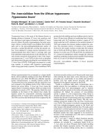
Figure 1
Study designStudy design. Treatment protocol for administration of free dexamethasone phosphate (DXM-P) and liposomal DXM-P. Blood samples for assess-
ment of the side-effect profile were collected 2 and 7 days after last treatment. An additional blood sample was collected from mice treated with free
DXM-P 2 days after start of the treatment. CFA, complete Freund's adjuvant; i.v., intravenous.
Injection of type II
bovine collagen in CFA
X1 X21 X25-35 1 2 3 4 5 6 7 8 9 10 11 12 13 14
7x i.v.
treatment
1x i.v.
treatment
Collection of blood
2 days after start
and end of treatment
Collection of blood
7 days after start
and end of treatment
Free
DXM-P
days
Liposomal
DXM-P
Collection of blood
7 days after end of
treatment
Collection of blood
2 days after end of
treatment
Collection of blood
2 days after start of
treatment
Available online />Page 3 of 9
(page number not for citation purposes)
Analytical measurements
Pyridinoline in urine was measured using a Metra PYD Elisa
8010 at 405 nm (Quidel Corp., San Diego, CA, USA) with cre-
atinine as the internal standard. Blood was analysed on
Abbott's CellDyn 3500 (Abbott Diagnostics, Abbott Park, IL,
USA). Corticosterone levels in plasma were determined by
ELISA (DE3600; R&D Systems, Wiesbaden, Germany) and
blood glucose was analysed using Accu-Chek Compact
(Roche, Basel, Switzerland).
Pharmacokinetic analysis of liposomal dexamethasone
phosphate
Radiolabelled liposomes (150 μmol lipid, 1.25 MBq
14
C-
DPPC) were processed as above in the presence of 27 MBq
3
H-Inulin. Obtained particles were 312 nm in size (polydisper-
sity <0.2) and were adjusted with PBS to 47 mM lipid.
For pharmacokinetic assays, 250 g male Wistar rats were
given a single intravenous injection of 0.5 ml liposomal prepa-
ration. Blood samples of ~0.2 ml were collected and mixed
with heparin, and 100 mg of each blood sample was analysed
by catalytic oxidation using Ox500 (Zinsser Analytics, Frank-
furt, Germany).
Statistical analysis
The nonparametric Mann-Whitney U test was applied to ana-
lyse differences between controls and individual treatments
using SPSS 13.0 (SPSS Inc., Chicago, IL, USA). Statistically
significant differences were accepted for P ≤ 0.05.
Results
Efficacy of liposomal and free dexamethasone
phosphate
Collagen-induced arthritis was established in mice, and
groups of 12 mice were treated with liposomal DXM-P or free
DXM-P, respectively. Single administrations of 0.4 to 4 mg/kg
liposomal DXM-P generated a rapid and substantial reduction
of the paw swelling. In addition, the highest dose resulted in a
persistent remission of arthritis for at least 7 days. In contrast,
a single dose of 1.6 mg/kg free DXM-P was ineffective in this
model, and daily injections of 0.4 or 1.6 mg/kg free drug were
required to suppress paw swelling over the treatment period
of 7 days. The onset of therapeutic improvement was substan-
tially delayed when using daily injections of 0.4 mg/kg free
DXM-P (Figure 2a).
We also determined concentrations of urinary pyridinoline to
monitor the arthritis-related degradation of bone and cartilage.
Mature collagen chains in these tissues are connected by 3-
hydroxypyridinium crosslinks, which become discharged and
excreted as pyridinoline upon disintegration of collagen [19].
Levels of urinary pyridinoline are about 500 nM in healthy ani-
mals and 2,000 nM in arthritic animals. Treatment with moder-
ate or high doses of liposomal DXM-P resulted in a reduction
well below 1,000 nM on day 2. In addition, the single adminis-
tration of 4 mg/kg liposomal DXM-P resulted in a sustained
reduction of this marker through to day 7. Daily use of free
DXM-P, although effective in the reduction of paw volumes
and arthritic scores, did not afford a significant inhibition of the
collagen breakdown (Figure 2b).
Side-effect profile of liposomal dexamethasone
phosphate
We next set out to directly compare glucocorticoid-mediated
side-effects for both treatment modalities. In the liposome arm
of the study, three groups of mice (n = 12) received a single
dose of 0.4, 1.6 or 4 mg/kg liposomal DXM-P on day 1 and the
side-effects were monitored 2 or 7 days later (n = 6 each).
In a second arm of the study, two groups of mice were treated
with daily injections of 0.4 or 1.6 mg/kg free DXM-P (n = 12).
Side-effects were monitored as above; that is, 2 or 7 days after
cessation of treatment (n = 6 each).
For corticosterone, base levels for all animals were taken on
day (X - 5) and all changes were expressed as animal-specific
values relative to this starting point (n = 12). Additional data
were obtained on day 3 for groups receiving daily free DXM-P
(n = 12) to facilitate a direct comparison between groups at
this point in time (Figure 1).
Control groups (n = 12) received single administrations of 1.6
mg/kg free drug or saline and were monitored 2 and 7 days
later (n = 6 each).
Blood corticosterone
Glucocorticoids inhibit the release of adrenocorticotropic hor-
mone from the pituitary gland, leading to a suppression of cor-
ticosterone production in the adrenal glands [20]. On the
contrary, increased corticosterone levels are caused by the
inflammation process itself [21]. We therefore analysed the
response of corticosterone to liposomal DXM-P or free DXM-
P in healthy and inflamed animals.
A low dose of 0.4 mg/kg liposomal DXM-P did not change the
level of corticosterone in arthritic mice, while improving the
clinical score on days 2 and 3. In contrast, a single administra-
tion of 1.6 mg/kg free DXM-P resulted in suppression of corti-
costerone at day 3 without delivering such therapeutic benefit
(P < 0.01 vs. saline) (Figure 3a).
Use of 1.6 or 4 mg/kg liposomal DXM-P also reduced serum
corticosterone levels (P ≤ 0.01 vs. saline); however, this effect
was transient since corticosterone production recovered by
day 9. The normalization was complete in healthy animals, but
not in inflamed mice. We thus attribute this incomplete rever-
sion to the therapeutic benefit of the liposomal drug rather
than to a long-term suppression of corticosterone (Figure 3a,
b).
Arthritis Research & Therapy Vol 11 No 6 Rauchhaus et al.
Page 4 of 9
(page number not for citation purposes)
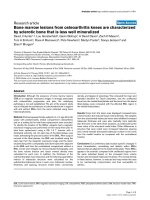
Figure 2
Therapeutic efficacy and persistence of liposomal dexamethasone phosphateTherapeutic efficacy and persistence of liposomal dexamethasone phosphate. (a) Joint swelling was assessed by clinical scoring after a single
injection of liposomal dexamethasone phosphate (DXM-P) (0.4 mg/kg, 1.6 mg/kg or 4 mg/kg) or after seven daily injections of free DXM-P (0.4 and
1.6 mg/kg). A single dose of free DXM-P at 1.6 mg/kg is also shown for comparison. Saline injections were used as controls. n = 12 for all groups;
means ± standard error of the mean. (b) Urine pyridinoline levels 2 and 7 days after cessation of the treatment. *P ≤ 0.05 Mann-Whitney U test ver-
sus saline-treated inflamed animals. n = 6 for all groups; means ± standard error of the mean.
A
B
5
6
7
8
9
10
11
12
13
02468101214
arthritis score
study period [day]
saline
liposomal DXM-P 0.4 mg/ kg
liposomal DXM-P 1.6 mg/ kg
liposomal DXM-P 4 mg/ kg
free DXM-P 0.4 mg/ kg (7 doses)
free DXM-P 1.6 mg/ kg (7 doses)
free DXM-P 1.6 mg/ kg (single dose)
0
500
1000
1500
2000
2500
3000
saline saline liposomal
DXM-P
1 x 0.4 mg/kg
liposomal
DXM-P
1 x 1.6 mg/kg
liposomal
DXM-P
1 x 4 mg/kg
f ree DXM-P
7 x 0.4 mg/kg
f ree DXM-P
7 x 1.6 mg/kg
f ree DXM-P
1 x 1.6 mg/kg
healthy
animals
inf lamed animals
pyridinoline[nmol/l]
2 days after withdrawal
7 days after withdrawal
*
*
Available online />Page 5 of 9
(page number not for citation purposes)
Animals treated with free DXM-P continued to display a signif-
icantly suppressed corticosterone level over the entire treat-
ment period. As with the liposomal form, these values
normalized after cessation of treatment.
Blood glucose
Daily administration of therapeutic amounts of free DXM-P led
to a significant increase of the blood glucose, with levels per-
sisting for at least 2 days after the last treatment - which is day
9 under this regimen. In contrast, a single injection of 4 mg/kg
liposomal DXM-P did not alter blood glucose levels 2 days
after the injection, which is day 3 in this group. Independent of
the respective treatments, no significant alterations of the
blood glucose levels were monitored at day 7 after the last
administration of drug (Figure 4a).
Liver enzymes
A slight, but nonsignificant, elevation of liver aspartate ami-
notransferase was connected to the use of free DXM-P, but
not of liposomal DXM-P. Any such elevation was no longer
detectable at day 7 after the termination of treatment (Figure
4b).
Haematology
Glucocorticoids are known to induce neutrophilia by stimulat-
ing the migration of immature polymorphonuclear neutrophils
from bone marrow into the circulation [22,23].
We confirmed this knowledge and observed a neutrophilia
upon daily administrations of 1.6 mg/kg free DXM-P. Treat-
ment with 4 mg/kg liposomal DXM-P resulted in a milder and
delayed increase of the number of neutrophils (Figure 4c),
which became significant only at day 7 after the cessation of
treatment.
The use of free DXM-P is lymphopenic; a significantly reduced
number of lymphocytes was thus observed during the treat-
ment with daily doses of 1.6 mg/kg free drug. In contrast, treat-
ment with 4 mg/kg liposomal DXM-P caused only a mild
lymphopenia (Figure 4c). Also, the lymphopenia became sig-
nificant only at the later time point. The reasons for these
delayed effects of the liposomal drug on neutrophils and lym-
phocytes are not known and require further investigation. No
other haematological alterations were observed in any of the
groups.
Body weight
A reduction in body weight is a common phenomenon of the
use of glucocorticoids in rodents [24]. In the model described
here, daily injections of 1.6 mg/kg free DXM-P reduced the
body weight at day 8 by 10%. The same reduction was
observed for a single injection of 4 mg/kg liposomal DXM-P.
The lower doses of 1.6 or 0.4 mg/kg of the liposomal form
resulted in less than 4% reduction of body weight.
Presence of free and liposomal dexamethasone
phosphate in plasma
We eventually analysed the pharmacokinetic of free and lipo-
somal DXM-P to correlate the presence of drug with the
observed side-effects. This analysis was performed in rats due
to the technical limitations in mice.
Free DXM-P is rapidly converted into dexamethasone immedi-
ately after injection, with a half-life of 1.3 minutes [25]. Dexam-
ethasone itself has a longer circulating half-life of 150 minutes
(Figure 5a, black diamonds).
Liposomal DXM-P circulates in an intact form, as demon-
strated by the constant sequestration of the aqueous phase
marker
3
H-inulin (an uncharged polyfructose; molecular
weight, ~5,000 Da) with respect to the lipid membrane marker
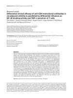
Figure 3
Impact of liposomal and free dexamethasone phosphate on corticosteroneImpact of liposomal and free dexamethasone phosphate on corti-
costerone. Corticosterone levels in the blood of (a) arthritic animals or
(b) healthy animals were measured following treatment with liposomal
dexamethasone phosphate (DXM-P), free DXM-P or saline. **P ≤ 0.01
Mann-Whitney U test versus saline. n ≥ 6 for all groups; mean ± stand-
ard error of the mean.
**
A
B
**
**
**
**
**
**
corticosterone under inflammatory conditions
1
10
100
1000
10000
02468101214
days
% of normal corticosterone
level
saline
liposomal DXM-P 1 x 0.4 mg/ kg
liposomal DXM-P 1 x 1.6 mg/ kg
liposomal DXM-P 1 x 4 mg/ kg
free DXM-P 1 x 1.6 mg/ kg
free DXM-P 7 x 0.4 mg/ kg
free DXM-P 7 x 1.6 mg/ kg
corticosterone in healthy animals
1
10
100
1000
0 2 4 6 8 101214
days
% of normal
corticosterone level
Arthritis Research & Therapy Vol 11 No 6 Rauchhaus et al.
Page 6 of 9
(page number not for citation purposes)
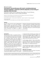
Figure 4
Effects of liposomal and free dexamethasone phosphate on blood glucose, liver enzymes and haematologyEffects of liposomal and free dexamethasone phosphate on blood glucose, liver enzymes and haematology. (a) Glucose levels as a percent-
age of the initial blood value in arthritic animals following treatment with liposomal dexamethasone phosphate (DXM-P), free DXM-P, or saline. (b)
Values of liver enzymes alkaline phosphatase (AP), alanine aminotransferase (ALAT) and aspartate aminotransferase (ASAT) after treatment with
liposomal DXM-P, free DXM-P, or saline. (c) Percentage of polymorphonuclear neutrophilic leukocytes (PMNs) and lymphocytes in the blood of
arthritic animals following treatment with liposomal DXM-P, free DXM-P, or saline. *P ≤ 0.05 Mann-Whitney U test versus saline-treated inflamed ani-
mals. n = 6 for all groups; mean ± standard error of the mean.
A
B
C
*
0
20
40
60
80
100
120
140
2 days 7 days
% of initial blood glucose level
0
100
200
300
400
500
2 days 7 days 2 days 7 days 2 days 7 days
AP ALAT ASAT
[U/l]
*
*
*
*
*
*
0
20
40
60
80
100
2 days 7 days 2 days 7 days
PMNs lymphocytes
% of cells in blood
saline lipos. DXM-P 1 x 4 mg/ kg free DXM-P 7 x 1.6 mg/ kg
Available online />Page 7 of 9
(page number not for citation purposes)
14
C-DPPC (Figure 5b) in a labelled probe of liposomal DXM-
P. The elimination of liposomes from the circulation followed a
two-phase elimination profile: 60% of the particles disappear
with a half-life of 44 minutes; the remainder circulates with a
half-life of 370 minutes.
A first portion of dexamethasone appeared shortly after the
injection (Figure 5a, squares), indicating a small fraction of free
DXM-P or drug associated with the outer layer of the lipo-
somes. The bulk of dexamethasone appeared in plasma slowly
and left the compartment with a terminal half-life of 520
minutes.
Both administrations of free DXM-P or liposomal DXM-P do
not yield persistent drug levels in plasma. Only low levels of the
drug are physically present at 24 hours, and certainly at 48
hours, after the last administration.
Discussion
In the present study, we used a mouse model of rheumatoid
arthritis to compare different treatment regimes for DXM-P in
its liposomal and free forms with respect to clinical outcome
and to the side-effects profile.
Liposomal DXM-P displayed a persistent therapeutic effect, as
a single injection of liposomal DXM-P at 4 mg/kg suppressed
established arthritis for at least 7 days. In contrast, treatment
with free DXM-P required daily administrations of 1.6 mg/kg to
achieve a continued suppression of the paw swelling; a single
treatment resulted in no detectable therapeutic benefit.
The liposomal formulation also afforded significant reduction
of pyridinoline, a marker for bone and cartilage degradation.
Such reduction of pyridinoline provides additional proof of the
strong therapeutic activity of liposomal DXM-P and correlates
to the reduced joint destruction observed for liposomal DXM-
P, but not free DXM-P as reported earlier [15].
The combined efficacy data provided herein support compara-
ble therapeutic benefit for a single administration of 4 mg/kg
liposomal DMX-P and daily administrations of at least 1.6 mg/
kg free glucocorticoid. This regimen is based on the limited
reduction of pyridinoline even after seven doses of 1.6 mg/kg
free drug, on the slower remission caused by the free drug and
on the absence of any therapeutic improvement after a single
administration of 1.6 mg/kg free DXM-P.
Glucocorticoids interfere with the hypothalamus-pituitary-
adrenal (HPA) axis, in that they compete with the natural ligand
corticosterone. The clinical manifestation of this interference is
Cushing syndrome, which is one of leading dose-limiting fac-
tors for glucocorticoids [1,26].
Figure 5
Pharmacokinetics of liposomal and free dexamethasone phosphate in ratsPharmacokinetics of liposomal and free dexamethasone phosphate in rats. (a) A dose of 4 mg/kg liposomal dexamethasone phosphate (DXM-
P) (open squares) or free DXM-P (diamonds) was injected intravenously into rats, and the appearance of the converted dexamethasone (DXM) was
monitored in plasma. In a separate experiment, radiolabelled liposomal DXM-P (triangles) was injected into rats and their pharmacokinetics was fol-
lowed using
14
C-1,2-dipalmitoyl-sn-glycero-3-phosphocholine (DPPC). (b) Integrity of liposomal DXM-P. Radiolabelled liposomal DXM-P was
injected as in (a) and the additional aqueous phase marker
3
H-inuline was analysed. The resulting
3
H/
14
C ratio was plotted with respect to the initial
ratio. The constant ratio indicates high carrier integrity in the circulation. Error bars indicate standard deviations.
Arthritis Research & Therapy Vol 11 No 6 Rauchhaus et al.
Page 8 of 9
(page number not for citation purposes)
Free DXM-P as well as higher doses of liposomal DXM-P did
show an adverse impact on the HPA axis on day 3. The
appearance of this corticosterone modulation is consistent
with the presence of residual amounts of DXM-P in the circu-
lation at this point in time. Our data, however, support a view
wherein the suppression of the HPA axis is substantially
shorter than the persistent therapeutic effect achieved with
liposomal DXM-P. Persistence may thus facilitate a separation
between therapeutic benefit and side-effects, at least in time.
In contrast to the liposomal dosage form, the free drug does
not allow such separation between HPA suppression and
therapeutic effect. Even worse, we found a sensitive suppres-
sion of the HPA axis using a single, therapeutically insufficient,
administration of free DXM-P. In contrast, no such suppres-
sion was monitored after treatment with a single dose of lipo-
somal DXM-P at 0.4 mg/kg, which was therapeutically active.
This finding provides a separation of the therapeutic benefit
from related side-effects using substantially reduced amounts
of drug.
Liposomal DXM-P did not induce hyperglycaemia across the
entire dose range, whereas daily treatments with the free drug
significantly increased blood glucose levels. This observation
is in line with the very low hepatic accumulation of the lipo-
somal drug that does not exceed peak plasma concentrations
[15]. In addition, any such uptake does mainly occur in the
phagocytic Kupffer cells - but not in hepatocytes, the main cell
type associated with gluconeogenesis (U Rauchhaus, unpub-
lished observations). We relate this to the large size of the lipo-
somes of about 300 nm, which is well above the exclusion limit
for the liver endothelium of about 100 nm [27].
Liposomal DXM-P also has a reduced impact on the complete
blood count. Exposure to free DXM-P resulted in significant,
but transient, neutrophilia and lymphopenia. The effect of the
liposomal DXM-P was modest and no significant changes
were observed on day 2. Long-term alterations in lymphocytes
and neutrophils, although significant by day 7, had small ampli-
tude. Although a mild lymphopenia was observed in the colla-
gen-induced arthritis model used here, the impact of liposomal
DXM-P on immune organs is generally quite modest. We
observed no pathological alterations in the spleen after admin-
istration of up to 30 mg/kg liposomal DXM-P into healthy rats.
Occasional hypotrophism of the thymus was observed with
doses of 10 mg/kg or more.
Conclusions
Glucocorticoids belong to the basic therapeutic arsenal in the
field of inflammatory and autoimmune diseases. Being power-
ful drugs, their clinical acceptance is mainly limited by side-
effects.
Using a liposomal dosage form of DXM-P we here observe a
separation between the therapeutic benefit and side-effects.
We attribute these improvements in the side-effect profile to a
targeted, cellular delivery of the liposomal dosage form to cells
and organs of the immune system, mainly the spleen, which we
hypothesize to drive the therapeutic persistence [15].
At the same time, the liposomal form excludes the drug from
reaching unwanted sites. This exclusion became most appar-
ent when a small, yet therapeutically active, dose of liposomal
DXM-P had no impact on the HPA axis, whereas even subther-
apeutic amounts of the free drug reduced the corticosterone
production.
Of note, the increased therapeutic potency of liposomal DXM-
P resulted also in a significant reduction of urinary pyridinoline,
indicating an inhibition of the ongoing, inflammation-mediated
breakdown of collagen. If translated into clinical practice, such
a feature would offer a window for regeneration of the inflamed
site, thus establishing a disease-modifying quality for
glucocorticoids.
Future developmental work should address the therapeutic
persistence, in particular. The intermittent use of liposomal
DXM-P has the potential for an improved therapeutic index in
that it combines an immediate but lasting therapeutic benefit
with side-effects that are restricted to the actual time of
treatment.
Competing interests
SP and UR are employees of Novosom AG - Novosom AG
funded this research. SP is founder and shareholder of the
Novosom AG - Novosom AG holds or has applied for patents
relating to the content of the manuscript (liposomal glucocor-
ticoids). No payments have been made to the authors.
Authors' contributions
UR and SP developed the liposomes used in this publication;
both conceived and coordinated the studies, interpreted the
data and prepared the manuscript. FWS performed experi-
mental work in the collagen-induced arthritis model.
Acknowledgements
D Pohlers (Experimental Rheumatology Unit, Department of Orthoped-
ics, University Hospital Jena) is gratefully acknowledged for the statisti-
cal analysis of the data. D Pohlers received funding from the DFG under
the project KI 439/6-3.
References
1. Hopkins RL, Leinung MC: Exogenous Cushing's syndrome and
glucocorticoid withdrawal. Endocrinol Metab Clin North Am
2005, 34:371-384. ix
2. Saklatvala J: Glucocorticoids: do we know how they work?
Arthritis Res 2002, 4:146-150.
3. Wang M: The role of glucocorticoid action in the pathophysiol-
ogy of the metabolic syndrome. Nutr Metab (Lond) 2005, 2:3.
4. Manolagas SC, Weinstein RS: New developments in the patho-
genesis and treatment of steroid-induced osteoporosis. J
Bone Miner Res 1999, 14:1061-1066.
Available online />Page 9 of 9
(page number not for citation purposes)
5. Buttgereit F, Wehling M, Burmester GR: A new hypothesis of
modular glucocorticoid actions: steroid treatment of rheu-
matic diseases revisited. Arthritis Rheum 1998, 41:761-767.
6. Buttgereit F, Straub RH, Wehling M, Burmester GR: Glucocorti-
coids in the treatment of rheumatic diseases: an update on the
mechanisms of action. Arthritis Rheum 2004, 50:3408-3417.
7. Song IH, Gold R, Straub RH, Burmester GR, Buttgereit F: New
glucocorticoids on the horizon: repress, don't activate! J
Rheumatol 2005, 32:1199-1207.
8. Schacke H, Hennekes H, Schottelius A, Jaroch S, Lehmann M,
Schmees N, Rehwinkel H, Asadullah K: SEGRAs: a novel class of
anti-inflammatory compounds. Ernst Schering Res Found
Workshop 2002, 40:357-371.
9. Avnir Y, Ulmansky R, Wasserman V, Even-Chen S, Broyer M, Bar-
enholz Y, Naparstek Y: Amphipathic weak acid glucocorticoid
prodrugs remote-loaded into sterically stabilized nanolipo-
somes evaluated in arthritic rats and in a Beagle dog: a novel
approach to treating autoimmune arthritis. Arthritis Rheum
2008, 58:119-129.
10. Metselaar JM, Wauben MH, Wagenaar-Hilbers JP, Boerman OC,
Storm G: Complete remission of experimental arthritis by joint
targeting of glucocorticoids with long-circulating liposomes.
Arthritis Rheum 2003, 48:2059-2066.
11. Metselaar JM, Berg WB van den, Holthuysen AE, Wauben MH,
Storm G, van Lent PL: Liposomal targeting of glucocorticoids to
synovial lining cells strongly increases therapeutic benefit in
collagen type II arthritis. Ann Rheum Dis 2004, 63:348-353.
12. Schmidt J, Metselaar JM, Wauben MH, Toyka KV, Storm G, Gold
R: Drug targeting by long-circulating liposomal glucocorticos-
teroids increases therapeutic efficacy in a model of multiple
sclerosis. Brain 2003, 126:1895-1904.
13. Barrera P, Mulder S, Smetsers A, Storm G, Beijnen J, Metselaar J,
van Riel P: Long-circulating liposomal prednisolone versus
pulse intramuscular methylprednisolone in patients with
active rheumatoid arthritis [abstract]. Proceedings of the Amer-
ican College of Rheumatology (ACR) Scientific Meeting; Oct 24-
29, 2008; San Francisco. Hall A, poster board 453 2008.
14. Ishida T, Ichihara M, Wang X, Yamamoto K, Kimura J, Majima E,
Kiwada H: Injection of PEGylated liposomes in rats elicits PEG-
specific IgM, which is responsible for rapid elimination of a
second dose of PEGylated liposomes. J Control Release 2006,
112:15-25.
15. Rauchhaus U, Kinne RW, Pohlers D, Wiegand S, Wolfert A, Gajda
M, Brauer R, Panzner S: Targeted delivery of liposomal dexam-
ethasone phosphate to the spleen provides a persistent ther-
apeutic effect in rat antigen-induced arthritis. Ann Rheum Dis
2009, 68:1933-1934.
16. Olson F, Hunt CA, Szoka FC, Vail WJ, Papahadjopoulos D: Prep-
aration of liposomes of defined size distribution by extrusion
through polycarbonate membranes. Biochim Biophys Acta
1979, 557:9-23.
17. Szoka F Jr, Papahadjopoulos D: Comparative properties and
methods of preparation of lipid vesicles (liposomes). Annu
Rev Biophys Bioeng 1980, 9:467-508.
18. Durie FH, Fava RA, Noelle RJ: Collagen-induced arthritis as a
model of rheumatoid arthritis. Clin Immunol Immunopathol
1994, 73:11-18.
19. Seibel MJ, Woitge H, Scheidt-Nave C, Leidig-Bruckner G, Duncan
A, Nicol P, Ziegler R, Robins SP: Urinary hydroxypyridinium
crosslinks of collagen in population-based screening for overt
vertebral osteoporosis: results of a pilot study. J Bone Miner
Res 1994, 9:1433-1440.
20. Kuperman H, Damiani D, Chrousos GP, Dichtchekenian V, Manna
TD, Filho VO, Setian N: Evaluation of the hypothalamic-pitui-
tary-adrenal axis in children with leukemia before and after 6
weeks of high-dose glucocorticoid therapy. J Clin Endocrinol
Metab 2001, 86:2993-2996.
21. Gaillard RC: Interaction between the hypothalamo-pituitary-
adrenal axis and the immunological system. Ann Endocrinol
(Paris) 2001, 62:155-163.
22. Claman HN: How corticosteroids work. J Allergy Clin Immunol
1975, 55:145-151.
23. Peng CT, Lin HC, Lin YJ, Tsai CH, Yeh TF: Early dexamethasone
therapy and blood cell count in preterm infants. Pediatrics
1999, 104:476-481.
24. Rooman R, Koster G, Bloemen R, Gresnigt R, van Buul-Offers SC:
The effect of dexamethasone on body and organ growth of
normal and IGF-II-transgenic mice. J Endocrinol 1999,
163:543-552.
25. Rohdewald P, Mollmann H, Barth J, Rehder J, Derendorf H: Phar-
macokinetics of dexamethasone and its phosphate ester.
Biopharm Drug Dispos 1987, 8:205-212.
26. Hatz H: Glukokortikoide. Immunologische Grundlagen, Pharma-
kologie und Therapierichtlinien 2nd edition. Stuttgart: Wissen-
schaftliche Verlagsgesellschaft mbH; 2005.
27. Braet F, Wisse E: Structural and functional aspects of liver
sinusoidal endothelial cell fenestrae: a review. Comp Hepatol
2002, 1:1.