Báo cáo y học: "B-lymphocyte stimulator/a proliferation-inducing ligand heterotrimers are elevated in the sera of patients with autoimmune disease and are neutralized by atacicept and B-cell maturation antigen-immunoglobulin" pdf
Bạn đang xem bản rút gọn của tài liệu. Xem và tải ngay bản đầy đủ của tài liệu tại đây (1.12 MB, 14 trang )
RESEARC H ARTIC L E Open Access
B-lymphocyte stimulator/a proliferation-inducing
ligand heterotrimers are elevated in the sera of
patients with autoimmune disease and are
neutralized by atacicept and B-cell maturation
antigen-immunoglobulin
Stacey R Dillon
1*
, Brandon Harder
1
, Kenneth B Lewis
1
, Margaret D Moore
1
, Hong Liu
1
, Thomas R Bukowski
1
,
Nels B Hamacher
1
, Megan M Lantry
1
, Mark Maurer
1
, Cecile M Krejsa
1
, Jeff L Ellsworth
1
, Susan Pederson
1
,
Keith B Elkon
2
, Mark H Wener
2
, Maria Dall’Era
3
, Jane A Gross
1
Abstract
Introduction: B-lymphocyte stimulator (BLyS) and a proliferation-inducing ligand (APRIL) are members of the
tumor necrosis factor (TNF) family that regulate B-cell maturation, survival, and function. They are overexpressed in
a variety of autoimmune diseases and reportedly exist in vivo not only as homotrimers, but also as BLyS/APRIL
heterotrimers.
Methods: A proprietary N-terminal trimerization domain was used to produce recombinant BLyS/APRIL
heterotrimers. Heterotrimer biologic activity was compared with that of BLyS and APRIL in a 4-hour signaling assay
by using transmembrane activator and CAML interactor (TACI)-transfected Jurkat cells and in a 4-day primary
human B-cell proliferation assay. A bead-based immunoassay was developed to quantify native heterotrimers in
human sera from healthy donors (n = 89) and patients with systemic lupus erythematosus (SLE; n = 89) or
rheumatoid arthritis (RA; n = 30). Heterotrimer levels were compared with BLyS and APRIL homotrimer levels in a
subset of these samples.
Results: The recombinant heterotrimers consisted mostly of one BLyS and two APRIL molecules. Heterotrimer
signaling did not show any significant difference compared with APRIL in the TACI-Jurkat assay. Heterotrimers were
less-potent inducers of B-cell proliferation than were homotrimeric BLyS or APRIL (EC
50
, nMol/L: BLyS, 0.02; APRIL,
0.17; heterotrimers, 4.06). The soluble receptor fusion proteins atacicept and B-cell maturation antigen (BCMA)-
immunoglobulin (Ig) neutralized the activity of BLyS, APRIL, and heterotrimers in both cellular assays, whereas B-cell
activating factor belonging to the TNF family receptor (BAFF-R)-Ig neutralized only the activity of BLyS. In human
sera, significantly more patients with SLE had detectable BLyS (67% versus 18%; P < 0.0001), APRIL (38% versus 3%;
P < 0.0002), and heterotrimer (27% versus 8%; P = 0.0013) levels compared with healthy donors. Significantly more
patients with RA had detectable APRIL, but not BLyS or heterotrimer, levels compared with healthy donors (83%
versus 3%; P < 0.0001). Heterotrimer levels weakly correlated with BLyS, but not APRIL, levels.
Conclusions: Recombinant BLyS/APRIL heterotrimers have biologic activity and are inhibited by atacicept and
BCMA-Ig, but not by BAFF-R-Ig. A novel immunoassay demonstrated that native BLyS/APRIL heterotrimers, as well
as BLyS and APRIL homotrimers, are elevated in patients with autoimmune diseases.
* Correspondence:
1
Preclinical Research and Development, ZymoGenetics, Inc., 1201 Eastlake
Ave East, Seattle, WA 98102, USA
Dillon et al. Arthritis Research & Therapy 2010, 12:R48
/>© 2010 Stacey Dillon et al.; licensee BioMed Central Ltd. This is an open access article distributed under the terms of the Creative
Commons Attribution License (http://creative commons.org/licenses/by/2.0), which permits unrestricted use, di stribution, and
reproduction in any medium, provided the original work is properly cited.
Introduction
B-lymphocyte stimulator (BLyS)-also called B cell-acti-
vating factor belonging to the tumor necrosis factor
family (BAFF)-and a proliferation-inducing ligand
(APRIL) are members of the tumor necro sis factor
(TNF) family and are important regulators of B-cell
maturation, survival, and function [1,2]. T he TNF
ligands generally form trimeric structures composed of
three monomers [3]. Heterotrimers of BLyS and APRIL
have also been shown to exist in vivo,andahigher-
order oligomer of BLyS homotrimers h as been reported
[4-6].
BLyS homotrimers bind to the B cell-expressed recep-
tors transmembrane activator and CAML interac tor
(TACI), B cell-maturation antigen (BCMA), and BAFF
receptor (BAFF-R), whereas APRIL homotrimers bind to
TACI, BCMA, and proteoglycans [7,8]. The binding of
BLyS and APRIL to these receptors activates specific
TNF receptor-associated factors (TRAFs), which regu-
late signal transduction in B cells. The interaction with
TRAFs induces the nuclear factor (NF)-B signaling
pathway, which plays a pivotal role in regulati ng diverse
aspects of immune function, including mediating inflam-
matory responses and facilitating adaptive immunity
[9-11]. The binding o f BLyS and APRIL to TACI,
BCMA, and BAFF-R receptors also triggers the upregu-
lation or downregulation of members of the Bcl-2 family
of proteins, which are involved in cell death, prolifera-
tion, survival, and cell-cell interactions [12]. It has been
propo sed that signaling through TACI in mature B cells
or plasmablasts requires higher-order BLyS oligomers or
the cross-linking of APRIL through its binding to pro-
teoglycans, whereas BAFF-R and TACI on primary B
cells can bind and respond to all forms of BLyS [4,8,13].
BLyS and APRIL are o verexpressed in the sera of
patients with a wide variety of autoimmune disorders,
including systemic lupus erythematosus (SLE) [14,15]. In
patients with rheumatoid arthritis (RA), BLyS and
APRIL are overexpressed in the synovial fluid as well as
in the sera [6,16]. P reliminary data suggest that BLyS/
APRIL heterotrimers also are elevated in patients with
various autoimmune conditions [6]. In light of their
roles in B-cell function and these clinical data, BLyS and
APR IL are targets for novel treatments for autoimmun e
diseases.
Atacicept is a fully human recombinant fusion protein
comp rising the extracellular portion of the TACI recep-
tor linked to an Fc domain of immunoglobulin (Ig)G.
Atacicept modulates B cells by neutralizing BLyS and
APRIL activity and is in clinical development for the
treatment of SLE and RA [17,18]. As BLyS/APRIL het-
erotrimers may also be elevated in patients with autoim-
mune diseases, it is important to determine whether
these heterotrimers play a particular biologic role, and if
therapies targeting BLyS and APRIL will also neutralize
BLyS/APRIL heterotrimers. This study investigated the
in vitro activity of recombinant heterotrimers in cell-sig-
naling and proliferation assays, and the ability of the
soluble B cell-expressed receptors atacicept, BCMA-Ig,
and BAFF-R-Ig to neutralize heterotrimer activity. A
bead-based immunoassay was developed for BLyS/
APRIL heterotrimers, and en dogenous levels of hetero-
trimers in the sera of healthy donors and patients with
SLE or RA were measured and compared with BLyS
and APRIL homotrimer levels in a subset of the same
samples.
Materials and methods
Production of recombinant BLyS and APRIL homotrimers
and BLyS/APRIL heterotrimers
BLyS and APRIL homotrimers were generated as pre-
viously described [19,20]. Recombinant BLyS/APRIL
heterotrimers were produced by using a proprietary N-
terminal trimerization domain [21]. The Flag-zippered
(zz) 12.6 form of APRIL [21] and the Hisx6-zz 12.6 form
of BLyS were made by overlap polymerase c hain
reaction (PCR) of human APRIL [amino acids 110-250]
template and human BLyS [amino acids 141-285] tem-
plate, respectively. The assembled cDNA was inserted
by homologous recombination into the vectors pZMP21
[22] and pZMP41z, respectively, downstream of the
optimized tissue plasminogen activator (otPA) leader
sequence. These vectors were t ransfected into protein-
free media-adapted Chinese hamster ovary (CHO)
DXB11 cell suspension by electr oporation, and the cells
were selected for growth in methotrexate- and copper-
chelated antibiotic (Zeocin)-containing medium.
Methotrexate- and Zeocin-resistant cells were stained
with fluorescein isothiocyanate-anti-CD8 and phycoery-
thrin-anti-CD4, and CD8
+
/CD4
+
cells sel ected by
fluorescence -activated cell sorting. Batches of the BLyS/
APRIL heterotrimer were produced in a 10-L WAVE
bioreactor. Both BLyS and APRIL were quantified by
Western blot with anti-His and anti-Flag antibodies,
respectively, as described later. The expression levels of
BLySNH6zz12.6andAPRILNF zz12.6 were approxi-
mately 10 mg/L and 33 mg/L, respectively.
Purification of recombinant BLyS/APRIL heterotrimers
Heterotrimers were purified from CHO-conditioned
media after buffer exchange to phosphate-buffered saline
(PBS) (pH 7.4) by ultrafiltration/diafiltration and loading
onto an immobilized metal affinity chromatography NI-
NTA His Bind Superflow Column (Novagen, Gibbstown,
NJ, USA). A heparin affinity column (HAC), Heparin AF-
650M (Tosoh Bioscience, Montgomeryville, PA, USA),
Dillon et al. Arthritis Research & Therapy 2010, 12:R48
/>Page 2 of 14
was used to resolve the heterotrimer from Flag-zz12.6
APRIL via NaCl gradient elution. The HAC eluate was
concentrated to < 8 ml and injected over a size-exclusion
chromatography (SEC) Superdex 200 Prep Grade Col-
umn (GE Healthcare, Piscataway, NJ, USA).
Generation of non-tagged recombinant BLyS/APRIL
heterotrimers
Non-tagged heterotrimers were produced by using a
limited proteolysis strategy with trypsin to cleave the
Flag and His tags from the zz12.6 heterotrimers. A
HAC (as earlier), was used to resolve non-tagged hetero-
trimer from undigested products by NaCl gradient elu-
tion. The HAC eluate was concentrated to < 3 ml and
injected over an SEC column (as earlier). The SEC elu-
ate was incubated and rocked slowly overnight with 1
ml of anti-Flag agaro se resin (Sigma, St Louis, MO,
USA). The resin was separated from solution via 0.22-
μm filtration. N-terminal sequence analysis, SEC with
multiangle light scattering (SEC-MALS), and Western
blot analyses were consistent with both tags having been
removed.
Protein detection
APRIL or BLyS protein samples were visualized by using
nonreducing sodium dode cylsulfate polyacrylamide gel
electrophoresis (SDS-PAGE). Analysis with Western
blotting was performe d by using standard methods. The
anti-APRIL blot comprised anti-Flag horseradish peroxi-
dase (HRP) for the Flag-zz12.6 form, and anti-APRIL
polyclonal antibody (pAb) followed by donkey anti-rab-
bit IgG-HRP for the trypsinized form of APRIL. The
anti-BLyS blot comprised anti-6x His HRP for the
Hisx6-zz12.6 form of BLyS, and an ti-BLyS pAb followed
by donkey anti-rabbit IgG-HRP for the trypsini zed form
of BLyS. The molecular mass of the heterotrimers was
confirmed by SEC-MALS mass distribution LS/UV/RI
3-detector analysis.
Binding kinetics and affinity studies
The binding affinities an d kinetics of BLyS (average
values were determined from three distinct lots of pro-
tein), APRIL (six lots), and heterotrimers (one lot) for
the receptor-Fc fusion proteins (atacicept, BCMA-Ig,
and BAFF-R-Ig) were assessed with Biacore surface plas-
mon resonance studies by using a Biacore 3000 analyzer
(GE Healthcare) equipped with Biacor e Control, Evalua-
tion, and Simulation software version 3.2.
Atacicept, BCMA-Ig, and BAFF-R-Ig were covalently
immobilized onto a Biacore CM4 sensor chip. Binding
affinity studies were performed at 25°C with a 50 μl/min
flow rate for varying ligand concentrations. Serial 1:2
dilutions of each ligand from ~0.05 to 20 nMol/L were
made in analysis buffer (20 mmol/L sodium phosphate,
150 mmol/L NaCl, 0.05% polysorbate 20, pH 7.5). Disso-
ciation constant (K
D
) values were determined from the
kineticrateconstants(k
a
and k
d
). The binding curves
were processed by double referencing and were globally
fitted to a 1:1 binding model. The stoichiometry of bind-
ing was not determined.
Biologic activity assays
A 4-hour signaling assay was performed by using TACI-
transfected Jurkat cells carrying an NF-B/luciferase
reporter gene (KZ142). TACI/KZ142-Jurkat cells (1 ×
10
5
cells/well) were incubated at 37°C for 4-6 hours
with recombinant BLyS, APRIL, or heterotrimer in a
totalwellvolumeof100μl in complete RPMI-1640
media without phenol red, and in the absence or pre-
sence of atacicept, BCMA-Ig, or BAFF-R-Ig in concen-
trations ranging from 0.001 to 100 nMol/L. After
incubation, 100 μl/well of Steady-Glo reagent was
added, and the plates were foil covered and agitated at
roomtemperaturefor10minutes.Theassayplatewas
then analyzed in a luminometer to measure luciferase
activity.
For the human B cell-proliferation assay, B cells from
two healthy donors were isolated from peripheral blood
mononuclear cells by negative selection w ith the human
B Cell Isolation Kit II from Miltenyi Biotec (Auburn, CA,
USA), according to the manufacturer’s instructions. Flow
cytometry confirmed that they were > 97% pure (CD19
+
).
The purified B cell s (5 × 10
4
cells/well) were plated in a
96-well flat-bottomed plate pre-coated with 5 μg/ml anti-
IgM monoclonal antibody (mAb) (Southern Biotech, Bir-
mingham, AL, USA) in media containing 10 ng/ml
recombinant human interleukin-4 (R&D Systems, Min-
neapolis, MN, USA) and BLyS, APRIL, or heterotrimer
at concentrations ranging from 0.001 to 100 nMol/L.
The plates were then incubated for 4 days at 37°C, and
proliferation was determined by
3
H-thymidine incorpora-
tion assay. To determine the relative neutralization with
the soluble receptors, atacicept, BCMA-Ig, or BAFF-R-Ig,
the assay was set up as previously described by using
50% effective concentrations ( EC
50
) of BLyS, APR IL, and
heterotrimers (1, 3, and 10 nMol/L, respectively). Starting
with a 300-fold molar excess, 1:4 serial dilutions of ataci-
cept, BCMA-Ig, or BAFF-R-Ig were then added to
the wells, and the assay run as described earlier. Fifty
percent inhibition concentration (IC
50
)valueswere
determined for the inhibition of ligand activity by each
soluble receptor by using purified B cells from a third
donor.
Please see Supplemental Methods in Additional file 1
for more-detailed descriptions of the generation, purifi-
cation, and characterization of the recombinant hetero-
trimers, and of the binding affinity and biologic activity
assays.
Dillon et al. Arthritis Research & Therapy 2010, 12:R48
/>Page 3 of 14
Generation of anti-APRIL and anti-BLyS monoclonal
antibodies
Anti-human APRIL mAbs were generated from BALB/c
mice immunized with 50 μg of recombinant Fla g-zz12.6
APRIL in combination with Ribi-CWS adjuvant (Co rixa-
Sigma, St Louis, MO, USA) every 2 weeks over an
8-week period. Serum titers were determined for the
presence of anti-APRIL antibodies. T he mice with the
most significant anti-APRIL serum titers were im mu-
nized a final time. Four days later, the spleen and lymph
nodes of the mice were harvested and fused to mouse
myeloma P3-X63-Ag8.653 cells (American Type Culture
Collection) at a 1:1 lymphocyte/myeloma ratio with
polyethylene glycol 1500 by using standard methods.
Wells of the fusion plates were fed 3 times with a 70%
replacement of media, and wells were assayed 10 and 12
days after the plating of the fusion for anti-APRIL anti-
bodies. Anti-human BLyS mAbs were prepared as
described earlier, exce pt that 20 μgofbaculovirus-pro-
duced His- zz12. 6 BLyS was used for the initial immuni-
zations followed by 10-μg maintenance boosts. Antibody
purifications were performed from hybridoma superna-
tant by Protein G (GE Healthcare) affinity chromatogra-
phy followed by pH elution.
Heterotrimer immunoassay
After pre-wetting and blocking a Luminex plate with
assay buffer (PBS with 0.05 % Tween 20, 1% bovine serum
albumin), 5 × 10
3
anti-APRIL capture mAb-coated
beads/well were added to the plate in 25 μl/well assay
buffer. To this, 25 μlofstandardplus25μlofnormal
human serum ( pre-screened for low heterotrimer levels)
or 25 μl of sample serum plus 25 μl of assay buffer was
added. The plate was incubated on a shaker for 1 ho ur at
room temperature, and then washed twice with 100 μlof
assa y buffer. Biotinylated anti-BLyS antibody (25 μl/well)
was added, and the plate was incubat ed again as
described earlier. Streptavidin-phycoerythrin (25 μl) was
then added at 1:200 in assay buffer and incubate d on a
shaker for 30 minutes at room temperature. The plate
was washed twice with 100 μl of assay buffer. Wells were
then resuspended in 100 μl of assay buffer and analyzed
by using a Luminex 100 mac hine (Luminex Corporation,
Austin, TX, USA). The total assay time was 2 hours. The
assayhadabroadrange(~100pg/mlto25ng/ml)with
an EC
50
of ~6 ng/ml.
Luminex-based assays may vary in sensitivity due to
differences in the capture-bead lots used. In this study,
the heterotrimer assay data were generated by using two
lots of beads with slightly different limits of quantitation
(LOQs), 0.100 ng/ml versus 0.313 ng/ml. To make use
of the combined dataset, the most conservative LOQ
(0.313 ng/ml) was applied to all heterotrimer data,
although some sa mple cohorts had reportable va lues
below this concentration (see Supplemental Tables 1
and 2 in Additional file 2).
Serum heterotrimer and BLyS and APRIL homotrimer
levels in patients with autoimmune diseases
Serum samples from 30 patients with SLE and 30
patients with RA were obtained from the University of
Washington (Seattle, WA, USA) serum repository. Dr.
Dall’Era and Dr. Wofsy from the University of Califor-
nia, San Francisco (UCSF) (San Francisco, CA, USA)
provided 59 serum samples from 47 patients with SLE
and from nine healthy donors. Twenty of the UCSF
serum samples from patients with SLE were collected
from the same eight patients over time (two to four
draws each, drawn a minimum of 1 month apart). All
patients fulfilled the revised American College of Rheu-
mato logy classification criteria for SLE and RA . Patients
were enrolled after obtaining their written informed
consent by using a protocol approved by the Human
Subjects Committee of the University o f Washington
and the Committee on Human Research at UCSF,
respectively. Serum samples from an additional 80
healthy donors were collected at ZymoGenetics, Inc.
(Seattle, WA, USA).
Serum samples from healthy donors (n = 89) and
patients with SLE (n = 89) or RA (n = 30) were col-
lected in et hylenedi aminetetraace tic acid (EDTA) tubes
and frozen at -80°C. Samples were thawed and assessed
for native heterotrimer levels by using the heterotrimer
immunoassay described earlier. BLyS and APRIL levels
were measured in a subset of the serum samples from
healthy donors (n = 40) and patients with SLE (n = 30).
BLyS levels were measured with enzyme-linked immu-
nosorbent assay (ELISA), as described previously [18].
All samples were measured in triplicate. As assay perfor-
mance criteria, a precision of 20% for the coefficient of
variati on in the patient samples was accepted. The LOQ
was 0.78 ng/ml of BLyS in the serum. APRIL levels were
determined by using an ELISA developed and validated
at ZymoGenetics, Inc. (Seattle, WA, USA) [20]. The
LOQ was 2 ng/ml of APRIL in the serum. Because of
insuff icient volume, one of 30 available SLE serum sam-
ples and one of 40 healthy donor samples could not be
included for assessment in the APRIL ELISA.
Statistical methods
All statistica l anal yses were performed by using Prism 4
for Windows (GraphPad Software, Inc., La Jolla, CA,
USA). The proportions of serum samples with BLyS or
APRILhomotrimerorBLyS/APRILheterotrimerlevels
above the assay LOQ in patien ts with SLE or RA were
compared with those f or the healthy donor cohort by
using Fisher’s exact test. A value of P < 0.05 was consid-
ere d to be statistically significant. To reduce the impact
Dillon et al. Arthritis Research & Therapy 2010, 12:R48
/>Page 4 of 14
of data skewing, correlation analysis for BLyS, APRIL,
and heterotrimers levels was assessed by using Spear-
man’s rank correlation coefficient. For the correlation
analysis and for data plotting, samples with serum ligand
levels below the assay LOQ were assigned values equal
to half of the LOQ for each assay (0.39, 1.0, an d 0.156
ng/ml for BLyS, APRIL, and heterotrimer, respectively).
Bivariate correlation plots were generated by using JMP
software (SAS Institute, Inc.).
Results
Characterization of heterotrimers
The recombinant BLyS/APRIL heterotrimers were char-
acterized by SDS-PAGE and Western-blot analyses
(Figure 1). The molecular mass and purity of the hetero-
trimers were confirmed by SEC-MALS analysis (Figure 2).
Because of unequal BLyS and APRIL expression plasmid
efficiencies (see Materials and methods), the recombinant
heterotrimers had a predominant stoichiometry o f
2APRILto1BLyS(A
2
B), and thus consisted of only
a small fraction of 1 APRIL to 2 BLyS (AB
2
) heterotri-
mers. The ratio of ~66.3% APRIL and 33.7% BLyS was
identified by Western blot, and confirmed by SEC-MALS
analysis.
The binding affinities of BLyS,APRIL,andheterotri-
mers for the soluble receptor fusion proteins atacicept,
BCMA-Ig, and BAFF-R-Ig, were determined from one
or two surface plasmon resonance experimen ts for each
ligand. BLyS (n = 2) was the only ligand that exhib ited
binding to BAFF-R-Ig (K
D
= 0.02-0.07 nMol/L). BLyS
(n = 2), APRIL (n = 2), and heterotrimers (n = 1) bound
to atacicept (K
D
= 0.02 nMo l/L [BLyS], 0.1-0.2 n Mol/L
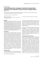
Figure 1 Sodium dodecylsulfate polyacrylamide gel electrophoresis and Western blot analysis of (a) BLyS/APRIL heterotrimer and (b)
trypsinized (nonzippered) heterotrimer. APRIL, a proliferation-inducing ligand; BLyS, B-lymphocyte stimulator.
Dillon et al. Arthritis Research & Therapy 2010, 12:R48
/>Page 5 of 14
[APRIL], and 0. 4 nMol/L [heterotrimers]) and BCMA-Ig
(K
D
= 0.3 nMol/L [BLyS], 0.0001-0.0003 nMol/L
[APRIL], and 0.01 nMol/L [heterotrimers]).
Biologic activity and neutralization of heterotrimers
Heterotrimer signaling was similar to that of APRIL in
the in vitro TACI-Jurkat assay (Figure 3a). Trypsinized
versions of the hete rotrimers and APRIL that lacked the
proprietary “zipper” trimerization domain needed for
their efficient expression were equally as active as the zz
versions. This suggested tha t the zipper domain did not
alter the biologic activity of these ligands in this assay.
Atacicept and BCMA-Ig neutralized the activity of BLyS,
APRIL, and heterotrimers in the TACI-Jurkat assay
(Figure 3b). As expected, BAFF-R-Ig only neutralized
the activity of BLyS.
The heterotrimers were less- potent inducers of B-cell
proliferation than were BLyS or APRIL, as evidenced by
the higher EC
50
values for heterotrimers than those of
BLyS or APRIL in the primary human B cell-prolifera-
tion assay (Figure 4a). In the neutralization assay, BAFF-
R-Ig inhibited BLyS but exhibited little to no inhibition
of heterotrimer or APRIL activity on human B cells
(Figure 4b).
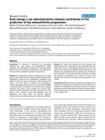
Heterotrimer immunoassay
To address whether endogenous heterotrimers are pre-
sent in the sera of patients with autoimmune diseases,
the recombinant heterotrimers were used as a standard
to develop a bead-based immunoassay by using anti-
APRIL capture mAb and fluorescenc e-labeled anti-BLyS
detection mAb to quantify native heterotrimer s in
human sera. The bead-based assay used a recombinant
protein heterotrimer that was heavily skewed toward
A
2
B BLyS/APRIL trimers as a reference standard, but
nevertheless, was also able to detect AB
2
trimers. During
the assay, beads conjugated with an anti-APRIL mAb
were incubated with the test sample and washed, and
then bead-bound heterotrimers were detected with a
biotinylated anti-BLyS detection mAb. This format
allowed the detection of both A
2
BandAB
2
native het-
erotrimers. We confirmed that the new assay detected
BLyS/APRIL heterotrimers but did not detect BLyS or
APRIL homotrimers (Figure 5).
Heterotrimer, BLyS, and APRIL levels in patients with
autoimmune diseases
The heterotrimer immunoassay and previously reported
ELISAs for BLyS and APRIL were used to measure
native heterotrimer, BLyS, and APRIL levels in serum
samples from healthy donors and patients with SLE or
RA (Figure 6a and Table 1). Significantly more patients
with SLE had detectable heterotrimers in sera (27%, P =
0.0013) compared with healthy donors (8%), whereas
detectable heterotrimer levels were found in only 7%
(P = nonsignificant) of the samples from patients with
RA (Table 1).
Analyses of BLyS and APRIL homotrimer levels were
performed in a subset o f the samples from healthy
donors and patients with SLE, and for all of the samples
from patients with RA (Figure 6a and Table 1). In sera
from patients with SLE, significantly more s amples had
detectable BLyS (67%; P < 0.0001) and APRIL (38%; P <
0.0002) levels compared with the healthy donor cohort
(18% (BLyS) and 3% (APRIL)) (Figure 6a and Table 1).
Strikingly, although APRIL was detectable in most sam-
ples for patients with RA (83%; P < 0.0001 ) compared
with healthy donors (3%), no statis tically significant dif-
ference in detectable levels of BLyS was found in
patients with RA (33%) compared with the healthy
donor samples (18%) (Table 1). Within the samples con-
taining detectable levels of the three ligands, no obvious
Figure 2 Size-exclusion chromatography with multiang le light-scattering mass distribution of purified HT. Red line, molecular weight
species by static light scattering; blue line, BLyS/APRIL HT; APRIL, a proliferation-inducing ligand; BLyS, B-lymphocyte stimulator; HT, heterotrimer;
MW, molecular weight.
Dillon et al. Arthritis Research & Therapy 2010, 12:R48
/>Page 6 of 14
differences were seen betwe en the mean values of BLyS,
APRIL, or heterotrimers levels in healthy donors com-
pared with the mean values of each ligand in patients
with SLE or RA, with the possible exception of elevated
APRIL levels in patients with RA (Table 1). The mean
detectable heterotrimers levels were generally similar to
or lower than the BLyS and APRIL levels in the same
patient cohorts (Table 1). Notably, in one sample from a
patient with RA in which all three ligands were detect-
able, heterotrimer levels ( 5.37 ng/ml) exceeded those of
APRIL (2.87 ng/ml), but not of BLyS (58.9 ng/ml).
A weak correlation was found between BLyS and het-
erotrimer levels (Spea rman r = 0.2328, P <0.02)in
samples for which data for both ligands were available
(Figure 6b, upper panel). No correlations were present
between APRIL and heterotrimer le vels or APRIL and
BLyS levels (Figure 6b, mid dle and lower panels, respec-
tively). Interestingly, in the 98 samples for which data
on all three ligands were available, 43 had detectable
levels of only one ligan d: 22 with APRIL, 16 with BLyS,
and five with heterotrimers. Two samples from patients
with RA and one sample from a patient with SLE had
detectable levels of all three ligands, whereas samples
from six patients with SLE had detectable levels of both
BLyS and heterotrimers. In contrast, levels were below
the LOQ for all three ligands in the majority of samples
Figure 3 (a) Biologic activity and (b) neutralization of BLyS/APRIL HTs compared with those of BLyS and APRIL in TACI-Jurkat
proliferation assays. APRIL, a proliferation-inducing ligand; BAFF-R, BAFF receptor; BCMA, B cell-maturation antigen; BLyS, B-lymphocyte
stimulator; EC
50
, 50% effective concentration; HT, heterotrimer; IC
50
, 50% inhibition concentration; Ig, immunoglobulin; nz, trypsinized trimer
without the “zipper” domain; TACI, transmembrane activator and CAML interactor; zz, trimer containing the “zipper” trimerization domain.
Dillon et al. Arthritis Research & Therapy 2010, 12:R48
/>Page 7 of 14
from healthy donors for which data from all three assays
were available (27 of 36, 75%) (Figure 6 and data not
shown).
Correlation between heterotrimer levels and disease
activity markers for patients with SLE
A subset of the samples from patients with S LE used in
the heterotrimer assay had previously been assessed for
various markers of disease activity, including patient
SLE Disease Activity Index (SLEDAI) scores at the time
of serum draw, erythrocyte sedimentation rate (ESR),
concentrations of anti-double stranded DNA (dsDNA)
antibodies, and levels of the complement components
C3 and C4. Although this information was available for
a relatively small group of these samples (n = 36), the
data were analyzed to seek any potential correlations
between serum heterotrimer levels and other markers of
SLE disease activity. The 36 samples from patients with
Figure 4 (a) Biologic activity and (b) neutralization of BLyS/APRIL HTs compared with those of BLyS and APRIL in B cell-proliferation
assays. BLyS, APRIL, and heterotrimers were used at concentrations of 1 nMol/L, 3 nMol/L, and 10 nMol/L, respectively. APRIL, a proliferation-
inducing ligand; BAFF-R, BAFF receptor; BCMA, B cell-maturation antigen; BLyS, B-lymphocyte stimulator; CPM, counts per minute; EC
50
, 50%
effective concentration; HT, heterotrimer; IC
50
, 50% inhibition concentration; Ig, immunoglobulin; TACI, transmembrane activator and CAML
interactor.
Dillon et al. Arthritis Research & Therapy 2010, 12:R48
/>Page 8 of 14
SLE were separated into three groups determined by the
heterotrimer levels found in each sample: undetectable,
low, and high heterotrimer levels. The median heterotri-
mer concentration in the 27 positive samples was 0.227
ng/ml; all samples with heterotrimers levels above this
valuewereassignedtothe“ high” heterotrimer group.
Typically, as the SLE disease state worsens , patients’
serum C3 and C4 le vels decrease, whereas SLEDAI
scores, anti-dsDNA antibody levels, and ESR all
increase. We observed a trend suggesting that heterotri-
mer levels increase along with SLEDAI, anti-dsDNA
antibodies, and possibly ESR (Supplemental Tables 1
and 2 in Additional file 2). C3 and C4 levels were also
lower in samples with detectable heterotrimer levels,
compared with those samples with undetectable hetero-
trimer levels.
Discussion
In this study, recombinant BLyS/APRIL heterotrimers
were produced by using a novel trimerization domain,
purified, and extensively characterized. Their biologic
activity was evaluated in vitro, and the effect of atacicept
and other related soluble recep tors on their activity was
determined. A novel immunoassay was developed, and
the occurrence of endogenous heterotrimers in healthy
donors and patients with SLE and RA was demon-
strated. Heterotrimer levels were compared in a subset
of the patient samples with levels of homotrimeric BLyS
and APRIL.
Several trimeric forms have been pr oposed for BLyS
and APRIL. In principle, BLyS/APRIL heterotrimers
could be any mix of stoichiometries (A
2
BorAB
2
). The
heterotrimers produced for this study were predomi-
nantly A
2
B, and their biologic activities were more simi-
lar to APRIL than to BLyS. The A
2
Bstoichiometrywas
obtained because of the characteristics of the two ve c-
tors that were used to express BLyS and APRIL in the
production cell lines. In vivo, BLyS/APRIL heterotrimers
are likely to be formed stochastically in cells in which
both BLyS and APRIL are generated, and the relative
production of these proteins may influence the propor-
tion of A
2
BandAB
2
heterotrimers that are endogen-
ously formed. Furthermore, BLyS and APRIL appear to
be differentially regulated [16,23-25], and their levels
may even be inversely correlated [23], which is also sug-
gested by a trend in our limited dataset of patient
samples.
Differential TACI, BCMA, and BAFF-R receptor
expression may favor biologic activity of the A
2
Bversus
the AB
2
heterotrimers. The highest-affinity binding was
observed between APRIL and BCMA-Ig, which was up
to three orders of magnitude greater than observed for
BLyS or heterotrimer binding to atacicept, BCMA-Ig, or
BAFF-R-Ig. In the TACI-Jurkat assay, heterotrimer sig-
naling was similar to that of APRIL homotrimers. The
reduced potency of the heterotrimer ligands in the pri-
mary human B cell-proliferation assay compared with
the homotrimeric BLyS may be explained by the predo-
minant expression on circulating B cells of BAFF-R, to
which our heterotrimers bind poorly owing to their pre-
dominantly A
2
B stoichiometry. Differences between
receptor expression and/or B cell composition in the
blood from different donors may explain the complex
differe nces in dose-response curves observed in the pri-
mary B-cell assay.
Roschke et al. [6] also investigated the ability of solu-
ble Ig fusion versions of TACI, BCMA, and BAFF-R to
neutralize BLyS and BLyS/APRIL heterotrimers in a
human B cell-proliferation assay. In that study, only
TACI-Ig inhibited the BLyS/APRIL heterotrimer, and
BCMA-Ig or BAFF-R-Ig was ineffective [6]. In our stu-
dies, both atacicept and BCMA-Ig neutralized the activ-
ity of BLyS, APRIL, and the recombinant BLyS/APRIL
heterotrimer in the TACI-Jurkat and human B cell-pro-
liferation assays, whereas BAFF-R-Ig inhi bited BLy S but
exhibited little or no inhibition of heterotrimer or
APRIL activity. This was expected and consistent with
the observed binding of BLyS, but not APRIL, to BAFF-
R. It could be hypothesized that the Roschke et al.het-
erotrimers were composed of predominantly AB
2
tri-
mers; however, it is difficult to explain why these
heterotrimers were not inhibited by soluble BAFF-R-Ig.
This discrepancy would best be ad dressed by specifically
purifying A
2
BandAB
2
heterotrimers, and assessing
their activity in the presence of each of the three soluble
receptors. We would speculate that native A
2
B trimers
are likely capable of binding to TACI and BCMA,
whereas AB
2
trimers should predominant ly bind to
TACI and possibly BAFF-R.
10 000
8000
6000
4000
2000
0
0.01 0.1 1
Concentration (ng/ml)
10 100 1000
Mean fluorescence intensity
BLyS
APRIL
HT
Figure 5 Detection of BLyS/APRIL HTs by using a bead-based
HT immunoassay. The assay has a broad detection range (~25 ng/
ml to ~100 pg/ml; see standard curve, inset), with an LOQ of ~100-
313 pg/ml in the presence of serum (EC
50
~6 ng/ml). The average
serum sample size required is 25 μl, and the total assay time is 2
hours. The assay does not detect BLyS or APRIL homotrimers. APRIL,
a proliferation-inducing ligand; BLyS, B-lymphocyte stimulator; EC
50
,
50% effective concentration; HT, heterotrimer; LOQ, limit of
quantitation.
Dillon et al. Arthritis Research & Therapy 2010, 12:R48
/>Page 9 of 14
75.0
A
B
50.0
10.0
5.0
0
Healthy donors
(n=40)
SLE patients
(n=30)
RA patients
(n=30)
BLyS concentration (ng/mL)
7.5
2.5
**
**
*
*
10
6
1
0.4
0.1
HT concentration (ng/mL)
BLyS concentration (ng/mL)
5
4
3
2
0.6
0.5
0.3
0.2
10
6
1
0.4
0.1
0.9 10
HT concentration (ng/mL)
APRIL concentration (ng/mL)
APRIL concentration (ng/mL)
BLyS concentration (ng/mL)
5
4
3
2
0.6
0.5
0.3
0.2
1 2 3 4 5 6 7 8 9
10
0.1 100
2
3
4
5
6
1
0.2
0.3
0.4
0.5
0.6 1 2 3 4 5 6 10 20 30
40
50
60
10.0
5.0
2.0
1.0
0.0
Healthy donors
(n=89)
SLE patients
(n=89)
RA patients
(n=30)
HT concentration (ng/mL)
1.5
0.5
10.0
7.5
0
Healthy donors
(n=39)
SLE patients
(n=29)
RA patients
(n=30)
APRIL concentration (ng/mL)
5.0
2.5
0.1 1000.2
0.3
0.4
0.5
0.6 1 2 3 4 5 6 10 20 30 405060
Figure 6 Serum levels of BLyS, APRIL, and HTs in healthy donors and patients with autoimmune diseases. (a) Serum concentrations of
HTs (upper panel), BLyS (middle panel), and APRIL (lower panel) for each patient. Horizontal bars depict median values for each serum donor
group: healthy donors (blue squares), patients with SLE (red triangles), and patients with RA (green inverse triangles). P values were determined
by using Fisher’s exact test. *P < 0.05, **P < 0.0001. (b) Bivariate plots showing serum levels of BLyS, APRIL, and HTs in healthy donors (blue
squares), patients with SLE (red triangles), and patients with RA (green inverse triangles). For data plotting, samples with serum ligand levels
below the LOQ were assigned values equal to half the LOQ for each assay (0.156, 0.39, and 1.0 ng/ml for HT, BLyS, and APRIL, respectively).
APRIL, a proliferation-inducing ligand; BLyS, B-lymphocyte stimulator; HT, heterotrimer; LOQ, limit of quantitation; RA, rheumatoid arthritis; SLE,
systemic lupus erythematosus.
Dillon et al. Arthritis Research & Therapy 2010, 12:R48
/>Page 10 of 14
Soluble BLyS has been reported to form higher-order
oligomers (for example, 60-mers) composed of multiple
homotrimers, a cluster formation mediated by a flap-like
region that is not present in APRIL [5]. Although some
early reports showed that BLyS existed in vivo only in
trimeric form [26,27], a more recent study suggested
that BLyS 60-mers may form naturally in vivo and have
biologic activity distinct from that of BLyS homotrimers
[4]. However, no reports have been published of native
oligomeric APRIL. It has been proposed that signaling
through TACI in mature B cells or plasmablasts
requires higher-order BLyS oligomers or the cross-link-
ing of APRIL through its binding to proteoglycans,
whereas BAFF-R and TACI on primary B cells can bind
and respond to all forms of BLyS [4,8,13]. Our BLyS
homotrimers signal less strongly than APRIL and the
heterotrime rs in the TACI-Jur kat assay, leadi ng to a 20-
to 25-fold difference in EC
50
values between BLyS and
APRIL or the heterotrimers. This finding appears to
support the contention that BLyS oligomers may be
required for optimal signaling through TACI. However,
after extensive evaluation of the recombinant BLyS,
APRIL, and heterotrimers using SEC-MALS and other
techniques, we found no evidence for higher-order mul-
timers or oligomerization of the ligands in this study
(data not shown).
The inhibition of BLyS, APRIL, and the heterotrimers
by atacicept is consistent with the observed effects of
atacicept and/or murine TACI-Ig in preclinical and clin-
ical studies. In mice and monkeys, atacicept reduces
serum IgM levels and inhibits t he IgM response to T-
dependent antigen [28]. It inhibits B-cell maturation and
survival, age-related T-cell activation, and the T cell-
independent marginal zone B-cell response, and signifi-
cantly decreases levels of plasma cells in the spleen and
bone marrow [28-30]. However, atacicept does not
reduce the numbers of B memory cells, which are active
in long-term humoral immunity, as their survival is
independent of BLyS or APRIL [31]. These biologic
changes in response to atacicept are associated with
reduced disease scores and prolonged survival in SLE-
prone mice [19,29,30,32]. In Phase Ib studies, subcuta-
neous atacicept treatment reduced serum Ig, mature B-,
and total B-cell levels in patients with RA or SLE
[17,18]. These actions were coupled with promising
exploratory effects on disease-activity measures
[17,18,33]. Phase II/III trials are currently assessin g the
efficacy and tolerability of atacicept in patients with
these conditions.
Our patient cohort data support and exp and on a pre-
vious report showing higher serum levels of BLyS/
APRIL heterotrimers in a limited sample of patients
with autoimmune diseases (n = 15) compared with
healthy controls (n = 6) [6]. Roschke et al. [6] investi-
gated whether BLyS/APRIL heterotrimers are elevated
in patients with autoimmune diseases, and reported
levels of up to ~230 ng/ml [6]. In the Roschke et al.
study, a mAb reagent capable of immunoprecipitating
the heterotrimers was identified, and an assay was per-
formed to measure heterotrimers by using an ELISA
strategy. Data were reported from two separate ELISA
assays, using either the anti-heterotrimer mAb or an
anti-BLyS pAb as capture antibodies to quantify the pro-
teins in patient sera. In several cases, data from the two
assays differed by an order of magnitude for the same
sample. The authors postulated that the higher hetero-
trimer levels were detected with the pAb assay because
of better capture abilities than the mAb-based assay, or
apossiblepreferenceforeithertheA
2
BortheAB
2
forms of the heterotrimers. In contrast, the immunoas-
say described in the current study was designed to
detect both the A
2
B and AB
2
forms of the heterotrimers,
and heterotrimer levels were quantified by using laser
detection of fluorescently labeled detection mAbs. The
results from our assay suggest that the levels of hetero-
trimers in vivo, even in very ill patients, are similar to or
lower than those of the homotrimeric forms of BLyS
and APRIL, with serum concentrations of native hetero-
trimers observed that were typically < 5 ng/ml. How-
ever, although heterotrimer levels are typically
somewhat lower than those of the homotrimers, in cer-
tain patients, t hey may be found in similar or even
Table 1 Serum heterotrimer, BLyS, and APRIL levels in healthy donors, and patients with SLE or RA
Heterotrimer BLyS APRIL
Patients Number of samples
with > LOQ
n/N (%)
Mean ± SD
a
(ng/ml)
Number of samples
with > LOQ
n/N (%)
Mean ± SD
a
(ng/ml)
Number of samples
with > LOQ
n/N (%)
Mean ± SD
a
(ng/ml)
Healthy donors 7/89 (7.8) 0.83 ± 0.61 7/40 (17.5) 3.91 ± 3.47 1/39 (2.6)
b
2.35 ± 0
SLE 24/89 (26.9) 0.52 ± 0.19 20/30 (66.7) 3.28 ± 2.35 11/29 (37.9)
b
2.31 ± 0.18
RA 2/30 (6.7) 2.85 ± 3.55 10/30 (33.3) 8.07 ± 18.0 25/30 (83.3) 3.15 ± 1.44
a
Mean concentration for serum samples with detectable ligand levels.
b
In the subset of serum samples available, one sample from a healthy donor and one from a patient with SLE were not assayed because of insufficient volume.
LOQ, limit of quantitation; RA, rheumatoid arthritis; SD, standard deviation; SLE, systemic lupus erythematosus.
Dillon et al. Arthritis Research & Therapy 2010, 12:R48
/>Page 11 of 14
greater concentrations. Larger studies with well-charac-
terized assays are needed to assess accurately the relative
levels of BLyS, APRIL, and heterotrimers, and to deter-
mine how common elevations are in their levels in clini-
cal populations.
In the serum samples from patients with SLE and RA
used in this study, levels of BLyS, APRIL, and heterotri-
mer were elevated in patients with SLE, compared with
the sera of healthy donors. The detection of a single
ligand (BLyS, APRIL, or heterotrimer) in more than one
third of the samples may reflect specific control mechan-
isms for these TNF family members. The available data
also suggest a trend toward correlation of BLyS and het-
erotrimer levels in patients with SLE, although this result
is based on a very small number of samples with levels
above the assay LOQ for both ligands.
It should be noted that, although our analysis shows
that APRIL is detectable in a higher fraction of patients
with SLE than in healthy controls, some controversy
exists with regard to the r ole of APRIL in SLE. Several
studies [6,14,24] have shown that APRIL levels are ele-
vated in patients with SLE. Koyama et al. [14] showed a
trend between APRIL levels and anti-dsDNA Ab levels
and a correlation with the British Isles Lupus Assess-
ment Group (BILAG) index score of musculoskeletal
disease. In contrast , Stohl et al. [24] reported an inverse
correlation between APRIL and anti-dsDNA Ab levels
and disease activity measured by SLEDAI score. Another
recent study reported high APRIL levels in the sera of
patients with SLE, but no correlatio n with SLEDAI
score[23].TheuseofdifferentAPRILassaysmaycon-
tribute to the disparate results currently in the literature,
as some assays (including ours) show serum APRIL con-
centrations of 2-8 ng/ml (see, for example, Planelles
2004 (34)), whereas other assays yield much higher
values, ≤ 2,500 ng/ml [14,16,23,35-37]. A possible expla-
nation for these discrepancies is the biochemical charac-
teristics of the recombinant APRIL used to generate
capture and detection reagents and to provide reference
standards for each assay. In our experience, some com-
mercially available forms of APRIL, when used as refer-
ence standards, yield inaccurate (high) or imprecise
determinations of native APRIL levels in sera (unpub-
lished observations).
The present analysis of our limited RA patient cohort
showsthatAPRILmaybespecificallyelevatedinRA,
whereas BLyS or heterotrimer levels do not appear to be
increased. Others have previously reported that levels of
both BLyS and APRIL in patients with RA are higher in
synovial fluid than in serum, suggesting that these
ligands play a n important role in the inflamed synovial
compartment [15,16,38]. It would be of interest to
repeat these studies of matched RA serum and synovial
fluid samples by using our APRIL assay.
Our analysis of the subset of serum samples from
patients with SLE for which corresponding disease-activity
data were available (that is, SLEDAI scores, anti-dsDNA
Abs, C3 and C4 levels, and ESR) indicates that elevated
heterotrimer levels may be associated with increasing SLE
disease activity (Supplemental Tables 1 and 2 in Addi-
tional file 2). Thus, further studies with larger group sizes
are warranted to pursue this possible correlation of het-
erotrimers with SLE disease activity. Indeed, a larger data-
set should be constructed to confirm data trends identified
in this study, and as disease levels may fluctuate for indivi-
dual patients, information on disease activity scores (at the
time of blood draw) should be collected, along with the
serum BLyS, APRIL, and heterotrimer levels.
In agreement with previous reports [15], our data also
show that BLyS is detectab le in a higher fraction of
patients with SLE than in healthy controls. BLyS levels
also reportedly correlate with clinical disease activity in
SLE [39] and RA [25]. Levels of BLyS have been shown
to increase during anti-CD20 mAb-mediated B-cell
depletion in patients with both SLE and RA, and to
decline with B-cell repopulation in patients with SLE
[25,40]. Similar elevations in BLyS have also been
observed in sera from patients with Sjögren’ ssyndrome
and non-Hodgkin lymph oma treated with an anti- CD20
mAb [41,42]. In contrast, APRIL levels were reported to
decrease during anti-CD20 mAb-mediated B-cell deple-
tion in patients with SLE, whereas no significant
changes in APRIL levels were observed in patients with
RA undergoing anti-CD20 therapy [25]. To address the
discrepancies in reported APRIL data from various
laboratories, these studies are currently being repeated
using our validated APRIL ELISA assay.
Given that heterotrimers have similar binding proper-
ties and in vitro activities to the BLyS and APRIL homo-
trimers, and may be present in sera in similar amounts to
APRIL and BLyS, we postulate that they may likely play
similar biologic roles to BLyS and APRIL in B-cell devel-
opment and differentiation. As our recombinant A
2
B het-
erotrimers behave much like APRIL in vitro,wealso
speculate that AB
2
heterotrimers would function more
like BLyS, for example, playing a role in early B-cell survi-
val and selection. BLyS exists in both soluble and trans-
membrane-bound forms, whereas APRIL is believed to
exist only in a so luble form, with the except ion of the
TWEAK-APRIL fusion protein TWE-PRIL [43]. We did
not test whether BLyS/APRIL heterotrimers were
expressed on the cell surface, but if so, their potential for
exerting biologic effects would presumably be expanded.
Whethernativeheterotrimersplayabiologicroledistinct
from their homotrimeric counterparts remains to be deter-
mined. Our data suggest that investigating forms of BLyS
and APRIL other than the conventional homotrimers in
patients with auto immune diseases may help to elucidate
Dillon et al. Arthritis Research & Therapy 2010, 12:R48
/>Page 12 of 14
the pathology of such disorders and may also reveal addi-
tional disease markers and targets for treatment.
Conclusions
Recombinant BLyS/APRIL heterot rimers are biologically
active in vitro on TACI-transfected cells and on primary
human B cells, and are inhibited by atacicept and
BCMA-Ig. A new heterotrimer assay that detects both
A
2
BandAB
2
forms of BLyS/APRIL heterotrimers
demonstrated that native heterotrimers are elevated in
patients with SLE.
Further investigation o f heterotrim er levels and t heir
apparent relation with disease activity is w arranted in a
large cohort of patients with autoimmune diseases.
Mechanistic studies to determine if BLyS/APRIL heterotri-
mersplayabiologicroledistinctfromBLySandAPRIL,
and whether native heterotrimers are inhibited by BLyS-
and APRIL-targeting agents, would also be of interest.
Additional file 1: Supplemental methods. Additional methodologic
details.
Additional file 2: Supplemental tables. Supplemental Tables 1 and 2
show serum heterotrimer levels and disease-activity markers in a subset
of samples from patients with SLE.
Abbreviations
APRIL: a proliferation-inducing ligand; BAFF: B cell-activating factor
belonging to the tumor necrosis factor family; BAFF-R: BAFF receptor; BCMA:
B cell-maturation antigen; BILAG: British Isles Lupus Assessment Group; BLyS:
B-lymphocyte stimulator; CHO: Chinese hamster ovary; dsDNA: double-
stranded DNA; EC
50
: 50% effective concentration; EDTA:
ethylenediaminetetraacetic acid; ELISA: enzyme-linked immunosorbent assay;
ESR: erythrocyte sedimentation rate; HAC: heparin affinity column; HRP:
horseradish peroxidase; IC
50
: 50% inhibition concentration; Ig:
immunoglobulin; K
D
: dissociation constant; LOQ: limit of quantitation; mAb:
monoclonal antibody; NF: nuclear factor; otPA: optimized tissue plasminogen
activator; pAb: polyclonal antibody; PBS: phosphate-buffered saline; PCR:
polymerase chain reaction; RA: rheumatoid arthritis; SDS-PAGE: sodium
dodecylsulfate polyacrylamide gel electrophoresis; SEC: size-exclusion
chromatography; SEC-MALS: SEC with multiangle light scattering; SLE:
systemic lupus erythematosus; SLEDAI: SLE Disease Activity Index; TACI:
transmembrane activator and CAML interactor; TNF: tumor necrosis factor;
TRAF: TNF receptor-associated factor; zz: “zipper” trimerization domain.
Acknowledgements
We gratefully acknowledge Shirley Rene (ZymoGenetics, Inc., Seattle, WA ,
USA) and Dr David Wofsy (UCSF, San Francisco, CA, USA), for their
contributions to this study. We thank David Burton and Gail Rickard, who
provided medical writing services on behalf of Merck Serono S.A Geneva, an
affiliate of Merck KGaA, Darmstadt, Germany.
Author details
1
Preclinical Research and Development, ZymoGenetics, Inc., 1201 Eastlake
Ave East, Seattle, WA 98102, USA.
2
Division of Rheumatology, School of
Medicine, University of Washington, 1959 NE Pacific Street, Box 356428,
Seattle, WA 98195-6428, USA.
3
Division of Rheumatology, Department of
Medicine, University of California, San Francisco, 533 Parnassus Avenue, Box
0633, San Francisco, CA 94143-0633, USA.
Authors’ contributions
SRD coordinated the data collection, helped design and interpret the study,
and co-wrote the manuscript. BH developed the heterotrimer assay,
measured heterotrimer and BLyS levels in serum samples, and ran the
heterotrimer signaling assays. KBL designed the Biacore studies, co-
developed the trypsin cleavage method, and contributed to data analysis.
MDM designed the heterotrimer expression vectors and co-invented the zz
trimerization domain. HL performed early-stage heterotrimer purity
assessment. TRB co-developed the heterotrimer purification process and
strategy. NBH co-developed and implemented the heterotrimer purification
process and the trypsin cleavage method. MML designed and performed
the SEC-MALS analyses of the heterotrimer. MM ran the human B cell-
proliferation assays. CMK performed the statistical analyses and correlation
studies for the BLyS, APRIL, and heterotrimer serum levels in patients with
SLE and RA, and co-wrote the manuscript. JLE helped design the
heterotrimer assay and assisted with data analysis. SP coordinated the
measurement of APRIL levels in serum samples and helped analyze data.
KBE, MHW, and MD provided serum samples for analysis. JAG helped design
the study and assisted with data analysis. All authors read and approved the
final manuscript.
Competing interests
SRD, TRB, NBH, MM, and SP are current employees and stockholders of
ZymoGenetics, Inc. MDM, BH, KBL, HL, MML, CMK, JLE, and JAG are
ZymoGenetics, Inc., stockholders and former employees. ZymoGenetics, Inc.,
has filed multiple patent applications on atacicept and BLyS/APRIL
heterotrimers. KBE and MHW received payment from ZymoGenetics, Inc.,
and Merck Serono S.A Geneva (an affiliate of Merck KGaA, Darmstadt,
Germany) for the serum samples they provided. MD declares that she has
no competing interests.
Received: 7 August 2009 Revised: 12 February 2010
Accepted: 19 March 2010 Published: 19 March 2010
References
1. Dillon SR, Gross JA, Ansell SM, Novak AJ: An APRIL to remember: novel
TNF ligands as therapeutic targets. Nat Rev Drug Discov 2006, 5:235-246.
2. Tangye SG, Bryant VL, Cuss AK, Good KL: BAFF, APRIL and human B cell
disorders. Semin Immunol 2006, 18:305-317.
3. Locksley RM, Killeen N, Lenardo MJ: The TNF and TNF receptor
superfamilies: integrating mammalian biology. Cell 2001, 104:487-501.
4. Bossen C, Cachero TG, Tardivel A, Ingold K, Willen L, Dobles M, Scott ML,
Maquelin A, Belnoue E, Siegrist CA, Chevrier S, Acha-Orbea H, Leung H,
Mackay F, Tschopp J, Schneider P: TACI, unlike BAFF-R, is solely activated
by oligomeric BAFF and APRIL to support survival of activated B cells
and plasmablasts. Blood 2008, 111:1004-1012.
5. Liu Y, Xu L, Opalka N, Kappler J, Shu HB, Zhang G: Crystal structure of
sTALL-1 reveals a virus-like assembly of TNF family ligands. Cell 2002,
108:383-394.
6. Roschke V, Sosnovtseva S, Ward CD, Hong JS, Smith R, Albert V, Stohl W,
Baker KP, Ullrich S, Nardelli B, Hilbert DM, Migone TS: BLyS and APRIL form
biologically active heterotrimers that are expressed in patients with
systemic immune-based rheumatic diseases. J Immunol 2002,
169:4314-4321.
7. Hendriks J, Planelles L, Jong-Odding J, Hardenberg G, Pals ST, Hahne M,
Spaargaren M, Medema JP: Heparan sulfate proteoglycan binding
promotes APRIL-induced tumor cell proliferation. Cell Death Differ 2005,
12:637-648.
8. Ingold K, Zumsteg A, Tardivel A, Huard B, Steiner QG, Cachero TG, Qiang F,
Gorelik L, Kalled SL, Cha-Orbea H, Rennert PD, Tschopp J, Schneider P:
Identification of proteoglycans as the APRIL-specific binding partners. J
Exp Med 2005, 201:1375-1383.
9. Miller JP, Stadanlick JE, Cancro MP: Space, selection, and surveillance:
setting boundaries with BLyS. J Immunol 2006, 176:6405-6410.
10. Morrison MD, Reiley W, Zhang M, Sun SC: An atypical tumor necrosis
factor (TNF) receptor-associated factor-binding motif of B cell-activating
factor belonging to the TNF family (BAFF) receptor mediates induction
of the noncanonical NF-kappaB signaling pathway. J Biol Chem 2005,
280:10018-10024.
11. Seshasayee D, Valdez P, Yan M, Dixit VM, Tumas D, Grewal IS: Loss of TACI
causes fatal lymphoproliferation and autoimmunity, establishing TACI as
an inhibitory BLyS receptor. Immunity 2003, 18:279-288.
Dillon et al. Arthritis Research & Therapy 2010, 12:R48
/>Page 13 of 14
12. Do RK, Hatada E, Lee H, Tourigny MR, Hilbert D, Chen-Kiang S: Attenuation
of apoptosis underlies B lymphocyte stimulator enhancement of
humoral immune response. J Exp Med 2000, 192:953-964.
13. Moreaux J, Sprynski AC, Dillon SR, Mahtouk K, Jourdan M, Ythier A, Moine P,
Robert N, Jourdan E, Rossi JF, Klein B: APRIL and TACI interact with
syndecan-1 on the surface of multiple myeloma cells to form an
essential survival loop. Eur J Haematol 2009, 83:119-129.
14. Koyama T, Tsukamoto H, Miyagi Y, Himeji D, Otsuka J, Miyagawa H,
Harada M, Horiuchi T: Raised serum APRIL levels in patients with systemic
lupus erythematosus. Ann Rheum Dis 2005, 64:1065-1067.
15. Zhang J, Roschke V, Baker KP, Wang Z, Alarcon GS, Fessler BJ, Bastian H,
Kimberly RP, Zhou T: Cutting edge: a role for B lymphocyte stimulator in
systemic lupus erythematosus. J Immunol 2001, 166:6-10.
16. Tan SM, Xu D, Roschke V, Perry JW, Arkfeld DG, Ehresmann GR, Migone TS,
Hilbert DM, Stohl W: Local production of B lymphocyte stimulator protein
and APRIL in arthritic joints of patients with inflammatory arthritis.
Arthritis Rheum 2003, 48:982-992.
17. Dall’Era M, Chakravarty E, Wallace D, Genovese M, Weisman M,
Kavanaugh A, Kalunian K, Dhar P, Vincent E, Pena-Rossi C, Wofsy D:
Reduced B lymphocyte and immunoglobulin levels after atacicept
treatment in patients with systemic lupus erythematosus: results of a
multicenter, phase Ib, double-blind, placebo-controlled, dose-escalating
trial. Arthritis Rheum 2007, 56:4142-4150.
18. Tak PP, Thurlings RM, Rossier C, Nestorov I, Dimic A, Mircetic V,
Rischmueller M, Nasonov E, Shmidt E, Emery P, Munafo A: Atacicept in
patients with rheumatoid arthritis: results of a multicenter, phase Ib,
double-blind, placebo-controlled, dose-escalating, single- and repeated-
dose study. Arthritis Rheum 2008, 58:61-72.
19. Gross JA, Johnston J, Mudri S, Enselman R, Dillon SR, Madden K, Xu W,
Parrish-Novak J, Foster D, Lofton-Day C, Moore M, Littau A, Grossman A,
Haugen H, Foley K, Blumberg H, Harrison K, Kindsvogel W, Clegg CH: TACI
and BCMA are receptors for a TNF homologue implicated in B-cell
autoimmune disease. Nature 2000, 404:995-999.
20. Gupta M, Dillon SR, Ziesmer SC, Feldman AL, Witzig TE, Ansell SM,
Cerhan JR, Novak AJ: A proliferation-inducing ligand mediates follicular
lymphoma B-cell proliferation and cyclin D1 expression through
phosphatidylinositol 3-kinase-regulated mammalian target of rapamycin
activation. Blood 2009, 113:5206-5216.
21. Moore MD, Fox BA: Trimerizing polypeptides. US 7,655,439 B2, Feb. 2, 2010
.
22. Moore MD: Hybrid vector having a cytomegalovirus enhancer and
myeloproliferative sarcoma virus promoter. US 7,262,025 B2, August 28.
2007 .
23. Morel J, Roubille C, Planelles L, Rocha C, Fernandez L, Lukas C, Hahne M,
Combe B: Serum levels of tumour necrosis factor family members a
proliferation-inducing ligand (APRIL) and B lymphocyte stimulator (BLyS)
are inversely correlated in systemic lupus erythematosus. Ann Rheum Dis
2009, 68:997-1002.
24. Stohl W, Metyas S, Tan SM, Cheema GS, Oamar B, Roschke V, Wu Y,
Baker KP, Hilbert DM: Inverse association between circulating APRIL levels
and serological and clinical disease activity in patients with systemic
lupus erythematosus. Ann Rheum Dis 2004, 63:1096-1103.
25. Vallerskog T, Heimburger M, Gunnarsson I, Zhou W, Wahren-Herlenius M,
Trollmo C, Malmstrom V: Differential effects on BAFF and APRIL levels in
rituximab-treated patients with systemic lupus erythematosus and
rheumatoid arthritis. Arthritis Res Ther 2006, 8:R167.
26. Kanakaraj P, Migone TS, Nardelli B, Ullrich S, Li Y, Olsen HS, Salcedo TW,
Kaufman T, Cochrane E, Gan Y, Hilbert DM, Giri J: BLyS binds to B cells
with high affinity and induces activation of the transcription factors NF-
kappaB and ELF-1. Cytokine 2001, 13:25-31.
27. Schneider P, Mackay F, Steiner V, Hofmann K, Bodmer JL, Holler N,
Ambrose C, Lawton P, Bixler S, Acha-Orbea H, Valmori D, Romero P,
Werner-Favre C, Zubler RH, Browning JL, Tschopp J: BAFF, a novel ligand
of the tumor necrosis factor family, stimulates B cell growth. J Exp Med
1999, 189:1747-1756.
28. Carbonatto M, Yu P, Bertolino M, Vigna E, Steidler S, Fava L, Daghero C,
Roattino B, Onidi M, Ardizzone M, Peano S, Visich J, Janszen D, Dillon S,
Ponce R: Nonclinical safety, pharmacokinetics, and pharmacodynamics of
atacicept. Toxicol Sci 2008, 105:200-210.
29. Ramanujam M, Wang X, Huang W, Schiffer L, Grimaldi C, Akkerman A,
Diamond B, Madaio MP, Davidson A: Mechanism of action of
transmembrane activator and calcium modulator ligand interactor-Ig in
murine systemic lupus erythematosus. J Immunol 2004, 173:3524-3534.
30. Ramanujam M, Wang X, Huang W, Liu Z, Schiffer L, Tao H, Frank D, Rice J,
Diamond B, Yu KO, Porcelli S, Davidson A: Similarities and differences
between selective and nonselective BAFF blockade in murine SLE. J Clin
Invest 2006, 116:724-734.
31. Benson MJ, Dillon SR, Castigli E, Geha RS, Xu S, Lam KP, Noelle RJ: Cutting
edge: the dependence of plasma cells and independence of memory B
cells on BAFF and APRIL. J Immunol 2008, 180:3655-3659.
32. Gross JA, Dillon SR, Mudri S, Johnston J, Littau A, Roque R, Rixon M,
Schou O, Foley KP, Haugen H, McMillen S, Waggie K, Schreckhise RW,
Shoemaker K, Vu T, Moore M, Grossman A, Clegg CH: TACI-Ig neutralizes
molecules critical for B cell development and autoimmune disease:
impaired B cell maturation in mice lacking BLyS. Immunity 2001,
15:289-302.
33. Pena-Rossi C, Nasonov E, Stanislav M, Yakusevich V, Ershova O, Lomareva N,
Saunders H, Hill J, Nestorov I: An exploratory dose-escalating study
investigating the safety, tolerability, pharmacokinetics and
pharmacodynamics of intravenous atacicept in patients with systemic
lupus erythematosus. Lupus 2009, 18:547-555.
34. Planelles L, Carvalho-Pinto CE, Hardenberg G, Smaniotto S, Savino W,
Gomez-Caro R, Alvarez-Mon M, de Jong J, Eldering E, Martinez A,
Medema JP, Hahne M: APRIL promotes B-1 cell-associated neoplasm.
Cancer Cell 2004, 6:399-408.
35. Kawasaki A, Tsuchiya N, Ohashi J, Murakami Y, Fukazawa T, Kusaoi M,
Morimoto S, Matsuta K, Hashimoto H, Takasaki Y, Tokunaga K: Role of
APRIL (TNFSF13) polymorphisms in the susceptibility to systemic lupus
erythematosus in Japanese. Rheumatology (Oxford) 2007, 46:776-782.
36. Moreaux J, Cremer FW, Reme T, Raab M, Mahtouk K, Kaukel P, Pantesco V,
De Vos J, Jourdan E, Jauch A, Legouffe E, Moos M, Fiol G, Goldschmidt H,
Rossi JF, Hose D, Klein B: The level of TACI gene expression in myeloma
cells is associated with a signature of microenvironment dependence
versus a plasmablastic signature. Blood 2005, 106:1021-1030.
37. Watanabe R, Fujimoto M, Yazawa N, Nakashima H, Asashima N, Kuwano Y,
Tada Y, Maruyama N, Okochi H, Tamaki K: Increased serum levels of a
proliferation-inducing ligand in patients with bullous pemphigoid. J
Dermatol Sci 2007, 46:53-60.
38. Carter RH: A role for BLyS in tissue inflammation? Arthritis Rheum 2003,
48:882-885.
39. Petri M, Stohl W, Chatham W, McCune WJ, Chevrier M, Ryel J, Recta V,
Zhong J, Freimuth W: Association of plasma B lymphocyte stimulator
levels and disease activity in systemic lupus erythematosus. Arthritis
Rheum 2008, 58:2453-2459.
40. Cambridge G, Stohl W, Leandro MJ, Migone TS, Hilbert DM, Edwards JC:
Circulating levels of B lymphocyte stimulator in patients with
rheumatoid arthritis following rituximab treatment: relationships with B
cell depletion, circulating antibodies, and clinical relapse. Arthritis Rheum
2006, 54:723-732.
41. Ansell SM, Novak AJ, Ziesmer S, Price-Troska T, LaPlant B, Dillon SR,
Witzig TE: Serum BLyS levels increase after rituximab as initial therapy in
patients with follicular Grade 1 non-Hodgkin lymphoma. Am J Hematol
2009, 84:71-73.
42. Pers JO, Devauchelle V, Daridon C, Bendaoud B, Le Berre R, Bordron A,
Hutin P, Renaudineau Y, Dueymes M, Loisel S, Berthou C, Saraux A,
Youinou P: BAFF-modulated repopulation of B lymphocytes in the blood
and salivary glands of rituximab-treated patients with Sjogren’s
syndrome. Arthritis Rheum 2007, 56:1464-1477.
43. Pradet-Balade B, Medema JP, Lopez-Fraga M, Lozano JC, Kolfschoten GM,
Picard A, Martinez A, Garcia-Sanz JA, Hahne M: An endogenous hybrid
mRNA encodes TWE-PRIL, a functional cell surface TWEAK-APRIL fusion
protein. EMBO J 2002, 21:5711-5720.
doi:10.1186/ar2959
Cite this article as: Dillon et al.: B-lymphocyte stimulator/a proliferation-
inducing ligand heterotrimers are elevated in the sera of patients with
autoimmune disease and are neutralized by atacicept and B-cell
maturation antigen-immunoglobulin. Arthritis Research & Therapy 2010 12:
R48.
Dillon et al. Arthritis Research & Therapy 2010, 12:R48
/>Page 14 of 14