Báo cáo y học: "The laminin b1-competing peptide YIGSR induces a hypercontractile, hypoproliferative airway smooth muscle phenotype in an animal model of allergic asthma" potx
Bạn đang xem bản rút gọn của tài liệu. Xem và tải ngay bản đầy đủ của tài liệu tại đây (992.14 KB, 11 trang )
RESEARC H Open Access
The laminin b1-competing peptide YIGSR induces
a hypercontractile, hypoproliferative airway
smooth muscle phenotype in an animal model of
allergic asthma
Bart GJ Dekkers
1*
, I Sophie T Bos
1
, Andrew J Halayko
2
, Johan Zaagsma
1
, Herman Meurs
1
Abstract
Background: Fibroproliferative airway remodelling, including increased airway smooth muscle (ASM) mass and
contractility, contributes to airway hyperresponsiveness in asthma. In vitro studies have shown that maturation of
ASM cells to a (hyper)contractile phenotype is dependent on laminin, which can be inhibited by the laminin-
competing peptide Tyr-Ile-Gly-Ser-Arg (YIGSR). The role of laminins in ASM remodelling in chronic asthma in vivo,
however, has not yet been established.
Methods: Using an established guinea pig model of allergic asthma, we investigated the effects of topical
treatment of the airways with YIGSR on features of airway remodelling induced by repeated allergen challenge,
including ASM hyperplasia and hypercontractility, inflammation and fibrosis. Human ASM cells were used to
investigate the direct effects of YIGSR on ASM proliferation in vitro.
Results: Topical administration of YIGSR attenuated allergen-induced ASM hyperplasia and pulmonary expression
of the proliferative marker proliferating cell nuclear antigen (PCNA). Treatment with YIGSR also increased both the
expression of sm-MHC and ASM contractility in saline- and allergen-challenged animals; this suggests that
treatment with the laminin-competing peptide YIGSR mimics rather than inhibits laminin function in vivo.In
addition, treatment with YIGSR increased allergen-induced fibrosis and submucosal eosinophilia. Immobilized YIGSR
concentration-dependently reduced PDGF-induced proliferation of cultured ASM to a similar extent as laminin-
coated culture plates. Notably, the effects of both immobilized YIGSR and laminin were antagonized by soluble
YIGSR.
Conclusion: These result s indicate that the laminin-competing peptide YIGSR promotes a contractile,
hypoproliferative ASM phenotype in vivo, an effect that appears to be linked to the microenvironment in which
the cells are exposed to the peptide.
Background
Airway inflammation, airway obstructive reactions and
development of transient ai rway hyperresponsiveness are
primary features of acute asthma [1,2]. In addition, struc-
tural changes in the airway wall are thought to contribute
to a decline of lung function and development of persistent
airway hyperresponsiveness in chronic asthma [1,3]. These
structural changes include goblet cell metaplasia and
mucous gland h yperplasia, increased vascularity, alte red
deposition of the extracellular matrix (ECM) proteins and
accumulation of contractile airway smooth muscle (ASM)
cells [1,4-7]. ASM cells can contribute to airway remodel-
ling as they retain the ability for reversible phenotypic
switching, enabling them to exhibit variable contractile, pro-
liferative, migra tory and synthetic states [8,9]. In vitro,mod-
ulation to a proliferative phenotype results from exposure
of ASM cells to mitogenic stimuli, leading t o i ncreased pro-
liferative activity and decreased contractile function [10-12].
* Correspondence:
1
Department of Molecular Pharmacology, University of Groningen,
Groningen, Netherlands
Full list of author information is available at the end of the article
Dekkers et al. Respiratory Research 2010, 11:170
/>© 2010 Dekkers et al; licensee BioMed Central Ltd. Thi s is an Open Access article distributed under the terms of the Creative Commons
Attribution License ( which p ermits unrestricted use, distribution, and reproduction in
any medium, provided the original work is prope rly cited.
Removal of growth factors, for example by serum depriva-
tion in the presence of insulin, results in maturation of the
cells to a contractile phenotype, characterized by increased
expression of contractile protein markers, incr eased con-
tractile function and increased expression of laminin a2, b1
and g1 chains [8,13-15].
Laminins are basement membrane ECM components
composed of heterotrimers of a, b and g chains. Five
laminin a-, three b-andthreeg-chains have been iden-
tified in mammals, which form at least fift een different
laminin isoforms [16]. Various laminin chains are
expressed in the lung and expression appears to be tis-
sue- and developmental stage-dependent [17]. In adult
asthmatics, expression of laminin a2andb2chainsin
the airways is increased [18,19]. In addition, asthmatics
with compromised epithelial integrity show increased
laminin g2 chain expression in the airways [19].
Laminins appear to be essential for lung development
and are important determinants of ASM function. Lami-
nin a1anda2 chains are required for pulmonary
branching and differentiation of naïve mesenchymal
cells into ASM [16,20,21]. Primary ASM cells cultured
on laminin-111 (laminin-1) are retained in a hypoproli-
ferative phenotype, associated with high expression
levels of contractile proteins [22]. This is of functional
relevance as the induction of a hypocontractile ASM
phenotype by PDGF can be prevented by co-incubation
with laminin-111 [11]. Increased expression of endogen-
ous laminin-211 (laminin-2) is essential for ASM cell
maturation [14], and studies from our laboratory show
that laminin-211 is essential for the induction of a
hypercontractile, hypoproliferative ASM phenotype by
prolonged insulin exposure [15].
Recently, in an animal model of chronic allergic asthma
we showed that ASM remodelling can be inhibited by the
integrin-blocking peptide Arg-Gly-Asp-Ser (RGDS) [23],
which contains the RGD-binding motif present in ECM
proteins like fibronectin, collagens and laminins [24,25].
The specific role of laminins in ASM remodelling in vivo,
however, remains to be determined. Therefore, using a
guinea pig model of chronic asthma, we explored the role
of laminins in ASM remodelling in vivo, by treating the
animals with the specific soluble laminin-competing pep-
tide Tyr-Ile-Gly-Ser-Arg (YIGSR), a binding motif pre-
sent in the b1 chain of laminins [26].
Methods
Animals
All protocols described in this study were app roved by
the University of Groningen Committee for Animal
Experimentation. Outbred, male, specified pathogen-fr ee
Dunkin Hartley guinea pigs (Harlan, Heathfield, UK)
weighing 150-250 g were sensitized to ovalbumin
(Sigma Chemical Co., St. Lou is, MO, USA), using Al
(OH)
3
as adju vant, as described previousl y [27]. In sho rt,
0.5 ml of an allergen solution containing 100 μg/ml oval-
bumin and 100 mg/ml Al(OH)
3
in saline was injected
intraperitoneally, while another 0.5 ml was divided over
seven intracutaneous injection sites in the proximity of
lymph nodes in the paws, lumbar regions and the neck.
The animals were group-housed in cages in climate con-
trolled animal quarters and given water and food ad libi-
tum, w hile a 12-hour on/12-hour off light cycle was
maintained.
Provocation Procedures
Four weeks after sensitization, allergen-provocations
were performed by inhalation of aerosolized solutions of
saline (control) or ovalbumin as described previously
[27]. Aerosols were produced by a DeVilbiss nebulizer
(type 646, DeVilbiss, Somerset, PA, USA). P rovocations
were carried out in a specially designed Perspex c age
(internal volume 9 L), in which the guinea pigs could
move freely . Before the start of the experimental proto-
col, the animals were habituated to the provocation pro-
cedures. After an adaptation period of 30 min, three
consecutive provocations with saline were performed,
each provocation lasting 3 min, separated by 7 min
intervals. Ovalbumin chal lenges were performed by
inhalation of increasing concentrations of ovalbumin
(0.5, 1.0, or 3.0 mg/ml) in saline. Allergen inhalations
were discontinued when the first signs of respiratory
distress were observed. No anti-histaminic was needed
to prevent the development of anaphylactic shock.
Study design
Guinea pigs w ere challenged with either saline or oval-
bumin once weekly for 12 consecutive weeks, as
described previously [23,28,29]. Animals were treated
with saline or YIGSR (Calbiochem, Nottingham, UK) by
intranasal instillation (2.5 mM, 200 μl), 0.5 hr prior to
and 5.5 hr after each challenge with saline or ovalbumin,
as described previously for RGDS [23]. T reatment
groups were as follows: saline-treated, saline-challenged
controls (n = 6); YIGSR-treated, saline-challenged ani-
mals (n = 5); saline-treated, ovalbumin-challenged ani-
mals (n = 7) and YIGSR-treated, ovalbumin-challenged
animals (n = 7). Data for the saline-treated animals
(controls) have been published previously as part of a
simultaneous parallel study [23]. During the 12-week
challenge protocol, guinea pig weight was monitored
weekly and no differences in weight gain between differ-
ent treatment groups were found
Tissue acquisition
Guinea pigs were sacrificed by experimental concussion,
followed by rapid exsanguination 24 h after the last
challenge. The lungs were immediately resected and
Dekkers et al. Respiratory Research 2010, 11:170
/>Page 2 of 11
kept on ice for further processing. The tra chea was
removed and transferred to a Krebs-Henseleit (KH) buf-
fer of the following composition (mM): 117.5 NaCl, 5.60
KCl, 1.18 MgSO
4
,2.50CaCl
2
,1.28NaH
2
PO
4
, 25.00
NaHCO
3
, and 5.50 glucose, pregassed with 5% CO
2
and
95% O
2
,pH7.4at37°C.Lungsweredividedintothree
parts and weighed. One part was snap frozen in liquid
nitrogen for the measurement of hydroxyproline con-
tent. One part was frozen at -80°C in isopentane and
stored at -80°C for histological purposes. The remaining
part was snap frozen in liquid nitrogen and stored at
-80°C to be used for Western analysis.
Isometric tension measurements
Isometric contraction experiments were performed as
described previously [23,28,29]. Briefly, the trachea was
prepared free of connective tissue. Single open-ring,
epithelium-den uded pre parations were mount ed for
isometric r ecord ing in organ baths, containing KH b uffer a t
37°C, continuously gassed with 5% CO
2
and 95% O
2
,pH
7.4. During a 90-min equilibration period, resting tension
was gra dually adjusted to 0.5 g. Subsequently, muscle strips
were precontracted with 20 mM and 40 mM KCl. Follow-
ing washouts, maximal relaxation was established by the
addition of 0.1 μM ( -)-isop rotereno l (Sigm a). After washout
and another 30 min equilibration period, cumulative con-
centration-response curves were constructed using stepwise
increasing concentrations of KCl (5.6-50 mM) or metha-
choline(1nM-0.1mM).Whenmaximaltensionwas
reached, the strips were washed several t imes and maximal
relaxation was established using 10 μM(-)-isoproterenol.
Histochemistry
Immunohistochemistry was performed as described pre-
viously [23,28,29]. Transverse cross-sections (8 μm) of the
main bronchi from both right and left lung lobes were
used for morphometric analyses. To identify smooth mus-
cle, the se ctions were stained fo r smooth-muscle-specific
myosin heavy chain (sm-MHC). Sections were dried, fixed
with acetone and washed in phosphate-buffered saline
(PBS). Subsequently, sections were incubated for 1 h in
PBS supplemented with 1% bovine serum albumin (BSA,
Sigma) and anti-sm-MHC (diluted 1:100, Neomarkers,
Fremont,CA,USA)atroomtemperature.Sectionswere
then washed with PBS, after which endogenous peroxidase
activity was blocked by trea tment with PBS containing
0.075% H
2
O
2
for 30 min. Sections were washed with PBS,
after which the horseradish peroxidase (HRP)-linked sec-
ondary antibody (rabbit anti-mouse IgG, Sigma, diluted
1:200) was applied for 30 min at room temperature. After
another three washes, sections were incubated with diami-
nobenzidin e (1 mg/ml) for 5 min in the dark, after which
sections were washed and stained with haematoxylin.
After rinsing with water the sections were embedded in
Kaisers glycerol gelatin. Airways within sections were digi-
tally photographed and subclassified as cartilaginous or
non-cartilaginous. A ll immunohistochemical measure-
ments were carried out digitally, using quantification soft-
ware (ImageJ). For this purpose, digital photographs of
lung sections were analyzed at a magnification of 40-100×.
For both types of airways, sm-MHC positive areas were
measured by a single observer in a blinded fashion. In
addition, haematoxylin-stained nuclei within t he ASM
bundle were counted. Of each animal, 4 lung sections
were prepared per immunohistochemical staining, in
which a total of 4 to 5 airways of each classification were
analyzed. Eosinophils w ere identified in haematoxylin-
and-eosin-stained lung sections.
Western analysis
Lung homogenates were prepared as described
previously [23,28,29]. Equal amounts of protein were
subjected to electrophoresis and transferred onto nitro-
cellulose membranes, followed by immunoblotting for
sm-MHC and PCNA (Neomarkers), using standard
techniques. Antibodies were visualized on film using
enhanced chemiluminesc ence reagents (Pierce, Rock-
ford, IL, USA) and analyzed by densi tometry (Totallab™ ,
Nonlinear dynamics, Newcastle, UK). All bands were
normalized to b-actin expression.
Hydroxyproline assay
Lungs were analyzed for hydroxyproline, an estimate of
collagen content, as described previously [23]. In short,
total lung homogenates were prepared by pulverizing tis-
sue under liquid nitrogen and sonification in PBS. Homo-
genates were incubated with 1,25 ml 5% trichl oroacetic
acid on ice for 20 min, after which the samples were cen-
trifuged. The pellet was resuspended i n 12 N hydrochlo-
ric acid (10 ml) and he ated ov ernight at 110°C. T he
samples were dissolved in 2 ml water by incubating for
72 h at room temperature. To determine hydroxyproline
concentrations, samples were incubated with 100 μl
chloramine T (1.4% chloramine T in 0.5 M sodium acet-
ate/10% isopropanol) for 30 min at room temperature.
Next, 100 μlEhrlich’ s solution (1.0 M 4-dimethylamino-
benzaldehyde in 70% isopropanol/30% perchloric acid)
was added and samples we re incubated at 65°C for 30
min. Samples were cooled to room temperature and
hydroxyproline concentrations were quantified by colori-
metric measurement (550 nm, Biorad 680 plate reader).
Cell culture
Three huma n bronchial smooth muscle cell lines,
immortalized by stable expression of human telomerase
reverse transcriptase (hTERT), were used for all experi-
ments. The primary cells used to generate each cell line
were prepared as we have described [30-32]. All
Dekkers et al. Respiratory Research 2010, 11:170
/>Page 3 of 11
procedures were approved by the Human Research
Ethics Board of the U niversity of M anitoba. For all
experiments, passages 26-34 myocytes grown on
uncoated plastic dishes in Dulbecco’s Modified Eagle’s
Medium (DMEM, Gibco BRL Life Technologies, Paisley,
U.K.) supplemented with 50 U/ml streptomycin, 50 μg/
ml penicillin, (Gibco) and 10% vol/vol Foetal Bovine
Serum (FBS, Gibco) were used.
Coating of culture plates with laminin and integrin-
blocking peptides
Dilutions of mouse Engelberth-Holm-Swarm (EHS) lami-
nin-111 (10 μg/ml, Invitrogen, Grand Island, NY, USA),
YIGSR (1-100 μM), Arg-Gly-Asp-Ser (RGDS, 100 μM,
Calbiochem) a nd Gl y-Arg-Ala-Asp-Se r-Pro (GR ADSP,
100 μM, Calbiochem) were prepared in PBS and absorbed
to 24-well culture plates overnight. Unoccupied protein-
binding sites were blocked by a 30-min incubation with
0.1% BSA in PBS. Subsequently, plates were washed twice
with plain DMEM and dried before further use.
[
3
H]-Thymidine incorporation
Cells in DMEM supplemented with streptomycin, penicil-
lin and 10% FBS were plated on uncoated or coated 24-
well culture plates at a density of 20,000 cells per well and
allowed to attach overnight. Subsequently, cells were
maintained in serum-free DMEM supplemented with anti-
biotics and 1% ITS ( Insulin, Transferrin and Selenium,
Gibco) for 3 days. Cells were then incubated with or with-
out PDGF-AB (10 ng/ml, human, Bachem, Weil am
Rhein, Germany) for 28 h, the last 24 h in the presence of
[methyl-
3
H]-thymidine (0.25 μCi/ml) in DMEM supple-
mented with antibiotics. After incubation, the cells were
washed twice with 0.5 ml PBS at room temperature.
Subsequently, the cells were treated with 0.5 ml ice-cold
5% trichloroacetic acid on ice for 30 min, and the acid-
insoluble fraction was di ssolved in 1 ml NaOH (1 M).
Incorporated [
3
H]-thymidine was quantified by liquid-
scintillation counting using a Beckman LS1701 b-counter.
Statistics
All da ta represent means ± SEM from n separate experi-
ments. Statistical significance of differences was evaluated
using one-way ANOVA, followed by a Newman-Keuls
multiple comparisons test. Differences were considered
to be statistically significant when P < 0.05.
Results
The laminin b1-competing peptide YIGSR inhibits
allergen-induced ASM accumulation in a guinea pig
model of chronic allergic asthma
In our guinea pig model repeated ovalbumin-challenge
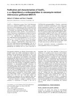
increased sm-MHC-positive area - corresponding to
ASM - predominantly in the cartilaginous airways by 1.9
±0.1-fold (P < 0.001) compared to saline-treated, saline-
challenged controls (Figure 1A). Topical treatment of
the airways with intranasally instilled YIGSR 0.5 h prior
to and 5.5 h after each ovalbumin-challenge nearly abro-
gated ovalbumin-induced increase in ASM mass (by 96
± 3%, P < 0.001). No significant effect of YIGSR treat-
ment was observed in saline-challenged animals.
To determine whether the changes in ASM content
were associated with changes in cell number and/or cell
size, the number of nuclei within the ASM layer were
counted and expressed relative to total ASM area.
Repeated ovalbumi n challenge did not change the num-
ber of nuclei per mm
2
of smooth muscle area (Figure
1B), indicating that the cell size is unchanged and oval-
bumin-induced increases in ASM mass were caused by
increased cell number (hyperplasia). YIGSR treatment
did not change ASM cell size in saline-challenged ani-
mals; however, a small, but significant (P < 0.05)
decrease in the number of nuclei/mm
2
was observed in
ovalbumin-challe nged animals (Figure 1B), suggesting
that this treatment may lead to some increase in cell
size (hypertrophy).
To assess whethe r the changes in ASM area were asso-
ciated with changes in proliferative responses, immuno-
blotting was used to determine expression of the
proliferation marker, PCNA, in whole lung homogenates.
After repeated ovalbumin-challenge, a considerable
increase (4.2 ± 0.2-fold, P < 0.001) in PCNA was observed
compared to saline-treated, saline-challenged controls
(Figure 1 C). Treatment with YIGSR fully normalized the
ovalbumin-induced increase in PCNA, when compared to
saline-challenged controls (P < 0.001). In the saline-
challenged animals, no significant effect of YIGSR treat-
ment on PCNA expression was observed. Unfortunately,
specific characterization of the proliferating cells in guinea
pig lung sections by immunohistochemistry was not possi-
ble with the antibody used. Collectively, these in vivo data
indicate that YIGSR treatment inhibits allergen-induced
ASM hyperplasia in association with suppressing prolifera-
tive responses of lung cells.
YIGSR treatment increases contractile protein
accumulation and ASM contractility
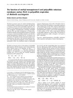
Previously, we showed t hat repeated ovalbumin-
exp osur e increased maximal methacholine- and KCl-
induced isometric contraction of epithelium-denuded, tra-
cheal smooth muscle preparations ex vivo [23,28,2 9].
Interestingly, treatment with the YIGSR peptide augmen-
ted the ovalbumin-induced increase in maximal metha-
choline- and KCl-induced contractions by 1.33 ± 0.08-fold
(P < 0.001) and 1.28 ± 0.11-fold (P < 0.05), respectively,
compared to saline-treated, ovalbumin-challenged controls
Dekkers et al. Respiratory Research 2010, 11:170
/>Page 4 of 11
(Figure 2A and Table 1). Similarly, in saline-challenged
animals YIGSR treatment increased methacholine- and
KCl-induced contraction (1.29 ± 0.03-fold and 1.39 ±
0.04-fold (P < 0.05), respectively). The sensitivity to either
contractile stimulus was unaffected b y treatme nt (Ta ble
1). Previously, we found that increased ASM contractility
induced by allergen challenge is associated with increased
pulmonary sm-MHC expression [23,28,29]. In saline-trea-
ted animals, re peated ovalbumin-challenge increased sm-
MHC by 2.5 ± 0.1-fold compared to saline-challenged
controls (P < 0.001, Figure 2B). In line with the in creased
methacholine- and KCl-induced contract ions, treatment
with YIGSR increased pulmonary sm-MHC expression in
saline-challenged animals (2.40 ± 0.28-fold, P < 0.001),
whereas in ov albumin-challenged animals the increase in
sm-MHC was increased further (1.37 ± 0.08-fold com-
pared to ovalbumin-challenged controls, P < 0.01). Collec-
tively, these data indicate that in vivo treatment with the
laminin-competing peptide YIGSR incre ases ASM con-
tractility and contractile protein expression both in saline-
and allergen-challenged animals.
Effects of YIGSR treatment on allergen-induced airway
inflammation
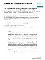
Infiltration of eosinophils into the airways is a charac-
teristic feature of allergic asthma and is generally con-
sidered to contribute to airway remodelling [2]. As
observed previously [23,28], repeated ovalbumin chal-
lenge increased the number of eosinophils in the sub-
mucosal and adventitial compartments of the airways
(P < 0.001 both, Figure 3A and 3B). No significant
effect of YIGSR on eosino phil number in the adventitial
compartment was observed in ovalbumin- and saline-
challenged animals (Figure 3B). However, YIGSR signifi-
cantly increased eosinophil number in the submucosal
airway compartment after repeated allergen challenge
(P < 0.05, Figure 3A).
Effects of YIGSR treatment on allergen-induced fibrosis
Aberrant deposition of ECM proteins, including col-
lagens, in the airway wall is another characteristic fea-
ture of chronic asthma [33,34]. As observed previously
[23], we demonstrated that lung hydroxyproline content,
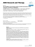
Figure 1 Increased ASM mass after repeated allergen challenge in vivo is inhibited by topical treatment with YIGSR. To assess the role
of laminins in increased ASM mass in asthma, the effects of treatment with YIGSR were evaluated in a guinea pig model of chronic allergic
asthma. (A) Treatment with YIGSR fully inhibited ovalbumin-induced increase in sm-MHC positive area in cartilaginous airways. (B) Changes in
ASM mass were mainly dependent on changes in ASM cell number, only a small increase in cell size was observed for the YIGSR-treated,
ovalbumin-challenged animals. (C) Increased pulmonary expression of the proliferative marker PCNA after repeated ovalbumin-challenges, was
almost fully reversed by YIGSR. Representative blots of PCNA and b-actin are shown. No effects of YIGSR were shown in saline-challenged
animals for any of the parameters. *P < 0.05, ***P < 0.001 compared to saline-treated, saline-challenged controls.
###
P < 0.001 compared to
saline-treated, ovalbumin-challenged controls. Data represent means ± SEM of 5-7 animals.
Dekkers et al. Respiratory Research 2010, 11:170
/>Page 5 of 11
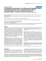
as an estimate of collagen, is increased after repeated
ovalbumin challenge (P < 0.001, Figure 4). Treatment
with YIGSR of the ovalbumin-challenged animals
further augmented the hydroxyproline content (P <
0.01), but did not change the hydroxyproline content in
saline-challenged animals. Collectively, our findings indi-
cate that YIGSR treatment increases allergen-induced
submucosal airway eosinophilia as well as collagen
deposition in the lung.
Immobilized YIGSR inhibits ASM cell proliferation in vitro
In comparison to the in vivo data from our current
study, it is paradoxical that previous in vitro studies
have indicated that soluble YIGSR inhibits ASM cell
Figure 2 Topical treatment of the airways with YIGSR increases ASM contractility and contractile protein accumulation. (A) Treatment
with YIGSR enhanced the maximal methacholine-induced isometric contraction of epithelium-denuded tracheal smooth muscle preparations
both in saline- and in ovalbumin-challenged animals. (B) Treatment with YIGSR increased pulmonary expression of sm-MHC, both in saline- and
in ovalbumin-challenged animals. Representative blots of sm-MHC and b-actin are shown. ***P < 0.001 compared to saline-treated, saline-
challenged controls.
##
P < 0.01 compared to saline-treated, ovalbumin-challenged controls. Data represent means ± SEM of 5-7 animals.
Table 1 Contractile responses of epithelium-denuded, tracheal smooth muscle preparations after repeated saline or
ovalbumin challenge of saline- or YIGSR-treated guinea pigs
Treatment Challenge Methacholine KCl n
E
max
(g) pEC
50
(- log M) E
max
(g) EC
50
(mM)
Saline Saline 1.42 ± 0.09 6.55 ± 0.18 1.02 ± 0.06 23.7 ± 0.9 6
YIGSR Saline 1.84 ± 0.04 6.82 ± 0.13 1.41 ± 0.04* 20.4 ± 2.2 5
Saline Ovalbumin 2.33 ± 0.22*** 6.28 ± 0.11 1.73 ± 0.13** 23.7 ± 1.2 7
YIGSR Ovalbumin 3.11 ± 0.18***
, ###
6.61 ± 0.08 2.12 ± 0.19***
,#
24.5 ± 1.1 7
Data represent means ± SEM. Abbreviations: E
max
: maximal contractile effect; EC
50
: concentration of the stimulus eliciting half-maximal response; pEC
50
: negative
logarithm of the EC
50
value. *P < 0.05, **P < 0.01, ***P < 0.001 compared to saline-treated, saline-challenged animals.
#
P < 0.05,
###
P < 0.001 compared to saline-
treated, ovalbumin-challenged animals.
Dekkers et al. Respiratory Research 2010, 11:170
/>Page 6 of 11
maturation and development of a hypercontractile,
hypoproliferative phenotype [14,15]. However, previous
in vitro experiments have revealed that YIGSR may both
mimic and inhibit laminin function, depending on the
physicochemical conditions [26,35,36]. Thus, when
immobilized, YIGSR promotes cell adhesion of various
cells, similar to laminin [26,35,36]. However, soluble
YIGSR blocks cell adhesion to laminin-111 [35]. To
further investigate whether this may also apply t o ASM
cells, the effects of immobilized and soluble YIGSR on
basal and growth factor-induced ASM cell proliferation
were compared in vitro.First,humanASMcellswere
cultured on 24 well plates coated with increasing con-
centrations of YIGSR (1-100 μM) and stimulated with
PDGF (10 ng/ml). Culturing the cells on immobilized
YIGSR concentration-dependently inhibited PDGF-
induced DNA synthesis (Figure 5A) and cell number
(not shown), but no ef fect was observed on basal DNA
synthesis. By contrast, culturing cells on immobilized
RGDS (100 μM) or its negative control peptide Gly-
Arg-Ala-Asp-Ser-Pro (GRADSP, 100 μM) did not affect
basal or PDGF-induced proliferation (Figure 5B).
To assess the effects of soluble YIGSR on proliferative
responses of human ASM, cells were cultured on immo-
bilized laminin-111 (10 μg/ml) or YIGSR (100 μM). Sub-
sequently, cells were stimulated with vehicle or PDGF in
the absence or presence of soluble YIGSR. As observed
previously [11,15], we found that culturing on laminin-
111 inhibited PDGF-induced DNA-synthesis (by 56 ±
11%, P < 0.05, Figure 5C) and cell number (not shown).
This inhibitory effect was fully reversed by soluble
YIGSR. Surprisingly , the inhibitory effect of coated
YIGSR on PDGF-induced proliferation w as also fully
normalized by soluble YIGSR. Of note, we have reported
previously that this peptide did not affect basal or
PDGF-induced proliferative responses in the absence of
laminin-111 [15]. Collectively, these results indicate that
the effects of the laminin-competing peptide YIGSR o n
ASM proliferative responses may depend on the peptide
microenvironment (i.e. soluble versus immobilized).
Discussion
In the current study, we demonstrate that treatment with
the laminin b1 chain-competing peptide YIGSR promotes
the formation of a hypercontractile, hypoproliferative
ASM phenotype in an animal model o f chronic asthma.
Topical application of YIGSR to the airways inhibited
ASM hyperplasia induced by repe ated allergen challenge.
However, ASM contractility and contractile protein
expression were increased under basal and allergen-
challenged conditions. These results appear to be in
contrast to previous in vitro studies, demonstrating that
Figure 3 YIGSR treatment increases allergen-induced eosinophilic inflammation in the submucosal airway compartment.
(A) Ovalbumin-induced eosinophil numbers in the submucosal compartment are increased by YIGSR treatment. (B) YIGSR treatment does not
affect eosinophilic cell number in the adventitial compartment. No effects of YIGSR were found in saline-challenged animals for any of the
conditions. ***P < 0.001 compared to saline-treated, saline-challenged controls.
#
P < 0.05 compared to saline-treated, ovalbumin-challenged
animals. Data represent means ± SEM of 5-7 animals.
Dekkers et al. Respiratory Research 2010, 11:170
/>Page 7 of 11
soluble YIGSR inhibits maturation of human ASM cells to
a h ypercontractile, h ypoproliferative ASM phenotype
[14,15].
Accumulation of ASM in the airway wall is a charac-
teristic feature of asthma, which may be due to an
increase in cell number (hyperplasia) [37,38] as well as
an increase in cell size (hypertrophy) [37,39]. This
ASM accumulation contributes importantly to
increased airway resistance and airway hyperrespon-
siveness [40,41]. Switching of the ASM phenotype
from a contractile to a proliferative state is thought to
contribute to the increased ASM mass in asthma [9].
In support, various mitogenic stimuli, including growth
factors and ECM proteins, induce a proliferative ASM
phenotype in vitro [10-12], an effect that can be inhib-
ited by culturing the cells on immobilized laminin-111
[11,22,23] or endogenously produced laminin-211 [15].
These inhibitory effects can be reversed using soluble
YIGSR [15], a binding motif present in the laminin b1
chain [26]. Similarly, in our study culturing human
ASM cells on laminin-111 reduced PDGF-induced pro-
liferation, an effect fully normalized by soluble YIGSR.
In contrast to this effect of soluble YIGSR, we also s how
that immobilized YIGSR concentration-dependently
inhibited growth factor-induced myocyte proliferation
to the same extent as laminin-111. Interestingly, pre-
vious work has also shown a disparate effect of immobi-
lized and soluble YIGSR, with the former promoting
attachment of various cells [26,35,36] whereas the latter
blocked attach ment to laminin-111 [35] or matrigel
[36]. The effects of immobilized YIGSR peptide are spe-
cific, as culturing on R GDS or GRADSP did not alter
proliferation. Of note, addition of soluble YIGSR nor-
malized the effects of immobilized YIGSR, an affect
consistent with studies using alveolar cells and a laminin
a chain peptide (Ser-Ile-Asn-Asn-Asn-Arg, or SINNNR)
[42]. Collectively, these findings suggest that the lami-
nin-competing peptide YIGSR may either promote or
inhibit ASM proliferative responses, depending on the
microenvironment of the peptide. The mechanisms
underlying these differential effects are unknown. How-
ever, since the anti-mitogenic effects of the peptide are
only observed when the peptide is immobilized, we
speculate that this may b e associated with bridging of
the 67 kDa laminin receptor LAMR1 - which has high
affinity to the YIGSR motif [43] - whereas soluble
YIGSR may competitively inhibit this type of interac-
tion. Similarly, it has been established that b inding of
ECM proteins such as fibronectin as a monovalent or
multivalent ligand to a5b1 integrin has diverse effects
on focal contacts, tyrosine kinase activation and cytos-
keletal dynamics [44]. Our data indicate that future stu-
dies of the ligation of soluble and immobilized YIGSR
peptides to specific cell surface receptors and resulting
intracellular signaling events are needed.
In addition to ASM accumulation, increased expres-
sion of contractile proteins and ASM contractility, and
ECM deposition are features of airway remodelling in
asthma [7]. In the airways of asthmatics increased
expression of laminin a2andb2chainsisobserved
[18,19], and laminin g2 chain expression inversely corre-
lates with epithelial integrity [19]. Laminins have not
only been shown to inhibit ASM proliferation, but also
to be critical in maintenance and induction of a (hyper)
contractile ASM phenotype. Indeed, culturing of ASM
cells on a laminin-111 matrix inhibits proliferation
[11,22,23], maintains contractile protein expression in
the presence of growth factors [22], and prevents induc-
tion of a hypocontractile phenotype by PDGF [11].
Induction of a contractile ASM phenotype in serum-free
culture supplemented with insulin is associated with
increased expression of laminin a2, b1 and g1 chains, all
found in the laminin-211 isoform [14,15]. Importantly,
the expression of endogenous laminin is required for
phenotype maturation, as soluble YIGSR prevents con-
tractile protein ac cumulation and hypercontractili ty
[14,15]. Recently , using our guinea pig model of chronic
asthma we showed that treatment with the RGD-
Figure 4 YIGSR treatment increases allergen-induced fibrosis in
the guinea pig lung. Hydroxyproline content in guinea pig lung
after repeated saline- or ovalbumin-challenges in saline- and YIGSR-
treated animals. ***P < 0.001 compared to saline-treated, saline-
challenged controls.
##
P < 0.01 compared to saline-treated,
ovalbumin-challenged animals. Data represent means ± SEM of 5-7
animals.
Dekkers et al. Respiratory Research 2010, 11:170
/>Page 8 of 11
Figure 5 Effects of immobilized and soluble YIGSR on basal and PDGF-induced human ASM cell proliferation . (A) Cu lturing of human
ASM cells on immobilized YIGSR matrices inhibits PDGF-induced thymidine-incorporation in a YIGSR concentration-dependent fashion. Under
unstimulated (Basal) conditions, no effects of immobilized YIGSR were observed. (B) Immobilized RGDS or its negative control GRADSP did not
affect basal or PDGF-induced thymidine-incorporation. (C) The inhibitory effects of immobilized laminin-111 and YIGSR matrices on PDGF-
induced thymidine-incorporation were normalized by soluble YIGSR. ***P < 0.001 compared to thymidine-incorporation of unstimulated cells
(basal) cultured on uncoated matrices (plastic).
#
P < 0.05 and
##
P < 0.01 compared to PDGF-induced thymidine-incorporation of cells cultured on
uncoated matrices. Data represent means ± SEM of 4-5 independent experiments of 3 different donors, performed in duplicate.
Dekkers et al. Respiratory Research 2010, 11:170
/>Page 9 of 11
containing RGDS peptide largely inhibits ASM hyperpla-
sia and hypercontractility [23]. The RGD sequence exists
in several ECM proteins [24,25], thus the specific contri-
bution of laminins cannot be discerned from these prior
studies. In the present study we found that in vivo treat-
ment with YIGSR inhibited allergen-induced ASM
hyperplasia, but increased both the expression of sm-
MHC a nd ASM contractility. In addition, a small
increase in cell size in the allergen-challenged YIGSR
treated animals was observed suggesting that hypertro-
phy may also have played a role in the observed effec ts.
Collectively, our results indicate that treatment with
YIGSR inhibits allergen-induced ASM hyperplasia and
increases ASM contractility in vivo,suggestingthat
YIGSR mimics and/or promotes rather than inhibit s
laminin function under this condition.
Eosinophils express a number of integrins, of which
the a6b1 mediates adhesion to laminin, but not to col-
lagen type I or type IV [45,46]. Eosinophils isolated
from allergic donors show higher adhesion to laminin
than those isolated from healthy subjects [46]. Migration
of eosinophils through matrigel, a base ment membrane
extract containing laminin-111, also requi res interaction
with b1-integrins [46]. These findings suggest that lami-
nin-competing peptides could affect allergen-induced
airway infiltration of inflammatory cells. To date no
reportsonYIGSReffectsoneosinophilmigrationare
available. In our study we noted that YIGSR increased
allergen-induced eosinophil cell numbers in the submu-
cosal compartment, without affecting eosinophil num-
bers in the adventitial compartment. The increased
number of eosinophils in the submucosa suggests that,
rather than, infiltration, retention time of the eosino-
phils in the compartment could be increased. Impor-
tantly, increased ECM depos ition may be secondary to
prolonged airway inflammation [2] and therefore
increased allergen-induced airway fibrosis in YIGSR-
treated animals could also indirectly result from
increas ed eosinophilia. As increased and altered depos i-
tion of ECM proteins, including laminins and collagens,
isafeatureofremodellinginchronicasthma[33,34]it
is important that further investigation focus on under-
standing the effects of YIGSR and laminins on ECM
deposition by fibroblasts and other structural cells.
In summary, our results indi cate that the laminin-
compe ting peptide YIGSR promotes a contractile, hypo-
proliferativ e ASM phenotype in vivo, an effect that is in
striking contrast to current and previously reported evi-
dence showing that soluble YIGSR prevents laminin-
dependent phenotype maturation. It appears that the
microenvironment of the peptide is a critical determi-
nant of its effect as immobilized YIGSR does mimic
the effects of laminin matrix on ASM in vitro.Ourdata
suggest that topically applied YIGSR mimics rather than
inhibits the effects of laminin in vivo,anditsuseis
linked to increased allergen-induced fibrosis, submuco-
sal eosinophilia, ASM hyperplasia and airway hypercon-
tractility. These data indicate that strategies to develop
capacity to use peptides that target ECM-cell interaction
to treat bronchial asthma need to be developed with
care, in particular with focus on understanding differ-
ences of such interventions that may exist between in
vitro and in vivo systems.
Acknowledgements
This work was financially supported by the Netherlands Asthma Foundation,
grant NAF 3.2.03.36. We are grateful to Dr. W.T. Gerthoffer (University of
Nevada-Reno) for preparation of the hTERT cell lines used in the study.
Author details
1
Department of Molecular Pharmacology, University of Groningen,
Groningen, Netherlands.
2
Department of Physiology, University of Manitoba,
Winnipeg, Manitoba, Canada.
Authors’ contributions
BGJD: design of the study, acquisition of data, data analysis and interpretation,
manuscript writing; ISTB: design of the study, acquisition of data, data analysis
and interpretation; AJH: preparation of ASM cell lines and critical revision of
the MS; JZ: design of the study, data interpretation and critical revision of the
MS; HM: design of the study, data interpretation and critical revision of the
MS. All authors have read and approved the manuscript.
Competing interests
The authors declare that they have no competing interests.
Received: 27 July 2010 Accepted: 3 December 2010
Published: 3 December 2010
References
1. Bousquet J, Jeffery PK, Busse WW, Johnson M, Vignola AM: Asthma. From
bronchoconstriction to airways inflammation and remodeling. Am J
Respir Crit Care Med 2000, 161:1720-1745.
2. Cockcroft DW, Davis BE: Mechanisms of airway hyperresponsiveness. J
Allergy Clin Immunol 2006, 118:551-559.
3. Davies DE, Wicks J, Powell RM, Puddicombe SM, Holgate ST: Airway
remodeling in asthma: new insights. J Allergy Clin Immunol 2003,
111:215-225.
4. Dunnill MS, Massarella GR, Anderson JA: A comparison of the quantitative
anatomy of the bronchi in normal subjects, in status asthmaticus, in
chronic bronchitis, and in emphysema. Thorax 1969, 24:176-179.
5. Parameswaran K, Willems-Widyastuti A, Alagappan VK, Radford K,
Kranenburg AR, Sharma HS: Role of extracellular matrix and its regulators
in human airway smooth muscle biology. Cell Biochem Biophys 2006,
44:139-146.
6. Fernandes DJ, Bonacci JV, Stewart AG: Extracellular matrix, integrins, and
mesenchymal cell function in the airways. Curr Drug Targets 2006,
7:567-577.
7. Dekkers BG, Maarsingh H, Meurs H, Gosens R: Airway structural
components drive airway smooth muscle remodeling in asthma. Proc
Am Thorac Soc 2009, 6:683-692.
8. Halayko AJ, Salari H, Ma X, Stephens NL: Markers of airway smooth muscle
cell phenotype. Am J Physiol 1996, 270:L1040-L1051.
9. Halayko AJ, Tran T, Ji SY, Yamasaki A, Gosens R: Airway smooth muscle
phenotype and function: interactions with current asthma therapies.
Curr Drug Targets 2006, 7:525-540.
10. Gosens R, Meurs H, Bromhaar MM, McKay S, Nelemans SA, Zaagsma J:
Functional characterization of serum- and growth factor-induced
phenotypic changes in intact bovine tracheal smooth muscle. Br J
Pharmacol 2002, 137:459-466.
Dekkers et al. Respiratory Research 2010, 11:170
/>Page 10 of 11
11. Dekkers BG, Schaafsma D, Nelemans SA, Zaagsma J, Meurs H: Extracellular
matrix proteins differentially regulate airway smooth muscle phenotype
and function. Am J Physiol Lung Cell Mol Physiol 2007, 292:L1405-L1413.
12. Gosens R, Roscioni SS, Dekkers BG, Pera T, Schmidt M, Schaafsma D,
Zaagsma J, Meurs H: Pharmacology of airway smooth muscle
proliferation. Eur J Pharmacol 2008, 585:385-397.
13. Schaafsma D, McNeill KD, Stelmack GL, Gosens R, Baarsma HA, Dekkers BG,
Frohwerk E, Penninks JM, Sharma P, Ens KM, et al: Insulin increases the
expression of contractile phenotypic markers in airway smooth muscle.
Am J Physiol Cell Physiol 2007, 293:C429-C439.
14. Tran T, McNeill KD, Gerthoffer WT, Unruh H, Halayko AJ: Endogenous
laminin is required for human airway smooth muscle cell maturation.
Respir Res 2006, 7:117.
15. Dekkers BG, Schaafsma D, Tran T, Zaagsma J, Meurs H: Insulin-induced
Laminin Expression Promotes a Hypercontractile Airway Smooth Muscle
Phenotype. Am J Respir Cell Mol Biol 2009, 41:494-504.
16. Nguyen NM, Senior RM: Laminin isoforms and lung development: all
isoforms are not equal. Dev Biol 2006, 294:271-279.
17. Virtanen I, Laitinen A, Tani T, Paakko P, Laitinen LA, Burgeson RE, Lehto VP:
Differential expression of laminins and their integrin receptors in
developing and adult human lung. Am J Respir Cell Mol Biol 1996,
15:184-196.
18. Altraja A, Laitinen A, Virtanen I, Kampe M, Simonsson BG, Karlsson SE,
Hakansson L, Venge P, Sillastu H, Laitinen LA: Expression of laminins in the
airways in various types of asthmatic patients: a morphometric study.
Am J Respir Cell Mol Biol 1996, 15:482-488.
19. Amin K, Janson C, Seveus L, Miyazaki K, Virtanen I, Venge P: Uncoordinated
production of Laminin-5 chains in airways epithelium of allergic
asthmatics. Respir Res 2005, 6:110.
20. Schuger L, Skubitz AP, Zhang J, Sorokin L, He L: Laminin alpha1 chain
synthesis in the mouse developing lung: requirement for epithelial-
mesenchymal contact and possible role in bronchial smooth muscle
development. J Cell Biol 1997, 139:553-562.
21. Relan NK, Yang Y, Beqaj S, Miner JH, Schuger L: Cell elongation induces
laminin alpha2 chain expression in mouse embryonic mesenchymal
cells: role in visceral myogenesis. J Cell Biol 1999, 147:1341-1350.
22. Hirst SJ, Twort CH, Lee TH: Differential effects of extracellular matrix
proteins on human airway smooth muscle cell proliferation and
phenotype. Am J Respir Cell Mol Biol 2000, 23:335-344.
23. Dekkers BG, Bos IS, Gosens R, Halayko AJ, Zaagsma J, Meurs H: The
Integrin-blocking Peptide RGDS Inhibits Airway Smooth Muscle
Remodeling in a Guinea Pig Model of Allergic Asthma. Am J Respir Crit
Care Med 2010, 181:556-565.
24. Plow EF, Haas TA, Zhang L, Loftus J, Smith JW: Ligand binding to
integrins. J Biol Chem
2000, 275:21785-21788.
25. Aumailley M, Gerl M, Sonnenberg A, Deutzmann R, Timpl R: Identification
of the Arg-Gly-Asp sequence in laminin A chain as a latent cell-binding
site being exposed in fragment P1. FEBS Lett 1990, 262:82-86.
26. Graf J, Ogle RC, Robey FA, Sasaki M, Martin GR, Yamada Y, Kleinman HK: A
pentapeptide from the laminin B1 chain mediates cell adhesion and
binds the 67,000 laminin receptor. Biochemistry 1987, 26:6896-6900.
27. Meurs H, Santing RE, Remie R, van der Mark TW, Westerhof FJ, Zuidhof AB,
Bos IS, Zaagsma J: A guinea pig model of acute and chronic asthma
using permanently instrumented and unrestrained animals. Nat Protoc
2006, 1:840-847.
28. Bos IS, Gosens R, Zuidhof AB, Schaafsma D, Halayko AJ, Meurs H,
Zaagsma J: Inhibition of allergen-induced airway remodelling by
tiotropium and budesonide: a comparison. Eur Respir J 2007, 30:653-661.
29. Gosens R, Bos IS, Zaagsma J, Meurs H: Protective effects of tiotropium
bromide in the progression of airway smooth muscle remodeling. Am J
Respir Crit Care Med 2005, 171:1096-1102.
30. Gosens R, Stelmack GL, Dueck G, McNeill KD, Yamasaki A, Gerthoffer WT,
Unruh H, Gounni AS, Zaagsma J, Halayko AJ: Role of caveolin-1 in p42/p44
MAP kinase activation and proliferation of human airway smooth
muscle. Am J Physiol Lung Cell Mol Physiol 2006, 291:L523-L534.
31. Gosens R, Dueck G, Gerthoffer WT, Unruh H, Zaagsma J, Meurs H,
Halayko AJ: p42/p44 MAP kinase activation is localized to caveolae-free
membrane domains in airway smooth muscle. Am J Physiol Lung Cell Mol
Physiol 2007, 292:L1163-L1172.
32. Gosens R, Dueck G, Rector E, Nunes RO, Gerthoffer WT, Unruh H,
Zaagsma J, Meurs H, Halayko AJ: Cooperative regulation of GSK-3 by
muscarinic and PDGF receptors is associated with airway myocyte
proliferation. Am J Physiol Lung Cell Mol Physiol 2007, 293:L1348-L1358.
33. Jeffery PK: Remodeling in asthma and chronic obstructive lung disease.
Am J Respir Crit Care Med 2001, 164:S28-S38.
34. Postma DS, Timens W: Remodeling in asthma and chronic obstructive
pulmonary disease. Proc Am Thorac Soc 2006, 3:434-439.
35. Graf J, Iwamoto Y, Sasaki M, Martin GR, Kleinman HK, Robey FA, Yamada Y:
Identification of an amino acid sequence in laminin mediating cell
attachment, chemotaxis, and receptor binding. Cell 1987, 48:989-996.
36. Grant DS, Tashiro K, Segui-Real B, Yamada Y, Martin GR, Kleinman HK: Two
different laminin domains mediate the differentiation of human
endothelial cells into capillary-like structures in vitro. Cell 1989,
58:933-943.
37. Ebina M, Takahashi T, Chiba T, Motomiya M: Cellular hypertrophy and
hyperplasia of airway smooth muscles underlying bronchial asthma. A
3-D morphometric study. Am Rev Respir Dis 1993,
148:720-726.
38. Woodruff PG, Dolganov GM, Ferrando RE, Donnelly S, Hays SR, Solberg OD,
Carter R, Wong HH, Cadbury PS, Fahy JV: Hyperplasia of smooth muscle in
mild to moderate asthma without changes in cell size or gene
expression. Am J Respir Crit Care Med 2004, 169:1001-1006.
39. Benayoun L, Druilhe A, Dombret MC, Aubier M, Pretolani M: Airway
structural alterations selectively associated with severe asthma. Am J
Respir Crit Care Med 2003, 167:1360-1368.
40. Lambert RK, Wiggs BR, Kuwano K, Hogg JC, Pare PD: Functional
significance of increased airway smooth muscle in asthma and COPD. J
Appl Physiol 1993, 74:2771-2781.
41. Oliver MN, Fabry B, Marinkovic A, Mijailovich SM, Butler JP, Fredberg JJ:
Airway hyperresponsiveness, remodeling, and smooth muscle mass:
right answer, wrong reason? Am J Respir Cell Mol Biol 2007, 37:264-272.
42. Matter ML, Laurie GW: A novel laminin E8 cell adhesion site required for
lung alveolar formation in vitro. J Cell Biol 1994, 124:1083-1090.
43. Nelson J, McFerran NV, Pivato G, Chambers E, Doherty C, Steele D,
Timson DJ: The 67 kDa laminin receptor: structure, function and role in
disease. Biosci Rep 2008, 28:33-48.
44. Miyamoto S, Akiyama SK, Yamada KM: Synergistic roles for receptor
occupancy and aggregation in integrin transmembrane function. Science
1995, 267:883-885.
45. Barthel SR, Johansson MW, McNamee DM, Mosher DF: Roles of integrin
activation in eosinophil function and the eosinophilic inflammation of
asthma. J Leukoc Biol 2008, 83:1-12.
46. Georas SN, McIntyre BW, Ebisawa M, Bednarczyk JL, Sterbinsky SA,
Schleimer RP, Bochner BS: Expression of a functional laminin receptor
(alpha 6 beta 1, very late activation antigen-6) on human eosinophils.
Blood 1993, 82:2872-2879.
doi:10.1186/1465-9921-11-170
Cite this article as: Dekkers et al.: The laminin b1-competing peptide
YIGSR induces a hypercontractile, hypoproliferative airway smooth
muscle phenotype in an animal model of allergic asthma. Respira tory
Research 2010 11:170.
Submit your next manuscript to BioMed Central
and take full advantage of:
• Convenient online submission
• Thorough peer review
• No space constraints or color figure charges
• Immediate publication on acceptance
• Inclusion in PubMed, CAS, Scopus and Google Scholar
• Research which is freely available for redistribution
Submit your manuscript at
www.biomedcentral.com/submit
Dekkers et al. Respiratory Research 2010, 11:170
/>Page 11 of 11