Báo cáo y học: " Antigen-Specific IgG ameliorates allergic airway inflammation via Fcg receptor IIB on dendritic cells" doc
Bạn đang xem bản rút gọn của tài liệu. Xem và tải ngay bản đầy đủ của tài liệu tại đây (2.18 MB, 10 trang )
RESEARC H Open Access
Antigen-Specific IgG ameliorates allergic airway
inflammation via Fcg receptor IIB on dendritic cells
Yumiko Ishikawa
1
, Kazuyuki Kobayashi
1*
, Masatsugu Yamamoto
1
, Kyosuke Nakata
1
, Tetsuya Takagawa
2
,
Yasuhiro Funada
1
, Yoshikazu Kotani
1
, Hajime Karasuyama
3
, Masaru Yoshida
2†
and Yoshihiro Nishimura
1†
Abstract
Background: There have been few reports on the role of Fc receptors (FcRs) and immunoglobulin G (IgG) in
asthma. The purpose of this study is to clarify the role of inhibitory FcRs and antigen presenting cells (APCs) in
pathogenesis of asthma and to evaluate antigen-transporting and presenting capacity by APCs in the
tracheobronchial mucosa.
Methods: In FcgRIIB deficient (KO) and C57BL/6 (WT) mice, the effects of intratracheal instillation of antigen-specific
IgG were analysed using the model with sensitization and airborne challenge with ovalbumin (OVA). Thoracic
lymph nodes instilled with fluorescein-conjugated OVA were analysed by fluorescence microscopy. Moreover, we
analysed the CD11c
+
MHC class II
+
cells which intaken fluorescein-conjugated OVA in thoracic lymph nodes by
flow cytometry. Also, lung-derived CD11c
+
APCs were analysed by flow cytometry. Effects of anti-OVA IgG1 on
bone marrow dendritic cells (BMDCs) in vitro were also analysed. Moreover, in FcgRIIB KO mice intravenously
transplanted dendritic cells (DCs) differentiated from BMDCs of WT mice, the effects of intratracheal instillation of
anti-OVA IgG were evaluated by bronchoalveolar lavage (BAL).
Results: In WT mice, total cells and eosinophils in BAL fluid reduced after instillation with anti-OVA IgG1. Anti-OVA
IgG1 suppressed airway inflammation in hyperresponsiveness and histology. In addition, the number of the
fluorescein-conjugated OVA in CD11c
+
MHC class II
+
cells of thoracic lymph nodes with anti-OVA IgG1 instillation
decreased compared with PBS. Also, MHC class II expression on lung-derived CD11c
+
APCs with anti-OVA IgG1
instillation reduced. Moreover, in vitro, we showed that BMDCs with anti-OVA IgG1 significantly decreased the T
cell proliferation. Finally, we demonstrated that the lacking effects of anti-OVA IgG1 on airway inflammation on
FcgRIIB KO mice were restored with WT-derived BMDCs transplanted intravenously.
Conclusion: Antigen-specific IgG ameliorates allergic airway inflammation via FcgRIIB on DCs.
Background
It is estimated that as many as 300 million people of all
ages suffer from bronchial asthma, and that asthmatic
patients are increasing by 50% per decade worldwide
[1]. The mucosa of respiratory tracts are replete with
organized follicles and scattered antigen reactive or sen-
sitized lymphoid elements, including B cells, T cells,
plasma cells, dendritic cells (DCs) and a variety of other
cellular elements against invading pathogens. The
mucosal surfa ces are also kno wn to possess critical
immunoglobulins, such as IgA, IgM and IgG.
Bronchial asthma is characterized by allergic inflamma-
tion of the bronchial mucosa, in addition to airway
hyperresponsiveness (AHR), and elevated titers of circu-
lating IgE. In asthmatic patients, antigen-specific IgE
binds to FcεRI on mast cells and FcεRII on eosinophils
and macrophages [2]. As a result of IgE cross-linking
after antigen inhalation, an immediate allergic reaction is
induced. On the other hand, the T helper 2 (Th2)-type
immune response plays an important role in the late-
phase reaction. Whe n the inhaled allergen is recognized
and presented by antigen presenting cells (APCs) in the
airway, T cells are activated and differentiate from Th0
cells into Th2-type cells. Th2-type cells produce Th2
* Correspondence:
† Contributed equally
1
Division of Respiratory Medicine, Department of Internal Medicine, Kobe
University Graduate School of Medicine, Kobe, Japan
Full list of author information is available at the end of the article
Ishikawa et al. Respiratory Research 2011, 12:42
/>© 2011 Ishikawa et al; licensee BioMed Central Ltd. This is an Open Access article distributed under the terms of the Creative
Commons Attribution License (ht tp://creativeco mmons.org/licenses/by/2.0), which permits unrestricted use, distribution, and
reproduction in any medium, provided the original work is properly cited.
cytokines such as IL-4, IL-5 and IL-13 [3]. IL-4 activates
the production of IgE in B cells, IL-5 increases the num-
ber of eosinophils in the airway, and IL-13 is involved in
AHR and mucus secretion in the airway.
With regard to the study of immunoglobulins in asthma,
there have been some reports on a novel anti-IgE therapy
that exerts its action by reducing the amount of free IgE to
bind to effector cells [4-6]. However, this approach cannot
completely reduce circulating IgE, and cannot control the
initial cascade of asthma pathogenesis. It is also known
that IgG is present in the airway lumen and submucosa
under normal conditions [7]. Although antigen-specific
IgG is induced after antigen inhalation, its role in bronchial
asthma remains unknown. OVA-specific IgG in rat-ser a,
such as IgG1 and IgG2a, is reported to increase on day 21
after OVA inhalation in asthmatic models induced by
OVA [8]. Platts-Mills et al. demonstrated a progressive
increase in specific serum IgG titers with extended expo-
sure and a prevalence of Th2 cytokine-dependen t IgG in
cats and dogs [9]. Immunotherapy by allergen vaccination
is reported to increase antigen-specific IgG titers in allergy
patients [10], thus suggesting that antige n-specific IgG m ay
exert a protective effect against allergies and bronchial
asthma. However, the mechanism that antigen-specific IgG
suppresses allergic airway i nflammation is unclear.
There have been many studies on Fc receptors (FcRs),
which are the receptors for the Fc portion of immuno-
globulin [11-13]. FcRs are known to be associated with
the immune responses in antibody-dependent cellular
cytotoxicity or hypersensitivity [12,13]. Activating type
FcRs consist of the FcR g-chain, which has an immunor-
eceptor tyrosine-based activation motif (ITAM) in cyto-
sol, while F cgRII B is the only immunosuppressive FcR
having an immunoreceptor tyrosine-based inhibitory
motif (ITIM) [14-16]. FcgRandFcgRIIB on effector
cells, such as macrophages or DCs regulate the immune
response by influencing one anot her, and Fc g RIIB on B
cells was recently reported to negatively regulate the
production of antibody [17]. FcgRIIB is present on var-
ious types of hematopoietic cells including macrophages,
neutrophilsandDCs[18].DCsplayanimportantrole
by presenting antigens to naive T cells in al lergic airway
inflammation [19-21]. Moreover, expression of FcgRs on
DCs is reported to be important during the sensitization
phase for the development of allergen-induced AHR and
inflammation as described previously [22]. Therefore,
anti-OVA IgG was intratracheally instilled during the
sensitization phase in allergic murine mo del to analy se
the role of FcRs and APCs in pathogenesis of asthma.
Methods
Mice
FcgRIIB-deficient [23] mice (FcgRIIB KO) of a C57BL/6J
(wild type; WT) background were kindly provided by Prof.
Toshiyuki Takai (Tohoku University). Mice transgenic for
the OVA
323-339
specific T cell receptor (OT-II mice) on a
C57BL/6J background [24] were used. WT mice were pur-
chased from CLEA Japan (Tokyo, Japan). All animal
experiments were performed in accordance with the
Guidelines for Animal Experimentation at Kobe University
Graduate School of Medicine. Our research was approved
by the Institutional Animal Care and Use Committee and
was carried out according to Kobe University Animal
Experimentation Regulations (P070703R).
Agents
The following drugs and chemicals were purchased com-
mercially: endotoxin-free OVA (Sigma-Aldrich, St. Louis,
MO, USA); aluminium hydroxide (alum) (Sigma-
Aldrich); acetyl-b-methylcholine chloride (Sigma-
Aldrich); fluorescein-conjugated OVA (Invitrogen,
Eugene, OR, USA); Albumin, from Bovine Serum (Wako
Pure Chemical Industries, Osaka, Japan); purified rat
anti-mouse CD16/CD32 a ntibody (BD Biosciences,
Franklin Lakes, N J, USA); phycoerythrin (PE)-hamster
anti-mouse CD11c antibody (BD Biosciences); biotin-
mouse anti-mouse I-A[b] antibody (BD Biosciences);
streptavidin allophycocyanin (BD Biosciences); Anti-
MHC class II-FITC (Miltenyi Biotec, Gladbach, Ger-
many); 7-Amino-Actinomycin D (7-AAD) (BD Bios-
ciences); collagenase D (Roche Molecular Biochemicals,
Mannheim, Germany); and deoxyribonuclease (DNase) I
(bovine pancreas; Wako); ethylenediaminetetraacetic acid
(EDTA), disodium salt (Wako); Roswell Park Memorial
Institute (RPMI)-1640 (Sigma-Aldri ch); 2-mercaptoetha-
nol (Sigma-Aldrich); penicillin-streptomycin solution
(Sigma-Aldrich); granulocyte macrophage colony-stimu-
lating factor (GM-CSF) (Wako Pure Chemical Indus-
tries); CD4 microbeads (Miltenyi Biotec, Gladbach,
Germany); and OVA peptide
323-339
(Genway Biotech,
Inc., San Diego, CA, USA).
Establishment of anti-OVA IgG
Anti-OVA IgG1 and anti-OVA IgG2a in endotoxin-free
condition were kindly provided by Hajime Karasuyama
(Tokyo Medical and Dental University Graduate School).
A panel of hybridomas secreting OVA-specific IgG
monoclonal antibodies was prepared from splenocytes
isolated from mice that immunized intraperitoneally 14
days before with OVA with injection alum plus B. pertus-
sis toxin or complete Freund’s adjuvant, as Ishikawa et al.
described previously [25]. Concentrations of anti-OVA
IgG1 and IgG2a were assayed by e nzyme-linked immu-
nosorbent assay (ELISA), as described previously [26].
Sensitization and antigen challenge
Six- to ten-week-old female mice were sensitized by
intraperitoneal injections of 10 μg of OVA with 1 mg of
Ishikawa et al. Respiratory Research 2011, 12:42
/>Page 2 of 10
alum on days 0, 7 a nd 14. The y were then exposed to
1% OVA diluted in sterile phosphate-buffered saline
(PBS) for 30 min on 2 consecutive days with an ultraso-
nic nebulizer (NE-U12) (OMRON, Tokyo, Japan). Each
subclass of anti-OVA IgG (10 μg/50 μl) was diluted in
sterile PBS and instilled intratracheally on 25th days
before OVA challenge. As compared to the controls,
non-specific IgG was intratracheally instilled instead of
anti-OVA IgG. All mice were analyzed 24 h after the
final OVA challenge.
Bronchoalveolar lavage (BAL)
BAL fluid was obtained by instilling 0.8 ml PBS and
aspirating three times (recovery >85%) on 24 h after the
final antigen challenge. BAL fluid was centrifuged at
3,000 × g for 5 min (at 4°C), and the supernatants were
stored at -80°C. Total cells in BAL fluid were counted
with a hemocytometer. On cytospin preparations stained
with Diff-Quick (Sysmex Corporatio n, Kobe, Japan), dif-
ferential cell counts were determi ned by classification of
200 cells based on standard morphology.
ELISA
Levels of IL-4, IL-5, IL-13 and IFN-g in BAL fluid were
determined using ELISA kits (Invitrogen, R&D systems,
Minneapolis, MN, USA) according to the manufacturer’s
instructions. The absorbance of each sample was mea-
suredat450nmwithiMark™ microplate reader
(BioRAD, Tokyo, Japan).
Histopathology
Murine lungs were intratracheally instilled with 10%
buffered formalin at a pressure of 20 cmH
2
Ofor24h
and then embedded in paraffin. Serial 4-μm sections
were stained with hematoxylin and eosin (H&E).
Measurement of airway hyperresponsiveness
At 24 h after the final aerosol challenge, AHR was assessed
in conscious and unrestrained mice by means of whole
body plethysmography (Buxco Electronics, Sharon, CT,
USA) as described previously [27]. Each mouse was placed
in a plastic chamber and exposed to aerosolized PBS, fol-
lowed by increasing concentration s of aerosolized acetyl-
b-methyl choline chloride solutions (methacholine) (1.56,
3.13, 6.25, 12.5, 25, 50 μg/ml) for 3 min each. Bronchocon-
striction was recorded for an additional 5 min for each
dose of methacholine. The highest enhanced pause (Penh)
value obtained during each methacholine challenge was
expressed as a ratio against the basal Penh value in
response to PBS challenge.
Analysis of OVA transport to thoracic lymph nodes
Atotalof50μl of 10 mg/ml fluorescein-conjugated
OVA was intratracheally instilled in sensitized WT mice
one day after intratracheal instillation of 10 μg/50 μl
anti-OVA IgG1 or PBS. Thoracic lymph nodes were
analyzed 24 h after OVA challenge for 30 min by fluor-
escence microscopy in order to verify whether anti-
OVA IgG1 inhibits antigen transport to thoracic lymph
nodes in allergic airway inflammation. Moreover, we
analysed the CD11c
+
cells which intaken fluorescein-
conjugated OVA in thoracic lymph nodes by flow cyto-
metry according to previous studies [28]. Lymph nodes
of OVA-sensitized and challenged mice were excised
after anti-OVA IgG1 or PBS instillation. Minced lymph
nodes were digested into single-cell suspensions with
RPMI 1640 medium supple mented with 5% fetal bovine
serum, 1 mg/ml collagenase D, 0.02 mg/ml DNase I and
5 mM EDTA for 30 min at 37°C. This was filtered
through a 70-μm cell s trainer. After blocking with 3%
BSA in PBS, single-cell suspensions were pre-incubated
with Fc-receptor blocking, purified rat anti-mouse
CD16/CD32 antibodies in order to reduce nonspecific
binding. PE-hamster anti-mouse CD11c antibodies, bio-
tin-mouse anti-mouse I-A[b] antibodies and streptavi-
din-allophycocyanin (SA-APC) were used to identify
CD11c
+
APCs populations in lymph nodes.
Flow cytometry of lung-derived CD11c
+
cells
Lungs of OVA-sensitized and challenged mice were
excised after anti-OVA IgG1 or PBS instillation. Minced
lung tissues were digested and incubated with RPMI
1640 medium supplemented with collagenase D, DNase
I, and EDTA. The digested cells were filtered through
cell strainer, and erythrocytes were lysed with a lysing kit
(Funakoshi, Tokyo, Japan). After blocking w ith 3% BSA
in PBS, single-cell suspensions were also pre-incubated
with purified rat anti-mouse CD16/CD32 antibodies. PE-
hamster anti-mouse CD11c antibo dies and anti-MHC
class II-FITC were used to identify murine lung CD11c
+
APC populations.
Preparation of BMDCs
BMDCs were differentiated from bone marrow cells in
WT mice as described previously [29,30]. C ell culture
medium was RPMI-1640 supplemented with penicillin-
streptomycin, 2-mercaptoethanol (50 μM) and 10% FCS.
BMDCsfromWTmicewereculturedwith20ng/ml
murine GM-CSF at day 0. On days 3 and 6, 20 ng/ml
GM-CSF was added t o the culture fluid. On day 6,
BMDCs were employed to examine whether anti-OVA
IgG1 ameliorated the activation of BMDCs.
Effects of anti-OVA IgG1 on BMDCs in vitro
In order to investigate the effects of anti-OVA IgG1 on
the activation of BMDCs, we cultured BMDCs in the
presence of GM-CSF for 5 days. On day 6, OVA peptide
(20 μM) with or without anti-OVA IgG1 (0.1 mg/ml)
Ishikawa et al. Respiratory Research 2011, 12:42
/>Page 3 of 10
wasaddedtoBMDCs(2×10
4
) per well. On day 7,
CD4
+
T-cells were isolated from the spleens of OT-II
mice using CD4 microbeads for auto-MACS (Miltenyi
Biotec, Gladbach, Germany) and CD4
+
T-cells (2 × 10
5
)
were added to each well after washing and were incu-
bated for 48 h. The number of viable cells was examined
by colorimetric assay using the WST-8 [2-(2-methoxy-4-
nitrophenyl)-3-(4-nitrophenyl)-5-(2,4-disulfophenyl)-2H-
tetrazolium, monosodium salt] cell-counting kit
(Dojindo, Kumamot o, Japan) accord ing to t he manufac-
turer’s protocols. At 2 h after incubation with WST-8
solution, the absorbance of each well was measured at
450 nm with a reference wavelength of 655 nm [31].
Transplantation of BMDCs into FcgR IIB KO mice
FcgRIIB KO mice were sensitized and c hallenged as
described a bove. To verify the function of DCs in aller-
gic airway inflammation, BMDCs (10
6
cells per mouse)
cultured from WT mice were transplanted intravenously
into FcgRIIB KO mice on day 24 before instillation of
anti-OVA IgG1.
Statistical analysis
All results are expressed as means ± standard error of the
mean (SEM). A t-test was conducted in order to deter-
mine differences between two groups. As measured
values were not distributed normally and the sample size
was small, nonparametric analysis using a Mann-Whitney
U test confirmed that differences remained significant,
even if the underlying distribution was uncertain. The p
values for significance were set at 0.05 for all tests.
Results
Intratracheal instillation of anti-OVA IgG1 ameliorated
Th2 response allergic airway inflammation to a greater
degree than anti-OVA IgG 2a
In WT mice, anti-OVA IgG subclass instillation signifi-
cantly decreased the number of total cells and eosinophils
in BAL fluid as compared with PBS group, although non-
specific IgG did not decrease (Figure 1A). In particular,
anti-OVA IgG1 significantly attenuated the number of
eosinophils in BAL fluid compared with anti-OVA IgG2a
(Figure 1A). Also, IL-4, IL-5, and IL-13 in BAL fluid of
anti-OVA IgG1 instillation significantly decreased com-
pared with PBS group, on the other hand, anti-OVA IgG1
instillation increased IFN-g cytokine levels in BAL fluid
compared with PBS (Figure 1B). IL-4, IL-5, and IL-13 in
BAL fluid with anti-OVA IgG2a instillation also reduced
compared with PBS group, not more than anti-OVA IgG1
(Figure 1B). IFN-g levels in BAL fluid by instillation of
anti-OVA IgG2a showed no change compared with PBS
group (Figure 1B). H&E-stained lung tissue sections
showed increased number of eosinophils in peribronchial
and perivascular areas of asthmatic WT mice (Figure 1C).
Instillation of anti-OVA IgG suppressed the number
of inflammatory cells in the air way compared to PBS
instillation (Figure 1C). Especially, the instillation of anti-
OVA IgG1 has the most suppression among every anti-
OVA IgG. With regard to AHR, mice i nstilled with
anti-OVA IgG1 also showed significant inhibition when
compared to anti-OVA IgG2a (Figure 1D).
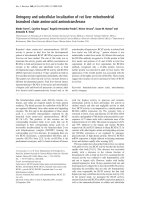
The numbers of OVA transport to thoracic lymph nodes
with anti-OVA IgG1 instillation reduced compared with PBS
In order to verify antigen transport of DCs in allergic air-
wayinflammation,weanalysedOVAtransportfromairway
mucosa to thoracic lymph nodes by fluorescence micro-
scopy. Fluorescein-conjugated OVA was seen in lymph
nodes of allergic mice instilled with P BS (Figure 2A). Fluor-
escence microscopy observation confirmed a reduction in
fluorescein-conjugated OVA in lymph nodes after instilla-
tion of anti-OVA IgG1 (Figure 2A). Additionally anti-OVA
IgG1 also reduced the number of the fluorescein-positive
CD11c
+
MHC class II
+
cells in thoracic lymph nodes com-
pared with PBS instilled group (Figure 2B). These results
show that anti-OVA IgG1 instillation in allergic inflamma-
tion reduced transport of OVA.
MHC class II expression on lung-derived CD11c
+
APCs
with anti-OVA IgG1 instillation reduced compared with
PBS
In order to clarify the role of CD11c
+
APCs in allergic
airway inflammation, we analyzed MHC class II expres-
sion on lung-derived CD11c
+
APCs by flow cytometry.
MHC class II expression on lung CD11c
+
APCs of WT
mice instilled with PBS increased as compared with
naïvemice(Figure3).Ontheotherhand,inWTmice
instilled with anti-OVA IgG1, it decreased (Figure 3).
These findings showed anti-OVA IgG1 instillation
resulted in the decrease of MHC class II
+
APCs, sug-
gesting its effects on lung APCs.
Anti-OVA IgG1 ameliorated the activation of BMDCs in vitro
TheopticaldensityofTcells, as measured with a cell
counting kit, increased by ad dition of OVA peptide in
co-culture medium with BMDCs compared to negative
control (OVA peptide free). AntiOVA-IgG1 group sig-
nificantly decreased the T cells proliferation (Figure 4).
It means that antiOVA-IgG1 prevented BMDCs from
presenting antigens, resulting in decreasing the number
of T cells.
Intratracheal instillation of anti-OVA IgG1 increases
allergic airway inflammation in FcgRIIB KO mice
Airway inflammation showed no changes in FcgRIIB KO
mice instilled with PBS, s imilarly to WT mice, while
Ishikawa et al. Respiratory Research 2011, 12:42
/>Page 4 of 10
FcgRIIB KO mice instilled with anti-OVA IgG1 signifi-
cantly deteriorated airway inflammation in BAL fluid
and histopathology (Figure 5A, B).
BMDCs transplantation into FcgRIIB KO mice attenuates
allergic airway inflammation by anti-OVA IgG1
In order to examine how DCs are related to allergic air-
way inflammation, BMDCs from WT mice were
intravenously transplanted into FcgRIIB KO mice and the
efficacy of anti-OVA IgG1 was analysed by BAL fluid.
Total cells and eosinophils were lower in FcgRIIB KO
mice transplanted intravenously and instilled with anti-
OVA IgG1 compared to FcgRIIB KO mice instilled with
PBS and anti-OVA IgG1, showing that WT BMDCs
restored the effect of anti-OVA IgG1 on airway inflam-
mation (Figure 5). These data indicate that anti-OVA
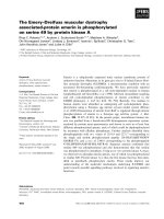
Figure 1 Anti-OVA IgG inhibited allergic airway inflammation of WT mice by BAL fluid, histopathology and AHR. A, Total cell counts
and cellular composition of BAL fluid obtained from OVA-sensitized WT mice instilled with PBS, anti-OVAIgG1 and IgG2a, and non-specific IgG. *:
p < 0.01 vs. WT/PBS; †: p < 0.05 vs. WT/IgG2a; TC: total cells; Eo: eosinophils; MF: macrophages; Neu: neutrophils; Lym: lymphocytes. B, Th1 and
Th2 cytokine levels in BAL fluid after instillation of PBS (black bars), anti-OVA IgG1 (gray bars) or anti-OVA IgG2a (dot bars) are shown. *: p < 0.05
vs. WT/PBS. C, Histological examination of lung tissues instilled with PBS and anti-OVA IgG subclasses. Micrographs (low and high magnification,
×100 and ×400, respectively) depict lung sections stained with H&E. D, AHR after instillation with PBS and anti-OVA IgG subclasses. Increased
Penh in response to inhaled methacholine was measured. Results are expressed as values relative to baseline. Data show means ± SEM pooled
from three independent experiments with 4-12 mice/group.
Ishikawa et al. Respiratory Research 2011, 12:42
/>Page 5 of 10
IgG1 attenuated allergic airway inflammation via FcgRIIB
in transplanted BMDCs.
Discussion
We demonstrated that antigen-specific IgG ameliorated
airway inflammation via FcgRIIB on CD11c
+
APCs
including DCs, but non-specific IgG did not. DCs are
the most potent APCs, and play a crucial role in pro-
moting develo pment of active immunity in allergic air-
way inflammation. Immature DCs sense the presence of
invading antigens and process the antigen intracellularly
in inflamed tissue, developing into mature DCs with
upregulated expression of MHC and costimulatory
molecules in inflammatory microenvironments [19-21,
32]. Subsequently, mature DCs home into secondary
lymphoid tissue, where they present the processed
antigens to naïve T-cells to generate effector T-cells
[19-21,32]. In vitro, there have been some reports that
human IgG inhibits the differentiation and maturation
of human monocyte-derived DCs [33,34]. Boruchov et
al. showed that the DCs maturation marker CD83 and
the costimulatory molecule CD86 on soluble human
IgG-treated DCs are decreased [34]. In addition,
although there are have been reports on the relationship
between IgG and DCs in vitro, there have been no pre-
vious studies of antigen-specific IgG and DCs in murine
asthmatic model. Therefore, we examined the role of
DCs and the mechanisms that antigen-specific IgG sup-
pressed allergic airway inflammation in mice.
CD11c
+
MHC class II
+
cells were previously con-
firmed as DCs, and were recently described as CD103
+
CD11b
-
and CD103
-
CD11b
+
subpopulations [35]. We
also used lung-derived CD11c
+
APCs to examine the
mechanisms related to allergen-specific IgG and DCs. In
the present study, anti-OVA IgG1 reduced the number
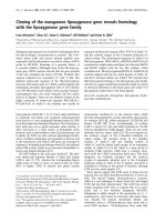
Figure 2 The numbers of OVA transport t o thoracic lymph
nodes with anti-OVA IgG1 instillation reduced compared with
PBS. (A) Fluorescence microscopy findings of thoracic lymph nodes
by instillation of OVA-fluorescein conjugate in OVA-sensitized WT
mice instilled with PBS and anti-OVA IgG1. (B) The CD11c
+
MHC
class II
+
cells which intaken fluorescein-conjugated OVA in thoracic
lymph nodes was analysed by flow cytometry. Data show mean
value pooled from three independent experiments.
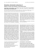
Figure 3 MHC class II expression on lung-derived CD11c
+
APCs
with anti-OVA IgG1 instillation reduced compared with PBS.
MHC class II expression on lung-derived CD11c
+
APCs purified from
naïve mice, mice instilled with PBS or anti-OVA IgG1 were analysed.
Values inside histograms represent the percentage of MHC class II
expression on lung-derived CD11c
+
APCs. The numbers on the
histogram show mean value of each percentage of gated cells from
three independent experiments.
Ishikawa et al. Respiratory Research 2011, 12:42
/>Page 6 of 10
of the fluorescein-positive CD11c
+
MHC class II
+
cells
in thoracic lymph nodes compared with PBS instilled
group, showing that anti-O VA IgG1 instillation in aller-
gic inflammation reduced transport of OVA. Our find-
ings also demonstrated that MHC class II expression on
CD11c
+
APCs was decreased in anti-OVA IgG1 instilled
mice. Moreover, WT-derived BMDCs transplantation
revealed the dependence of the anti-OVA IgG1 effect
on FcgRIIB on transplanted BMDCs. In addition, CD11c
+
DCs, not macrophages, have been reported to play cru-
cial role in development of allergen-induced airway
inflammation [21]. From above, anti-OVA IgG1 instilla-
tion suggested to modify the functions of CD11c
+
lung
APCs, including DCs. In vitro, we also indicated that
anti-OVA IgG1 significantly ameliorated the activation of
DCs, showing a reduction of the proliferation of T cells.
Our study thus demonstrated for the first time that anti-
gen-specific IgG ameliorates the act ivation and matura-
tion of CD11c
+
APCs including DCs in allergic mice and
in vitro.
Mice have four different classes of FcRs; FcgRI, FcgRII,
FcgRIII and FcgRIV. Functionally, FcRs are classified in
two types: activat ing FcgRs that possess an ITAM in the
cytoplasmic domain, including FcgRI, FcgRIIA, FcgRIII,
FcgRIV [13,15]; and inhibitory FcgRs such as FcgRIIB,
which exerts activity via ITIM. The composite expres-
sion of activating and inhibitory FcgRs thus regulates
the immune response [15]. Also, in the previous studies,
thepresenceofFcgRI-III on pulmonary macrophages
and high RNA levels of FcgRIIB relative to FcgRI and
FcgRIII on lung-derived CD11c
+
MHC class II
+
DCs are
demonstrated [35] . In a previous study, Kitamura et al.
reported that expression of FcgRsonDCsisimportant
during the sensitization for the development of allergen-
induced AHR and inflammation [22]. In vitro experi-
ments, expression levels of CD86 and MHC class II
wereshowntobehigherinBMDCinmiceloadedwith
OVA-immune complex (IC) than in those loaded with
OVA; however, no significant differ ences were seen in
FcgR KO mice [36], suggesting more efficient matura-
tion by OVA-IC than by OVA alone through FcgRs on
DCs. Moreover, allergen-specific IgG, which is generated
during sensitization, lead to IC formation upon antigen
challenge and result in enhanced FcgR-mediated antigen
presentation as previously described [35]. However, in
our study, intratracheal instillation of anti-OVA IgG1
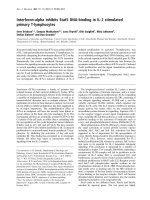
Figure 4 Anti-OVA IgG1 inhibited the cell proliferation of
BMDCs in vitro. BMDCs (2 × 10
4
cells per well) were purified from
WT mice and pulsed with OVA peptide (20 μM) or anti-OVA IgG1
(0.1 mg/ml) containing the same amount of OVA. Negative control
means no addition of OVA peptide in culture medium. Cells were
washed and used to stimulate CD4
+
T-cells (2 × 10
5
cells per well).
The proliferation of T cells after DCs treatment with anti-OVA IgG1
was quantified in duplicate by measuring the optical density using
the WST-8 cell-counting kit after 48 h. Data show means ± SEM.
*p < 0.05.
Figure 5 Anti-OVA IgG1 increased allergic airway inflammation
in FcgRIIB KO mice, and BMDCs transplantation reversed its
effects. A, Total cell, eosinophil and the others cell counts of BAL
fluid obtained from OVA-sensitized and challenged FcgRIIB KO mice
(IIBKO) instilled with PBS or anti-OVAIgG1. Moreover, FcgRIIB KO
mice intravenously transplanted with WT-derived BMDCs (IIBKO/DC
i.v. IgG1) were sensitized and challenged with OVA, and instilled
intratracheally with anti-OVA IgG1. The number of BAL fluid was
analysed. Data show mean ± SEM. #: p < 0.05 vs. WT/PBS, ‡:p<
0.05 vs. IIBKO/PBS. B, Histological examination of lung tissues of
FcgRIIB KO mice instilled with PBS and anti-OVA IgG1. Micrographs
(high magnification, ×400) depict lung sections stained with H&E.
Ishikawa et al. Respiratory Research 2011, 12:42
/>Page 7 of 10
separately from OVA ameliorated allergic airway inflam-
mation. This difference may be the result of differences
in FcR affinity for IgG alone or IC. FcgRmaymore
readily ligate to IC, while FcgRIIBmaymorereadily
ligate to separate IgG than IC. In the previous studies,
soluble monomeric IgG decreased FcgRIIA expression
and increased FcgRIIB on immature DCs in human per-
ipheral blood mononuclear cells when soluble mono-
meric IgG was added to cultures of immature DCs [34].
Also, high expression levels of FcgRIIB in immature
human DCs were detected, while i n mature DCs
FcgRIIBwasmarkedlydown-regulatedaspreviously
described [37], indicating that engagement of FcgRIIB by
IC can inhibit DCs activation and antigen uptake. In
mice, we suggested that FcgRIIB on immature DCs
increases during sensitization by instillation of anti-
OVA IgG1 and are more easily ligated with anti-OVA
IgG1 alone. Moreover, Hartwig et al. demonstrated that
anti-OVA IgG alone ameliorated the number of total
cells and eosinophils in BAL, and peribronchial and
perivascular cell counts of inflammatory cells in histol-
ogy when compared with OVA-challenged mice [35]. In
this point, their results correspond to our results that
anti-OVA IgG alone ameliorated allergic airway inflam-
mation and AHR, suggesting that anti-OVA IgG1
directly binds with FcgRIIB on DCs during allergic air-
way inflammation.
Various studies have examined the role of FcgRIIB in
down-regulating specific allergic inflammatory cells in
vitro. Dharajiya et al. also reported that FcgRIIB inhib-
ited allergic lung inflammation in a murine model of
allergic asthma, although our protocol was different
from their properties of the chall enged allergen or the
methods of challenge. They examined the role of
FcgRIIB in allergic lung inflammation using FcgRIIB KO
mice sensitized to and challenged with ragweed. Rag-
weed challenge in sensitized mice up-regulated FcgRIIB
in the lungs; however, disruption of the IFN-g gene
abrogated this up-regulation. These results indicate that
ragweed challenge up-regulates FcgRIIB in the lungs via
IFN-g and Th1-depende nt mechanisms. They indicated
that FcgRIIB physiologically regulated allergic airway
inflammation by the up-regulation of FcgRIIB on pul-
monary CD14
+
/MHC class II
+
mononuclear cells and
CD11c
+
cells by an IFN-g and Th1-dependent mechan-
ism[38].Moreover,wehavepreviouslydemonstrated
that intrav enous IgG ameliorated allergic airway inflam-
mation via FcgRIIB on CD11c
+
DCs [39]. In another
study, Sehra et al. reported that airway IgG counteracted
allergen-triggered pulmonary inflammation and sho wed
that this treatment increased Th1 reactivity and IFN-g
levels in BAL [40]. They showed that airway IgG appli-
cation increased allergen capture by activating FcgRon
alveolar macrophages and led to increased Th1 reactivity
and IFN-g, resulting in sup pression of airway eosinophil
infiltration. In this study, antigen-specific IgG was found
to ameliorate antigen uptake and presentation by DCs,
and suppress allergic airway inflammation via FcgRIIB,
not FcgR, on DCs. In addition, anti-OVA IgG1 instilla-
tion significantly decreased IL-4, IL -5, and IL-1 3 cyto-
kine levels and increased IFN-g in BAL fluid compared
with PBS. These results indicated that intratracheal
instillation of anti-OVA IgG1 alone caused a shift from
Th2 to Th1 in allergic airway inflammation. We indicate
that anti-OVA IgG1 attenuate the activation of DCs
which induces Th2 response, by binding to FcgRIIB on
DCs.
We demonstrated that intratracheal instillation of
antigen-specific IgG ameliorated allergic airway inflam-
mation via FcgRIIB on CD11c
+
APCs including DCs.
Mouse IgG1 is reported to have higher affinity for
FcgRIIB than IgG2a [16]. We found that anti-OVA IgG1
most strongly ameliorated airway inflammation among
the anti-OVA IgG subclasses in total cells, eosinophils,
Th2 cytokine levels in BAL fluid, AHR, and histology.
The present studies in FcgRIIB KO mice instilled with
PBS didn’t show increased inflammation compar ed with
WT asthmatic mice. However, our previous studies
demonstrated that FcgRIIB KO mice instilled with PBS
showed allergic airway inflammation as WT asthmatic
mice [39]. Challenged Fcg RIIB KO mice instilled wit h
PBS have i ncreased inflammation, indicating that the
balance between FcgRIIB and FcgR which existed in its
cytoplasm had a tendency to FcgR of activating type
FcR. Moreover, FcgRIIB KO mice instilled with anti-
OVA IgG1 significantly deteriorated airway inflamma-
tion in BAL fluid and histopathology. These data suggest
the balance with a further tendency to activating Fc gR
caused by intratracheal instillation of anti-OVA IgG1 in
FcgRIIB KO.
In a murine model, intratracheally transplanted mye-
loid DCs from the airway are known to induce Th2
reactivity after antigen sensitization and inhalation, and
leading to eosinophilic airway inflammation [41].
In vitro, we also indicated that anti-OVA IgG1 signifi-
cantly ameliorated the activation of DCs, showing a
reduction of the proliferation of T cells. Furthermore,
we demonstrated that intravenous transplantation of
BMDCs and intratracheal instillation of anti-OV A IgG1
ameliorated the cellular i nfiltration to BAL fluid com-
pared to FcgRIIB KO mice instilled with PBS and anti-
OVAIgG1,showingthati.v.transplantedBMDCshad
its local effects on allergic inflammation. Our data in
vivo and in vitro showed that myeloid DCs play impor-
tant roles to develop Th2 response inflammation
as previously reported. Our findings suggested that
Ishikawa et al. Respiratory Research 2011, 12:42
/>Page 8 of 10
antigen-specific IgG ameliorated the antigen transport-
ing a nd presenting capacity on CD11c
+
APCs including
DCs, resulting in attenuating al lergic airway i nflamma-
tion via FcgRIIB on DCs by shifting from Th2 to Th1
immune response.
Conclusions
Weconcludedthatanti-OVAIgGamelioratedallergic
airway inflammation via FcgRIIB on DCs in murine
asthmatic model. Further studies for the function of
FcgRIIB in human airway inflammation would be
required. Our findings have important implications for
elucidating the pathophysiology of asthmatic diseases
and engineering agents to target IgG-FcR on DCs.
Acknowledgements
The author would like to thank the members of the Division of Respiratory
Medicine and Division of Molecular and Cellular Biology for fruitful
discussions and technical assistance. This study was supported by a grant for
Research Fellows of the Global COE Program “Global Center of Excellence
for Education and Research on Signal Transduction Medicine in the Coming
Generation” from the Ministry of Education, Culture, Sports, Science, and
Technology of Japan [F031 to M.Y]. This study was also supported, in part,
by a grant from the Japan Society for the Promotion of Science [21590811
to M.Y.], by a grant from the Japan Chemical Industry Association, by a grant
from The Mother and Child Health Foundation, and KAKENHI [21790769 to
M.Y/19790557 to K. K].
Author details
1
Division of Respiratory Medicine, Department of Internal Medicine, Kobe
University Graduate School of Medicine, Kobe, Japan.
2
Division of
Metabolomics Research, Division of Gastroenterology, The Integrated Center
for Mass Spectrometry, Kobe University Graduate School of Medicine, Kobe,
Japan.
3
Department of Immune Regulation, Tokyo Medical and Dental
University Graduate School, Tokyo, Japan.
Authors’ contributions
YI, KK, & MY designed the protocol, HK, TT, MY, & KN assisted to acquire the
data, YI, MY, KK, & MY interpreted the data, YI draft ed the manuscript but all
of the authors contributed to the manuscript. All authors read and approved
the final manuscript.
Competing interests
We can state that this research is original and has not been submitted for
publication elsewhere, and that no part of the research presented has been
funded by tobacco industry sources.
Received: 30 September 2010 Accepted: 10 April 2011
Published: 10 April 2011
References
1. Masoli M, Fabian D, Holt S, Beasley R: The global burden of asthma:
executive summary of the GINA Dissemination Committee report. Allergy
2004, 59:469-478.
2. Kinet JP: The high-affinity IgE receptor (Fc epsilon RI): from physiology to
pathology. Annu Rev Immunol 1999, 17:931-972.
3. Wills-Karp M: Immunologic basis of antigen-induced airway
hyperresponsiveness. Annu Rev Immunol 1999, 17:255-281.
4. Avila PC: Does anti-IgE therapy help in asthma? Efficacy and
controversies. Annu Rev Med 2007, 58:185-203.
5. Strunk RC, Bloomberg GR: Omalizumab for asthma. N Engl J Med 2006,
354:2689-2695.
6. Hanania NA: Targeting airway inflammation in asthma: current and
future therapies. Chest 2008, 133:989-998.
7. Ogra PL, Faden H, Welliver RC: Vaccination strategies for mucosal
immune responses. Clin Mivrobiol Rev 2001, 14:430-445.
8. Miyagawa N, Iwasaki H, Kato T, Tanaka M, Shibata T, Wakitani K: Induction
of late airway response was involved in serum antigen-specific
immunoglobulin G in rats. Int Immunopharmacol 2008, 8:1848-1853.
9. Platts-Mills T, Vaughan J, Squillace S, Woodfolk J, Sporik R: Sensitisation,
asthma, and a modified Th2 response in children exposed to cat
allergen: a population-based cross-sectional study. Lancet 2001,
357:752-756.
10. Aalberse RC, van der Gaag R, van Leeuwen J: Serologic aspects of IgG4
antibodies. I. Prolonged immunization results in an IgG4-restricted
response. J Immunol 1983, 130:722-726.
11. Ravetch JV, Kinet JP: Fc receptors. Annu Rev Immunol 1991, 9:457-492.
12. Takai T: Roles of Fc receptors in autoimmunity. Nat Rev Immunol 2002,
2:580-592.
13. Hulett MD, Hogarth PM: Molecular basis of Fc receptor function. Adv
Immunol 1994, 57:1-127.
14. Masuda A, Yoshida M, Shiomi H, Morita Y, Kutsumi H, Inokuchi H, Mizuno S,
Nakamura A, Takai T, Blumberg RS, Azuma T: Role of Fc Receptors as a
therapeutic target. Inflamm Allergy Drug Targets 2009, 8:80-86.
15. Masuda A, Yoshida M, Shiomi H, Ikezawa S, Takagawa T, Tanaka H,
Chinzei R, Ishida T, Morita Y, Kutsumi H, Inokuchi H, Wang S, Kobayashi K,
Mizuno S, Nakamura A, Takai T, Blumberg RS, Azuma T: Fcgamma receptor
regulation of Citrobacter rodentium infection.
Infect Immun 2008,
76:1728-1737.
16.
Nimmerjahn F, Ravetch JV: Fcgamma receptors: old friends and new
family members. Immunity 2006, 24:19-28.
17. McGaha TL, Sorrentino B, Ravetch JV: Restoration of tolerance in lupus by
targeted inhibitory receptor expression. Science 2005, 307:590-593.
18. Qiu WQ, de Bruin D, Brownstein BH, Pearse R, Ravetch JV: Organization of
the human and mouse low-affinity Fc gamma R genes: duplication and
recombination. Science 1990, 248:732-735.
19. Lambrecht BN, Hammad H: Taking our breath away: dendritic cells in the
pathogenesis of asthma. Nat Rev Immunol 2003, 3:994-1003.
20. Lambrecht BN: Allergen uptake and presentation by dendritic cells. Curr
Opin Allergy Clin Immunol 2001, 1:51-59.
21. Van Rijt LS, Jung S, KleinJan A, Vos N, Willart M, Duez C, Hoogsteden HC,
Lambrecht BN: In vivo depletion of lung CD11c dendritic cells during
allergen challenge abrogates the characteristic features of asthma. JEM
2005, 201:981-991.
22. Kitamura K, Takeda K, Koya T, Miyahara N, Kodama T, Dakhama A, Takai T,
Hirano A, Tanimoto M, Harada M, Gelfand EW: Critical role of the Fc
receptor gamma-chain on APCs in the development of allergen-induced
airway hyperresponsiveness and inflammation. J Immunol 2007,
178:480-488.
23. Takai T, Ono M, Hikida M, Ohmori H, Ravetch JV: Augmented humoral and
anaphylactic responses in Fc gamma RII-deficient mice. Nature 1996,
379:346-349.
24. Barnden MJ, Allison J, Heath WR, Carbone FR: Defective TCR expression in
transgenic mice constructed using cDNA-based alpha- and beta-chain
genes under the control of heterologous regulatory elements. Immunol
Cell Biol 1998, 76:34-40.
25. Ishikawa R, Tsujimura Y, Obata K, Kawano Y, Minegishi Y, Karasuyama H:
IgG-mediated systemic anaphylaxis to protein antigen can be induced
even under conditions of limited amounts of antibody and antigen.
BBRC 2010, 402:742-746.
26. Renz H, Saloga J, Bradley KL, Loader JE, Greenstein JL, Larsen G, Gelfand EW:
Specific V beta T cell subsets mediate the immediate hypersensitivity
response to ragweed allergen. J Immunol 1993, 151:1907-1917.
27. Hamelmann E, Schwarze J, Takeda K, Oshiba A, Larsen GL, Irvin CG,
Gelfand EW: Noninvasive measurement of airway responsiveness in
allergic mice using barometric plethysmography. Am J Respir Crit Care
Med 1997, 156:766-775.
28. Vermaelen KY, Carro-Muino I, Lambrecht BN, Pauwels RA: Specific
migratory dendritic cells rapidly transport antigen from the airways to
the thoracic lymph nodes. JEM 2001, 193:51-60.
29. Inaba K, Inaba M, Romani N, Aya H, Deguchi M, Ikehara S, Muramatsu S,
Steinman RM: Generation
of large numbers of dendritic cells from
mouse bone marrow cultures supplemented with granulocyte/
macrophage colony-stimulating factor. J Exp Med 1992, 176:1693-1702.
Ishikawa et al. Respiratory Research 2011, 12:42
/>Page 9 of 10
30. Lutz MB, Kukutsch N, Ogilvie AL, Rossner S, Koch F, Romani N, Schuler G:
An advanced culture method for generating large quantities of highly
pure dendritic cells from mouse bone marrow. J Immunol Methods 1999,
223:77-92.
31. Ishiyama M, Miyazono Y, Sasamoto K, Ohkura Y, Ueno K: A highly water-
soluble disulfonated tetrazolium salt as a chromogenic indicator for
NADH as well as cell viability. Talanta 1997, 44:1299-1305.
32. Sato K, Fujita S: Dendritic cells: nature and classification. Allergol Int 2007,
56:183-191.
33. Bayry J, Lacroix-Desmazes S, Carbonneil C, Misra N, Donkova V, Pashov A,
Chevailler A, Mouthon L, Weill B, Bruneval P, Kazatchkine MD, Kaveri SV:
Inhibition of maturation and function of dendritic cells by intravenous
immunoglobulin. Blood 2003, 101:758-765.
34. Boruchov AM, Heller G, Veri MC, Bonvini E, Ravetch JV, Young JW:
Activating and inhibitory IgG Fc receptors on human DCs mediate
opposing functions. J Clin Invest 2005, 115:2914-2923.
35. Hartwig C, Mazzega M, Constabel H, Krishnaswamy JK, Gessner JE, Braun A,
Tschernig T, Behrens GM: Fcgamma receptor-mediated antigen uptake by
lung DC contributes to allergic airway hyper-responsiveness and
inflammation. Eur J Immunol 2010, 40:1284-1295.
36. Akiyama K, Ebihara S, Yada A, Matsumura K, Aiba S, Nukiwa T, Takai T:
Targeting apoptotic tumor cells to Fc gamma R provides efficient and
versatile vaccination against tumors by dendritic cells. J Immunol 2003,
170:1641-1648.
37. Liu Y, Gao X, Masuda E, Redecha PB, Blank MC, Pricop L: Regulated
expression of FcgammaR in human dendritic cells controls cross-
presentation of antigen-antibody complexes. J Immunol 2006,
177:8440-8447.
38. Dharajiya N, Vaidya SV, Murai H, Cardenas V, Kurosky A, Boldogh I, Sur SA:
FcgammaRIIb inhibits allergic lung inflammation in a murine model of
allergic asthma. PLoS One 2010, 5:e9337.
39. Yamamoto M, Kobayashi K, Ishikawa Y, Nakata K, Funada Y, Kotani Y,
Masuda A, Takai T, Azuma T, Yoshida M, Nishimura Y: The inhibitory effects
of intravenous administration of rabbit IgG on airway inflammation are
dependent on Fcγ receptor IIb on CD11c
+
dendritic cells in murine
model. Clin Exp Immunol 2010, 162:315-324.
40. Sehra S, Pynaert G, Tournoy K, Haegeman A, Matthys P, Tagawa Y,
Pauwels R, Grooten J: Airway IgG counteracts specific and bystander
allergen-triggered pulmonary inflammation by a mechanism dependent
on Fc gamma R and IFN-gamma. J Immunol 2003, 171:2080-2089.
41. Lambrecht BN, Veerrman MD, Coyle AJ, Gutierrez-Ramos JC, Thielemans K,
Pauwels RA: Myeloid dendritic cells induce Th2 responses to inhaled
antigen, leading to eosinophilic airway inflammation. J Clin Invest 2000,
106:551-559.
doi:10.1186/1465-9921-12-42
Cite this article as: Ishikawa et al.: Antigen-Specific IgG ameliorates
allergic airway inflammation via Fcg receptor IIB on dendritic cells.
Respiratory Research 2011 12:42.
Submit your next manuscript to BioMed Central
and take full advantage of:
• Convenient online submission
• Thorough peer review
• No space constraints or color figure charges
• Immediate publication on acceptance
• Inclusion in PubMed, CAS, Scopus and Google Scholar
• Research which is freely available for redistribution
Submit your manuscript at
www.biomedcentral.com/submit
Ishikawa et al. Respiratory Research 2011, 12:42
/>Page 10 of 10