Báo cáo y học: " CD39+ Regulatory T cells suppress generation and differentiation of Th17 cells in human malignant pleural effusion via a LAP-dependent mechanism" pps
Bạn đang xem bản rút gọn của tài liệu. Xem và tải ngay bản đầy đủ của tài liệu tại đây (1.06 MB, 10 trang )
RESEARC H Open Access
CD39
+
Regulatory T cells suppress generation
and differentiation of Th17 cells in human
malignant pleural effusion via a LAP-dependent
mechanism
Zhi-Jian Ye
1†
, Qiong Zhou
1†
, Jian-Chu Zhang
1
, Xiao Li
1
, Cong Wu
2
, Shou-Ming Qin
2
, Jian-Bao Xin
1
and
Huan-Zhong Shi
1*
Abstract
Background: Both regulatory T cells (Tregs) and T helper IL-17-producing cells (Th17 cells) have been found to be
involved in human malignancies, however, the possible implication of Tregs in regulating generation and
differentiation of Th17 cells in malignant pleural effusion remains to be elucidated.
Methods: The numbers of both CD39
+
Tregs and Th17 cells in malignant pleural effusion and peripheral blood
from patients with lung cancer were determined by flow cytometry. The regulation and mechanism of Tregs on
generation and differentiation of Th17 cells were explored.
Results: Both CD39
+
Tregs and Th17 cells were increased in malignant pleural effusion when compared with blood,
and the numbers of CD39
+
Tregs were correlated negatively with those of Th17 cells. It was also noted that high
levels of IL-1b, IL-6, and TGF-b1 could be observed in malignant pleural effusion when compared the
corresponding serum, and that pleural CD39
+
Tregs could express latency-associated peptide on their surface. When
naïve CD4
+
T cells were cocultured with CD39
+
Tregs, Th17 cell numbers decreased as CD39
+
Treg numbers
increased, addition of the anti-latency-associated peptide mAb to the coculture reverted the inhibitory effect
exerted by CD39
+
Tregs.
Conclusions: Therefore, the above results indicate that CD39
+
Tregs inhibit generation and differentiation of Th17
cells via a latency-associated peptide-dependent mechanism.
Keywords: latency-associated peptide, malignant pleural effusion, regulatory T cells, Th17 cells, transfer growth
factor
Introduction
It has been well documented that CD4
+
T lymphocyte
dominance occurs in malignant pleural effusion (MPE)
[1,2]. On encountering an antigen, naïve CD4
+
T-helper
precursor cells enact a specific process that results in
differentiation toward the T-helper type 1 (Th1) or Th2
lineage. Early studies have suggested that Th1/Th2 cell
balance in MPE may influe nce pathophysiologic process
of pleural disease [3,4]. Two additional CD4
+
T cell sub-
sets, regulatory T cells (Tregs) and T helper IL-17-pro-
ducing cells (Th17 cells), have been described more
recently. Tregs are characterized by the expression of
the lineage-specific transcription factor FOXP3, which i s
involved both in their development and in their suppres-
sor functions [5,6]. Th17 cells are now defined as a
separate subset distinct from the Th1, Th2, and Tregs,
in terms of developmental regulation and function [7,8].
Our previous studies showed that increased Tregs w ere
found in MPE, and these Tregs w ere recruited into
pleural space induced by chemokine CCL22 [9,10].
* Correspondence:
† Contributed equally
1
Department of Respiratory Diseases, Key Laboratory of Pulmonary Diseases
of Health Ministry, Union Hospital, Tongji Medical College, Huazhong
University of Science and Technology, China
Full list of author information is available at the end of the article
Ye et al. Respiratory Research 2011, 12:77
/>© 2011 Ye et al; licensee BioMed Cent ral Ltd. This is an Open Access article distributed under the terms of the Creative Comm ons
Attribution License ( which permits unrestricted use, distribution, and reprod uction in
any medium, provide d the original work is properly cited.
More recently, we have demonstrated that due to local
differentiation and expansion stimulated by cytokines
and to recruitment from peripheral blood induced by
chemokines, the numbers of Th17 cells were signifi-
cantly increased in MPE, and that the accumulation of
Th17 cells in MPE predicted improved patient survival
[11]. I t has been reported that human Tregs can differ-
entiate into Th17 cells, when stimulated by allogeneic
antigen-presenting cells in the presence of IL-2 or/and
IL-15 [12].
It has been well documented that TGF-b is synthe-
sized in cells as a pro-TGF-b precursor. Following
homodimerization, pro-TGF-b is cleaved into two frag-
ments: the C-terminal ho modimer corresponds to
mature TGF-b, while the N-terminal homodimer is
latency-associated peptide (LAP) [13]. Mature TGF-b
and LAP remain non-covalently bound to each other in
a complex called latent TGF-b. Latent TGF-b is inactive
because LAP prevents mature TGF-b from binding to
its receptor, and hence from transducin g a signal [14].
The role of TGF-b in the differentiation of human Th17
cells is still co ntroversial. Some studies demonstrated
that TGF-b is required for human Th17 cell differentia-
tion [15,16], however, the other data showed that TGF-
b suppresses the differentiation of Th17 cells [17,18].
Because TGF-b induces FOXP3 expression in naïve
CD4
+
T cells and converts them to Tregs [19], while
Tregs express cell surface or secrete TGF-b [20,21], this
introduces the possibility that Tregs may be playing a
role in generation and differentiation of Th17 cells via a
LAP-dependent mechanism. In the present study, we
were prompted to investigate whether CD39
+
Tregs are
capable of suppressing generation and differentiation of
Th17 cells, as well as whether LAP is involved in such a
possible suppression in MPE.
Methods
Subjects
The study protocol was approved by our institutional
reviewboardforhumanstudies,andinformedconsent
was obtained from all subjects. Pleural fluid samples
were collected fro m 16 patients (age range: 31 to 76 yr)
with newly diagnosed lung cancer with MPE. Histologi-
cally, 11 cases were adenocarcinoma and 5 were squa-
mous cell carcinoma. A diagnosis of MPE was
established by demonstration of malignant cells in
pleural fluid or/and on closed pleural biopsy specimen.
The patients were excluded if they had received any
invasive procedures directed into the pleural cavit y or if
they had suffered chest trauma within 3 mo prior to
hospitalization. At the time of sample collection, none
of the patients had received any anti-c ancer therapy,
corticosteroids, or other nonsteroid anti-inflammatory
drugs.
Sample Collection and Processing
The pleural fluid samples were collected in heparin-trea-
ted tubes from each subject, using a standard thoraco-
centesis technique within 24 h after hospitalization.
Twenty milliliters of peripheral blood were drawn simul-
taneously. MPE specimens were immersed in ice imme-
diately and were then centrifuged at 1,200 g for 5 min.
The cell-free supernatants of MPE and serum were fro-
zen at -80°C immediately after centrifuge for later deter-
mining cytokine concentrations. The cell pellets of MPE
were resuspended in HBSS, and mononuclear cells were
isolated by Ficoll-Hypaque gradient centrifugation
(Pharmacia, Uppsala, Sweden) to determine the T cell
subsets within 1 h. A pleural biopsy was performed
when the results of pleural fluid analysis were suggestive
of malignancy.
Flow Cytometry
The expression markers on T cells from MPE and blood
were determined by flow cytometry after surface stain-
ing or intracellular staining with anti-human-specific
Abs conjugated with either phycoerythrin or fluor escein
isothiocyanate. These human Abs included anti-CD3,
anti-CD4, anti-CD39, anti-CD45RA, anti-CD45RO, anti-
CD127, anti-LAP, anti-IL-17, and anti-FOXP3 mAbs,
which were purchased from BD Biosciences or
eBioscience (San Diego, CA). Intracellular staining for
IL-17 or FOXP3 was performed on T cells stimulated
with phorbol myristate acetate (50 ng/ml; Sigma-
Aldrich) and ionomycin (1 μM; Sigma-Aldrich) in the
presence of GolgiSt op (BD Biosciences) for 5 h, and the
intracellular IL-17 or FOXP3 was then stained with
anti-IL-17 or -FOXP3 conjugated with phycoerythrin
(eBioscience). Flow cytometry was performed on a BD
FACSCalibur flowcytometer using FCS ExpressV3
software.
Cell Isolation
Bulk CD4
+
T cells from pleural fluid and blood were
isolated by negative selection ( by depletion of CD8
+
,
CD11b
+
,CD16
+
,CD19
+
,CD36
+
,andCD56
+
cells) with
the Untouched CD4
+
cell isolation kit ( Miltenyi Biotec,
Auburn, CA) according to the manufacturer’sinstruc-
tions. After isolation of bulk CD4
+
T cells, the naïve
CD4
+
T cells (CD45RA
+
CD45RO
-
) were further purified
by EasySep enrichment kits (StemCell Technologies,
Vancouver, British Columbia, Canada) according to the
manufacturer’s instructions. The purity of naïve CD4
+
T
cells was > 97%, as measured by flow cytometry.
CD4
+
T cells were also stained with CD4-PerCP-
Cy5.5, CD25-PE, and CD39-FITC, (eBiosciences), and
CD4
+
CD25
high
CD39
+
T cells and CD4
+
CD25
-
responder
T cells were sorted using a Beckman Coulter cell sorter.
Purity of the sorted populations was > 97%.
Ye et al. Respiratory Research 2011, 12:77
/>Page 2 of 10
Generation and Differentiation of Th17 Cells and Tregs in
MPE
Purified naïve CD4
+
T cells (5 × 10
5
) were cultured in 1
ml of complete medium co ntaining human IL-2 (2 ng/
ml) in 48-well plates and stimu lated with plate-bound
anti-CD3 (OKT3; 1 μg/ml) and soluble anti-CD28 mAbs
(1 μg/ml) for 7 d. The exogenous cytokines used were
TGF-b1(5ng/ml),IL-1b (10 ng/ml), IL-6 (100 ng/ml),
and IL-23 (10 ng/ml). Recombinant human IL-1b,IL-2,
IL-6, IL-23, and TGF-b1, were purchased from R&D
Systems. In some experiments, designated numbers of
CD39
+
Tregs were added into the cultures. To demon-
strate that LAP was responsible for the inhibitive effects
of CD39
+
Tregs, blocking experiments were performed
by mixing the MPE with 500 ng/ml of anti-LAP mAb
(Clone 27235) or mouse IgG irrelevant isotype control
(R&D Systems). The culture supernatants were co llected
for determining IL-17 concentration.
Measurement of Cytokines
The concentrations of IL-1b, IL-6, IL-23, and TGF-b1in
both pleural fluids and sera, as well as IL-17 in culture
supernatants, were measured by sandwich ELISA kits
according to the manufacturer’s protocols (all kits were
purchased from R & D Systems Inc., Minneapolis, MN,
USA). All samples were assayed in duplicate. The lower
detection limits of IL-1b, IL-6, IL-23, TGF-b1, and IL-
17 were 1 pg/ml, 0.70 pg/ml, 6.8 pg/ml, 4.61 pg/ml, and
15 pg/ml, respectively.
Statistics
Data are expressed as mean ± SEM. Comparisons of the
data between different groups were performed using a
Kruskal-Wallis one-way analysis of variance on ranks.
For data in MPE and in the corresponding blood, paired
data comparisons were made using a Wilcoxon signed-
rank test. Analysis was completed with SPSS version
16.0 Statistical Software (Chicago, IL, USA), and p
values of less than 0.05 were considered to indicate sta-
tistical significance.
Results
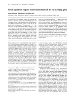
Tregs and Th17 Cells Were Significantly Increased in MPE
We first used flow cytometry to identify both CD39
+
Tregs and Th17 cells in CD4
+
T cells in MPE (Figure
1A,B,C)andperipheralblood.WithgatingonCD4
+
CD25
high
subset, we once again observed in the present
study that a significant increase in CD39
+
Tregs were
was observed in MPE (7.5 ± 1.0%) compared with blood
(4.4 ± 0.5%) (n = 16, Wilcoxon signed-rank test, p <
0.001) (Figure 1D). Consistent with our previous find-
ings [14], we noted that percentages of Th17 cells repre-
sented the higher values in MPE (3.7 ± 0.4%), showing a
significant increase in comparison with those in the
corresponding blood (0.6 ±0.1%)(n=16,Wilcoxon
signed-rank test, p < 0.001) (Figure 1E). We further
found that the ratios of CD39
+
Tregs/Th17 cells were
significantly lower in MPE (3.3 ± 1.0) than in blood
(11.5 ± 2.4, n = 16 , Wilcoxon signed-rank test, p <
0.001) (Figure 1F). In addition, pleural Th17 cell num-
bers were correlated negatively with Treg numbers (r =
-0.804, p < 0.001) (Figure 1G).
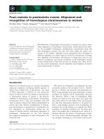
As show in Figure 2, majority of CD4
+
CD25
high
T
cells were CD39 positive (86 - 94%) and were CD127
negative (88 - 95%), possessing typical phenotypes of
Tregs.
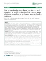
Impacts of Cytokines on Tregs and Th17 Cells in MPE
We determined some cytokines that reported be
involved in generation and differenti ation of Tregs or
Th17 cells, and observed that high levels of IL-1b,IL-6,
and TGF-b1, but not of IL-23, in MPE when compared
with the corresponding sera (Figure 3), suggesting that
these proinflammatory cytokines might affect the gen-
eration and differentiation of Tregs or/and Th17 cells in
MPE.
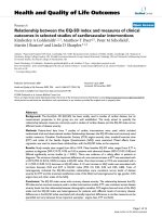
To evaluate the contribution of cytokines to the num-
bers of pleural C D39
+
Tregs and Th17 cells , we purified
naïve CD4
+
T cells from MPE and blood and cultured
the m in the pr esence of one or more of IL-1b, IL-6, IL-
23, and TGF-b. With IL-2-containing medium provided
a baseline for comparison, IL-1b, IL-6, or IL-23, but not
TGF-b, could promote the differentiation of Th17 cells
from naïve CD4
+
T cells (Figure 4). The combination of
IL-1b plus IL-6, IL-1b plus IL-23, IL-6 plus IL-23, or IL-
1b plus IL-6 plus IL-23, significantly increased the per-
centage of Th17 cells at higher extents compared with
any single one of above cytokines. Although a significant
high concentration of TGF-b was found in MPE, it did
not promote the generation and differentiation and of
Th17 cells; in contrast, TGF-b could reduce the
increased percentage of Th17 cells stimulated by the
above cytokines. On the other hand, TGF-b was capab le
of promoting the differentiation of CD39
+
Tregs during
the 7-day culture, and that any one or their various
combinations of IL-1b , IL-6, and IL-23 did not affect
the i ncrease in CD39
+
Treg numbers induced by TGF-b
(Figure 4).
Inhibition of Generation and Differentiation of Th17 Cells
by CD39
+
Tregs
On the basis of our observation that both CD39
+
Tregs
and Th17 cells were significantly increased in MPE, and
that pleural CD39
+
Treg numbers were correlated nega-
tively with pleural Th17 cell numbers, we therefore
investigated the impacts of Tregs on generation and dif-
ferentiation of Th17 cells in vitro. We purified CD39
+
CD4
+
CD25
high
T cells from both MPE and blood, and
Ye et al. Respiratory Research 2011, 12:77
/>Page 3 of 10
Figure 1 Both CD39
+
regulatory T (Tregs) cells and Th17 cells increased in malignant pleural effusion (MPE). (A) Lymphocytes were
identified based on their characteristic properties shown in the forward scatter (FSC) and sideward scatter (SSC). (B) A representative gating was
set for CD4
+
T cells from pleural lymphocytes. (C) A representative dot plots showing expression of CD39 and IL-17 in pleural CD4
+
T cells.
Comparisons of percentages of CD39
+
Tregs (D), Th17 cells (E), and ratios of CD39
+
Tregs/Th17 cells (F) in MPE and blood from patients with lung
cancer (n = 16). The percentages of CD39
+
Tregs and Th17 cells were determined by flow cytometry. Horizontal bars indicate medians.
Comparison was made using a Wilcoxon signed-rank test. (G) The percentages of Th17 cells correlated with CD39
+
Tregs cells in MPE.
Correlations were determined by Spearman’s rank correlation coefficients.
Figure 2 CD39 and CD127 expr essed on CD4
+
CD25
high
Tcells. The subset of pleural CD4
+
CD25
high
T cells (A) was identified by flow
cytometry for determining the surface expression of CD39 (B) and CD127 (C), data for one representative donor of 16 tested are shown.
Ye et al. Respiratory Research 2011, 12:77
/>Page 4 of 10
found that this subset of T cells were almost FOXP3
positive (> 97%) (Figure 5A) and were almost CD127
negative (> 97%) (Figure 5B), possessing typical phe-
notypes of Tregs. As shown in Figure 5C, generation
and differentiation of Th17 cells were observed when
the purified naïve CD4
+
T cells were cultured for 7 d
in presence of IL-1b and IL-6. When CD39
+
Tregs
were added into the coculture, Th17 cell numbers
decreased as CD39
+
Treg numbers increased. Likewise,
IL-17 concentrations in the cultured supernatants
decreased as CD39
+
Treg numbers increased (Figure
5D). There were no differences in inhibiting effects on
both Th17 cell numbers and IL-17 concentrations
between pleural CD39
+
Tregs and blood CD39
+
Tregs
(Figure 5C and 5D).
LAP Mediates Treg-Induced Inhibition of Th17 Cells
Since high concentration of TGF-b1wasfoundinMPE
(Figure 2D), we explored whether LAP w as involved in
the observed suppressive effect by CD39
+
Tregs on the
generation and differentiation of Th17 cells. We deter-
mined LAP expression of on the cell surface of CD39
+
Tregs by flow cytometry and found that there was
some an extent of LAP surface expression on fleshly
purified CD39
+
Tregs (6.2 - 12.4%) (Figure 6A); when
CD39
+
Tregs were cultured with plate-bound anti-CD3
and soluble anti-CD28 mAbs in the presence of IL-1b
and IL-6 for 7 d, the expression of LAP increased signif-
icantly (47.2 - 56.3%) (Figure 6B). We included a block -
ing mAb against LAP in the above coculture of naïve
CD4
+
T cells and CD39
+
Tregs from MPE or blood. As
shown in Figure 6C, addition of the anti-LAP mAb to
the cultures markedly reverted the inhibitory effect
exerted by CD39
+
Tregs. Therefore, the above results
indicate that C D39
+
Tregs inhibit generation and differ-
entiation of Th17 cells via a LAP-dependent mechanism.
In addition, a similar inhibitory effect of CD39
+
Tregs on
IL-17 production was also observed (Figure 6D).
Discussion
In the previous studies, we have reported that i ncreased
Tregs and Th17 cells could be f ound in MPE [9-11]. In
the present study, we have extended the previ ous works
anddemonstratedthatCD39
+
Tregs play an important
role in regulating g eneration and differentiation of Th17
cells in human MPE.
It has been reported by the other groups [15,22-24]
that Tregs can differentiate into Th17 ce lls. In the ani-
mal studies , the development and differentiation of
Th17 cells was described t o be linked to that of Tregs
in a reciprocal fashion, both TGF-b and IL-6 appeared
obligatory for t his differentiation process [25,26]. Mur-
ine activated Tregs promoted Th17 cell differentiation
from CD4
+
T cells likely through their production of
TGF-b [26,27]. However, the process of human Tregs
differentiating into Th17 cells was enhanced by exogen-
ous cytokines, such as IL-1b, IL-6, IL-21, and IL-23, and
inhibited by TGF-b [15,22]. The IL-17-producing Tregs
strongly inhibit the proliferation of CD4
+
responder T
cells, and maintain their suppressive funct ion via a cell-
Figure 3 Proinflammatory cytokines increased in malignant pleural effusion (MPE). Comparisons of concentrations of IL-1b (A), IL-6 (B), IL-
23 (C), and TGF-b1 (D) in both MPE and sera from patients with lung cancer (n = 16). The cytokines were determined by ELISA, and
comparisons of cytokine concentrations were made using a Wilcoxon signed-rank test.
Ye et al. Respiratory Research 2011, 12:77
/>Page 5 of 10
Figure 4 Generation and differentiation of human CD39
+
Tregs and Th17 cells from malignant pleural effusion regulated by different
cytokines. (A) The representative dot plots of freshly isolated naïve CD4
+
T cells from malignant pleural effusion were determined for expression
of CD39 and IL-17 by flow cytometry. (B) The representative dot plots of CD39
+
Tregs and Th17 cells detected in naïve CD4
+
T cells after
culturing in presence both IL-1b and IL-6. (C) The representative dot plots of CD39
+
Tregs and Th17 cells detected in naïve CD4
+
T cells after
culturing in presence of TGF-b1. (D) The mean ± SEM of CD39
+
Tregs (open bars) and Th17 cells (closed bars) detected in naïve CD4
+
T cells
from 5 independent experiments. The purified naïve CD4
+
T cells were stimulated with plate-bound anti-CD3 and soluble anti-CD28 mAbs in the
presence of the indicated cytokines, either alone or in various combinations for 7 d. * p < 0.01 compared with their corresponding controls with
no cytokines.
Ye et al. Respiratory Research 2011, 12:77
/>Page 6 of 10
cell contact mechani sm [23,24]. These data suggest that
in addition to their well-known suppressive functions,
these Tregs likely play additional, as yet undescribed,
proinflammatory functions. The ability of Tregs to
secrete IL-17 may represent inherent plasticity in this
population to convert to effector T cells under condi-
tions of inflammation, such as in the presence of IL-2
or IL-15 (14), IL-1b and IL-6 [22], IL-1b and I L-2 (28),
or dendritic cells activated under specific conditions
[29,30].
Consistent with the findings reported by other authors
[31,32], we also found fewer Th17 cells than CD39
+
Tregs in MPE, although the numbers of both CD39
+
Tregs and Th17 cells were increased in MPE when
compared with peripheral blood. Interestingly, we
further noted that the numbers of CD39
+
Tregs and
Th17 cells are inversely correlated in MPE, and that the
ratios of CD39
+
Tregs/Th17 cells were significantly lower
in MPE than in blood, suggesting that t here could be a
dynamic interaction between Th17 cells and CD39
+
Tregs in the tumor microenvironment. Therefore, we
were prompted to investigate whether CD39
+
Tregs are
capable of suppressing generation and differentiation of
Th17 cells. It was quite well documented that various
cytokines contribute to the generation and differentia-
tion of Tregs or Th17 cells. In the present study, high
levels of IL-1b, IL-6, TGF-b1, but not of IL-23, could be
found in MPE, moreover, CD39
+
Tregs could express
LAP on their cell surface. Our r esults suggested that
these proinflammatory cytokines, especially TGF-b,
mightaffectthegenerationanddifferentiationofTregs
or/and Th17 cells in MPE. Indeed, we found that IL-1b,
IL-6, or IL-23, but not TGF-b, could promote the differ-
entiation of Th17 cells from naïve CD4
+
T cells, and the
combination of IL-1b plus IL-6, IL-1b plus IL-23, IL-6
plus IL-23, or IL-1b plus IL-6 plus IL-23, significantly
incr eased the percentage of Th17 cells at higher extents
compared with any single one of above cytokines. On
Figure 5 CD39
+
Tregs inhibit generation and differentiation of Th17 cells. The representativ e dot plots showing isolated pleural CD39
+
CD4
+
CD25
high
T cells are almost CD39 positive (A) and CD127 negative (B). Naïve CD4
+
T cells isolated from malignant pleural effusion (open bars)
and blood (closed bars) were cultured in the conditions described in Figure 4 with indicated ratio of CD39
+
Tregs, Th17 cell numbers were
determined by flow cytometry (C) and IL-17 concentrations in the cultured supernatants were determined by ELISA (D). The results are reported
as mean ± SEM from 5 independent experiments. * p < 0.01 compared with naïve CD4
+
T cells without CD39
+
Tregs.
Ye et al. Respiratory Research 2011, 12:77
/>Page 7 of 10
the other hand, TGF-b could reduce the increased per-
centage of Th17 cells stimulated by the above cytokines.
In contrast, TGF-b could promote the differentiation of
CD39
+
Tregs under the same conditions.
Tregs in human studies have been being identified
mostly based on high expression of CD25 and FOXP3
and, in some cases, low expression of CD127 [9,10,33].
However, FOXP3 mRNA expression could be induced
in human CD25
-
and CD8
+
peripheral blood mono nuc-
lear cells, which were both negative for FOXP3 mRNA
expression after isolation, indicating that FOXP3 expres-
sion in humans, unlike mice, may not be specific for
Tregsandmaybeonlyaconsequenceofactivationsta-
tus [34]. F urthermo re, these markers cannot be used to
identify Treg poststimulation in vitro, since their expres-
sion patterns change toward t he Treg phenotype upon
activation of effector T cells. Recently, CD39 was found
to be expressed on a subpopulation of Tregs [35,36].
The technique of isolating human Tregs based on the
CD39 expression has be en proved to be highly desirable
Figure 6 LAP m ediates Treg-induced inhibition of T h17 cells. Freshly purified pleural CD39
+
Tregs (A) and cultured CD39
+
Tregs (B) were
analyzed by flow cytometry for determining the surface expression of LAP, data for one representative donor of 5 tested are shown. Naïve CD4
+
T cells isolated from malignant pleural effusion (open bars) and blood (closed bars) were cultured CD39
+
Tregs (ratio, 1 : 1), an anti-LAP mAb or
isotype control IgG was added into the coculture, Th17 cell numbers were determined by flow cytometry (C) and IL-17 concentrations in the
cultured supernatants were determined by ELISA (D). The results are reported as mean ± SEM from 5 independent experiments. * p < 0.01
compared with isotype control.
Ye et al. Respiratory Research 2011, 12:77
/>Page 8 of 10
[37]. The advantage of this marker is that it recognizes
Tregs with suppressor activity mediated via pericellular
adenosine, which is the end product of enzymatic degra-
dation of ATP [38]. Thus, CD39 defines Treg based not
only on the phenotypic but also functional characteris-
tics. In the present study, we isolated Tregs from MPE
and blood based CD39 expression and found that the
purified CD39
+
CD4
+
CD25
high
Tcellswerealmost
FOXP3 p ositive and were almost CD127 negative, indi-
cating that these T cells were Tregs. The most impor-
tant finding in the present study was that CD39
+
Tregs
could be able to inhibit the generation and differentia-
tion of Th17 cells in a dose-dependent manner.
The mechanism by which human Tregs inhibi t the
generat ion and differentiation of Th17 cells is unknown.
It was reported that murine Tregs inhibit Th17 cell
responses in vivo in a signal transducer and activator of
transcription-3-dependent manner, and Treg cell-speci-
fic ablation of signal transducer and activa tor of tran-
scription-3 leads to the loss of their suppressive
functions [39]. Fletcher et al [40] hav e demonstra ted for
the first time that human Tregs can suppress IL-17 pro-
duction by responder T cells, their data suggested that
CD39 molecule might be involved in the mechanism by
which Tregs suppress generation and differentiation o f
Th17, since the hydrolysis of ATP by CD39 could
reduce IL-17 production by CD4
+
T cells, and an analog
of adenosine, the final breakdown product of ATP effec-
tively inhibited IL-17. As above mentioned, high con-
centration of TGF-b was found in MPE, and majority of
pleural CD39
+
Tregs expressed LAP on their surface, we
thus explored whether LAP was involved in the
observed suppressive effect by CD39
+
Tregs on the gen-
eration and differentiation of Th17 cells. In the in vitro
coculture of naïve CD4
+
T cells and CD39
+
Tregs, We
added a blocking mAb against LAP and observed that
this mAb was able to revert the inhibitory effect exerted
by CD39
+
Tregs. Thus, we herein provided the direct
evidence for the first time that CD39
+
Tregs inhibit gen-
eration and differentiation of Th17 cells via a LAP-
dependent mechanism.
In conclusion, our data showed that both CD39
+
Tregs
and Th17 c ells were increased in MPE when compared
with blood, the numbers of CD39
+
Tregs were co rrelated
negatively with those of Th17 cells, and that CD39
+
Tregs inhibit generation and differentiation of pleural
Th17 cells via a LAP-dependent mechanism.
Conclusions
This study showed that both CD39
+
Tregs and Th17
cells were increased in MPE when compared with
blood, the numbers of CD39
+
Tregs were correlated
negatively with those of Th17 cells, and that CD39
+
Tregs inhibit generation and differentiation of pleural
Th17 cells via a LAP-dependent mechanism.
Funding
ThisstudywassupportedbyagrantfromNational
Science Fund for Distinguished Young Scholars (No.
3092 5032) and by grants from National Natural Science
Foundation of China (No. 30872343).
Author details
1
Department of Respiratory Diseases, Key Laboratory of Pulmonary Diseases
of Health Ministry, Union Hospital, Tongji Medical College, Huazhong
University of Science and Technology, China.
2
Institute of Respiratory
Diseases, First Affiliated Hospital, Guangxi Medical University, China.
Authors’ contributions
YZJ, QZ, and HZS designed the study design and the experiments. JCZ and
XL were responsible for flow cytometry and data collection. CW and SMQ
analyzed the data. JBX and HZS drafted the manuscript. YZJ, QZ, JCZ and XL
read, critically revised and all authors approved the final manuscript.
Competing interests
The authors declare that they have no competing interests.
Received: 30 March 2011 Accepted: 10 June 2011
Published: 10 June 2011
References
1. RW Light, Clinical practice. Pleural effusion. N Engl J Med. 346, 1971–1977
(2002). doi:10.1056/NEJMcp010731
2. G Lucivero, G Pierucci, L Bonomo, Lymphocyte subsets in peripheral blood
and pleural fluid. Eur Respir J. 1, 337–340 (1988)
3. H Ikeda, K Chamoto, T Tsuji, Y Suzuki, D Wakita, T Takeshima, T Nishimura,
The critical role of type-1 innate and acquired immunity in tumor
immunotherapy. Cancer Sci. 95, 697–703 (2004). doi:10.1111/j.1349-
7006.2004.tb03248.x
4. D Atanackovic, A Block, WA de, C Faltz, DK Hossfeld, S Hegewisch-Becker,
Characterization of effusion-infiltrating T cells: benign versus malignant
effusions. Clin Cancer Res. 10, 2600–2608 (2004). doi:10.1158/1078-0432.
CCR-03-0239
5. S Hori, T Nomura, S Sakaguchi, Control of regulatory T cell development by
the transcription factor Foxp3. Science. 299, 1057–1061 (2003). doi:10.1126/
science.1079490
6. JD Fontenot, MA Gavin, AY Rudensky, Foxp3 programs the development
and function of CD4
+
CD25
+
regulatory T cells. Nat Immunol. 4, 330–336
(2003)
7. KH Mills, Induction, function and regulation of IL-17-producing T cells. Eur J
Immunol. 38, 2636–2649 (2008). doi:10.1002/eji.200838535
8. CT Weaver, RD Hatton, Interplay between the T
H
17 and T
Reg
cell lineages: a
(co-)evolutionary perspective. Nat Rev Immunol. 9, 883–889 (2009).
doi:10.1038/nri2660
9. YQ Chen, HZ Shi, XJ Qin, WN Mo, XD Liang, ZX Huang, HB Yang, C Wu,
CD4
+
CD25
+
regulatory T lymphocytes in malignant pleural effusion. Am J
Respir Crit Care Med. 172, 1434–1439 (2005). doi:10.1164/rccm.200504-
588OC
10. XJ Qin, HZ Shi, QL Liang, GN Liu, J Jiang, SM Qin, JM Deng, ZJ Ye, CCL22
recruits CD4-positive CD25-positive regulatory T cells into malignant pleural
effusion. Clin Cancer Res. 15, 2231–2237 (2009). doi:10.1158/1078-0432.CCR-
08-2641
11. ZJ Ye, Q Zhou, YY Gu, SM Qin, WL Ma, JB Xin, XN Tao, HZ Shi, Generation
and differentiation of interleukin-17-producing CD4
+
T cells in malignant
pleural effusion. J Immunol. 185, 6348–6354 (2010). doi:10.4049/
jimmunol.1001728
12. HJ Koenen, RL Smeets, PM Vink, E van Rijssen, AM Boots, I Joosten, Human
CD25
high
Foxp3
pos
regulatory T cells differentiate into IL-17-producing cells.
Blood. 112, 2340–2352 (2008). doi:10.1182/blood-2008-01-133967
Ye et al. Respiratory Research 2011, 12:77
/>Page 9 of 10
13. PE Gleizes, JS Munger, I Nunes, JG Harpel, R Mazzieri, I Noguera, DB Rifkin,
TGF-beta latency: biological significance and mechanisms of activation.
Stem Cells. 15, 190–197 (1997). doi:10.1002/stem.150190
14. DA Lawrence, Latent-TGF-beta: an overview. Mol Cell Biochem. 219,
163–170 (2001). doi:10.1023/A:1010819716023
15. E Volpe, N Servant, R Zollinger, SI Bogiatzi, P Hupé, E Barillot, V Soumelis, A
critical function for transforming growth factor-β, interleukin 23 and
proinflammatory cytokines in driving and modulating human T
H
-17
responses. Nat Immunol. 9, 650–657 (2008)
16. L Yang, DE Anderson, C Baecher-Allan, WD Hastings, E Bettelli, M Oukka, VK
Kuchroo, DA Hafler, IL-21 and TGF-β are required for differentiation of
human T
H
17 cells. Nature. 454, 350–352 (2008). doi:10.1038/nature07021
17. EV Acosta-Rodriguez, G Napolitani, A Lanzavecchia, F Sallusto, Interleukins
1β and 6 but not transforming growth factor-β are essential for the
differentiation of interleukin 17-producing human T helper cells. Nat
Immunol. 8, 942–949 (2007)
18. Y Miyahara, K Odunsi, W Chen, G Peng, J Matsuzaki, RF Wang, Generation
and regulation of human CD4
+
IL-17-producing T cells in ovarian cancer.
Proc Natl Acad Sci USA. 105, 15505–15510 (2008). doi:10.1073/
pnas.0710686105
19. W Chen, JE Konkel, TGF-β and ‘adaptive’ Foxp3
+
regulatory T cells. J Mol
Cell Biol. 2,30–36 (2010). doi:10.1093/jmcb/mjp004
20. K Nakamura, A Kitani, W Strober, Cell contact-dependent
immunosuppression by CD4
+
CD25
+
regulatory T cells is mediated by cell
surface-bound transforming growth factor beta. J Exp Med. 194, 629–644
(2001). doi:10.1084/jem.194.5.629
21. T Oida, L Xu, HL Weiner, A Kitani, W Strober, TGF-β-mediated suppression
by CD4
+
CD25
+
T cells is facilitated by CTLA-4 signaling. J Immunol. 177,
2331–2339 (2006)
22. G Beriou, CM Costantino, CW Ashley, L Yang, VK Kuchroo, C Baecher-Allan,
DA Hafler, IL-17-producing human peripheral regulatory T cells retain
suppressive function. Blood. 113, 4240–4249 (2009). doi:10.1182/blood-2008-
10-183251
23. KS Voo, YH Wang, FR Santori, C Boggiano, YH Wang, K Arima, L Bover, S
Hanabuchi, J Khalili, E Marinova, B Zheng, DR Littman, YJ Liu, Identification
of IL-17-producing FOXP3
+
regulatory T cells in humans. Proc Natl Acad Sci
USA. 106, 4793–4798 (2009). doi:10.1073/pnas.0900408106
24. M Ayyoub, F Deknuydt, I Raimbaud, C Dousset, L Leveque, G Bioley, D
Valmori, Human memory FOXP3
+
Tregs secrete IL-17 ex vivo and
constitutively express the T
H
17 lineage-specific transcription factor RORγt.
Proc Natl Acad Sci USA. 106, 8635–8640 (2009). doi:10.1073/
pnas.0900621106
25. E Bettelli, Y Carrier, W Gao, T Korn, TB Strom, M Oukka, HL Weiner, VK
Kuchroo, Reciprocal developmental pathways for the generation of
pathogenic effector T
H
17 and regulatory T cells. Nature. 441, 235–238
(2006). doi:10.1038/nature04753
26. M Veldhoen, RJ Hocking, CJ Atkins, RM Locksley, B Stockinger, TGF-β in the
context of an inflammatory cytokine milieu supports de novo
differentiation of IL-17-producing T cells. Immunity. 24, 179–189 (2006).
doi:10.1016/j.immuni.2006.01.001
27. L Xu, A Kitani, I Fuss, W Strober, Cutting edge: regulatory T cells induce
CD4
+
CD25
+
Foxp3
+
T cells or are self-induced to become Th17 cells in the
absence of exogenous TGF-β. J Immunol. 178, 6725–6729 (2007)
28. F Deknuydt, G Bioley, D Valmori, M Ayyoub, IL-1β and IL-2 convert human
Treg into T
H
17 cells. Clin Immunol. 131, 298–307 (2009). doi:10.1016/j.
clim.2008.12.008
29. F Osorio, S LeibundGut-Landmann, M Lochner, K Lahl, T Sparwasser, G
Eberl, C Reis e Sousa, DC activated via dectin-1 convert Treg into IL-17
producers. Eur J Immunol. 38, 3274–3281 (2008). doi:10.1002/eji.200838950
30. S Radhakrishnan, R Cabrera, EL Schenk, P Nava-Parada, MP Bell, VP Van
Keulen, RJ Marler, SJ Felts, LR Pease, Reprogrammed FoxP3
+
T regulatory
cells become IL-17
+
antigen-specific autoimmune effectors in vitro and in
vivo. J Immunol. 181, 3137–3147 (2008)
31. TJ Curiel, G Coukos, L Zou, X Alvarez, P Cheng, P Mottram, M Evdemon-
Hogan, JR Conejo-Garcia, L Zhang, M Burow, Y Zhu, S Wei, I Kryczek, B
Daniel, A Gordon, L Myers, A Lackner, ML Disis, KL Knutson, L Chen, W Zou,
Specific recruitment of regulatory T cells in ovarian carcinoma fosters
immune privilege and predicts reduced survival. Nat Med. 10, 942–949
(2004). doi:10.1038/nm1093
32. I Kryczek, M Banerjee, P Cheng, L Vatan, W Szeliga, S Wei, E Huang, E
Finlayson, D Simeone, TH Welling, A Chang, G Coukos, R Liu, W Zou,
Phenotype, distribution, generation, and functional and clinical relevance of
Th17 cells in the human tumor environments. Blood. 114, 1141–1149
(2009). doi:10.1182/blood-2009-03-208249
33. W Liu, AL Putnam, Z Xu-Yu, GL Szot, MR Lee, S Zhu, PA Gottlieb, P
Kapranov, TR Gingeras, B Fazekas de St Groth, C Clayberger, DM Soper, SF
Ziegler, JA Bluestone, CD127 expression inversely correlates with FoxP3 and
suppressive function of human CD4
+
T reg cells. J Exp Med. 203,
1701–1711 (2006). doi:10.1084/jem.20060772
34. ME Morgan, JH van Bilsen, AM Bakker, B Heemskerk, MW Schilham, FC
Hartgers, BG Elferink, L van der Zanden, RR de Vries, TW Huizinga, TH
Ottenhoff, RE Toes, Expression of FOXP3 mRNA is not confined to CD4
+
CD25
+
T regulatory cells in humans. Hum Immunol. 66,13–20 (2005)
35. G Borsellino, M Kleinewietfeld, D Di Mitri, A Sternjak, A Diamantini, R
Giometto, S Höpner, D Centonze, G Bernardi, ML Dell’Acqua, PM Rossini, L
Battistini, O Rötzschke, K Falk, Expression of ectonucleotidase CD39 by
Foxp3
+
Treg cells: hydrolysis of extracellular ATP and immune suppression.
Blood. 110, 1225–1232 (2007). doi:10.1182/blood-2006-12-064527
36. S Deaglio, KM Dwyer, W Gao, D Friedman, A Usheva, A Erat, JF Chen, K
Enjyoji, J Linden, M Oukka, VK Kuchroo, TB Strom, SC Robson, Adenosine
generation catalyzed by CD39 and CD73 expressed on regulatory T cells
mediates immune suppression. J Exp Med. 204, 1257–1265 (2007).
doi:10.1084/jem.20062512
37. M Mandapathil, S Lang, E Gorelik, TL Whiteside, Isolation of functional
human regulatory T cells (Treg) from the peripheral blood based on the
CD39 expression. J Immunol Methods. 346,55–63 (2009). doi:10.1016/j.
jim.2009.05.004
38. SC Robson, J Sévigny, H Zimmermann, The E-NTPDase family of
ectonucleotidases: Structure function relationships and pathophysiological
significance. Purinergic Signal. 2, 409–430 (2006). doi:10.1007/s11302-006-
9003-5
39. A Chaudhry, D Rudra, P Treuting, RM Samstein, Y Liang, A Kas, AY
Rudensky, CD4
+
regulatory T cells control T
H
17 responses in a Stat3-
dependent manner. Science. 326, 986–991 (2009). doi:10.1126/
science.1172702
40. JM Fletcher, R Lonergan, L Costelloe, K Kinsella, B Moran, C O’Farrelly, N
Tubridy, KH Mills, CD39
+
Foxp3
+
regulatory T Cells suppress pathogenic
Th17 cells and are impaired in multiple sclerosis. J Immunol. 183,
7602–7610 (2009). doi:10.4049/jimmunol.0901881
doi:10.1186/1465-9921-12-77
Cite this article as: Ye et al.: CD39
+
Regulatory T cells suppress
generation and differentiation of Th17 cells in human malignant pleural
effusion via a LAP-dependent mechanism. Respiratory Research 2011
12:77.
Submit your next manuscript to BioMed Central
and take full advantage of:
• Convenient online submission
• Thorough peer review
• No space constraints or color figure charges
• Immediate publication on acceptance
• Inclusion in PubMed, CAS, Scopus and Google Scholar
• Research which is freely available for redistribution
Submit your manuscript at
www.biomedcentral.com/submit
Ye et al. Respiratory Research 2011, 12:77
/>Page 10 of 10