Báo cáo y học: " Sildenafil attenuates pulmonary inflammation and fibrin deposition, mortality and right ventricular hypertrophy in neonatal hyperoxic lung injury" pps
Bạn đang xem bản rút gọn của tài liệu. Xem và tải ngay bản đầy đủ của tài liệu tại đây (1.38 MB, 16 trang )
BioMed Central
Page 1 of 16
(page number not for citation purposes)
Respiratory Research
Open Access
Research
Sildenafil attenuates pulmonary inflammation and fibrin deposition,
mortality and right ventricular hypertrophy in neonatal hyperoxic
lung injury
Yvonne P de Visser
1
, Frans J Walther
1,3
, El Houari Laghmani
1
,
Hester Boersma
1
, Arnoud van der Laarse
2
and Gerry TM Wagenaar*
1
Address:
1
Department of Pediatrics, Division of Neonatology, Leiden University Medical Center, 2300 RC Leiden, the Netherlands,
2
Department
of Cardiology, Leiden University Medical Center, 2300 RC Leiden, the Netherlands and
3
Department of Pediatrics, Los Angeles Biomedical
Research Institute at Harbor-UCLA Medical Center, Torrance, CA 90502, USA
Email: Yvonne P de Visser - ; Frans J Walther - ; El Houari Laghmani - ;
Hester Boersma - ; Arnoud van der Laarse - ; Gerry TM Wagenaar* -
* Corresponding author
Abstract
Background: Phosphodiesterase-5 inhibition with sildenafil has been used to treat severe pulmonary
hypertension and bronchopulmonary dysplasia (BPD), a chronic lung disease in very preterm infants who
were mechanically ventilated for respiratory distress syndrome.
Methods: Sildenafil treatment was investigated in 2 models of experimental BPD: a lethal neonatal model,
in which rat pups were continuously exposed to hyperoxia and treated daily with sildenafil (50–150 mg/kg
body weight/day; injected subcutaneously) and a neonatal lung injury-recovery model in which rat pups
were exposed to hyperoxia for 9 days, followed by 9 days of recovery in room air and started sildenafil
treatment on day 6 of hyperoxia exposure. Parameters investigated include survival, histopathology, fibrin
deposition, alveolar vascular leakage, right ventricular hypertrophy, and differential mRNA expression in
lung and heart tissue.
Results: Prophylactic treatment with an optimal dose of sildenafil (2 × 50 mg/kg/day) significantly
increased lung cGMP levels, prolonged median survival, reduced fibrin deposition, total protein content in
bronchoalveolar lavage fluid, inflammation and septum thickness. Treatment with sildenafil partially
corrected the differential mRNA expression of amphiregulin, plasminogen activator inhibitor-1, fibroblast
growth factor receptor-4 and vascular endothelial growth factor receptor-2 in the lung and of brain and
c-type natriuretic peptides and the natriuretic peptide receptors NPR-A, -B, and -C in the right ventricle.
In the lethal and injury-recovery model we demonstrated improved alveolarization and angiogenesis by
attenuating mean linear intercept and arteriolar wall thickness and increasing pulmonary blood vessel
density, and right ventricular hypertrophy (RVH).
Conclusion: Sildenafil treatment, started simultaneously with exposure to hyperoxia after birth, prolongs
survival, increases pulmonary cGMP levels, reduces the pulmonary inflammatory response, fibrin
deposition and RVH, and stimulates alveolarization. Initiation of sildenafil treatment after hyperoxic lung
injury and continued during room air recovery improves alveolarization and restores pulmonary
angiogenesis and RVH in experimental BPD.
Published: 29 April 2009
Respiratory Research 2009, 10:30 doi:10.1186/1465-9921-10-30
Received: 7 August 2008
Accepted: 29 April 2009
This article is available from: />© 2009 de Visser et al; licensee BioMed Central Ltd.
This is an Open Access article distributed under the terms of the Creative Commons Attribution License ( />),
which permits unrestricted use, distribution, and reproduction in any medium, provided the original work is properly cited.
Respiratory Research 2009, 10:30 />Page 2 of 16
(page number not for citation purposes)
Introduction
Pharmacological and technical advances in neonatal
intensive care medicine have greatly improved the sur-
vival and morbidity of premature infants. The preterm
lung is highly susceptible to injury during resuscitation
and mechanical ventilation and to pro-inflammatory
mediators interfering with signaling required for normal
late gestational lung development [1]. Preterm infants of
< 30 weeks of gestation and a birth weight of < 1,200 g are
at high risk for perinatal lung injury, that can progress to
chronic lung disease (bronchopulmonary dysplasia,
BPD). BPD is characterized by an arrest in alveolar and
vascular lung development, complicated by inflamma-
tion, abnormal coagulation and fibrinolysis with intra-
alveolar fibrin accumulation, oxidative stress, and at later
stages by pulmonary hypertension and right ventricular
hypertrophy [1,2].
Pharmacological treatment of BPD has relied upon sys-
temic glucocorticoid administration, but has been refuted
because of a higher incidence of neurological morbidity in
long-term survivors. Theophylline, a non-selective phos-
phodiesterase (PDE) inhibitor, is widely used in neonatal
intensive care to treat apnea of prematurity and wean pre-
term infants at risk for developing BPD from the ventila-
tor, because it increases respiratory drive and has an
immunomodulatory effect [3,4]. Since inflammation and
unbalanced coagulation and fibrinolysis, leading to
extravascular fibrin deposition in the lung, are two inter-
related processes that play a pivotal role in the pathophys-
iology of inflammatory lung disease, we investigated
whether the development of BPD can be interrupted by
intervening in the vicious cycle of inflammation and coag-
ulation. We have previously shown that anti-inflamma-
tory agents, including the PDE4 inhibitors pentoxifylline,
rolipram and piclamilast, and inhaled nitric oxide (NO)
reduce fibrin deposition, pulmonary inflammation and
prolong survival in rats with neonatal hyperoxic lung
injury [5-7], a suitable in vivo model for experimental BPD
[8]. PDEs exert their biological function by inactivating
the intracellular messenger cAMP and cGMP by hydrolysis
[9,10]. PDE5, a cGMP-specific inactivator, is expressed in
smooth muscle cells, vascular endothelium, and platelets
[9]. Inhibition of PDE5 increases intracellular cGMP lev-
els. Inhibition of PDE5 promotes alveolar growth and
angiogenesis, and attenuates inflammation and airway
reactivity in animal models [11-15]. PDE5 inhibition also
improves pulmonary vascular physiology in infants with
persistent pulmonary hypertension, which may lead to
prevention of right ventricular hypertrophy (RVH)
[16,17].
To elucidate the role of PDE5 inhibition in the vicious cir-
cle of inflammation and coagulation in neonatal hyper-
oxic lung disease, we investigated the effect of sildenafil, a
selective PDE5 inhibitor [18], using two different treat-
ment strategies: a prophylactic strategy in a lethal model
and a more clinically relevant strategy in which treatment
was started after injury was induced in a non-lethal lung
injury-recovery model. In the lethal model we show that
sildenafil administration throughout the experimental
period reduces inflammation, attenuates pulmonary
fibrin deposition, improves alveolarization and angiogen-
esis, prevents RVH and prolongs survival of rat pups with
hyperoxia-induced BPD. In the lung injury-recovery
model we show that sildenafil treatment improves alveo-
larization and restores angiogenesis and RVH by reducing
MLI, arteriolar wall thickness and increasing pulmonary
vessel density and reducing right ventricular free wall
thickness in rat pups with hyperoxia-induced BPD.
Materials and methods
Animals
The research protocol was approved by the Institutional
Animal Care and Use Committee of the Leiden University
Medical Center. Timed-pregnant Wistar rats were kept in a
12 h dark/light cycle and fed a standard chow diet (Special
Diet Services, Witham, Essex, England) ad libitum. Breed-
ing pairs were allowed access for one hour on the day
female rats showed very specific sexual behaviour: lordo-
sis, hopping and air-flapping. After a gestation of approx-
imately 21
1/2
days pregnant rats were killed by
decapitation (spontaneous birth occurs 22 days after con-
ception) and pups were delivered by hysterectomy
through a median abdominal incision to ensure that the
delay in birth between the first and the last pup is only 5
min. Immediately after birth, pups were dried and stimu-
lated. Pups from four litters were pooled and distributed
over two experimental groups: the oxygen (O
2
) and the
oxygen-sildenafil (sildenafil) group, and a room air-
exposed (RA) control group. Litter size was 12 pups per
litter in the experimental groups. Pups were kept in a
transparent 50 × 50 × 70 cm Plexiglas chamber for 10 days
or until death occurred (survival experiments). In this way
influences of the birth process within and between litters
can be avoided and exposure to hyperoxia can be started
within 30 min after birth. Pups were fed by lactating foster
dams, which were rotated daily to avoid oxygen toxicity.
Foster dams were exposed to 100% oxygen for 24 h and
next to room air for 48 h. The oxygen concentration was
kept at 100% using a flow of 2.5 L/min. Oxygen concen-
trations were monitored daily with an oxygen sensor
(Drägerwerk AG, Lübeck, Germany). Weight, evidence of
disease, and mortality were also checked daily.
Lethal neonatal hyperoxia model
In this model neonatal lung injury was induced by contin-
uous exposure to 100% oxygen for 10 days. Starting on
day 2, hyperoxia-exposed pups were injected daily subcu-
taneously with a 0.5 mL syringe (U-100 Micro-Fine insu-
Respiratory Research 2009, 10:30 />Page 3 of 16
(page number not for citation purposes)
lin 29G syringe, Becton Dickinson, Franklin Lakes, NJ,
USA) at the lower back. Pups received either 150 μL silde-
nafil citrate (a gift from Pfizer Limited, Sandwich, Kent,
UK) in 0.9% saline or 150 μL 0.9% saline (age-matched
control). In a pilot experiment in which rats were treated
with 50–150 mg/kg/day sildenafil (25–75 mg/kg twice a
day) under hyperoxia, we found that pups treated with
150 mg/kg/day sildenafil showed severe growth retarda-
tion and increased mortality. Therefore, experiments were
performed with 50 and 100 mg/kg/day sildenafil. Sepa-
rate experiments were performed for (1) survival studies,
(2) collection of lung and heart tissue for fibrin deposi-
tion and RT-PCR, (3) histology, and (4) collection of
bronchoalveolar lavage fluid.
Neonatal lung injury-recovery model
The effect of sildenafil on lung injury and recovery was
investigated by exposing newborn rat pups to hyperoxia
for 9 days, followed by recovery in room air for 9 days.
After 6 days of exposure to hyperoxia daily subcutaneous
injections with 100 mg/kg/day sildenafil were started and
continued throughout the 9-day recovery period in room
air. Lung and heart tissue was collected for histology at the
end of the 9-day hyperoxia period and after the 9-day
recovery period in room air.
Tissue preparation
Pups were anesthetized with an intraperitoneal injection
of ketamine (25 mg/kg body weight; Nimatek, Eurovet
Animal Health BV, Bladel, The Netherlands) and xylazine
(50 mg/kg body weight; Rompun, Bayer, Leverkusen, Ger-
many) on day 10. To avoid postmortem fibrin deposition
in the lungs, heparin (100 units; Leo Pharma, Breda, The
Netherlands) was injected intraperitoneally. After 5 min,
pups were exsanguinated by transection of the abdominal
blood vessels. The thoracic cavity was opened, and the
lungs and heart were removed, snap-frozen in liquid
nitrogen, and stored at -80°C until analysis by real-time
RT-PCR, fibrin deposition or the cyclic GMP assay. For
histology studies, the trachea was cannulated (Bioflow 0.6
mm intravenous catheter, Vygon, Veenendaal, The Neth-
erlands), and the lungs and heart were fixed in situ via the
trachea cannula with buffered formaldehyde (4% parafor-
maldehyde in PBS, pH 7.4) at 25 cm H
2
O pressure for 5
min. Lungs and hearts were removed, fixed (additionally)
in formaldehyde for 24 h at 4°C, and embedded in paraf-
fin after dehydration in a graded alcohol series and xylene.
To quantify the degree of right ventricular hypertrophy
(RVH), hearts were harvested, followed by the removal of
left and right atria. Hereafter the right ventricular free wall
(RV) was dissected, weighed separately from the interven-
tricular septum (IVS) and left ventricle (LV), frozen imme-
diately in liquid nitrogen, and stored at -80°C for real time
RT-PCR. As an indicator of RVH the weight ratio RV/(LV +
IVS) was calculated.
Bronchoalveolar lavages
Pups were anesthetized with an intraperitoneal injection
of ketamine and xylazine and injected intraperitoneally
with heparin on day 10. A cannula (Bioflow 0.6 mm intra-
venous catheter, Vygon, Veenendaal, The Netherlands)
was positioned in the trachea, and the pups were exsan-
guinated by transection of the abdominal blood vessels.
Lungs were slowly lavaged two times with 500 μL 0.15 M
NaCl, 1 mM EDTA (pH 8.0), without opening the thorax.
Samples were pooled, stored temporarily at 4°C and cen-
trifuged for 10 min at 5,000 rpm. Supernatants were
stored at -20°C until further use.
Histology
Paraffin sections (5 μm) were cut and mounted onto
SuperFrost plus-coated slides (Menzel, Braunschweig,
Germany). After deparaffinization, lung sections were
stained with hematoxylin and eosin (HE) or with mono-
clonal anti-ED-1 antibody that specifically recognizes rat
monocytes and macrophages [19], with polyclonal (rab-
bit) anti-myeloperoxidase (MPO) antibody [20], with
monoclonal anti-alpha smooth muscle actin (ASMA) to
visualize the pulmonary medial arterial walls or with pol-
yclonal (rabbit) anti-von Willebrand Factor (vWF) as a
marker for pulmonary blood vessels. Heart sections were
stained with hematoxylin and eosin or with polyclonal
(rabbit) anti-tenascin-C antibody, as an indicator for car-
diac tissue damage [21]. For immunohistochemistry, sec-
tions were incubated with 0.3% H
2
O
2
in methanol to
block endogenous peroxidase activity. After a graded alco-
hol series, sections were boiled in 0.01 M sodium citrate
(pH 6.0) for 10 min. Sections were incubated overnight
with monoclonal anti-ED-1, polyclonal anti-MPO
(Thermo Fisher Scientific, Fremont, CA, USA), mono-
clonal anti-ASMA (A2547, Sigma-Aldrich, St. Louis, MO,
USA), polyclonal anti-vWF (A0082, Dako Cytomation,
Glostrup, Denmark) or polyclonal anti-tenascin-C anti-
body (SC-20932, Santa Cruz Biotechnology, Santa Cruz,
CA, USA), stained with EnVision-HRP (Dako, Glostrup,
Denmark) using NovaRed (Vector, Burlingame, CA, USA)
as chromogenic substrate, and counterstained briefly with
hematoxylin. For morphometry of the lung, an eye piece
reticle with a coherent system of 21 lines and 42 points
(Weibel type II ocular micrometer; Paes, Zoeterwoude,
The Netherlands) was used. Mean linear intercept (MLI),
an indicator of mean alveolar diameter, was assessed in 10
non-overlapping fields at a 200× magnification in one
HE-section for each animal. The density of ED-1 positive
monocytes and macrophages or MPO-positive neu-
trophilic granulocytes was determined by counting the
number of cells per field. Fields containing large blood
vessels or bronchioli were excluded from the analysis.
Results were expressed as cells per mm
2
. Per experimental
animal 20 fields in one section were studied at a 400×
magnification. Pulmonary alveolar septum thickness was
Respiratory Research 2009, 10:30 />Page 4 of 16
(page number not for citation purposes)
assessed in HE-stained lung sections at a 400× magnifica-
tion by averaging 100 measurements per 10 representative
fields. Capillary density was assessed in lung sections
stained for vWF at a 200× magnification by counting the
number of vessels per field. At least 10 representative
fields per experimental animal were investigated. Results
were expressed as number of vessels per field. Pulmonary
arteriolar wall thickness was assessed in lung sections
stained for ASMA at a 1000× magnification by averaging
at least 10 vessels with a diameter of less than 15 μm per
animal. Fields containing large blood vessels or bronchi-
oli were excluded from the analysis. Thickness of the right
and left ventricular free walls and interventricular septum
(IVS) was assessed in a transversal section taken halfway
the long axis at a 40× magnification by averaging 6 meas-
urements per structure. For morphometric studies in lung
and heart at least 6 rat pups per experimental group were
studied. Quantitative morphometry was performed by
two independent researchers blinded to the treatment
strategy.
Fibrin detection assay
Fibrin deposition was detected in lung homogenates by
Western blotting as described previously [8]. Tissue sam-
ples, dissolved in reducing sample buffer (10 mM Tris pH
7.5, 2% SDS, 5% glycerol, 5% β-mercaptoethanol, and 0.4
mg/mL bromophenol blue) were subjected to SDS-PAGE
(7.5%; 5% stacking) and blotted onto PVDF membrane
(Immobilon-P, Millipore, Bredford, MA, USA). The 56-
kDa fibrin β-chains were detected with monoclonal 59D8
(Oklahoma Medical Research Foundation, Oklahoma
City, OK, USA), which specifically recognizes β-fibrin
[8,22], using ECL plus Western blotting detection system
and Hyperfilm ECL (Amersham Biosciences, Arlington
Heights, IL, USA). Exposures were quantified with a Bio-
Rad GS-800 calibrated densitometer using the Quantity
One, version 4.4.1 software package (Bio-Rad,
Veenendaal, the Netherlands). Fibrin deposition was
quantified in lungs of at least ten rats per experimental
group using rat fibrin as a reference.
Cyclic GMP assay
Lung tissue samples were homogenized in 10 volumes of
5% trichloroacetic acid (TCA) at 4°C. Samples were cen-
trifuged at 1,500 g for 10 minutes. TCA was extracted from
the supernatant by adding 5 volumes of water-saturated
ether for 3 times. Residual ether was removed from the
aqueous layer by heating at 70°C for 10 minutes. Cyclic
GMP was detected in non-acetylated samples using a
cyclic GMP EIA Kit (581021, Cayman Chemical Com-
pany, Ann Arbor, MI, USA) according to manufacturer's
instructions.
Real-time RT-PCR
Total RNA was isolated from lung and heart tissue
homogenates using guanidium-phenol-chloroform
extraction and isopropanol precipitation (RNA-Bee, Tel-
Test Inc, Bio-Connect BV, Huissen, the Netherlands). The
RNA sample was dissolved in RNase-free water and quan-
tified spectrophotometrically. The integrity of the RNA
was studied by gel electrophoresis on a 1% agarose gel,
containing ethidium bromide. Samples did not show deg-
radation of ribosomal RNA by visual inspection under
ultraviolet light. First-strand cDNA synthesis was per-
formed with the SuperScript Choice System (Life Technol-
ogies, Breda, the Netherlands) by oligo(dT)12–18
priming as described previously [8]. For real-time quanti-
tative PCR, 1 μL of first-strand cDNA diluted 1:10 in
RNase-free water was used in a total volume of 25 μL, con-
taining 12.5 μL 2× SYBR Green PCR Master Mix (Applied
Biosystems, Foster City, CA, USA) and 200 ng of each
primer. Primers, designed with the Primer Express soft-
ware package (Applied Biosystems), are listed in Table 1.
Hyperoxia-induced lung injury induces alterations in
inflammation, coagulation, fibrinolysis, alveolar enlarge-
ment, and edema. Therefore, we studied differential
Table 1: Sequences of oligonucleotides used as forward and reverse primers for real-time RT-PCR.
Gene Product Forward Primer Reverse Primer
Amphiregulin 5'-TTTCGCTGGCGCTCTCA-3' 5'-TTCCAACCCAGCTGCATAATG-3'
ANP 5'-CCAGGCCATATTGGAGCAAA-3' 5'-AGGTTCTTGAAATCCATCAGATCTG-3'
BNP 5'-GAAGCTGCTGGAGCTGATAAGAG-3' 5'-TGTAGGGCCTTGGTCCTTTG-3'
CNP 5'-AGGCAGCTGGTGGCAATC-3' 5'-GCGATCGGTCTCCCTTGAG-3'
FGFR4 5'-GTTGGCACGCAGCTCCTT-3' 5'-GCAGGACCTTGTCCAGAGCTT-3'
IL-6 5'-ATATGTTCTCAGGGAGATCTTGGAA-3' 5'-TGCATCATCGCTGTTCATACAA-3'
NPR-A 5'-CCTCCTGACGTCCCTAAATGTG-3' 5'-CCAGTGTGGAAAAGTGGTCTTG-3'
NPR-B 5'-TGAGCAAGCCACCCACTTC-3' 5'-CAGCGGGCCGCAGATATA-3'
NPR-C 5'-ACCAACAGCTCTCCTTGCAAA-3' 5'-AGGGCCCCCACAACAATT-3'
PAI-1 5'-AGCTGGGCATGACTGACATCT-3' 5'-GCTGCTCTTGGTCGGAAAGA-3'
TF 5'-CCCAGAAAGCATCACCAAGTG-3' 5'-TGCTCCACAATGATGAGTGTT-3'
VEGFR2 5'-CCACCCCAGAAATGTACCAAAC-3' 5'-AAAACGCGGGTCTCTGGTT-3'
β-actin 5'-TTCAACACCCCAGCCATGT-3' 5'-AGTGGTACGACCAGAGGCATACA-3'
Respiratory Research 2009, 10:30 />Page 5 of 16
(page number not for citation purposes)
expression of key genes of these pathways, previously
characterized in this rat model for experimental BPD [8],
in lungs of pups exposed to room air, 100% oxygen, or
100% oxygen with 100 mg/kg/day sildenafil on postnatal
day 10. PCR reactions consisting of 95°C for 10 min (1
cycle), 94°C for 15 s, and 60°C for 1 min (40 cycles), were
performed on an ABI Prism 7900 HT Fast Real Time PCR
system (Applied Biosystems) of the Leiden Genome Tech-
nology Center (Leiden, The Netherlands). Data were ana-
lyzed with the ABI Prism 7900 sequence detection system
software (version 2.2) and quantified with the compara-
tive threshold cycle method with β-actin as a housekeep-
ing gene reference [23]. In a DNA array experiment we
demonstrated that β-actin was not differentially expressed
in lungs of hyperoxic rat pups compared to room air con-
trols [8]. In addition β-actin was not differentially
expressed in left and right ventricle in both control and
experimental rat pups. In the heart samples mRNA expres-
sion in the RV was quantified relative to the expression in
the LV and IVS.
Protein assay
Total protein concentration was measured in bronchoal-
veolar lavage fluid (BALF) using the Dc protein assay (Bio-
Rad, Veenendaal, the Netherlands), according to the man-
ufacturer's instructions with bovine serum albumin, frac-
tion V (Roche Diagnostics, Almere, The Netherlands) as a
standard. The detection limit was 31 μg/mL.
Statistical analysis
Values are expressed as mean ± SEM. Differences between
groups (> 3) were analyzed with analysis of variance
(ANOVA), followed by Tukey's multiple comparison test.
For comparison of survival curves, Kaplan-Meier analysis
followed by a log rank test was performed. Differences at
p values < 0.05 were considered statistically significant.
Results
Lethal neonatal hyperoxia model
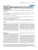
Fibrin deposition
Because fibrin deposition is a sensitive marker for tissue
damage in hyperoxia-induced neonatal lung disease, pul-
monary fibrin deposition was studied in homogenates as
a read-out for lung damage using Western blot analysis
(Figure 1A) and quantified after treatment with two differ-
ent sildenafil concentrations (50 and 100 mg/kg/day; Fig-
ure 1B). Fibrin deposition was at reference levels during
normal neonatal pulmonary development on day 10
(18.4 ± 1.8 ng fibrin/mg tissue) and increased more than
13-fold to 239 ± 34.8 ng fibrin/mg tissue in lungs of pups
exposed to 100% oxygen for 10 days (p < 0.001). Com-
pared to oxygen-exposed controls, sildenafil treatment
attenuated fibrin deposition in a concentration-depend-
ent way by 62.5% to 89.8 ±10.3 ng fibrin/mg tissue for
100 mg/kg/day sildenafil (p < 0.05). Because 100 mg/kg/
day of sildenafil was the most effective dose, additional
experiments were limited to this dosage.
Cyclic GMP
To establish that sildenafil is a specific cyclic GMP
dependent PDE inhibitor cyclic GMP levels were deter-
mined in lung tissue homogenates (Figure 1C). Exposure
to hyperoxia for 10 days did not change cyclic GMP levels
in lung homogenates compared to room air controls.
Western blot analysis of fibrin deposition in lung homogenates of rat pups exposed to room air (RA), oxygen (O
2
) and O
2
in combination with 100 mg/kg/day of sildenafil (Sil
100
) for 10 days (panel A)Figure 1
Western blot analysis of fibrin deposition in lung homogenates of rat pups exposed to room air (RA), oxygen
(O
2
) and O
2
in combination with 100 mg/kg/day of sildenafil (Sil
100
) for 10 days (panel A). Panel B shows quantifica-
tion of fibrin deposition in lung homogenates on day 10. Experimental groups include room air-exposed controls (RA, white
bar), age-matched O
2
-exposed controls (O
2
, black bar) and sildenafil-treated rat pups (50 mg/kg/day: Sil
50
, striped bar; 100 mg/
kg/day: Sil
100
, gray bar) under hyperoxia. Quantification of cyclic GMP in lung homogenates (panel C) in room air-exposed lit-
termates (white bars), O
2
-exposed control pups (black bars) and 100 mg/kg/day sildenafil-treated pups (Sil
100
, gray bars). Data
are expressed as mean ± SEM of at least 6 pups per experimental group. *p < 0.05 and ***p < 0.001 versus age-matched O
2
-
exposed controls.
Δ
p < 0.05 versus room air-exposed controls.
Respiratory Research 2009, 10:30 />Page 6 of 16
(page number not for citation purposes)
Treatment with sildenafil resulted in a significant increase
in cyclic GMP by 102% (p < 0.05) compared to oxygen-
exposed controls.
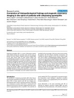
Growth and survival
At birth, on postnatal day 1, mean body weight of the rat
pups was 5.0 ± 0.18 g (Figure 2A). Body weight increased
to approximately 8 grams on day 5 in oxygen exposed
pups and room air controls. Hereafter, room air control
pups grew slightly faster than oxygen-exposed pups.
Growth of pups treated with 100 mg/kg/day sildenafil was
not different from oxygen-exposed controls. Median sur-
vival of oxygen-exposed controls was 12 days and was
prolonged with 4 days in pups treated with 100 mg/kg/
day sildenafil and hyperoxia (Figure 2B; p < 0.001). After
13 days of oxygen exposure, 92% of the controls and only
25% of the sildenafil-treated pups had died. Room air-
exposed pups did not show signs of illness or mortality
during the first 4 weeks after birth.
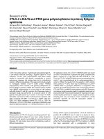
Lung histology
Lung development proceeds from the saccular stage at
birth towards the alveolar stage on day 10 (Figure 3A).
Oxygen exposure for 10 days resulted in edema, a reduc-
tion in pulmonary vessel density (Figure 3, panels B and
D), a heterogeneous distribution of enlarged air-spaces
with increased mean linear intercept (Figure 3E), which
were surrounded by septa with increased thickness (Figure
3F) and an increase in pulmonary arteriolar medial wall
thickness (Figure 3, panels H and J). Sildenafil treatment
improved alveolarization and angiogenesis during hyper-
oxia exposure by increasing pulmonary vessel density
(47.9%, p < 0.01; Figure 3, panels C and D), decreasing
mean linear intercept (12.5%, p < 0.001; Figure 3E), thin-
ning of alveolar septa (34.2%, p < 0.01; Figure 3F) and
reducing arteriolar medial wall thickness (38.8%, p <
0.001; Figure 3, panels I and J) compared to oxygen expo-
sure for 10 days.
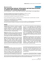
Hyperoxia led to a massive inflammatory reaction, charac-
terized by an overwhelming influx of inflammatory cells,
including macrophages (Figure 4B) and neutrophils (Fig-
ure 4E), compared to room air-exposed controls (Figure 4,
panels A and D). Resident ED-1-positive monocytes and
macrophages were present at 548 cells per mm
2
in septa
and alveoli of control lungs, whereas lungs of oxygen-
exposed pups contained 2.9 times as many (p < 0.001; Fig-
ure 4G). Sildenafil treatment reduced the influx of ED-1-
positive cells by 38.7% (p < 0.001; Figure 4, panels C and
G) compared to oxygen-exposed controls. Resident MPO-
positive neutrophils were present at 68 cells per mm
2
in
septa and alveoli of control lungs, whereas lungs of oxy-
gen-exposed pups contained 7.3 times as many (p < 0.001;
Figure 4H). Sildenafil treatment reduced the influx of
MPO-positive cells by 67.3% (p < 0.001; Figure 4, panels
F and H) compared to oxygen-exposed controls.
Growth in sildenafil-treated rat pups (100 mg/kg/day, black circle), age-matched O
2
-exposed controls (open triangle) and room air exposed controls (open square) during the first 16 days after birthFigure 2
Growth in sildenafil-treated rat pups (100 mg/kg/day, black circle), age-matched O
2
-exposed controls (open
triangle) and room air exposed controls (open square) during the first 16 days after birth. Data are expressed
as mean ± SEM (panel A). Kaplan-Meier survival curve of sildenafil-treated rat pups (black circle), age-matched O
2
-exposed
controls (open triangle) and room air exposed controls (open square) during the first 19 days after birth (panel B). Data are
expressed as percentage ± SEM of pups surviving at the observed time point. At least 12 pups per experimental group were
studied. ***p < 0.001 for sildenafil-treated pups versus age-matched O
2
-exposed controls.
Survival
0 2 4 6 8 10 12 14 16 18 20
0
20
40
60
80
100
***
Post-natal days
Survival (%)
Growth
1 3 5 7 9 11 13 15 17
0
4
8
12
16
20
24
28
32
Post-natal days
Body weight (gram)
AB
Respiratory Research 2009, 10:30 />Page 7 of 16
(page number not for citation purposes)
Protein in bronchoalveolar lavage fluid
Total protein concentration in bronchoalveolar lavage
fluid (BALF) was measured to establish the inhibitory
effect of sildenafil on pulmonary edema by capillary-alve-
olar leakage (Figure 4I). Protein concentration on postna-
tal day 10 increased 9.4-fold after hyperoxia and had
decreased by 52.5% after treatment with sildenafil (p <
0.05; hyperoxia versus sildenafil).
mRNA expression in lung tissue
Ten days of oxygen exposure resulted in an increase in
mRNA expression of the pro-inflammatory cytokine IL-6
(133-fold; p < 0.001, Figure 5A), the procoagulant factor
Paraffin lung sections stained with polyclonal anti-vWF antibody (panels A-C) to visualize the endothelium of pulmonary vessels for the quantification of pulmonary vessel density (panel D) of room-air (RA, panel A) and O
2
-exposed controls (panel B), and age-matched pups treated with sildenafil (100 mg/kg/day) under hyperoxia (panel C) at 10 days of ageFigure 3
Paraffin lung sections stained with polyclonal anti-vWF antibody (panels A-C) to visualize the endothelium of
pulmonary vessels for the quantification of pulmonary vessel density (panel D) of room-air (RA, panel A) and
O
2
-exposed controls (panel B), and age-matched pups treated with sildenafil (100 mg/kg/day) under hyperoxia
(panel C) at 10 days of age. Pictures were taken at a 200× magnification. Arrows in panels A-C indicate vWF-positive blood
vessels. Quantification of pulmonary vessel density (panel D), mean linear intercept (panel E), alveolar septum thickness (panel
F) and medial wall thickness (panel J) in room air-exposed littermates (white bars), O
2
-exposed control pups (black bars) and
100 mg/kg/day sildenafil-treated pups (Sil
100
, gray bars). Paraffin lung sections stained with monoclonal anti-ASMA antibody for
the visualization of medial wall thickness in pulmonary arterioles (panels G-I) of room-air (RA, panel G) and O
2
-exposed con-
trols (panel H), and age-matched pups treated with sildenafil (100 mg/kg/day) under hyperoxia (panel I) at 10 days of age. Pic-
tures were taken at a 1000× magnification. The enlargements shown in the lower right parts of panels A, B and C are indicated
in the boxed areas. Values are expressed as mean ± SEM in at least 6 different rat pups per group. a = alveolus **p < 0.01 and
***p < 0.001 versus age-matched O
2
-exposed controls.
ΔΔΔ
p < 0.001 versus room air-exposed controls.
Respiratory Research 2009, 10:30 />Page 8 of 16
(page number not for citation purposes)
tissue factor (TF, 3.0-fold; p < 0.001, Figure 5B), the fibri-
nolytic factor plasminogen activator inhibitor-1 (PAI-1,
50-fold; p < 0.001, Figure 5C) and the growth factor
amphiregulin (5.2-fold; p < 0.001, Figure 5D), and a
decrease in the expression of vascular endothelial growth
factor receptor-2 (VEGFR2, 3.5-fold; p < 0.001, Figure 5E)
and fibroblast growth factor receptor-4 (FGFR4, 9.0-fold;
p < 0.001, Figure 5F) in lungs of oxygen-exposed com-
pared to room air-exposed pups. Treatment with 100 mg/
kg/day sildenafil resulted in a reduction in PAI-1 (by
26.8%; p < 0.05, Figure 5C) and amphiregulin (by 33.3%;
p < 0.05, Figure 5D) mRNA expression, whereas sildenafil
treatment showed only a tendency towards lower IL-6 and
TF mRNA expression compared to oxygen-exposed con-
Paraffin lung sections stained with monoclonal anti-ED-1 antibody (panels A-C) or polyclonal anti-MPO antibody (panels D-F) of room-air (RA, panels A and D) and O
2
-exposed controls (panels B and E), and age-matched pups treated with sildenafil (100 mg/kg/day) under hyperoxia (panels C and F) at 10 days of ageFigure 4
Paraffin lung sections stained with monoclonal anti-ED-1 antibody (panels A-C) or polyclonal anti-MPO anti-
body (panels D-F) of room-air (RA, panels A and D) and O
2
-exposed controls (panels B and E), and age-
matched pups treated with sildenafil (100 mg/kg/day) under hyperoxia (panels C and F) at 10 days of age. Pic-
tures were taken at a 200× magnification. Quantification of ED-1-positive monocytes and macrophages (panel G), MPO-posi-
tive neutrophilic granulocytes (panel H) and total protein concentration in bronchoalveolar lavage fluid (BALF; panel I) in room
air-exposed littermates (white bars), O
2
-exposed control pups (black bars) and 100 mg/kg/day sildenafil-treated O
2
-exposed
pups (Sil
100
, gray bars) for 10 days. Values are expressed as mean ± SEM in at least 6 different rat pups per group. Note the
presence of large numbers of leukocytes, including macrophages and neutrophils in thickened septa and in the enlarged alveolar
lumen in panels B and E in hyperoxia-exposed controls, and low numbers of pulmonary inflammatory cells after sildenafil treat-
ment (panels C and F). a = alveolus. *p < 0.05 and ***p < 0.001 versus age-matched O
2
-exposed controls.
Δ
p < 0.05 and versus
room air-exposed controls.
BALF
RA O
2
Sil
100
0
150
300
450
600
750
900
*
***
Protein (mg/ml)
ED-1
RA O
2
Sil
100
0
300
600
900
1200
1500
1800
***
***
'
Number of cells per mm
2
MPO
RA O
2
Sil
100
0
100
200
300
400
500
600
***
***
Number of cells per mm
2
HGI
a
a
a
ABC
a
a
a
D
EF
100 μm 100 μm 100 μm
100 μm 100 μm 100 μm
Respiratory Research 2009, 10:30 />Page 9 of 16
(page number not for citation purposes)
trols. In lung tissue of sildenafil-treated rat pups expres-
sion of VEGFR2 and FGFR4 mRNA was increased by
37.5% (p < 0.001) and by 32.6% (p < 0.05), respectively,
compared to oxygen-exposed pups (Figure 5, panels E and
F).
Right ventricular hypertrophy
Exposure to hyperoxia for 10 days resulted in RVH as
demonstrated by a 1.4-fold increase in the weight ratio
RV/(LV + IVS) compared to room air controls (p < 0.001;
Table 2; Figure 6A). Treatment with sildenafil resulted in
a significant regression of RVH (Figure 6A) and a decrease
of the RV wall thickness by 26.8% compared to the oxy-
gen-exposed controls (p < 0.05, Figure 6B). Extracellular
expression of tenascin-C, a marker of myocardial over-
load, was visible in the RV only after exposure to hyper-
oxia. Tenascin-C expression was absent in room air
exposed controls, as well as after treatment with sildenafil
in experimental BPD (Figure 6, panels C-E).
Relative mRNA expression, determined with RT-PCR, of genes related to inflammation; interleukin-6 (IL-6; panel A), coagula-tion; tissue factor (TF; panel B), fibrinolysis; plasminogen activator inhibitor-1 (PAI-1; panel C) and alveolar growth; amphiregu-lin (panel D), vascular endothelial growth factor receptor-2 (VEGFR2; panel E) and fibroblast growth factor receptor-4 (FGFR4; panel F) in room air-exposed controls (RA, white bars), age-matched O
2
-exposed controls (O
2
, black bars) and sildenafil-treated rat pups (100 mg/kg/day [Sil
100
], gray bars) on day 10Figure 5
Relative mRNA expression, determined with RT-PCR, of genes related to inflammation; interleukin-6 (IL-6;
panel A), coagulation; tissue factor (TF; panel B), fibrinolysis; plasminogen activator inhibitor-1 (PAI-1; panel
C) and alveolar growth; amphiregulin (panel D), vascular endothelial growth factor receptor-2 (VEGFR2;
panel E) and fibroblast growth factor receptor-4 (FGFR4; panel F) in room air-exposed controls (RA, white
bars), age-matched O
2
-exposed controls (O
2
, black bars) and sildenafil-treated rat pups (100 mg/kg/day
[Sil
100
], gray bars) on day 10. Data are expressed as mean ± SEM of 10 rat pups. *p < 0.05 and ***p < 0.001 versus age-
matched O
2
-exposed controls.
ΔΔΔ
p < 0.001 versus room air-exposed controls.
FGFR4
RA O
2
Sil
100
0.0
0.2
0.4
0.6
0.8
1.0
1.2
***
'''
*
relat ive expr ession
VE GF R2
RA O
2
Sil
100
0.0
0.2
0.4
0.6
0.8
1.0
1.2
'''
***
***
relat ive expr ession
Amphiregulin
RA O
2
Sil
100
0
1
2
3
4
5
6
***
'''
*
relat ive expr ession
PAI-1
RA O
2
Sil
100
0
10
20
30
40
50
60
***
'''
*
relat ive expression
IL6
RA O
2
Sil
100
0
30
60
90
120
150
180
***
'''
relat ive expr ession
TF
RA O
2
Sil
100
0.0
0.5
1.0
1.5
2.0
2.5
3.0
3.5
***
'''
relat ive expr ession
AB
C
D
E
F
Table 2: Cardiac characteristics
RA O
2
Sil
100
RV free wall thickness (μm) 240 ± 6 310 ± 34 197 ± 11*
LV free wall thickness (μm) 575 ± 13 568 ± 39 515 ± 34
IVS thickness (μm) 563 ± 67 568 ± 102 454 ± 62
RV/(LV+IVS) 0.302 ± 0.02*** 0.412 ± 0.02 0.343 ± 0.01*
*** p < 0.001 and * p < 0.05 versus age-matched O
2
exposed controls.
Respiratory Research 2009, 10:30 />Page 10 of 16
(page number not for citation purposes)
mRNA expression in the heart
Increased right ventricular mRNA expression was
observed for the natriuretic peptides ANP (2.5-fold; p <
0.01, Figure 7A) and BNP (3.3-fold; p < 0.001, Figure 7B),
whereas expression was decreased for CNP (5.5-fold; p <
0.001, Figure 7C) and for the natriuretic peptide receptors
(NPR) -A (1.7-fold; p < 0.001, Figure 7D) and NPR-B (2.1-
fold; p < 0.001, Figure 7E) after exposure to hyperoxia for
10 days compared to room air controls. Treatment with
sildenafil decreased the expression of BNP (by 36.3%; p <
0.01) and increased the expression of CNP (by 267%; p <
0.001), NPR-A (by 24.7%; p < 0.05), NPR-B (by 35.7%; p
< 0.05) and NPR-C (by 39.2%; p < 0.05, Figure 7F) com-
pared to oxygen-exposed controls.
Neonatal lung injury-recovery model
Lung histology
Continuous neonatal exposure to hyperoxia for 9 days
resulted in a 2.5-fold reduction in blood vessel density (p
< 0.001; Figure 8 panels B and G) and enlarged alveoli
(Figure 8B), demonstrated by an increased MLI (p < 0.001,
Figure 8H) and a 3.1-fold increase in medial wall thick-
ness (p < 0.001; Figure 9, panels B and G) compared to
room air controls. Sildenafil treatment during the last 3
days of the injurous hyperoxic period decreased medial
wall thickness by 27.4% (p < 0.05 vs O
2
; Figure 9, panels
C and G), but did not affect alveolar enlargement and
blood vessel density (Figure 8, panels C, G and H). A
recovery period of 9 days in room air after hyperoxia-
induced lung injury (Figure 8E) reduced MLI (Figure 8H)
and increased blood vessel density (Figure 8G), but alve-
oli continued to be enlarged (Figure 8E). Treatment with
Right ventricular hypertrophy is depicted as the increase in the ratio RV/(LV+IVS) compared to the room air control (panel A) and ventricular wall thickness, indicated as the RV/LV ratio (panel B) in room air-exposed controls (RA, white bars), age-matched O
2
-exposed controls (O
2
, black bars) and sildenafil-treated rat pups (100 mg/kg/day [Sil
100
], gray bars) under hyper-oxia on day 10Figure 6
Right ventricular hypertrophy is depicted as the increase in the ratio RV/(LV+IVS) compared to the room air
control (panel A) and ventricular wall thickness, indicated as the RV/LV ratio (panel B) in room air-exposed
controls (RA, white bars), age-matched O
2
-exposed controls (O
2
, black bars) and sildenafil-treated rat pups
(100 mg/kg/day [Sil
100
], gray bars) under hyperoxia on day 10. Cardiac characteristics are presented in table 2. Paraffin
sections of the right ventricular wall stained with polyclonal tenascin C (panels C-E) of room-air (RA, panel C) and O
2
-exposed
controls (panel D), and age-matched pups treated with sildenafil (100 mg/kg/day) under hyperoxia (panel E) at 10 days of age.
Note the extravascular expression of tenascin C in the right ventricle in oxygen-exposed pups (panel D) and the absence of
staining after treatment with sildenafil (panel E) and in room air controls (panel C). Pictures were taken at a 400× magnifica-
tion.
RV/LV wall thickness ratio
RA O
2
Sil
100
0.0
0.1
0.2
0.3
0.4
0.5
0.6
*
ratio RV/LV
Right ventricular hypertrophy
RA O
2
Sil
100
0
10
20
30
40
50
***
*
increase vs. RA contr ols (% )
A
B
CD
E
50 μm 50 μm 50 μm
Respiratory Research 2009, 10:30 />Page 11 of 16
(page number not for citation purposes)
sildenafil restored blood vessel density (p < 0.05 vs O
2
;
Figure 8, panels F and G) and reduced MLI by 11.8% (p <
0.001 vs O
2
, Figure 8H) compared to non-treated experi-
mental BPD pups. However, medial wall thickness was
only reduced in sildenafil-treated pups by 47% (p < 0.001;
Figure 9, panels D-G) after a 9-day recovery period in
room air.
Nine days of hyperoxic lung injury resulted in a 1.4-fold
increase in the ratio RV/LV wall thickness, which was sig-
nificantly reduced after sildenafil treatment for 3 days
(42.2%; p < 0.001, Figure 9N). A recovery period of 9 days
did not reduce RVH in the non-treated experimental BPD
pups, but the RV/LV wall thickness ratio was completely
restored after sildenafil treatment.
Discussion
Prophylactic sildenafil therapy prolonged survival,
improved lung histopathology, reduced RVH, and
increased lung cGMP levels in neonatal rat pups exposed
to continuous and prolonged hyperoxia, a suitable in vivo
model for experimental BPD [8], by inhibiting inflamma-
tion, reducing capillary-alveolar protein leakage, alveolar
septum thickness, and alveolar enlargement and by atten-
uating alveolar fibrin deposition in neonatal rat pups
exposed to prolonged hyperoxia. Inhibition of lung
inflammation was demonstrated by a reduction in the
influx of inflammatory cells, including macrophages and
neutrophilic granulocytes. Sildenafil therapy started after
the initiation of hyperoxia-induced lung injury improved
alveolarization and angiogenesis by attenuating alveolar
enlargement and arteriolar medial wall thickness, and
restoring pulmonary bloodvessel density and RVH in a
lung injury-recovery model, demonstrating its therapeutic
potential for treatment of BPD in the neonatal intensive
care unit.
mRNA expression in the right ventricle, relative to the expression in the left ventricle and interventricular septum, determined with RT-PCR, of atrial natriuretic peptide (ANP; panel A), brain natriuretic peptide (BNP; panel B), c-type natriuretic peptide (CNP; panel C), natriuretic peptide receptor (NPR) -A (panel D), NPR-B (panel E) and NPR-C (panel F) in room air-exposed controls (RA, white bars), age-matched O
2
-exposed controls (O
2
, black bars) and sildenafil-treated rat pups (100 mg/kg/day [Sil
100
], gray bars) under hyperoxia on day 10Figure 7
mRNA expression in the right ventricle, relative to the expression in the left ventricle and interventricular
septum, determined with RT-PCR, of atrial natriuretic peptide (ANP; panel A), brain natriuretic peptide
(BNP; panel B), c-type natriuretic peptide (CNP; panel C), natriuretic peptide receptor (NPR) -A (panel D),
NPR-B (panel E) and NPR-C (panel F) in room air-exposed controls (RA, white bars), age-matched O
2
-
exposed controls (O
2
, black bars) and sildenafil-treated rat pups (100 mg/kg/day [Sil
100
], gray bars) under
hyperoxia on day 10. Data are expressed as mean ± SEM of 10 rat pups. *p < 0.05, **p < 0.01 and ***p < 0.001 versus age-
matched O
2
-exposed controls.
Δ
p < 0.05,
ΔΔ
p < 0.01 and
ΔΔΔ
p < 0.001 versus room air-exposed controls.
NPR-C
RA O
2
Sil
100
0.0
0.2
0.4
0.6
0.8
1.0
1.2
1.4
*
relat ive expr ession
NPR-B
RA O
2
Sil
100
0.0
0.2
0.4
0.6
0.8
1.0
1.2
1.4
***
*
''
relat ive expr ession
NPR-A
RA O
2
Sil
100
0.0
0.2
0.4
0.6
0.8
1.0
1.2
***
*
relat ive expr ession
CNP
RA O
2
Sil
100
0.0
0.2
0.4
0.6
0.8
1.0
1.2
1.4
***
***
relative expr ession
BNP
RA O
2
Sil
100
0.0
0.5
1.0
1.5
2.0
2.5
3.0
3.5
4.0
***
'''
**
relative expr ession
AB
C
D
E
F
ANP
RA O
2
Sil
100
0.0
0.5
1.0
1.5
2.0
2.5
3.0
3.5
**
'
relative expr ession
Respiratory Research 2009, 10:30 />Page 12 of 16
(page number not for citation purposes)
Paraffin lung sections stained with polyclonal anti-vWF antibody (panels A-F) after hyperoxic injury for 9 days (panels A-C) and subsequent recovery in room air for 9 days (panels D-F) of room-air (RA, panel A), O
2
-exposed (panel B) and age-matched pups treated with sildenafil (100 mg/kg/day) under hyperoxia (panel C), and of RA (panel D), O
2
-exposed (panel E) and age-matched O
2
-exposed pups treated with sildenafil (100 mg/kg/day, panel F) after recoveryFigure 8
Paraffin lung sections stained with polyclonal anti-vWF antibody (panels A-F) after hyperoxic injury for 9 days
(panels A-C) and subsequent recovery in room air for 9 days (panels D-F) of room-air (RA, panel A), O
2
-
exposed (panel B) and age-matched pups treated with sildenafil (100 mg/kg/day) under hyperoxia (panel C),
and of RA (panel D), O
2
-exposed (panel E) and age-matched O
2
-exposed pups treated with sildenafil (100 mg/
kg/day, panel F) after recovery. Pictures were taken at a 200× magnification. Quantification of pulmonary vessel density
(panel G) and mean linear intercept (panel H) after hyperoxic lung injury for 9 days (Hyp in panels G and H) and after recovery
in room air for 9 days (Hyp + Rec in panels G and H) in room air-exposed (white bars), O
2
-exposed (black bars) and O
2
-
exposed pups treated with 100 mg/kg/day sildenafil (Sil
100
, gray bars). The enlargements shown in the lower left parts of panels
A-F are indicated in the boxed areas. *p < 0.05 and ***p < 0.001 versus age-matched O
2
-exposed controls.
ΔΔΔ
p < 0.001 versus
room air-exposed controls.
δδδ
p < 0.001 versus own treatment controls in hyperoxia period (hyp).
MLI
RA O
2
Sil
100
RA O
2
Sil
100
0
20
40
60
80
Hyp Hyp + Rec
***
'''
***
***
'''
GGG
GGG
GGG
m
Pulmonary vessel density
RA O
2
Sil
100
RA O
2
Sil
100
0
5
10
15
***
***
'''
*
GGG
GGG
Hyp Hyp + Rec
number of vessels/field
G
H
E
A
BC
DF
100 μm 100 μm 100 μm
100 μm 100 μm 100 μm
Hyp
Hyp + Rec
Respiratory Research 2009, 10:30 />Page 13 of 16
(page number not for citation purposes)
In vitro studies of lipopolysaccharide (LPS) mediated
cytokine production in alveolar epithelial cells and in vivo
studies on the influx of macrophages and neutrophils in a
rat model of airway hyperreactivity have demonstrated
the anti-inflammatory properties of PDE5 inhibition on
pulmonary inflammatory processes [15,24]. Increased
neo-vascularization in chicken chorioallantoic mem-
branes has shown that sildenafil stimulation angiogenesis
[25]. The improvement of alveolarization after sildenafil
treatment in our study confirms, in part, the findings of
Ladha et al, who investigated the effects of prophylactic
sildenafil treatment in a similar rat model using quantita-
tive histopathological techniques [14]. Lung injury in
hyperoxia-exposed pups in this study was more severe as
we used a different rat strain (Wistar instead of Sprague-
Dawley rats, which are more resistant against hyperoxic
Paraffin lung sections stained with monoclonal anti-ASMA antibody (panels A-F) and paraffin heart sections stained with HE (panels H-M) after hyperoxic injury for 9 days (panels A-C and H-J) and subsequent recovery in room air for 9 days (panels D-F and K-M) of room-air (RA, panels A and H), O
2
-exposed (panels B and I) and age-matched pups treated with sildenafil (100 mg/kg/day) under hyperoxia (panels C and J), and of RA (panels D and K), O
2
-exposed (panels E and L) and age-matched O
2
-exposed pups treated with sildenafil (100 mg/kg/day, panels F and M) after recoveryFigure 9
Paraffin lung sections stained with monoclonal anti-ASMA antibody (panels A-F) and paraffin heart sections
stained with HE (panels H-M) after hyperoxic injury for 9 days (panels A-C and H-J) and subsequent recovery
in room air for 9 days (panels D-F and K-M) of room-air (RA, panels A and H), O
2
-exposed (panels B and I) and
age-matched pups treated with sildenafil (100 mg/kg/day) under hyperoxia (panels C and J), and of RA (panels
D and K), O
2
-exposed (panels E and L) and age-matched O
2
-exposed pups treated with sildenafil (100 mg/kg/
day, panels F and M) after recovery. Pictures were taken at a 1000× magnification (panels A-F) or at a 40× magnification
(panels H-M). Quantification of pulmonary arteriolar medial wall thickness (panel G) and right ventricular hypertrophy (RV/LV
wall thickness ratio, panel N) after hyperoxic lung injury for 9 days (Hyp in panels G and N) and after recovery in room air for
9 days (Hyp + Rec in panels G and N) in room air-exposed (white bars), O
2
-exposed (black bars) and O
2
-exposed pups treated
with 100 mg/kg/day sildenafil (Sil
100
, gray bars). LV = left ventricle and RV = right ventricle. *p < 0.05, **p < 0.01 and ***p <
0.001 versus age-matched O
2
-exposed controls.
ΔΔ
p < 0.01 and
ΔΔΔ
p < 0.001 versus room air-exposed controls.
RV/LV wall thickness ratio
RA O
2
Sil
100
RA O
2
Sil
100
0.0
0.1
0.2
0.3
0.4
0.5
0.6
0.7
**
*
***
Hyp Hyp + Rec
**
ratio RV/LV
Medial wall thickness
RA O
2
Sil
100
RA O
2
Sil
100
0.0
0.3
0.6
0.9
1.2
1.5
1.8
***
***
'''
***
Hyp Hyp + Rec
*
''
m
GA
B
C
D
E
F
10 μm 10 μm 10 μm
10 μm
10 μm 10 μm
Hyp
Hyp + Rec
Fi
N
I
HJ
K
L
M
RV
Hyp
Hyp + Rec
400 μm400 μm
400 μm400 μm
RV
RV
RV
RV
RV
LV
LV
LV
LV
LV
LV
Respiratory Research 2009, 10:30 />Page 14 of 16
(page number not for citation purposes)
lung injury), 100% instead of 95% oxygen and differences
in the onset of lung injury.
We have previously shown that the specific inhibition of
PDE4 with rolipram or piclamilast reduces alveolar fibrin
deposition, inflammation and vascular alveolar leakage,
and prolongs survival in rats with neonatal hyperoxic lung
injury [6]. PDE4 inhibitors can exert their protective effect
in inflammatory lung diseases by increasing intracellular
cAMP levels [26]. PDEs belong to an enzyme family with
11 different members, designated PDE1-11, which exert
their biological function by inactivating the intracellular
messengers cAMP and/or cGMP by hydrolysis [26-28].
The beneficial effects of PDE5 inhibition by sildenafil on
hyperoxia-induced lung injury may, at least in part, be due
to higher intracellular cGMP levels as demonstrated by
increased cGMP levels in lung homogenates (this study).
In contrast to previous studies in which hyperoxic lung
injury resulted in either increased [14,29] or decreased
cGMP levels [30] we did not observe differences in cGMP
levels in experimental BPD. This may be explained by dif-
ferences in tissue source: plasma [14] versus lung tissue
(this study) and the duration of the injurious hyperoxic
response [30].
We have recently demonstrated that inhaled NO therapy
improves lung pathology, reduces fibrin deposition and
pulmonary inflammation, and prolongs survival in an
animal model of BPD [7]. NO plays an important role in
regulating pulmonary vascular tone and alveolar capillary
development and in reducing inflammation in the devel-
oping lung [7,31,32]. Inhaled NO can exert its biological
effects via the S-nitrosylation or via the NO-cGMP path-
way [31,33,34]. The similarity of beneficial effects by
inhaled NO and sildenafil treatment in experimental BPD
suggests that the NO-cGMP pathway plays an important
role in the pathogenesis of experimental BPD. Sildenafil-
treated pups survived longer than pups treated with
inhaled NO, but the effects of sildenafil treatment on pul-
monary fibrin deposition and inflammation were less
pronounced than the effects of inhaled NO. Intervention
studies in hyperoxic lung injury with inhaled NO and
(selective) PDE inhibitors have demonstrated less inflam-
mation, but, incomplete restoration of lung development
resulting in persistent enlarged alveoli [6,7,14,33]. Alveo-
lar enlargement was accompanied by a downregulation of
FGFR-4 which was partially restored after treatment with
sildenafil. This confirms the observation that lungs of
FGFR-3(-/-)/FGFR-4(-/-) mice are normal at birth, but
have a complete block in alveogenesis and do not form
secondary septa, demonstrating their cooperative func-
tion to promote the formation of alveoli [35].
NO stimulates the formation of cGMP in the endothelium
and smooth muscle cells [14,36], whereas sildenafil pro-
tects cGMP from degradation by inhibiting PDE5 activity,
but both modalities result in increased intracellular cGMP
levels in these cells. Enhanced cGMP levels reduce pulmo-
nary vascular resistance by relaxation of vascular smooth
muscle cells and induce redistribution of pulmonary
blood flow to ventilated lung regions, thereby preventing
further lung injury [11,17,37]. Sildenafil and inhaled NO
have both been used in term newborns with severe per-
sistent pulmonary hypertension [16,17,37,38], a late
complication of BPD. Early use of inhaled NO may
improve the chances of survival without BPD in ventilated
preterm infants [39], but data on sildenafil use in this
group are not available. In addition, enhanced cGMP lev-
els in endothelial cells improves angiogenesis and alveo-
larization via the vascular endothelial growth factor
(VEGF)-NO-cGMP pathway [40,41]. Recombinant
human VEGF treatment enhances alveolarization and ves-
sel growth and improves lung structure in hyperoxia-
induced neonatal lung injury [42,43]. On the contrary,
VEGF blockade in newborn rats impairs alveolarization
and vessel growth [44]. In experimental BPD in newborn
rats alveolar enlargement and loss of lung capillaries are
associated with decreased expression of lung VEGF and
VEGF receptor-2 (VEGFR2) [44], whereas sildenafil
improves alveolarization and angiogenesis [14], and
reduces pulmonary fibrin deposition, inflammation and
vascular alveolar leakage, resulting in prolonged survival
in the present study. In lung injury-recovery models of
experimental BPD alveoli are still enlarged after recovery
in non-treated pups [42,44], but alveolarization and ang-
iogenesis are almost completely restored after treatment
with pro-angiogenic factors, such as VEGF [42,44] and
sildenafil (this study). These results strongly suggest that
sildenafil treatment of preterm infants may reverse the
arrest in lung development which is typical for those
developing BPD.
Sildenafil treatment improved hyperoxia-induced RVH in
experimental BPD (this study and [14]), reduced extracel-
lular tenascin-C expression in the RV, a marker that is
upregulated under myocardial stress conditions [45,46],
and reduced the thickness of the RV. The beneficial effect
of sildenafil on the heart can be explained either directly
or indirectly by a reduction of pulmonary hypertension
resulting in reduced RVH. This is supported by a sildena-
fil-induced reduction in pulmonary arteriolar wall thick-
ness (this study) and by similar beneficial effects of PDE5-
inhibitors in experimental models of lung disease, includ-
ing monocrotaline-induced pulmonary hypertension and
bleomycin-induced pulmonary fibrosis [47-49]. A direct
beneficial effect of sildenafil is supported by an induction
of PDE5 in the myocardium of the hypertrophied LV or
RV in patient material and in the RV after monocrotaline-
induced RVH in rats [50]. In addition, Nagendran et al.
have demonstrated that sildenafil treatment restored the
Respiratory Research 2009, 10:30 />Page 15 of 16
(page number not for citation purposes)
upregulated cGMP-PDE activity in RV of rats with
monocrotaline-induced pulmonary artery hypertension
and increased RV contractility of these rats.
The natriuretic peptides atrial natriuretic peptide (ANP)
and brain natriuretic peptide (BNP) are synthesized and
released in response to atrial pressure and ventricular
overload, respectively, and their plasma concentrations
are related to ventricular dysfunction and severity of car-
diac pathology [51,52]. Occupation of the natriuretic pep-
tide receptor (NPR) -A, activated by ANP, BNP and DNP,
and NPR-B, which is specific to CNP, induces cellular
responses via activation of particulate guanylate cyclase,
in contrast to soluble guanylate cyclase that is activated by
NO, thereby elevating the intracellular levels of cGMP
[53,54]. As markers for RVH we studied the differential
expression of ANP, BNP, CNP and the natriuretic peptide
receptors NPR-A, NPR-B and NPR-C at the mRNA level.
Hyperoxia-induced RVH resulted in reduced expression of
the guanylate cyclase-coupled natriuretic peptide recep-
tors NPR-A and NPR-B in cardiomyocytes. Signaling after
activation of these receptors by natriuretic peptides is
mediated by cGMP [54]. This suggests that the intracellu-
lar cGMP concentration in the hypertrophic RV cardiomy-
ocyte is not only lowered by increased PDE5 expression,
but may also be reduced due to decreased levels of NPR-A
and NPR-B, which can be restored, at least in part, by
sildenafil treatment.
Conclusion
The beneficial effects of sildenafil on alveolarization, lung
inflammation and extravascular fibrin deposition, right
ventricular hypertrophy and survival in neonatal rats with
hyperoxia-induced lung injury emphasise the potential of
phosphodiesterase 5 inhibitors as treatment for bron-
chopulmonary dysplasia in premature infants.
Abbreviations
ANP: atrial natriuretic peptide; ASMA: alpha smooth mus-
cle actin; BNP: brain natriuretic peptide; BALF: bronchoal-
veolar lavage fluid; BPD: bronchopulmonary dysplasia;
cAMP: cyclic adenosine monophosphate; cGMP: cyclic
guanosine monophosphate; CNP: c-type natriuretic pep-
tide; FGFR4: fibroblast growth factor receptor-4; IL: inter-
leukin; IVS: interventricular septum; LV: left ventricle;
MLI: mean linear intercept; MPO: myeloperoxidase; NO:
nitric oxide; NPR: natriuretic peptide receptor; O
2
: oxy-
gen; PAI-1: plasminogen activator inhibitor-1; PDE: phos-
phodiesterase; RA: room air; RT-PCR: reverse transcriptase
polymerase chain reaction; RV: right ventricular free wall;
TF: tissue factor; VEGFR2: vascular endothelial growth fac-
tor (VEGF) receptor-2; vWF: Von Willebrand Factor.
Competing interests
The authors declare that they have no competing interests.
Authors' contributions
YPV, EHL and HB carried out the experimental studies.
YPV drafted the manuscript. GTMW, FJW and AL designed
the experimental setup and provided intellectual input in
the manuscript preparation. GTMW supervised the work.
Acknowledgements
The authors gratefully acknowledge Professor J.C.M. Meijers and Professor
T. van der Poll for providing the 59D8 antibody and Dr. E. de Heer for pro-
viding the ED-1 antibody.
This study was supported by grant 1R01 HL092158 from the National Insti-
tutes of Health (F. J. Walther).
References
1. Jobe AH, Ikegami M: Mechanisms initiating lung injury in the
preterm. Early Hum Dev 1998, 53:81-94.
2. Goodman G, Perkin RM, Anas NG, Sperling DR, Hicks DA, Rowen M:
Pulmonary hypertension in infants with bronchopulmonary
dysplasia. J Pediatr 1988, 112:67-72.
3. Aranda JV, Grondin D, Sasyniuk BI: Pharmacologic considera-
tions in the therapy of neonatal apnea. Pediatr Clin North Am
1981, 28:113-133.
4. Harris MC, Baumgart S, Rooklin AR, Fox WW: Successful extuba-
tion of infants with respiratory distress syndrome using ami-
nophylline. J Pediatr 1983, 103:303-305.
5. ter Horst SA, Wagenaar GT, de Boer E, van Gastelen MA, Meijers JC,
Biemond BJ, et al.: Pentoxifylline reduces fibrin deposition and
prolongs survival in neonatal hyperoxic lung injury. J Appl
Physiol 2004, 97:2014-2019.
6. de Visser YP, Walther FJ, Laghmani EH, van Wijngaarden S, Nieuw-
land K, Wagenaar GT: Phosphodiesterase 4 inhibition attenu-
ates pulmonary inflammation in neonatal lung injury. Eur
Respir J 2007, 31:633-644.
7. ter Horst SA, Walther FJ, Poorthuis BJ, Hiemstra PS, Wagenaar GT:
Inhaled nitric oxide attenuates pulmonary inflammation and
fibrin deposition and prolongs survival in neonatal hyperoxic
lung injury. Am J Physiol Lung Cell Mol Physiol 2007, 293:L35-L44.
8. Wagenaar GT, ter Horst SA, van Gastelen MA, Leijser LM, Mauad T,
Velden PA van der, et al.: Gene expression profile and histopa-
thology of experimental bronchopulmonary dysplasia
induced by prolonged oxidative stress. Free Radic Biol Med 2004,
36:782-801.
9. Essayan DM: Cyclic nucleotide phosphodiesterases. J Allergy Clin
Immunol 2001, 108:671-680.
10. Torphy TJ: Phosphodiesterase isozymes: molecular targets
for novel antiasthma agents. Am J Respir Crit Care Med 1998,
157:351-370.
11. Hemnes AR, Champion HC: Sildenafil, a PDE5 inhibitor, in the
treatment of pulmonary hypertension. Expert Rev Cardiovasc
Ther 2006, 4:293-300.
12. Liu H, Liu ZY, Guan Q: Oral sildenafil prevents and reverses the
development of pulmonary hypertension in monocrotaline-
treated rats. Interact Cardiovasc Thorac Surg 2007, 6:608-613.
13. Hemnes AR, Zaiman A, Champion HC: PDE5A inhibition attenu-
ates bleomycin-induced pulmonary fibrosis and pulmonary
hypertension through inhibition of ROS Generation and
RhoA/Rho kinase Activation. Am J Physiol Lung Cell Mol Physiol
2007, 294(1):L24-33.
14. Ladha F, Bonnet S, Eaton F, Hashimoto K, Korbutt G, Thebaud B:
Sildenafil improves alveolar growth and pulmonary hyper-
tension in hyperoxia-induced lung injury. Am J Respir Crit Care
Med 2005, 172:750-756.
15. Toward TJ, Smith N, Broadley KJ: Effect of phosphodiesterase-5
inhibitor, sildenafil (Viagra), in animal models of airways dis-
ease. Am J Respir Crit Care Med 2004, 169:227-234.
16. Baquero H, Soliz A, Neira F, Venegas ME, Sola A: Oral sildenafil in
infants with persistent pulmonary hypertension of the new-
born: a pilot randomized blinded study. Pediatrics 2006,
117:1077-1083.
17. Juliana AE, Abbad FC: Severe persistent pulmonary hyperten-
sion of the newborn in a setting where limited resources
Respiratory Research 2009, 10:30 />Page 16 of 16
(page number not for citation purposes)
exclude the use of inhaled nitric oxide: successful treatment
with sildenafil. Eur J Pediatr 2005, 164:626-629.
18. Boolell M, Allen MJ, Ballard SA, Gepi-Attee S, Muirhead GJ, Naylor
AM, et al.: Sildenafil: an orally active type 5 cyclic GMP-specific
phosphodiesterase inhibitor for the treatment of penile
erectile dysfunction. Int J Impot Res 1996, 8:47-52.
19. Dijkstra CD, Dopp EA, Joling P, Kraal G: The heterogeneity of
mononuclear phagocytes in lymphoid organs: distinct mac-
rophage subpopulations in the rat recognized by monoclonal
antibodies ED1, ED2 and ED3. Immunology 1985, 54:589-599.
20. Liao L, Ning Q, Li Y, Wang W, Wang A, Wei W, et al.: CXCR2
blockade reduces radical formation in hyperoxia-exposed
newborn rat lung. Pediatr Res 2006, 60:299-303.
21. Hessel MHM, Steendijk P, den Adel B, Schutte CI, Laarse A van der:
Pressure Overload-Induced Right Ventricular Dilatation is
Associated with Re-Expression of Myocardial Tenascin-C
and Increased Plasma Levels of Tenascin-C. Circulation 2006,
114(II):133.
22. Hui KY, Haber E, Matsueda GR: Monoclonal antibodies to a syn-
thetic fibrin-like peptide bind to human fibrin but not fibrin-
ogen. Science 1983, 222:1129-1132.
23. Pfaffl MW: A new mathematical model for relative quantifica-
tion in real-time RT-PCR. Nucleic Acids Res 2001, 29(9):e45.
24. Haddad JJ, Land SC, Tarnow-Mordi WO, Zembala M, Kowalczyk D,
Lauterbach R: Immunopharmacological potential of selective
phosphodiesterase inhibition. I. Differential regulation of
lipopolysaccharide-mediated proinflammatory cytokine
(interleukin-6 and tumor necrosis factor-alpha) biosynthesis
in alveolar epithelial cells. J Pharmacol Exp Ther 2002,
300:559-566.
25. Pyriochou A, Zhou Z, Koika V, Petrou C, Cordopatis P, Sessa WC, et
al.: The phosphodiesterase 5 inhibitor sildenafil stimulates
angiogenesis through a protein kinase G/MAPK pathway. J
Cell Physiol 2007, 211:197-204.
26. Houslay MD, Schafer P, Zhang KY:
Keynote review: phosphodi-
esterase-4 as a therapeutic target. Drug Discov Today 2005,
10:1503-1519.
27. Conti M, Richter W, Mehats C, Livera G, Park JY, Jin C: Cyclic AMP-
specific PDE4 phosphodiesterases as critical components of
cyclic AMP signaling. J Biol Chem 2003, 278:5493-5496.
28. Lugnier C: Cyclic nucleotide phosphodiesterase (PDE) super-
family: a new target for the development of specific thera-
peutic agents. Pharmacol Ther 2006, 109:366-398.
29. Potter CF, Kuo NT, Farver CF, McMahon JT, Chang CH, Agani FH, et
al.: Effects of hyperoxia on nitric oxide synthase expression,
nitric oxide activity, and lung injury in rat pups. Pediatr Res
1999, 45:8-13.
30. Sopi RB, Haxhiu MA, Martin RJ, Dreshaj IA, Kamath S, Zaidi SI: Dis-
ruption of NO-cGMP signaling by neonatal hyperoxia
impairs relaxation of lung parenchyma. Am J Physiol Lung Cell
Mol Physiol 2007, 293:L1029-L1036.
31. Lin YJ, Markham NE, Balasubramaniam V, Tang JR, Maxey A, Kinsella
JP, et al.: Inhaled nitric oxide enhances distal lung growth after
exposure to hyperoxia in neonatal rats. Pediatr Res 2005,
58:22-29.
32. McCurnin DC, Pierce RA, Chang LY, Gibson LL, Osborne-Lawrence
S, Yoder BA, et al.: Inhaled NO improves early pulmonary func-
tion and modifies lung growth and elastin deposition in a
baboon model of neonatal chronic lung disease. Am J Physiol
Lung Cell Mol Physiol 2005, 288:L450-L459.
33. Auten RL, Mason SN, Whorton MH, Lampe WR, Foster WM, Gold-
berg RN, et al.: Inhaled Ethyl Nitrite Prevents Hyperoxia-
impaired Postnatal Alveolar Development in Newborn Rats.
Am J Respir Crit Care Med 2007, 176:291-299.
34. Gaston B, Singel D, Doctor A, Stamler JS: S-nitrosothiol signaling
in respiratory biology. Am J Respir Crit Care Med 2006,
173:1186-1193.
35. Weinstein M, Xu X, Ohyama K, Deng CX: FGFR-3 and FGFR-4
function cooperatively to direct alveogenesis in the murine
lung. Development 1998, 125:3615-3623.
36. Humbert M, Sitbon O, Simonneau G: Treatment of pulmonary
arterial hypertension. N Engl J Med 2004, 351:1425-1436.
37. Leibovitch L, Matok I, Paret G: Therapeutic applications of silde-
nafil citrate in the management of paediatric pulmonary
hypertension. Drugs 2007, 67:57-73.
38. Abman SH: Recent advances in the pathogenesis and treat-
ment of persistent pulmonary hypertension of the newborn.
Neonatology 2007, 91:283-290.
39. Barrington KJ, Finer NN: Inhaled nitric oxide for preterm
infants: a systematic review. Pediatrics 2007, 120:1088-1099.
40. Hoeben A, Landuyt B, Highley MS, Wildiers H, Van Oosterom AT, De
Bruijn EA: Vascular endothelial growth factor and angiogen-
esis. Pharmacol Rev 2004, 56:549-580.
41. Voelkel NF, Vandivier RW, Tuder RM: Vascular endothelial
growth factor in the lung. Am J Physiol Lung Cell Mol Physiol 2006,
290:L209-L221.
42. Kunig AM, Balasubramaniam V, Markham NE, Morgan D, Mont-
gomery G, Grover TR, et al.: Recombinant human VEGF treat-
ment enhances alveolarization after hyperoxic lung injury in
neonatal rats. Am J Physiol Lung Cell Mol Physiol 2005,
289:L529-L535.
43. Kunig AM, Balasubramaniam V, Markham NE, Seedorf G, Gien J,
Abman SH: Recombinant human VEGF treatment transiently
increases lung edema but enhances lung structure after neo-
natal hyperoxia. Am J Physiol Lung Cell Mol Physiol 2006,
291:L1068-L1078.
44. Thebaud B, Ladha F, Michelakis ED, Sawicka M, Thurston G, Eaton F,
et al.: Vascular endothelial growth factor gene therapy
increases survival, promotes lung angiogenesis, and prevents
alveolar damage in hyperoxia-induced lung injury: evidence
that angiogenesis participates in alveolarization. Circulation
2005, 112:2477-2486.
45. Boerma M, Wees CG van der, Vrieling H, Svensson JP, Wondergem
J, van der LA, et al.: Microarray analysis of gene expression pro-
files of cardiac myocytes and fibroblasts after mechanical
stress, ionising or ultraviolet radiation. BMC Genomics 2005,
6(1):6.
46. Yamamoto K, Dang QN, Kennedy SP, Osathanondh R, Kelly RA, Lee
RT: Induction of tenascin-C in cardiac myocytes by mechan-
ical deformation. Role of reactive oxygen species. J Biol Chem
1999, 274:21840-21846.
47. Hemnes AR, Zaiman A, Champion HC: PDE5A inhibition attenu-
ates bleomycin-induced pulmonary fibrosis and pulmonary
hypertension through inhibition of ROS generation and
RhoA/Rho kinase activation. Am J Physiol Lung Cell Mol Physiol
2008, 294:L24-L33.
48. Itoh T, Nagaya N, Fujii T, Iwase T, Nakanishi N, Hamada K, et al.: A
combination of oral sildenafil and beraprost ameliorates pul-
monary hypertension in rats. Am J Respir Crit Care Med 2004,
169:34-38.
49. Schermuly RT, Kreisselmeier KP, Ghofrani HA, Yilmaz H, Butrous G,
Ermert L, et al.: Chronic sildenafil treatment inhibits monocro-
taline-induced pulmonary hypertension in rats. Am J Respir Crit
Care Med 2004, 169:39-45.
50. Nagendran J, Archer SL, Soliman D, Gurtu V, Moudgil R, Haromy A,
et al.: Phosphodiesterase type 5 is highly expressed in the
hypertrophied human right ventricle, and acute inhibition of
phosphodiesterase type 5 improves contractility. Circulation
2007, 116:238-248.
51. Felker GM, Petersen JW, Mark DB: Natriuretic peptides in the
diagnosis and management of heart failure. CMAJ 2006,
175:611-617.
52. Yoshimura M, Yasue H, Okumura K, Ogawa H, Jougasaki M,
Mukoyama M, et al.: Different secretion patterns of atrial natri-
uretic peptide and brain natriuretic peptide in patients with
congestive heart failure. Circulation 1993, 87:464-469.
53. Potter LR, Abbey-Hosch S, Dickey DM:
Natriuretic peptides,
their receptors, and cyclic guanosine monophosphate-
dependent signaling functions. Endocr Rev 2006, 27:47-72.
54. Woodard GE, Rosado JA: Recent advances in natriuretic pep-
tide research. J Cell Mol Med 2007, 11:1263-1271.