Báo cáo y học: " Oxidative stress augments toll-like receptor 8 mediated neutrophilic responses in healthy subjects" pps
Bạn đang xem bản rút gọn của tài liệu. Xem và tải ngay bản đầy đủ của tài liệu tại đây (448.36 KB, 13 trang )
BioMed Central
Page 1 of 13
(page number not for citation purposes)
Respiratory Research
Open Access
Research
Oxidative stress augments toll-like receptor 8 mediated
neutrophilic responses in healthy subjects
Satoru Yanagisawa, Akira Koarai, Hisatoshi Sugiura, Tomohiro Ichikawa,
Masae Kanda, Rie Tanaka, Keiichiro Akamatsu, Tsunahiko Hirano,
Kazuto Matsunaga, Yoshiaki Minakata and Masakazu Ichinose*
Address: Third Department of Internal Medicine, Wakayama Medical University, School of Medicine, Wakayama, Japan
Email: Satoru Yanagisawa - ; Akira Koarai - ; Hisatoshi Sugiura - sugiura@wakayama-
med.ac.jp; Tomohiro Ichikawa - ; Masae Kanda - ; Rie Tanaka - ;
Keiichiro Akamatsu - ; Tsunahiko Hirano - ;
Kazuto Matsunaga - ; Yoshiaki Minakata - ;
Masakazu Ichinose* -
* Corresponding author
Abstract
Background: Excessive oxidative stress has been reported to be generated in inflamed tissues and
contribute to the pathogenesis of inflammatory lung diseases, exacerbations of which induced by
viral infections are associated with toll-like receptor (TLR) activation. Among these receptors,
TLR8 has been reported as a key receptor that recognizes single-strand RNA virus. However, it
remains unknown whether TLR8 signaling is potentiated by oxidative stress. The aim of this study
is to examine whether oxidative stress modulates TLR8 signaling in vitro.
Methods: Human peripheral blood neutrophils were obtained from healthy non-smokers and
stimulated with TLR 7/8 agonist imidazoquinoline resiquimod (R848) in the presence or absence of
hydrogen peroxide (H
2
O
2
). Neutrophilic responses including cytokine release, superoxide
production and chemotaxis were examined, and the signal transduction was also analyzed.
Results: Activation of TLR8, but not TLR7, augmented IL-8 release. The R848-augmented IL-8
release was significantly potentiated by pretreatment with H
2
O
2
(p < 0.01), and N-acetyl-L-cysteine
reversed this potentiation. The combination of H
2
O
2
and R848 significantly potentiated NF-kB
phosphorylation and IkBα degradation. The H
2
O
2
-potentiated IL-8 release was suppressed by MG-
132, a proteosome inhibitor, and by dexamethasone. The expressions of TLR8, myeloid
differentiation primary response gene 88 (MyD88), and tumor necrosis factor receptor-associated
factor 6 (TRAF6) were not affected by H
2
O
2
.
Conclusion: TLR8-mediated neutrophilic responses were markedly potentiated by oxidative
stress, and the potentiation was mediated by enhanced NF-kB activation. These results suggest that
oxidative stress might potentiate the neutrophilic inflammation during viral infection.
Published: 15 June 2009
Respiratory Research 2009, 10:50 doi:10.1186/1465-9921-10-50
Received: 10 January 2009
Accepted: 15 June 2009
This article is available from: />© 2009 Yanagisawa et al; licensee BioMed Central Ltd.
This is an Open Access article distributed under the terms of the Creative Commons Attribution License ( />),
which permits unrestricted use, distribution, and reproduction in any medium, provided the original work is properly cited.
Respiratory Research 2009, 10:50 />Page 2 of 13
(page number not for citation purposes)
Introduction
Reactive oxygen species (ROS) such as hydrogen peroxide
(H
2
O
2
) and superoxide anion are generated in inflamed
tissues and are reported to contribute to the pathogenesis
of inflammatory lung diseases including chronic obstruc-
tive pulmonary diseases (COPD) [1,2], bronchial asthma
[3,4], cystic fibrosis [5,6], and idiopathic pulmonary
fibrosis [7,8]. Large amounts of ROS derived from inflam-
matory cells cause pro-inflammatory cytokine produc-
tion. In fact, H
2
O
2
has been reported to augment cytokine
production in previous studies [9,10]. Among inflamma-
tory cells, neutrophils are a key player in the inflammatory
lung diseases. It is well-known that excessive infiltration
of neutrophils is observed in the airways during exacerba-
tions induced by viral infections [11-14].
Toll-like receptors (TLRs) are simple pattern recognition
receptor systems and are known to react with conserved
molecular patterns of pathogens [15]. The innate immu-
nity cells also act against viral infections through TLRs
including TLR3, TLR7 and TLR8. Human neutrophils pos-
sess all functional TLRs except TLR3 [16], and their ago-
nists enhance neutrophil functions such as cytokine
release, superoxide generation and phagocytosis [16].
TLR7 and TLR8, located in the endosome, act as anti-viral
receptors for recognizing single strand RNA (ssRNA) [17-
19], which is present at various phases of viral infection
from viral entry to replication. After TLR7 and TLR8 are
activated by ssRNA, their signals are transduced through
myeloid differentiation primary response gene 88 (MyD-
88) and tumor necrosis factor (TNF) receptor-associated
factor 6 (TRAF6) leading to enhanced nuclear factor-
kappa B (NF-kB) DNA binding activity [20]. Activation of
NF-kB leads to increased inflammatory gene products
such as interleukin-8 (IL-8) and GM-CSF causing neu-
trophilic inflammation during viral infection. Resiqui-
mod (R848), a potent synthetic agonist of TLR 7/8 has
been reported to simulate the effects of ssRNA viruses on
TLR 7/8, to prime human neutrophils [16,21], and then
increase the biosynthesis of lipid mediators through NF-
kB activation [22] suggesting that TLR7 and TLR8 activa-
tion might affect the neutrophilic responses.
Although excessive oxidative stress occurs in the airways
of inflammatory lung diseases during exacerbations, it
remains unclear whether oxidative stress potentiates the
neutrophilic responses against viral infection. Therefore,
by using human peripheral neutrophils from healthy
never-smoking subjects, the present study was designed to
clarify whether oxidative stress can potentiate the TLR8-
mediated neutrophilic responses, including cytokine pro-
duction, chemotaxis and superoxide generation. Further-
more, we also investigated what signal transductions are
associated with this potentiation of the neutrophilic
responses.
Materials and methods
Reagents
Commercially available reagents were obtained as fol-
lows: Mono-Poly Resolving Medium was from Dainippon
Pharmaceutical Co. Ltd. (Osaka, Japan); fetal calf serum
(FCS) and RPMI medium 1640 (RPMI 1640) were from
Invitrogen (Carlsbad, California, USA); R848 (resiqui-
mod: 4-amino-2-etoxymethyl-α,α-dimethyl-1H-imidazo
[4,5-c]quinolin-1-ethanol), bafilomycin and 12-o-tetra-
decanoylphorbol 13-acetate were from Alexis Biochemi-
cals (San Diego, California, USA); R837 (Imiquimod: 1-
isobutyl-1H-imidazo [4,5-c]quinolin-4-amine) was from
Biomol (Plymouth Meeting, Pennsylvania, USA); N-ace-
thyl-
L-cysteine, MG-132, dexamethasone and anti-β-actin
antibody were from Sigma (St. Louis, Missouri, USA);
anti-TLR8 rabbit polyclonal antibody was from Abgent
(San Diego, California, USA); Cellfix solution was from
Becton Dickinson (San Jose, California, USA); phyco-
erythrin (PE)- conjugated anti-TLR8 antibody solution
was from Imgenex (San Diego, California, USA); dihydro-
rhodamine-123 (DHR-123) was from Cayman Chemical
(Ann Arbor, Michigan, USA); human recombinant IL-8
was from Acris antibodies (Hiddenhausen, Germany);
anti-human MyD88 antibody, anti-human TRAF6, and
anti-human IkBα were from Santa Cruz (San Diego, Cali-
fornia, USA); peroxidase-conjugated secondary antibod-
ies were from Rockland Immunochemicals (Gilbertsville,
Pennsylvania, USA)
Isolation of peripheral blood neutrophils
Healthy subjects participated in the present study. They
were never-smokers and had had no infection for 4 weeks
preceding the study. Human peripheral blood neutrophils
were isolated from whole blood by a density gradient
technique using Mono-Poly Resolving Medium as previ-
ously reported [23]. Briefly, whole blood was collected by
vein puncture into tubes containing EDTA anticoagulant.
Then, each blood sample was gently mounted onto the
same volume of Mono-Poly Resolving Medium without
mixing. The samples were centrifuged at 400 × g for 20
min at room temperature. The blood was separated into
four layers from the top, plasma, lymphocytes/mononu-
clear cells, neutrophils, and red blood cells. The neu-
trophil layer was gently collected by a pasteur pipette
without aspirating the other layers and put into fresh 20
ml tubes. This procedure allowed us to obtain neutrophils
with over 95% purity and viability as determined by
trypan blue staining. After washing by phosphate-buff-
ered saline (PBS) solution and counting the cell numbers,
neutrophils were suspended in 10% FCS in RPMI 1640 at
a concentration of 1 × 10
6
cells/ml. The neutrophils were
isolated before each experiment and used immediately.
All replicate experiments in the current study were per-
formed by using neutrophils from different donors. This
study was approved by the local ethics committee of
Respiratory Research 2009, 10:50 />Page 3 of 13
(page number not for citation purposes)
Wakayama Medical University School of Medicine.
Informed written consent was obtained from all subjects.
Immunocytochemistory
100 μl of the neutrophil suspension containing 1 × 10
5
cells were centrifuged by a Cytospin 4 cytocentrifuge
(ThermoShandon, ThermoBioAnalysis, Tokyo, Japan) at
25 × g for 5 min. The preparation was fixed in 4% parafor-
maldehyde fixative solution for 30 min. Endogeneous
peroxidase activity was blocked by incubation in 0.3%
H
2
O
2
in PBS for 15 min at room temperature. After wash-
ing, the cells were incubated with anti-TLR8 rabbit poly-
clonal antibody (1:100 dilution) for 12 hrs at 4°C. Non-
specific binding to the antibody was prevented by pre-
incubation with 2% bovine serum albumin in PBS con-
taining 0.3% Triton-X for 30 min. The immunoreactions
were visualized by the indirect immunoperoxidase
method using Envision polymer reagent, which is goat
anti-rabbit IgG conjugated with peroxidase labeled dex-
tran (Dako Japan Ltd, Kyoto, Japan), for 1 hour at room
temperature. Diaminobenzidine reaction was performed,
followed by counterstaining with hematoxirin. The slides
were viewed with a microscope (BX-50, Olympus Corpo-
ration, Tokyo, Japan) and photographed with a digital
camera (c-5050, Olympus Corporation, Tokyo, Japan).
Flow cytometry analysis
The expression of TLR8 in neutrophils was assessed by a
FACS calibur flow cytometer (Becton Dickinson, San Jose,
CA) according to the manufacturer's instructions. Briefly,
200 μl of the neutrophil suspension containing 2 × 10
6
neutrophils were first permeabilized by 1 × permeabiliz-
ing solution (Becton Dickinson, San Jose, California,
USA) for 30 min on ice to stain not only cell surface TLR8
but also endosomal TLR8, and then incubated with 4 μl of
PE-conjugated anti-TLR8 antibody solution or its isotype-
control for 20 min at 4°C. After washing, the samples
were fixed by 500 μl of 1% paraformaldehyde for 10 min.
Binding of each antibody was detected using CellQuest
analysis software on a FACS Calibur (Becton Dickinson,
San Jose, California, USA). Specific binding of each anti-
body was expressed as relative fluorescence that was calcu-
lated by the ratio of the mean fluorescence intensity for
TLR8 to the mean fluorescence intensity for the isotype
control.
TLR stimulation
Isolated neutrophils were stimulated in 24-well tissue cul-
ture plates with various concentrations of R848, a ligand
for TLR 7/8, or R837, a ligand for TLR7, for 24 hr at 37°C
in a humidified atmosphere of 5% CO
2
. Cells were pre-
treated with various concentrations of H
2
O
2
for 30 min
prior to the stimulation with R848 [24]. To investigate the
effects of the inhibitors or a scavenger on the IL-8 release,
cells were further pretreated with each agent prior to the
treatment with H
2
O
2
as follows: bafilomycin, an inhibitor
of endosomal acidification, for 15 min; N-acethyl-
L-
cysteine was for 10 min; MG-132, a proteosome inhibitor,
for 60 min; and dexamethasone for 30 min. Media were
harvested at 24 hours after treatment with R848 for subse-
quent enzyme-linked immunosorbent assays (ELISA) to
measure various cytokine levels. Similarly, cells were har-
vested at the same time for flow-cytometry analysis, or
western blotting.
Measurement of cytokines
IL-8 expression was measured by sandwich ELISA (R&D
System Europe, Abingdon, UK) according to the manufac-
turer's instructions. The lower detection limit was 16 pg/
ml. The levels of IL-1β, IL-6, IL-10, IL-12 and TNF-α were
measured by a Human Inflammation Cytokine Beads
array kit (Becton Dickinson, San Jose, California, USA)
according to the manufacturer's instructions.
Measurement of superoxide generation
Neutrophils were pre-incubated with or without 50 μM
H
2
O
2
, and then stimulated with various concentrations of
R848 for 1 hr at 37°C. Cells were harvested, washed twice
and resuspended in 10% FCS in RPMI 1640 at a concen-
tration of 1 × 10
6
cells/ml. One ml cell suspensions were
cultured at 37°C with 3 μM DHR-123 for 5 min and then
with 12-o-tetradecanoylphorbol 13-acetate for 30 min at
37°C. The cells were cooled on ice, centrifuged, and resus-
pended in PBS. Stained cells were assessed by a flow-
cytometer (Becton Dickinson, San Jose, California, USA).
The amount of superoxide generation was evaluated by
the relative fluorescence intensity of DHR-123 compared
with that of the control group.
Chemotaxis assay
Neutrophils were pre-incubated with or without 50 μM
H
2
O
2
and then stimulated with various concentrations of
R848 for 1 hr. Cells were harvested, washed twice and
resuspended in 10% FCS in RPMI 1640 at a concentration
of 2 × 10
6
cells/ml. Chemotaxis assays were performed on
plastic chemotaxis chambers (pore size: 3 μm; Kurabou,
Osaka, Japan) according to the manufacturer's instruc-
tions. Briefly, 250 μl of RPMI 1640 containing IL-8 (0.3
ng/ml) were placed into the bottom wells and 100 μl of
the neutrophil suspension were added into the top wells.
The chambers were then incubated in a tissue-culture
incubator at 37°C for 1 hr. The numbers of neutrophils
that transmigrated to the bottom wells were counted
using a flow-cytometer (Becton Dickinson, San Jose, Cali-
fornia, USA). Results are shown as the ratio of the
migrated cell number of each group to that of the control
group.
Respiratory Research 2009, 10:50 />Page 4 of 13
(page number not for citation purposes)
Elastase assay
Elastase release from the neutrophils was measured by a
human PMN elastase ELISA kit (Bender Medsystems,
Vienna, Austria) according to the manufacturer's instruc-
tions.
Phosflow analysis of phosphorylated NF-kB p65
1 × 10
6
neutrophils were incubated with or without 50 μM
H
2
O
2
and stimulated with various concentrations of R848
for 1 hr. The phosphorylated NF-kB p65 levels were meas-
ured by the BD phosflow method (Becton Dickinson, San
Jose, CA) according to the manufacturer's instructions.
Western blotting
After stimulation, the neutrophils were centrifuged at 400
× g for 10 seconds and incubated on ice for 30 min with
cold Triton buffer (1% Triton X-100, 150 mM NaCl, 20
mM Tris-HCl, pH 7.4, 1 mM EDTA, 2 mM diisopro-
pylfluorophosphate, 5 μg/ml pepstatin A and 1 mM phe-
nylmethylsulfonylfluoride). Then, the cell lysates were
centrifuged at 12,000 × g for 10 min, collected and stored
at -80°C. Cell lysates were mixed with the same volume of
2 × SDS loading buffer and separated with 12.5% gradient
polyacrylamide gel (DRC Co. Ltd., Tokyo, Japan). After
electrophoresis, the proteins were transferred to a nitrocel-
lulose membrane and incubated with anti-human MyD88
antibody (1:200 dilution), anti-human TRAF6 (1:200
dilution), or anti-human IkBα (1:200 dilution) overnight.
To standardize the expression of each protein, the mem-
branes were stripped off and re-probed with anti-β-actin
antibody (1:10000 dilution). The membranes were then
incubated with the appropriate peroxidase-conjugated
secondary antibodies (1:2000 dilution). The bound anti-
bodies were visualized with an ECL-plus detection system
(Amersham, Backinghamshire, UK) and photographed by
an ECL minicamera (Amersham, Backinghamshire, UK).
Stastical analysis
Data are expressed as mean values ± SEM. Data were ana-
lyzed by one way analysis of variance (ANOVA) followed
by Bonferroni's test or Sheffe's test to adjust for multiple
comparisons. An unpaired two-tailed Student's t-test was
used for single comparisons. Probability values of less
than 0.05 were considered significant.
Results
Detection of toll-like receptor (TLR) 8 in human
polymorphonuclear cells (PMNs) and its reaction to R848
To determine whether human neutrophils express TLR8,
we first investigated the expression of TLR8 in neutrophils
by immunocytochemistry and flow-cytometry. As shown
in Figure 1A, TLR8 was detected by immunocytochemis-
try. To examine the cellular localization of TLR8, we per-
formed flow-cytometry analysis against TLR8. TLR8 was
stained with or without cell membrane permeabilization,
indicating that TLR8 exists not only in the cytosol such as
the endosome but also on the cell surface (Figure 1B).
We next investigated the effect of TLR7 ligand R837 or TLR
7/8 ligand R848 on the release of IL-8 from neutrophils.
R848 increased IL-8 release in a time-dependent manner
(Figure 1C). As shown in figure 1D, R848 dose-depend-
ently augmented the release of IL-8 at 24 hr, whereas R837
had no effect. To confirm whether this augmentation of
IL-8 release is mediated by TLR signaling, the cells were
pretreated with bafilomycin, an inhibitor of endosomal
acidification. Pretreatment with bafilomycin significantly
inhibited the R848-augmented IL-8 release in a dose-
dependent manner (Figure 1E). Dexamethasone also sig-
nificantly inhibited the R848-augmented IL-8 release (Fig-
ure 1F).
Effect of H
2
O
2
on R848-augmented cytokine release,
superoxide generation, elastase release, and chemotaxis in
human PMNs
To examine whether oxidative stress potentiates the R848-
augmented IL-8 release, we examined the effects of H
2
O
2
on the IL-8 release from neutrophils. Pretreatment with
H
2
O
2
significantly potentiated the R848-augmented IL-8
release in a dose-dependent manner (Figure 2A). Pre-
incubation with 50 μM H
2
O
2
shifted the dose-response
curve leftward (Log EC
50
2.757 vs. 1.775 μM, p < 0.01, Fig-
ure 2B). In addition, the maximal response by R848 was
also significantly potentiated compared with control (Fig-
ure 2B). This potentiation was abolished by an antioxi-
dant, N-acetyl-
L-cysteine, compared with the vehicle-
pretreatment group (Figure 2C). The effect of R848 on the
release of cytokines and the potentiation by H
2
O
2
were
also examined. As shown in Figure 2D–F, R848 signifi-
cantly augmented TNF-α, IL-6 and IL-1β release from neu-
trophils. H
2
O
2
potentiated the R848-augmented TNF-α
(Figure 2D) and IL-6 release (Figure 2E) as well as IL-8,
but H
2
O
2
caused no potentiation of the IL-1β release (Fig-
ure 2F). Furthermore, we investigated whether H
2
O
2
potentiated the R848-induced neutrophilic responses,
including superoxide generation, elastase release, and
chemotaxis. Neither H
2
O
2
nor R848 stimulated superox-
ide production on their own, but the combination of the
two did (Figure 3A), whereas H
2
O
2
did not cause any
potentiation of the elastase release and chemotactic capac-
ity (Figure 3B and 3C).
Effect of H
2
O
2
on the R848-mediated TLR8 signaling
To clarify the mechanisms of the potentiation of the
R848-induced neutrophilic responses by H
2
O
2
, we inves-
tigated whether H
2
O
2
modulates the NF-kB activation
induced by R848, which is a key signaling in TLR activa-
tion. Although R848 or H
2
O
2
enhanced the phosphoryla-
tion of NF-kB p65, the phosphorylation was significantly
augmented by the combination of R848 and H
2
O
2
(Figure
Respiratory Research 2009, 10:50 />Page 5 of 13
(page number not for citation purposes)
Figure 1 (see legend on next page)
(A)
Isotype Control
Anti-TLR8
(B)
Permeabilized
Unpermeabilized
Fluorescence Intensity
Fluorescence Intensity
IgG
Anti-TLR8
IgG
Anti-TLR8
Cell Count
Cell Count
(C)
0 0.1 0.3 1.0 3.0 10 30
R837
0
5000
10000
15000
R848( M)
**
**
**
(10 M)
IL-8 (pg/ml)
(D)
0 1.0 3.0 10 30 100
0
5000
10000
15000
20000
Control
10 M R848
Bafilomycin( M)
++
IL-8(pg/ml)
++
(E) (F)
0 10 9 8 7 6
0
5000
10000
15000
20000
25000
Control
10 M R848
+
+
+
Dexamethasone(-lo
g
,M)
IL-8(pg/ml)
0 4 8 12 24
0
5000
10000
15000
20000
*
**
**
Time(hr)
IL-8 (pg/ml)
Respiratory Research 2009, 10:50 />Page 6 of 13
(page number not for citation purposes)
4A). To investigate the mechanisms in the enhancement
of NF-kB p65 phosphorylation by H
2
O
2
, we examined the
effect of H
2
O
2
on IkBα expression in the presence of R848.
As shown in Figure 4B, R848 treatment dose-dependently
reduced the IkBα protein levels. Furthermore, 50 μM
H
2
O
2
significantly reduced the IkBα protein level in the
R848-treated cells, suggesting that H
2
O
2
could modulate
the NF-kB activity through the regulation of IkBα expres-
sion. Because NF-kB regulates IL-8 gene expression, we
examined the effect of MG-132, a proteosome inhibitor,
on the IL-8 release in the presence of R848 and H
2
O
2
. Pre-
treatment with MG-132 dose-dependently inhibited IkBα
degradation as estimated by western blotting (Additional
file 1). MG-132 also significantly reduced the augmented
IL-8 release by treatment with R848 and H
2
O
2
(Figure
4C). Furthermore, we evaluated whether H
2
O
2
affected
the amounts of TLR8, MyD88 and TRAF6, which are
thought to be key molecules in TLR8 signaling. H
2
O
2
did
not affect these protein amounts in the presence of R848
(data not shown).
Effect of dexamethasone on the H
2
O
2
-potentiated IL-8
release
Because steroids have been used for viral infection-
induced exacerbations of various pulmonary diseases
such as bronchial asthma or COPD, we examined the
effect of dexamethasone on the H
2
O
2
-potentiated IL-8
release in the R848 treated cells. As shown in Figure 5,
dexamethasone dose-dependently reduced the H
2
O
2
-
potentiated IL-8 release in the presence of R848. However,
the inhibitory effects of dexamethasone were lower in the
H
2
O
2
and R848 combination treatment group than in the
R848 treatment group.
Discussion
In the current study, we have shown that peripheral blood
neutrophils from healthy never-smoking subjects
expressed TLR8, and that the TLR 7/8 ligand R848, but not
the TLR7 ligand, induced IL-8 release from neutrophils.
H
2
O
2
potentiated the R848-augmented IL-8 release, and
this potentiation was reversed by N-acetyl-
L-cysteine. In
addition, H
2
O
2
potentiated the release of TNF-α and IL-6,
and the superoxide generation in the R848 treated neu-
trophils. Although the expressions of TLR8, MyD88 and
TRAF6 were not affected by H
2
O
2
, H
2
O
2
enhanced the
phosphorylation of NF-kB and potentiated the IkBα deg-
radation in the R848 treated cells. Furthermore, MG-132,
a proteosome inhibitor, reversed the H
2
O
2
-potentiated IL-
8 release in the R848 treated neutrophils. These results
suggested that oxidative stress potentiated the release of
various R848-induced cytokines and superoxide genera-
tion in human neutrophils through NF-kB activation.
Previous reports have demonstrated that human periph-
eral blood neutrophils possessed all known TLRs except
TLR3, but the expression levels of TLR7 and its reponses
are extremely limited [16]. In the present study, R848, a
potent synthetic agonist of TLR 7/8, but not the TLR7 lig-
and R837, enhanced the neutrophilic responses including
the cytokine production (IL-8, TNF-α, IL-6 and IL-1β), the
superoxide generation and the chemotaxis of neutrophils.
This is consistent with a previous study, which showed
that the influenza virus and R848 stimulated the IL-8
release in neutrophils through the activation of TLR 7/8
[21]. It was also shown that TLR7 knockout neutrophils
respond poorly to both the TLR 7/8 ligand and the influ-
enza virus in comparison with wild type neutrophils, sug-
gesting that TLR7 plays an essential role in murine
neutrophils. These results are inconsistent with our cur-
rent study. However, several studies have reported that
TLR7 stimulation affects the cytokine release not in
human neutrophil, but in murine neutrophils [25,26].
These results suggest that the discrepancy of the findings
with the previous report might be due to differences in the
species.
In the current study, we showed that H
2
O
2
potentiated the
cytokine release including IL-8, TNF-α, and IL-6, and the
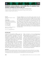
Detection of toll-like receptor (TLR) 8 in human polymorphonuclear cells (PMNs), and the effects of TLR 7/8 ligand R848 on interleukin(IL)-8 releaseFigure 1 (see previous page)
Detection of toll-like receptor (TLR) 8 in human polymorphonuclear cells (PMNs), and the effects of TLR 7/8
ligand R848 on interleukin(IL)-8 release. (A) TLR8 in PMN was detected by immunocytochemistry. Left panel indicates
isotype control. Right panel shows TLR8 immunoreactivity in PMN. (Original magnification: × 400, Scale bars = 10 μm). (B)
TLR8 expression was analyzed by flow-cytometry. PMNs were stained by anti-human TLR8 (solid lines) or the isotype control
(gray histograms) in the permeabilized (left panel) and unpermeabilized condition (right panel). Left panel indicates both inter-
cellular and cell surface expression of TLR8. Right panel shows cell surface expression alone. (C-F) Effect of R848 on the
release of IL-8, and effect of bafilomycin or dexamethasone on the R848-induced IL-8 release from PMN. (C) PMNs were
treated with 10 μM R848. The media were harvested at various time points and assayed for IL-8 by ELISA. (D) PMNs were
treated for 24 hrs with R837, a ligand of TLR7, or various concentrations of R848, a ligand of TLR 7/8. Media were assayed for
IL-8 by ELISA. (E, F) PMNs were treated with 10 μM R848 or vehicle in the presence of various concentrations of bafilomycin,
an inhibitor of endosomal acidification (E), or dexamethasone (F). Media were assayed for IL-8 by ELISA. All values are mean
values ± SEM of three to four separate experiments. *p < 0.05, **p < 0.01, compared with the values of control; +p < 0.05, ++p
< 0.01, compared with the values of the vehicle-pretreated and 10 μM R848-treated group.
Respiratory Research 2009, 10:50 />Page 7 of 13
(page number not for citation purposes)
Figure 2 (see legend on next page)
0 0.3 1 3 10
0
50
100
150
200
Control
zz
+
50
M H
2
O
2
++
++
zz
zz
zz
N.S
N.S
R848( M)
IL-1 (pg/ml)
0 0.3 1 3 10
0
500
1000
1500
Control
50 M H
2
O
2
zz
zz
zz
+
++
++
p<0.01
p<0.01
R848( M)
IL-6(pg-ml)
0 0.1 0.3 1 3 10 30 50 100
0
2000
4000
6000
Control
1 M R848
**
**
H
2
O
2
( M)
IL-8(pg/ml)
(A)
0
5000
10000
15000
20000
25000
0.1 1 10
Control
R848( M)
50
M H
2
O
2
IL-8(pg/ml)
(B)
0
1000
2000
3000
4000
Control
NAC(+)
p<0.01
p<0.01
p<0.01
-
-
++
++
1.0
M R848
50
M H2O2
IL-8(pg/ml)
(C)
0 0.3 1 3 10
0
50
100
150
200
250
Control
50 M H
2
O
2
zz
++
++
p<0.01
zz
zz
p<0.01
R848( M)
TNF (pg/ml)
(D)
(E) (F)
Respiratory Research 2009, 10:50 />Page 8 of 13
(page number not for citation purposes)
superoxide generation in R848-treated neutrophils. In
addition, this potentiation was reversed by N-acethyl-
L-
cysteine suggesting that oxidative stress is associated with
the potentiation of the R848-mediated neutrophilic
response. A previous report has shown that H
2
O
2
pre-
incubation potentiated lipopolysaccharide-induced IL-8
production, and that hydroxy radical scavengers markedly
suppressed this potentiation [9,10,27]. These results are
consistent with our findings. Although H
2
O
2
potentiated
the R848-augmented neutrophilic responses, the potenti-
ation seemed to be heterogeneous. Indeed, H
2
O
2
potenti-
ated the R848-augmented IL-8, TNF-α, and IL-6 release,
but did not potentiate the IL-1β release. This was an inter-
esting finding because the degree of oxidative stress may
modulate the profile of inflammatory mediators during
viral infection. In the current study, it remained unclear
why the potentiation by oxidative stress was heterogene-
ous. A future study is needed to explore this issue.
Hydrogen peroxide enhanced the R848-induced phos-
phorylation of NF-kB, and potentiated the degradation of
IkBα. In addition, a proteosome inhibitor, MG-132,
inhibited the H
2
O
2
-augmented IL-8 release in the R848-
treated neutrophils. Considering that H
2
O
2
did not affect
the expression levels of TLR8 or other signaling molecules
such as MyD88 or TRAF6, these results suggested that the
H
2
O
2
-potentiated NF-kB activation could play a central
role in the augmentation of the neutrophilic responses.
This was consistent with previous reports, which have
shown that oxidative stress cooperatively activated NF-kB
with other mediators such as TNF-α [28-30].
In Figure 4A and 4B, the phosphorylation of NF-kB p65 in
the vehicle-pretreated and R848-treated group was less
than in the H
2
O
2
-pretreated and vehicle-treated group. In
theory, the phosphorylation in the vehicle-pretreated and
R848-treated group should be greater than in the H
2
O
2
-
pretreated and vehicle-treated group. There is a possible
explanation for this discrepancy. Generally, NF-kB is
phosphorylated by NF-kB kinase and IkBα kinases when
NF-kB is dissociated from IkBα and translocated into the
nucleus in various types of cells [31,32]. There is no report
that explored the interaction between NF-kB phosphor-
ylation and IkBα degradation in neutrophils under TLR8
activation. Therefore, the finding observed in the current
study may be due to an unknown signaling in the R848-
treated neutrophils.
Steroids have been reported to reduce the severity and
duration of admission in exacerbations of COPD and
asthma. In this study, dexamethasone inhibited the R848-
augmented IL-8 release from neutrophils in a dose-
dependent manner, and this inhibition was observed in
the presence or absence of H
2
O
2
. These results might indi-
cate that steroids are useful therapeutic agents to attenuate
the viral-induced neutrophilic inflammation. However,
the pretreatment with H
2
O
2
attenuated the effect of dex-
amethasone, suggesting that oxidative stress induced the
steroid resistance. It has been reported that oxidative
stress attenuates the effects of steroids in macrophages
and epithelial cells through histone deacetylase 2 inactiva-
tion [24,33]. This mechanism may also explain the results
observed in the present study.
There are several limitations in the current study. First, we
used H
2
O
2
as a model of oxidative stress. Many previous
reports used this in vitro model to mimic the pathophysi-
ological condition of oxidative stress observed in inflam-
matory lung diseases including COPD and asthma. We
used H
2
O
2
at 0.1 – 100 μM in the current study and these
concentrations are the same range as in previous reports
[24,34]. However, we should be careful when extrapolat-
ing the findings obtained in this in vitro model to the
"real" pathophysiological conditions in inflammatory
lung diseases. Second, we used neutrophils isolated from
healthy subjects, not from smokers or patients with lung
diseases. According to previous reports, the characteristics
of neutrophils are altered in patients with COPD com-
pared with healthy subjects [23,35]. The neutrophilic
responses to TLR activation may be altered in patients
with inflammatory lung disease. Third, we used R848 as a
synthetic ligand for TLR 7/8. Many reports have used R848
Effect of H
2
O
2
on the R848-induced cytokine release from human PMNs, and effect of N-acethyl-L-cysteine on the potentiation of cytokine release by H
2
O
2
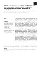
Figure 2 (see previous page)
Effect of H
2
O
2
on the R848-induced cytokine release from human PMNs, and effect of N-acethyl-L-cysteine on
the potentiation of cytokine release by H
2
O
2
. (A) PMNs were incubated with various concentrations of H
2
O
2
for 30 min,
and then treated with R848 for 24 hrs. Media were assayed for IL-8 by ELISA. (B) Various concentrations of R848 were added
to PMNs in the presence or absence of 50 μM H
2
O
2
. After 24 hrs, IL-8 levels in media were measured by ELISA. Dose-
response curve of IL-8 release from PMNs was plotted against the R848 concentration. (C) Ten mM N-acethyl-
L-cysteine
(NAC) was added 10 min before H
2
O
2
or vehicle treatment, then the PMNs were cultured for 24 hrs in the presence or
absence of R848. (D-F) Effects of H
2
O
2
on TNF-α (D), IL-6 (E) and IL-1β (F) release from the R848-treated PMNs were
assessed by Cytokine-Beads Array. All values are mean values ± SEM of three to five separate experiments. **p < 0.01, com-
pared with the values of vehicle-pretreated 1 μM R848-treated group;
××
p < 0.01, compared with the values of control; ≠≠p <
0.01, compared with the values of vehicle treated group; +p < 0.05, ++p < 0.01, compared with the values of 50 μM H
2
O
2
-pre-
treated and vehicle-treated group.
Respiratory Research 2009, 10:50 />Page 9 of 13
(page number not for citation purposes)
Effect of H
2
O
2
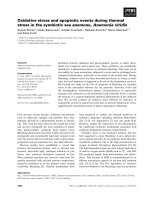
on the R848-induced superoxide generation, elastase release and chemotaxis in human PMNsFigure 3
Effect of H
2
O
2
on the R848-induced superoxide generation, elastase release and chemotaxis in human PMNs.
(A) PMNs were preincubated for 30 min with or without 50 μM H
2
O
2
, and treated with vehicle or R848. Cells were then har-
vested and incubated with dihydro-rhodamine-123 (DHR-123) for 5 min. The amount of superoxide generation was indicated
as the relative fluorescence intensity of DHR-123. (B) After incubation with or without 50 μM H
2
O
2
, PMNs were stimulated
with various concentrations of R848 for 24 hrs. The media were assayed for elastase release by ELISA. (C) After one hour
treatment with various concentrations of R848 with or without 50 μM H
2
O
2
, chemotactic capacity toward IL-8 was assessed
by a modified boyden chamber method. Vertical axis: Relative ratio of the PMN counts (-fold increase). Relative ratio of the
PMN counts was calculated as the ratio of the migrated cell count of each group to that of the control group. All values are
mean values ± SEM of three to four separate experiments. *p < 0.05, compared with the values of vehicle-treated group; +p <
0.05, compared with the values of 50 μM H
2
O
2
-pretreated and vehicle-treated group; MFI = mean fluorescence intensity.
0 1 3 10
0
10
20
30
40
50
Control
50 M H
2
O
2
R848( M)
Elastase(ng/ml)
0 0.1
1
0.0
0.5
1.0
1.5
2.0
Control
50 M H
2
O
2
*
+
R848(
M)
Relative ratio
of PMN count
0.0
0.5
1.0
1.5
p<0.01
p<0.01
p<0.01
p<0.01
-
-
++
++
1.0 M R848
50
M H
2
O
2
Relative MFI
(A) (B)
(C)
Respiratory Research 2009, 10:50 />Page 10 of 13
(page number not for citation purposes)
Figure 4 (see legend on next page)
0.0
0.5
1.0
1.5
2.0
2.5
p<0.01
p<0.05
-
-
++
++
1.0
M R848
50
M H
2
O
2
**
**
**
p<0.01
NF-kB p65
Relative MFI
0
2000
4000
6000
8000
10000
Control
10 M MG-132
-
-
++
++
1.0 M R848
50
M H
2
O
2
p<0.01
p<0.05
p<0.01
**
**
IL-8(pg/ml)
(A)
(B)
(C)
0.0
0.5
1.0
1.5
**
**
++
++
p<0.05
-
-
1
R848(
M)
H
2
O
2
( M)
10
-
5050
-
50
101
-
-actin
IkB
Relative Density
(IkB
)
Respiratory Research 2009, 10:50 />Page 11 of 13
(page number not for citation purposes)
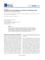
Effect of H
2
O
2
on the R848-induced nuclear factor-kappa B (NF-kB) activationFigure 4 (see previous page)
Effect of H
2
O
2
on the R848-induced nuclear factor-kappa B (NF-kB) activation. Cells were treated with or without
50 μM H
2
O
2
, and then further treated with various concentrations of R848 for 60 min. Phosphorylated NF-kB p65 was assayed
by a flow cytometer (A), and nuclear factor of kappa light polypeptide gene enhancer in B-cells inhibitor, alpha (IkBα) protein
levels were assayed by western blotting (B). Each band intensity was assessed by densitometry. Relative intensity was calculated
as the ratio of the specific band intensity to that of each appropriate β-actin band intensity. (C) PMNs were treated with 1 μM
R848 with or without 50 μM H
2
O
2
in the presence or absence of MG-132, a proteosome inhibitor. After 24 hrs, the media
were assayed for IL-8 by ELISA. All values were mean values ± SEM of three to five separate experiments, and analyzed by
ANOVA followed by Bonferroni's test. **p < 0.01, compared with the values of control; ++p < 0.01, compared with the values
of H
2
O
2
-pretreated and vehicle-treated group; NF-kB p65 = nuclear factor-kappa B p65; IkBα = nuclear factor of kappa light
polypeptide gene enhancer in B-cells inhibitor, alpha.
Effect of dexamethasone on the H
2
O
2
-potentiated IL-8 release in the R848-treated PMNsFigure 5
Effect of dexamethasone on the H
2
O
2
-potentiated IL-8 release in the R848-treated PMNs. PMNs were treated
with or without dexamethasone for 30 min before treatment with or without 50 μM H
2
O
2
. Then, cells were treated with
R848 for 24 hours. Media were assayed for IL-8 by ELISA. Vertical axis: IL-8 relative release (-fold increase). IL-8 relative ratio
was calculated as follows: IL-8 relative ratio = IL-8 levels in the various conditions/IL-8 levels in the vehicle-pretreated and
R848-treated condition. All values are mean values ± SEM of six separate experiments. ++p < 0.01, compared with the values
of vehicle pretreated R848 treated group; **p < 0.01, compared to the values of H
2
O
2
pretreated R848 treated group.
Respiratory Research 2009, 10:50 />Page 12 of 13
(page number not for citation purposes)
as the ligand [16,21,22]. The stimulation of TLR 7/8 by
R848 might be different from that of single strand RNA
virus infection. In the current study, we attempted to elu-
cidate the effects of oxidants on the TLR8 signaling. To
accomplish this, we used R848 for the following reasons.
First, R848 is a stable agent and is easy to handle com-
pared with single strand RNA. Second, R848 does not
have any other effect except TLR 7/8 stimulation. Indeed,
the R848 signaling was abolished by treatment with
bafilomycin, an inhibitor of endosomal acidification.
Therefore, the findings in the current study seemed to be
mediated by TLR8 signaling.
In conclusion, we have shown that the TLR8-mediated
neutrophilic responses in healthy never-smoking subjects
were markedly potentiated by oxidative stress, and this
potentiation was mediated by enhanced NF-kB activation.
These results suggested that oxidative stress might poten-
tiate the neutrophilic inflammation during viral infection.
Abbreviations
COPD: Chronic obstructive pulmonary disease; TLR8:
Toll-like receptor 8; H
2
O
2
: Hydrogen peroxide; NF-kB
p65: Nuclear factor-kappa B p65; IkBα: Nuclear factor of
kappa light polypeptide gene enhancer in B-cells inhibi-
tor, alpha; MyD88: Myeloid differentiation primary
response gene 88; TRAF6: Tumor necrosis factor receptor-
associated factor 6.
Competing interests
The authors declare that they have no competing interests.
Authors' contributions
SY carried out the data analysis and drafted the manu-
script. AK, HS, and MI participated in the design of the
original study, and contributed substantially to the man-
uscript. TI, MK, RT, KA, TH, KM and YM assisted with data
analysis and interpretation, and supervised statistical
analysis.
Additional material
Acknowledgements
We acknowledge Mr. Brent Bell for reading this manuscript.
References
1. MacNee W: Pulmonary and systemic oxidant/antioxidant
imbalance in chronic obstructive pulmonary disease. Proc Am
Thorac Soc 2005, 2(1):50-60.
2. Rahman I, Adcock IM: Oxidative stress and redox regulation of
lung inflammation in COPD. Eur Respir J 2006, 28(1):219-242.
3. Barnes PJ, Chung KF, Page CP: Inflammatory mediators of
asthma: an update. Pharmacol Rev 1998, 50(4):515-596.
4. Sugiura H, Ichinose M: Oxidative and nitrative stress in bron-
chial asthma. Antioxid Redox Signal 2008, 10(4):785-797.
5. Brown RK, Kelly FJ: Evidence for increased oxidative damage in
patients with cystic fibrosis. Pediatr Res 1994, 36(4):487-493.
6. Starosta V, Rietschel E, Paul K, Baumann U, Griese M: Oxidative
changes of bronchoalveolar proteins in cystic fibrosis. Chest
2006, 129(2):431-437.
7. Cantin AM, North SL, Fells GA, Hubbard RC, Crystal RG: Oxidant-
mediated epithelial cell injury in idiopathic pulmonary fibro-
sis. J Clin Invest 1987, 79(6):1665-1673.
8. Kinnula VL, Fattman CL, Tan RJ, Oury TD: Oxidative stress in pul-
monary fibrosis: a possible role for redox modulatory ther-
apy. Am J Respir Crit Care Med 2005, 172(4):417-422.
9. DeForge LE, Fantone JC, Kenney JS, Remick DG: Oxygen radical
scavengers selectively inhibit interleukin 8 production in
human whole blood. J Clin Invest 1992, 90(5):2123-2129.
10. Tanaka C, Kamata H, Takeshita H, Yagisawa H, Hirata H: Redox reg-
ulation of lipopolysaccharide (LPS)-induced interleukin-8
(IL-8) gene expression mediated by NF kappa B and AP-1 in
human astrocytoma U373 cells. Biochem Biophys Res Commun
1997, 232(2):568-573.
11. Fahy JV, Kim KW, Liu J, Boushey HA: Prominent neutrophilic
inflammation in sputum from subjects with asthma exacer-
bation. J Allergy Clin Immunol
1995, 95(4):843-852.
12. Wark PA, Johnston SL, Moric I, Simpson JL, Hensley MJ, Gibson PG:
Neutrophil degranulation and cell lysis is associated with
clinical severity in virus-induced asthma. Eur Respir J 2002,
19(1):68-75.
13. Drost EM, Skwarski KM, Sauleda J, Soler N, Roca J, Agusti A, MacNee
W: Oxidative stress and airway inflammation in severe exac-
erbations of COPD. Thorax 2005, 60(4):293-300.
14. Papi A, Bellettato CM, Braccioni F, Romagnoli M, Casolari P, Cara-
mori G, Fabbri LM, Johnston SL: Infections and airway inflamma-
tion in chronic obstructive pulmonary disease severe
exacerbations. Am J Respir Crit Care Med 2006, 173(10):1114-1121.
15. Underhill DM, Ozinsky A: Toll-like receptors: key mediators of
microbe detection. Curr Opin Immunol 2002, 14(1):103-110.
Additional file 1
Effect of MG-132 on the R848-induced nuclear factor of kappa light
polypeptide gene enhancer in B-cells inhibitor, alpha (IkB
α
) degrada-
tion. PMNs were incubated with or without 10
μ
M MG-132, a proteo-
some inhibitor, and then further treated with various concentrations of
R848 for 60 min. The cytoplasmic fraction of cell lysates were used for
estimating the protein levels of IKB
α
by western blotting. Each band
intensity was assessed by densitometry. Relative intensity was calculated
as the ratio of specific band intensity to that of each appropriate
β
-actin
band intensity. All values are mean values ± SEM of three separate exper-
iments. **p < 0.01; compared with the values of vehicle-treated group,
IkB
α
= nuclear factor of kappa light polypeptide gene enhancer in B-cells
inhibitor, alpha, n.s. = not significant.
Click here for file
[ />9921-10-50-S1.pdf]
Publish with Bio Med Central and every
scientist can read your work free of charge
"BioMed Central will be the most significant development for
disseminating the results of biomedical research in our lifetime."
Sir Paul Nurse, Cancer Research UK
Your research papers will be:
available free of charge to the entire biomedical community
peer reviewed and published immediately upon acceptance
cited in PubMed and archived on PubMed Central
yours — you keep the copyright
Submit your manuscript here:
/>BioMedcentral
Respiratory Research 2009, 10:50 />Page 13 of 13
(page number not for citation purposes)
16. Hayashi F, Means TK, Luster AD: Toll-like receptors stimulate
human neutrophil function. Blood 2003, 102(7):2660-2669.
17. Heil F, Hemmi H, Hochrein H, Ampenberger F, Kirschning C, Akira
S, Lipford G, Wagner H, Bauer S: Species-specific recognition of
single-stranded RNA via toll-like receptor 7 and 8. Science
2004, 303(5663):1526-1529.
18. Diebold SS, Kaisho T, Hemmi H, Akira S, Reise Sousa C: Innate anti-
viral responses by means of TLR7-mediated recognition of
single-stranded RNA. Science 2004, 303(5663):1529-1531.
19. Lund JM, Alexopoulou L, Sato A, Karow M, Adams NC, Gale NW,
Iwasaki A, Flavell RA: Recognition of single-stranded RNA
viruses by Toll-like receptor 7. Proc Natl Acad Sci USA 2004,
101(15):5598-5603.
20. Takeda K, Akira S: Toll-like receptors in innate immunity. Int
Immunol 2005, 17(1):1-14.
21. Wang JP, Bowen GN, Padden C, Cerny A, Finberg RW, Newburger
PE, Kurt-Jones EA: Toll-like receptor-mediated activation of
neutrophils by influenza A virus. Blood 2008, 112(5):2028-2034.
22. Hattermann K, Picard S, Borgeat M, Leclerc P, Pouliot M, Borgeat P:
The Toll-like receptor 7/8-ligand resiquimod (R-848) primes
human neutrophils for leukotriene B4, prostaglandin E2 and
platelet-activating factor biosynthesis. FASEB J 2007,
21(7):1575-1585.
23. Yamagata T, Sugiura H, Yokoyama T, Yanagisawa S, Ichikawa T,
Ueshima K, Akamatsu K, Hirano T, Nakanishi M, Yamagata Y, Matsu-
naga K, Minakata Y, Ichinose M: Overexpression of CD-11b and
CXCR1 on circulating neutrophils: its possible role in COPD.
Chest 2007, 132(3):890-899.
24. Ito K, Hanazawa T, Tomita K, Barnes PJ, Adcock IM: Oxidative
stress reduces histone deacetylase 2 activity and enhances
IL-8 gene expression: role of tyrosine nitration. Biochem Bio-
phys Res Commun 2004, 315(1):240-245.
25. Hemmi H, Kaisho T, Takeuchi O, Sato S, Sanjo H, Hoshino K, Hori-
uchi T, Tomizawa H, Takeda K, Akira S: Small anti-viral com-
pounds activate immune cells via the TLR7 MyD88-
dependent signaling pathway. Nat Immunol 2002, 3(2):196-200.
26. Jurk M, Heil F, Vollmer J, Schetter C, Krieg AM, Wagner H, Lipford
G, Bauer S:
Human TLR7 or TLR8 independently confer
responsiveness to the antiviral compound R-848. Nat Immunol
2002, 3(6):499.
27. DeForge LE, Preston AM, Takeuchi E, Kenney J, Boxer LA, Remick
DG: Regulation of interleukin 8 gene expression by oxidant
stress. J Biol Chem 1993, 268(34):25568-25576.
28. Schreck R, Rieber P, Baeuerle PA: Reactive oxygen intermedi-
ates as apparently widely used messengers in the activation
of the NF-kappa B transcription factor and HIV-1. Embo J
1991, 10(8):2247-2258.
29. Janssen-Heininger YM, Macara I, Mossman BT: Cooperativity
between oxidants and tumor necrosis factor in the activa-
tion of nuclear factor (NF)-kappaB: requirement of Ras/
mitogen-activated protein kinases in the activation of NF-
kappaB by oxidants. Am J Respir Cell Mol Biol 1999, 20(5):942-952.
30. de Oliveira-Marques V, Cyrne L, Marinho HS, Antunes F: A quanti-
tative study of NF-kappaB activation by H2O2: relevance in
inflammation and synergy with TNF-alpha. J Immunol 2007,
178(6):3893-3902.
31. Kamata H, Manabe T, Oka S, Kamata K, Hirata H: Hydrogen per-
oxide activates IkappaB kinases through phosphorylation of
serine residues in the activation loops. FEBS Lett 2002, 519(1–
3):231-237.
32. Takada Y, Mukhopadhyay A, Kundu GC, Mahabeleshwar GH, Singh S,
Aggarwal BB: Hydrogen peroxide activates NF-kappa B
through tyrosine phosphorylation of I kappa B alpha and ser-
ine phosphorylation of p65: evidence for the involvement of
I kappa B alpha kinase and Syk protein-tyrosine kinase. J Biol
Chem 2003, 278(26):24233-24241.
33. Ito K, Lim S, Caramori G, Chung KF, Barnes PJ, Adcock IM: Ciga-
rette smoking reduces histone deacetylase 2 expression,
enhances cytokine expression, and inhibits glucocorticoid
actions in alveolar macrophages. FASEB J 2001,
15(6):1110-1112.
34. Powers KA, Szaszi K, Khadaroo RG, Tawadros PS, Marshall JC, Kapus
A, Rotstein OD: Oxidative stress generated by hemorrhagic
shock recruits Toll-like receptor 4 to the plasma membrane
in macrophages. J Exp Med 2006, 203(8):
1951-1961.
35. Yanagisawa S, Sugiura H, Yokoyama T, Yamagata T, Ichikawa T, Aka-
matsu K, Koarai A, Hirano T, Nakanishi M, Matsunaga K, Minakata Y,
Ichinose M: The possible role of hematopoietic cell kinase in
the pathophysiology of COPD. Chest 2009, 135(1):94-101.