Báo cáo y học: "Physiological tonicity improves human chondrogenic marker expression through nuclear factor of activated T-cells 5 in vitro" docx
Bạn đang xem bản rút gọn của tài liệu. Xem và tải ngay bản đầy đủ của tài liệu tại đây (1.99 MB, 12 trang )
van der Windt et al. Arthritis Research & Therapy 2010, 12:R100
/>Open Access
RESEARCH ARTICLE
BioMed Central
© 2010 van der Windt et al.; licensee BioMed Central Ltd. This is an open access article distributed under the terms of the Creative Com-
mons Attribution License ( which permits unrestricted use, distribution, and reproduc-
tion in any medium, provided the original work is properly cited.
Research article
Physiological tonicity improves human
chondrogenic marker expression through nuclear
factor of activated T-cells 5
in vitro
Anna E van der Windt
1
, Esther Haak
1
, Ruud HJ Das
1
, Nicole Kops
1
, Tim JM Welting
2
, Marjolein MJ Caron
2
, Niek P van
Til
3
, Jan AN Verhaar
1
, Harrie Weinans
1
and Holger Jahr*
1
Abstract
Introduction: Chondrocytes experience a hypertonic environment compared with plasma (280 mOsm) due to the
high fixed negative charge density of cartilage. Standard isolation of chondrocytes removes their hypertonic matrix,
exposing them to nonphysiological conditions. During in vitro expansion, chondrocytes quickly lose their specialized
phenotype, making them inappropriate for cell-based regenerative strategies. We aimed to elucidate the effects of
tonicity during isolation and in vitro expansion on chondrocyte phenotype.
Methods: Human articular chondrocytes were isolated and subsequently expanded at control tonicity (280 mOsm) or
at moderately elevated, physiological tonicity (380 mOsm). The effects of physiological tonicity on chondrocyte
proliferation and chondrogenic marker expression were evaluated. The role of Tonicity-responsive Enhancer Binding
Protein in response to physiological tonicity was investigated using nuclear factor of activated T-cells 5 (NFAT5) RNA
interference.
Results: Moderately elevated, physiological tonicity (380 mOsm) did not affect chondrocyte proliferation, while higher
tonicities inhibited proliferation and diminished cell viability. Physiological tonicity improved expression of
chondrogenic markers and NFAT5 and its target genes, while suppressing dedifferentiation marker collagen type I and
improving type II/type I expression ratios >100-fold. Effects of physiological tonicity were similar in osteoarthritic and
normal (nonosteoarthritic) chondrocytes, indicating a disease-independent mechanism. NFAT5 RNA interference
abolished tonicity-mediated effects and revealed that NFAT5 positively regulates collagen type II expression, while
suppressing type I.
Conclusions: Physiological tonicity provides a simple, yet effective, means to improve phenotypical characteristics
during cytokine-free isolation and in vitro expansion of human articular chondrocytes. Our findings will lead to the
development of improved cell-based repair strategies for chondral lesions and provides important insights into
mechanisms underlying osteoarthritic progression.
Introduction
Hyaline articular cartilage is a connective tissue covering
the ends of bones in joints and is composed of specialized
cells, chondrocytes that produce a large amount of extra-
cellular matrix. This matrix is crucial for the unique bio-
mechanical properties of this tissue and is composed of a
collagen fiber network, providing tensile strength and
flexibility, and abundant ground matrix rich in proteogly-
cans [1].
The glycosaminoglycan (GAG) side chains of the pro-
teoglycans are sulfated and responsible for a characteris-
tic high fixed negative charge density [2], which binds
mobile cations (mainly sodium). This binding determines
the physiological tonicity (that is, osmotic pressure) of
the extracellular fluid around chondrocytes in vivo, but
the tonicity indirectly also largely depends on the quality
of the collagen network. Extracellular tonicity in healthy
cartilage ranges between 350 and 480 mOsm [3,4]. In
* Correspondence:
1
Department of Orthopaedics, Erasmus MC, University Medical Center
Rotterdam, Dr. Molewaterplein 50, 3015 GE Rotterdam, The Netherlands
Full list of author information is available at the end of the article
van der Windt et al. Arthritis Research & Therapy 2010, 12:R100
/>Page 2 of 12
vivo, tonicity of the extracellular fluid is dynamic and
changes due to alterations in matrix hydration [5]. During
cartilage degeneration (that is, in osteoarthritis (OA)), the
collagen matrix degrades and the GAG concentration
diminishes, resulting in a severity-dependent decreased
tonicity of between 280 and 350 mOsm [3,6]. Currently,
chondrocyte isolation and in vitro expansion culture are
performed in medium of nonphysiological tonicity (270 ±
20 mOsm). Several studies have already shown that chon-
drocytes are tonicity responsive [7-9] and react with
changes in matrix synthesis [4,8,10,11], but focused on
aggrecan (AGC1) core protein mRNA levels, AGC1 pro-
moter activity and GAG production.
Molecular mechanisms involved in the hypertonic
response of human articular chondrocytes (HACs) are
poorly understood. Hypertonicity perturbs cells by caus-
ing osmotic efflux of water, resulting in cell shrinkage
[12,13]. Cells react by a rapid uptake of ions, which
increase cellular ionic strength [14] with potentially detri-
mental effects [15-17]. The initial, rapid response is the
activation of transporters that exchange these ions for
compatible osmolytes [16,18]. This process is controlled
by Tonicity-responsive Enhancer Binding Protein
(TonEBP/NFAT5), which mediates transcriptional activa-
tion of these transporters [16]. Nuclear factor of activated
T-cells 5 (NFAT5) is a member of the Rel family of tran-
scription factors [19] and targets sodium/myo-inositol
cotransporter (SMIT) [20,21], sodium/chloride-coupled
acid transporters (BGT1/SLC6A12) [20], aquaporin
channels (AQP1 and AQP2) [22], and calcium-binding
proteins (S100A4) [23-25]. Upon hypertonic stress, tran-
scription of NFAT5 itself is upregulated in several cell
types [26-28], but the tonicity threshold and cell signaling
pathways required to activate NFAT5 may be cell type
specific [29]. Nothing is currently known about the
expression or function of NFAT5 in HACs.
Chondral lesions from, for example, trauma or overuse,
can cause joint pain, immobility and eventually OA. The
associated high prevalence - 60% of all patients undergo-
ing knee arthroscopy are diagnosed with a chondral
lesion [30] - and loss of quality of life makes cartilage
damage a major personal and economical burden. Treat-
ment options for chondral lesions are limited, and autolo-
gous chondrocyte implantation is the currently most
developed hyaline repair technique for the knee [31].
Characterized chondrocyte implantation, employing
phenotypical prescreening prior to implantation, has
recently improved structural repair [32].
Chondrocyte dedifferentiation during in vitro expan-
sion for autologous chondrocyte implantation is detri-
mental; but almost inevitably in standard monolayer
culture, spherical chondrocytes will gradually convert
into fibroblast-like cells [33,34]. This morphological
change is accompanied by a shift in collagen expression
towards less collagen type II (COL2) and more collagen
type I (COL1) [34,35]. Consequently, dedifferentiated
chondrocytes produce fibrocartilage in vivo, with an
extracellular matrix of inferior biomechanical properties
due to higher collagen (especially type I) content and less
proteoglycans compared with native hyaline cartilage
[36]. Three-dimensional culture systems can partially
prevent dedifferentiation, but are labor intensive and
essentially impair propagation. Chondrocyte dedifferen-
tiation might also play a role in the pathogenesis of OA,
as the ability of aging chondrocytes to produce and repair
the extracellular matrix is compromised [37] and as
COL1 is shown to be present in chondrocyte clusters in
fibrillated areas of late-stage OA cartilage while it is
absent in healthy cartilage [38].
In the present article we report that physiological tonic-
ity (380 mOsm) during isolation and monolayer expan-
sion can suppress chondrocyte dedifferentiation and that
expression of the extracelluar matrix components colla-
gen type I and collagen type II as well as aggrecan is
NFAT5 dependent. We further show that NFAT5 con-
tributes to the differential regulation of both collagen
types. This study provides a simple, yet novel and effec-
tive, means to improve cell-based repair strategies for
chondral lesions and contribute to our understanding of
OA progression.
Materials and methods
Cartilage and chondrocyte isolation
After informed consent was obtained, human articular
cartilage was explanted from macroscopically normal
areas of the femoral condyles and tibial plateau of nine
patients undergoing total knee replacement surgery for
OA (medical ethical approval MEC2004-322). In addition
to preparation of cartilage explants and isolation of HACs
under standard conditions (DMEM, 280 mOsm) as
described by Das and colleagues [39], medium tonicity
was also adjusted to 380 mOsm, 480 mOsm or 580
mOsm by addition of sterile NaCl. Enzymatic digestion,
removal of undigested fragments and subsequent chon-
drocyte culture were all reported earlier [39]. The 280
mOsm and 380 mOsm isolations were also performed
with cartilage obtained from the femoral condyles and
tibial plateau of two non-OA donors (further referred to
as normal donors) undergoing above-knee amputation
surgery after trauma.
Chondrocyte proliferation and DNA measurements
Primary (P0), passage 1 (P1), passage 2 (P2) and passage 3
(P3) HACs were monolayer expanded in medium corre-
sponding to their isolation tonicity (280 mOsm, 380
mOsm, 480 mOsm or 580 mOsm), with an initial seeding
density of 6,000 cells/cm
2
. Cells were harvested daily for
cell counts and DNA assay between days 2 and 6. Experi-
van der Windt et al. Arthritis Research & Therapy 2010, 12:R100
/>Page 3 of 12
ments were performed in duplicate from three OA
donors (n = 6). At each passage, growth curves were
established by cell counts using Trypan Blue (catalogue
number T8154; Sigma-Aldrich, St. Louis, MO, USA) and
DNA quantification. DNA measurements were per-
formed according to Karsten and Wollenberger [40] with
slight modifications [41]. Doubling times within each
passage were calculated from the trend line of the expo-
nential growth phase using the equation:
where k is the growth constant and T is the doubling
time.
Chondrocyte expansion
Primary HACs were cultured for expansion in monolay-
ers at a seeding density of 7,500 cells/cm
2
in medium cor-
responding to their isolation tonicity (280 mOsm, 380
mOsm, 480 mOsm or 580 mOsm). P0 cells to P3 cells
were seeded in high-density monolayers (20,000 cells/
cm
2
) and were cultured for an additional 5 days and 7
days before analysis of mRNA (quantitative RT-PCR) and
protein expression (Western blotting), respectively.
Experiments were performed in triplicate from four OA
donors (n = 12). Additional experiments were performed
in triplicate from two healthy donors (n = 6) to investigate
whether the hypertonic stress response is specific for
pathologically altered cells. To exclude sodium-specific or
chloride-specific effects, we performed experiments
using N-methyl-d-glucamine chloride (NMDG-Cl) or
sucrose to adjust the medium tonicity to 380 mOsm.
Lentiviral NFAT5 gene knockdown
We used lentiviral vectors for nontransient shRNA-medi-
ated gene silencing in primary chondrocytes [42].
BamHI/MunI restriction fragments of the parental
pLKO.1-puro vector - each containing the U6 promotor
and one out of five different, sequence-verified anti-
human NFAT5 shDNAs (MISSION shRNA library [43]) -
were subcloned into corresponding restriction sites of
recipient vector pRRL.PPT.PGK.GFPpre. This vector was
kindly provided by L Naldini (San Raffaele Telethon Insti-
tute for Gene Therapy, Milan, Italy) [44,45] and was opti-
mized by A Schambach (Department of Experimental
Hematology, Hannover Medical School, Hannover, Ger-
many) [46] to express enhanced green fluorescent protein
(eGFP) from the phosphoglycerate kinase promoter. Len-
tiviral particles were produced in HEK293T cells by tran-
sient transfection using a calcium phosphate protocol
[47]. Cells transduced with a lentiviral vector lacking the
NFAT5-specific shRNA expression cassette served as
controls. All cells were grown in monolayers.
TRCN0000020020 was identified as the best performing
anti-NFAT5 shRNA clone by quantitative PCR-based
knockdown efficiency determination, and was used in
subsequent experiments.
P1 OA HACs from two donors were seeded (15,000
cells/cm
2
) and cultured for 4 days in control medium (280
mOsm). Three hours prior to transduction, cells were
deprived of antibiotics, and then were transduced for ±
18 hours, refreshed with control medium with antibiotics
and cultured for an additional 4 days before harvesting
for fluorescence-activated cell sorting (FACS) analyses.
Cells were resuspended in PBS with 10% FCS and antibi-
otics, and were washed. Cells were collected and stained
with Hoechst 33258 (1 mg/ml; Molecular Probes/Invitro-
gen Corp., Carlsbad, CA, USA) to discriminate between
dead cells and live cells. FACS was performed on the
FACSAria (Becton Dickinson BV, Breda, The Neder-
lands), and eGFP-expressing cells were collected (>50%,
multiplicity of infection ~1) and reanalyzed for purity
(>95%) using Cell Quest Pro Software (Becton Dickinson
Biosciences BV, Breda, The Nederlands).
The eGFP-expressing populations were seeded (10,000
cells/cm
2
) and cultured in control medium up to 80%
confluency. Cells were then switched to medium of 380
mOsm or were kept on control medium for 24 hours
prior to RNA analysis.
RNA expression analysis
RNA isolation, purification, quantification and cDNA
synthesis are described elsewhere [48]. Expression levels
of AGC1, SOX9 and COL2 were studied as chondrogenic
markers, while COL1 was studied as a dedifferentiation
marker [34,35,49,50]. Quantitative PCR assays for COL2,
SOX9, AGC1 and COL1 have been reported earlier [51].
To qu antif y expressi on o f NFAT5 and its target genes,
the following primers were tested for similar amplifica-
tion efficiency and specificity according to Das and col-
leagues [39], and were used as 20 μl SYBR
®
Green I
reactions: HsNFAT5_Fw, GGGTCAAACGACGAGATT-
GTG and HsNFAT5_Rv, TTGTCCGTGGTAAGCT-
GAGAA; HsS100A4_Fw, GTCCACCTTCCACAAGTAC
TCG and HsS100A4_Rv, TCATCTGTCCTTTTC-
CCCAAG; and HsSLC6A12_Fw, ACACAGAGCATTG-
CACGGACT and HsSLC6A12_Rv, CCAGAACTCGTC
TCTCCCAGAA. Data were normalized to an index of
three reference genes (GAPDH, UBC, HPRT1) that were
pre-evaluated to be stably expressed across samples [39].
Relative expression was calculated according to the 2
-ΔCT
method [52].
Western blot analysis
Cells seeded at high densities were washed twice with
PBS and were lysed in RIPA buffer [53] with addition of
protease inhibitors. The total protein concentration was
quantified by the bicinchoninic acid assay according to
the manufacturer's protocol (#23225; Thermo Fisher Sci.,
Rockford, IL, USA). Aliquots (10 μg) were subjected to
yx kxt k T=
() ()
()
=0 exp with ln 2/
van der Windt et al. Arthritis Research & Therapy 2010, 12:R100
/>Page 4 of 12
10% SDS-PAGE prior to electroblotting onto nitrocellu-
lose membranes (Protran BA83; Schleicher & Schuell BV,
s-Hertogenbosch, The Netherlands). Blots were blocked
in 5% low-fat dry milk in 1× PBS, 0.05% v/v NP-40, were
incubated with primary antibodies - anti-type II collagen
and anti-type I collagen, both 1:100 (SouthernBiotech,
Birmingham, Alabama, USA), or 1:10,000 anti-α-Tubulin
(Sigma) - were washed, were incubated with secondary
antibodies (both 1:1,000; Dako Cytomation, Heverlee,
Belgium) and were chemiluminescently detected. Signals
were quantified using ImageJ 1.42 software [54].
Statistical analysis
Statistical analysis was performed using SPSS 13.0 soft-
ware (SPSS Inc., Chicago, IL, USA). Data were compared
between groups by Kruskall-Wallis H test and post-hoc
Mann-Whitney U test. Results represent the mean ±
standard deviation, and P < 0.05, P < 0.01 and P < 0.001
were considered to indicate levels of statistically signifi-
cant difference.
Results
Hypertonicity influences proliferation and survival of
chondrocytes
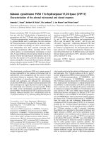
We first determined the influence of tonicity on prolifera-
tion: OA HACs isolated at 580 mOsm hardly attached or
proliferated (Figure 1d), and 2 days after seeding no viable
cells were recovered. At 280 mOsm, 380 mOsm and 480
mOsm, respectively, cells did adhere but increasing tonic-
ity induced marked morphological changes: at 280
mOsm, cells appeared fibroblast-like, stretched out and
flattened with long filopodia (Figure 1a); while at 380
mOsm, cells were more sphere-shaped and had shorter
filopodia (Figure 1b). At 480 mOsm, cells showed few
filopodia and appeared spherical (Figure 1c). The differ-
ences in appearance remained throughout the dedifferen-
tiation period (P0 to P3), but were most apparent at
earlier passages.
Using cell counts and DNA assays, doubling times were
calculated from growth curves established from each pas-
sage at three different tonicities (280 mOsm, 380 mOsm
and 480 mOsm). Throughout dedifferentiation, OA
HACs isolated at 480 mOsm showed severely inhibited
proliferation compared with cells at 280 mOsm and 380
mOsm (Table 1). In contrast, doubling times of OA HACs
at 280 mOsm and 380 mOsm never significantly differed
(Table 1). All further experiments were therefore per-
formed at 380 mOsm (as high tonicity condition) and
compared with 280 mOsm (control condition).
Isolation and expansion of chondrocytes under hypertonic
conditions improves their phenotype
Next, we set out to determine whether expansion culture
in physiological tonicity improves the chondrocytic phe-
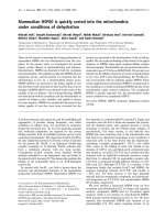
notype. Physiological tonicity (380 mOsm) during isola-
tion and subsequent passaging of OA HACs significantly
increased mRNA levels of both AGC1 (Figure 2a) and
SOX9 (Figure 2b) at all passages. In expanded P3 chon-
drocytes in physiological culture, AGC1 levels were still
higher than in unpassaged P0 chondrocytes cultured
under the standard culture conditions (280 mOsm).
Physiological tonicity also significantly upregulated
COL2 levels from 8.5-fold in P0 to 11.6-fold in expanded
P3 chondrocytes (Figure 2c) compared with controls. In
contrast, COL1 expression was significantly suppressed
in physiological conditions throughout culture. Conse-
quently, we found a significantly improved COL2/COL1
ratio during chondrocyte expansion (Figure 2d), from
sevenfold in P0 cells to 100-fold in expanded P3 cells.
Physiological tonicity also upregulated COL2 protein
expression (Figure 2e): levels significantly increased
(between 1.5-fold and 2.2-fold) in P0, P1 and P2 chondro-
cytes. In contrast, physiological tonicity significantly
decreased COL1 protein expression (Figure 2f), from
twofold in P0 cells to 13-fold in P1 cells.
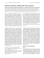
Physiological tonicity also significantly increased AGC1
(Figure 3a) and SOX9 (Figure 3b) mRNA levels in nonos-
teoarthritic human articular chondrocytes (NHACs).
Furthermore, COL2 mRNA levels were significantly
upregulated, from 5.8-fold in P0 cells to 270-fold in
expanded P3 NHACs (Figure 3c). As in OA HACs, hyper-
tonicity also downregulated COL1 expression with
increasing passage number in NHACs: the COL2/COL1
ratios increased during expansion (Figure 3d), from 6.8-
fold in P0 cells to 355-fold in expanded P3 cells. Corre-
spondingly, COL2 protein levels increased under these
conditions (4.8-fold in P1 cells and 2.9-fold in P2 cells),
while the amount of COL1 diminished (by 4.7-fold in P1
cells and fivefold in P2 cells) (Figure 3e, f).
Hypertonicity activates NFAT5 in human articular
chondrocytes
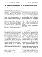
Compared with 280 mOsm controls, NFAT5 mRNA lev-
els were significantly increased in 380 mOsm OA HAC
cultures (Figure 4a), as was the expression of established
NFAT5 target genes S100A4 (in all passages; Figure 4b)
and SLC6A12 (until P2; Figure 4c). Similar effects were
found in NHACs (data not shown).
NFAT5 knockdown inhibits hypertonicity-induced
chondrogenic marker expression
Upon transduction, sorted eGFP-coexpressing OA HACs
were switched to 380 mOsm for 24 hours. In controls not
expressing NFAT5-specific shRNAs, an approximately
twofold increase in NFAT5 mRNA levels was observed
upon hypertonic stimulation (Figure 4a, P1). In contrast,
likewise challenged cells expressing anti-NFAT5 shRNAs
showed an approximately 75% reduction in NFAT5 levels
van der Windt et al. Arthritis Research & Therapy 2010, 12:R100
/>Page 5 of 12
(Figure 5a). Following NFAT5 knockdown, the NFAT5
targets S100A4 and SLC6A12 were also no longer hyper-
tonically inducible: S100A4 expression decreased twofold
and SLC6A12 was virtually undetectable upon NFAT5
RNAi (Figure 5a), confirming a functional NFAT5 knock-
down. At 380 mOsm, NFAT5 RNAi also downregulated
chondrogenic markers: AGC1 by 80%, SOX9 by 32% and
COL2 by 84%, as compared with non-RNAi controls (Fig-
ure 5b). Interestingly, expression of COL1 increased after
NFAT5 RNAi in OA HACs to ~300% of control levels
(Figure 5b).
Discussion
Isolation and expansion of adult HACs under physiologi-
cal tonicity (380 mOsm) improves expression of chondro-
genic markers on mRNA and protein levels. While other
studies partially confirm that nonhuman chondrocytes
respond to tonicity with altered aggrecan and SOX9
expression [4,8,10], we are reporting beneficial effects of
Figure 1 Hypertonic isolation and expansion of chondrocytes changes chondrocyte morphology. Representative images (200×) of chondro-
cytes cultured for 2 days at (a) 280 mOsm, (b) 380 mOsm, (c) 480 mOsm and (d) 580 mOsm.
Table 1: Proliferation of chondrocytes isolated and cultured at 280 mOsm, 380 mOsm and 480 mOsm
Chondrocyte proliferation (%)
Culture condition Passage 0 Passage 1 Passage 2 Passage 3
280 mOsm 100 (68 ± 28 hours) 100 (89 ± 54 hours) 100 (67 ± 48 hours) 100 (57 ± 11 hours)
380 mOsm 113 ± 18 89 ± 25 99 ± 9 154 ± 41
480 mOsm 675 ± 405* 180 ± 24* 168 ± 28* 165 ± 81*
Data presented as relative doubling times in percentage of cells cultured at 280 mOsm, mean ± standard deviation. The absolute doubling
time ± standard deviation in hours is displayed in brackets. n = 6. mOsm, milliosmoles per kilogram of water. *P < 0.05.
van der Windt et al. Arthritis Research & Therapy 2010, 12:R100
/>Page 6 of 12
Figure 2 Hypertonic isolation and expansion increased marker gene expression in osteoarthritis human articular chondrocytes. Relative ex-
pression of (a) AGC1, (b) SOX9, (c) COL2 and (d) COL2:COL1 ratio in primary (P0) and passaged (P1 to P3) chondrocytes cultured at 380 mOsm compared
with 280 mOsm. (e) COL2 protein expression and (f) COL1 protein expression in P0 and P1 osteoarthritis human articular chondrocytes. Protein levels
normalized to α-tubulin. Data are mean ± standard deviation, n = 12. Differences from cells cultured at 280 mOsm are indicated: *P < 0.05, **P < 0.01
and ***P < 0.001.
van der Windt et al. Arthritis Research & Therapy 2010, 12:R100
/>Page 7 of 12
Figure 3 Hypertonic isolation and expansion increased chondrogenic marker expression in nonosteoarthritic human articular chondro-
cytes. Relative expression of (a) AGC1, (b) SOX9, (c) COL2 and (d) COL2:COL1 ratio in primary (P0) and passaged (P1 to P3) nonosteoarthritic human
articular chondrocytes (NHACs) cultured at 380 mOsm compared with cells cultured at 280 mOsm. (e) COL2 protein expression and (f) COL1 protein
expression in P1 and P2 NHACs, normalized to α-tubulin. Data are mean ± standard deviation, n = 6. Differences from 280 mOsm controls are indicated:
*P < 0.05 and **P < 0.01.
van der Windt et al. Arthritis Research & Therapy 2010, 12:R100
/>Page 8 of 12
isolating and expanding human normal and OA articular
chondrocytes at physiological levels (380 mOsm). In
addition, we also studied collagen type II expression, gen-
erally acknowledged to be the most important chondro-
genic marker. As fibrocartilaginous collagen type I and
hyaline collagen type II expression are differentially regu-
lated in chondrocytes [34], analyzing the collagen type II/
type I expression ratios is informative of chondrogenic
potential [51]. Interestingly, NFAT5 seems to be crucially
involved in this differential regulation upon hypertonic
challenge: it positively regulates collagen type II, while
suppressing collagen type I (Figure 5b). Fibrocartilage,
occurring in areas subject to frequent stress like interver-
tebral discs and tendon attachment sites, is more rich in
collagen type I than is hyaline cartilage [55]. Tonicity may
thus provide a simple means to manipulate expression of
these two collagens for broader applications than regen-
erative chondrocyte implantations (autologous chondro-
cyte implantation or characterized chondrocyte
implantation) alone [56].
Under our conditions, COL2 mRNA abundances mea-
sured by quantitative PCR correlated well with protein
synthesis as determined by Western blots (Figures 2 and
3). The same observation holds for COL1 expression in
the early passages, but not for COL1 expression in the
later passages.
Hypertonicity induced an increase in NFAT5 abun-
dance, and protein synthesis rates were found to be pro-
portional to the increase in mRNA in MDCK cells [28]
and mIMCD3 cells [27]. NFAT5 mRNA is expressed
abundantly in chondrocytes throughout passages and is
further induced by hypertonicity. However, we failed to
show NFAT5 protein expression by Western blotting.
Whether this failure is due to low protein abundance in
our cells or technical issues such as poor extraction effi-
ciency of this very large transcription factor remains to be
elucidated in future experiments.
Hypertonicity induces cell shrinkage, which may acti-
vate Na
+
, K
+
, or 2Cl
-
co-transport, allowing cellular accu-
mulation of NaCl and KCl. The beneficial effects on
chondrogenic marker gene expression therefore could
have been caused by accumulation of specific inorganic
ions or specific channel activity rather than primarily
tonicity-mediated effects. We used NMDG-Cl, a bulky
substitute for small cations that is impermeable to almost
all known channels [57], and sucrose to exclude sodium-
Figure 4 Hypertonic conditions activate nuclear factor of activated T-cells 5 in osteoarthritis human articular chondrocytes. Relative expres-
sion of (a) nuclear factor of activated T-cells 5 (NFAT5) and its target genes (b) S100A4 and (c) SLC6A12 in primary (P0) and passaged (P1 to P3) chon-
drocytes cultured at 380 mOsm compared with 280 mOsm. Data are mean ± standard deviation, n = 12. Differences are indicated: *P < 0.05, **P < 0.01
and ***P < 0.001.
Figure 5 Nuclear factor of activated T-cells 5 knockdown inhibits
hypertonicity-induced chondrogenic marker expression. (a) Rela-
tive expression of nuclear factor of activated T-cells 5 (NFAT5) and its
target genes S100A4 and SLC6A12 in transduced chondrocytes either
expressing (NFAT5 shRNA) or not expressing (control) NFAT5-specific
shRNAs, 24 hours after increasing tonicity to 380 mOsm. (b) Effects of
NFAT5 knockdown on chondrogenic markers AGC1, SOX9, COL2 and
COL1. Data are mean ± standard deviation, n = 6. Differences from cells
transduced with control virus are indicated: *P < 0.05 and **P < 0.01.
van der Windt et al. Arthritis Research & Therapy 2010, 12:R100
/>Page 9 of 12
specific or chloride-specific effects. We were not able to
detect any significant differences in gene expression pat-
terns between the NaCl, NMDG-Cl or sucrose methods
of tonicity alteration (data not shown).
As our initial studies concerned adult HACs obtained
from OA knee joints, we aimed at eliminating interpreta-
tion bias due to the pathological state of these cells. Using
identically challenged NHACs, we showed that these
chondrocytes react similarly to the same order of tonicity
with respect to our marker genes: 380 mOsm signifi-
cantly delayed the phenotypical deterioration of NHACs
as observed in control medium. This may imply that
physiological tonicity, postulated to be around 380
mOsm for chondrocytes, is sensed by OA cells and nor-
mal cells in a similar fashion. We observed a slightly faster
decrease in AGC1 and COL2 mRNA levels in P2 and P3
NHACs as compared with OA HACs. Late-stage OA
chondrocytes from fibrillated areas are dedifferentiated,
flattened cells. The loss of a proper spherical shape as an
integral part of the chondrocytes phenotype [58,59]
involves cytoskeletal changes [60]. Exposing these cells to
physiological tonicity as a redifferentiation stimulus
probably induces a more enduring response as compared
with spherical, normal chondrocytes. Cell-based thera-
pies using the latter are usually restricted to younger indi-
viduals after traumatic insults. Autologous chondrocyte
implantation employing OA cells may benefit relatively
more from a hypertonic treatment protocol.
The precise molecular mechanism by which tonicity is
sensed by cells is still poorly understood. Hypertonicity-
increased NFAT5 mRNA abundances have been shown
for other cell types [26-28]. NFAT5 is thus accepted as
key transcription factor participating in the mammalian
hypertonic stress response. Our study is the first showing
the functional expression of NFAT5 in HACs. In both OA
and normal chondrocytes, cellular NFAT5 mRNA levels
are increased by 380 mOsm. In addition, mRNA levels of
the generally accepted NFAT5 target genes, S100A4 and
SLC6A12 [20,61], were induced accordingly after hyper-
tonic challenge, underscoring an involvement of NFAT5.
It has recently been suggested that guanine nucleotide
exchange factors near the plasma membrane may be acti-
vated through cytoskeleton changes or by changes in
interactions with putative osmosensors at the cell mem-
brane in other cells [62]. The sensation of such basic
responses might not be different in chondrocytes than in
other cells. Rho-type small G proteins [63] and p38
kinases [64,65] might also act upstream of NFAT5 in
chondrocytes. In IMCD cells, p38 mitogen-activated pro-
tein kinase (MAPK) signaling was recently also shown to
be involved in the NFAT5-mediated hypertonic induction
of the osmosensitive [66,67] serine-threonine protein
kinase Sgk-1 [68,69]. As p38 MAPK plays important roles
in chondrocytes and seems to be necessary for NFAT5
expression [20], further experiments employing pharma-
cological inhibition or knockdown experiments in HACs
will hopefully shed more light into this signaling cascade
in chondrocytes.
An increase in NFAT5 mRNA is usually transient with a
cell type-dependent time course and a twofold to fourfold
upregulation [26,28], which fits with our data. NFAT5
mRNA abundance might rapidly increase upon hyper-
tonic stress by a transient increase in its mRNA stability,
mediated by its 5'-untranslated region [27]. Whether 380
mOsm is a sufficiently high tonicity to explain our
increase in mRNA by this phenomenon, or whether
active transcription is involved, has to be addressed in
other studies. Interestingly, Tew and colleagues showed
very recently that the mRNA of SOX9, an important reg-
ulator of COL2 expression, is stabilized by supraphysio-
logical tonicity [70]. Therefore, 380 mOsm might also
directly contribute to SOX9 mRNA stability and abun-
dance in our experiment, rather than elevating promoter
activity. COL2 regulation could thus be an indirect effect
of tonicity.
Interestingly, AGC1 seems to be more stably expressed
in cultures maintained at 280 mOsm compared with 380
mOsm, with a lower overall expression in the former con-
dition. Effects of tonicity on promoter activity and mRNA
stability of AGC1 are incompletely understood. Other
groups have described the complexity of osmotic stress
on gene expression [71,72]. It is tempting to speculate
that gene expression may be influenced by morphological
changes between our conditions: while cells cultured at
380 mOsm are rather round, cells cultured in monolayer
at 280 mOsm are rather flat and more fibroblast-like (see
Figure 1). Although we did not investigate actin stress
fiber formation in the present study, they are usually
more pronounced in fibroblastic cells and have been
shown to suppress SOX9 mRNA levels in chondrocytes
[50].
Aggrecan expression, however, has been reported to be
influenced by both hypertonicity and hypotonicity [4,8].
The promoter regions of both collagen type II and AGC1
contain a plethora of potential other binding sites for
transcriptional enhancers and suppressors, such as
SOX5/6 [73,74], Barx2 [75], β-catenin [76], c-Maf [77],
PIAS [78], TRAP230 [79], Bapx1 [80], and C/EBP and
NF-κB [81]. Chondrogenic differentiation and the SOX9
dependency of aggrecan and collagen expression may
also be differentially modulated by these transcriptional
cofactors under different tonicities. Interestingly, while
the SOX9 dependency of COL2A1 expression has been
unequivocally shown, it may not actually be a key regula-
tor of COL2A1 promoter activity in human adult articular
chondrocytes [82]. Of note, the human aggrecan pro-
moter sequence has been shown to contain a conserved
NFAT5 binding site [83]. In nucleus pulposus cells,
van der Windt et al. Arthritis Research & Therapy 2010, 12:R100
/>Page 10 of 12
SOX9-mediated aggrecan expression has recently been
shown to critically depend on PI3K/AKT signaling [84].
Moreover, while high NaCl rapidly activates p38 MAPK,
its action can be isoform specific and may exert opposing
effects on NFAT5 [85], which in turn may influence
COL2A1 and AGC1 transcription differently in a tonicity-
dependent manner. We are therefore currently looking
into the underlying molecular mechanisms regulating
AGC1 and COL2 expression in both conditions.
With respect to regenerative medical applications, the
high-end hypertonic conditions used by Tew and col-
leagues can be considered a limitation of that study. In
our hands, these tonicity levels (≥ 480 mOsm) induced
chondrocyte death within 48 hours (Figure 1d) and are
probably not applicable for chondrocyte expansion cul-
ture. To ensure sufficient cell numbers for cell-based
repair techniques, the proliferation capacity of the iso-
lated chondrocytes should not be compromised. Cell
numbers generally need to be increased during two pas-
sages (>4 to 10 times) for clinical application [86,87]. We
found that supraphysiological conditions (480 mOsm and
580 mOsm) clearly compromised survival rates, which is
in agreement with data by Racz and colleagues [17]. From
our data, we conclude that about 380 mOsm is optimal
for both isolation and in vitro expansion culture of HACs.
NFAT5 knockdown downregulates its own transcrip-
tion by 75% and compromises target gene induction (Fig-
ure 5), being in line with functionally active NFAT5 in
chondrocytes. Constitutive homodimeric NFAT5 mole-
cules encircle DNA rather independently of tonicity in
solution [88], enabling NFAT5 to exert its biological
activity over a wide tonicity range [89,90]. It is thus rea-
sonable to assume that NFAT5 activity is not generally
compromised at 380 mOsm. However, other aspects are
involved in the regulation of NFAT5 as well as its target
genes. Like other proteins larger than 50 kDa [91],
NFAT5 depends on nuclear localization and export
sequences for its nuclear translocation [26,88,91]. In most
cells, NFAT5 is equally distributed between the cyto-
plasm and the nucleus at physiological tonicity (± 300
mOsm), whereas at 500 mOsm most of it localizes to the
nucleus [19,26,89].
To demonstrate that the hypertonicity-induced chon-
drogenic marker expression was indeed mediated by
NFAT5, we used RNAi to confirm that knockdown of
NFAT5 significantly inhibited hypertonic induction of its
own transcription as discussed before, significantly sup-
pressed the tonicity-mediated induction of known
NFAT5 targets, and, most importantly, significantly elim-
inated the hypertonicity-mediated mRNA expression of
chondrogenic marker genes (COL2, AGC1, SOX9 and
COL1).
Conclusions
We have shown that isolation and expansion of adult
HACs in culture medium of physiological tonicity (380
mOsm) improves chondrogenic marker expression and
extracellular matrix production through NFAT5. We
identified NFAT5 as a novel molecular target preserving
chondrocytic marker expression. Our data provide valu-
able insights for the development of strategies for cell-
based repair of chondral lesions, and contribute to the
understanding of mechanisms involving OA.
Abbreviations
DMEM: Dulbecco's modified Eagle's medium; eGFP: enhanced green fluores-
cent protein; FACS: fluorescence-activated cell sorting; FCS: fetal calf serum;
HAC: human articular chondrocyte; MAPK: mitogen-activated protein kinase;
mOsm: milliosmoles per kilogram of water; NF: nuclear factor; NFAT: nuclear
factor of activated T cells; NHAC: nonosteoarthritic human articular chondro-
cyte; NMDG-Cl: N-methyl-d-glucamine chloride; OA: osteoarthritis; PBS: phos-
phate-buffered saline; PCR: polymerase chain reaction; P: passage; RNAi: RNA
interference; RT: reverse transcriptase; TonEBP: Tonicity-responsive Enhancer
Binding Protein.
Competing interests
The authors declare that they have no competing interests.
Authors' contributions
HJ conceived the study. AEvdW, HW, JANV and HJ designed the study. AEvdW,
EH and RHJD analyzed the data. AEvdW, EH, NK, TJMW and MMJC performed
the experiments. NPvT, TJMW and MMJC contributed the reagents/materials/
analysis tools. AEvdW and HJ wrote the paper. All authors read and approved
the final manuscript.
Acknowledgements
The authors thank A Prins (Erasmus Medical Center Rotterdam, Department of
Hematology, The Netherlands) for his expert FACS sorting and analysis, and Dr
Peter de Boer (UMC St Radboud, Department of Obstetrics and Gynaecology,
The Netherlands) for critical reading of the manuscript. The present work was
supported by the Dutch Program for Tissue Engineering (project RGT6738) and
the Dutch Arthritis Association (project LLP11).
Author Details
1
Department of Orthopaedics, Erasmus MC, University Medical Center
Rotterdam, Dr. Molewaterplein 50, 3015 GE Rotterdam, The Netherlands,
2
Department of Orthopaedic Surgery, GROW school for Oncology and
Developmental Biology, Maastricht University Medical Center,
Universiteitssingel 40, 6202 AZ Maastricht, The Netherlands and
3
Department
of Hematology, Erasmus MC, University Medical Center Rotterdam, Dr.
Molewaterplein 50, 3015 GE Rotterdam, The Netherlands
References
1. Mow VC, Hayes WC, editors: Basic Orthopaedic Biomechanics Lippincott-
Raven, Philadelphia, PA; 1997.
2. Lesperance LM, Gray ML, Burstein D: Determination of fixed charge
density in cartilage using nuclear magnetic resonance. J Orthop Res
1992, 10:1-13.
3. Maroudas AI: Balance between swelling pressure and collagen tension
in normal and degenerate cartilage. Nature 1976, 260:808-809.
4. Urban JP, Hall AC, Gehl KA: Regulation of matrix synthesis rates by the
ionic and osmotic environment of articular chondrocytes. J Cell Physiol
1993, 154:262-270.
5. Urban JP: The chondrocyte: a cell under pressure. Br J Rheumatol 1994,
33:901-908.
6. Bank RA, Soudry M, Maroudas A, Mizrahi J, TeKoppele JM: The increased
swelling and instantaneous deformation of osteoarthritic cartilage is
Received: 9 February 2010 Revised: 28 April 2010
Accepted: 21 May 2010 Published: 21 May 2010
This article is available from: 2010 van der Windt et al.; licensee BioMed Central Ltd. This is an open access article distributed under the terms of the Creative Commons A ttribution License ( which permits unrestricted use, distribution, and reproduction in any medium, provided the original work is properly cited.Arthritis R esearch & Thera py 2010, 12:R100
van der Windt et al. Arthritis Research & Therapy 2010, 12:R100
/>Page 11 of 12
highly correlated with collagen degradation. Arthritis Rheum 2000,
43:2202-2210.
7. Bush PG, Hall AC: Passive osmotic properties of in situ human articular
chondrocytes within non-degenerate and degenerate cartilage. J Cell
Physiol 2005, 204:309-319.
8. Palmer GD, Chao Ph PH, Raia F, Mauck RL, Valhmu WB, Hung CT: Time-
dependent aggrecan gene expression of articular chondrocytes in
response to hyperosmotic loading. Osteoarthritis Cartilage 2001,
9:761-770.
9. Bush PG, Hall AC: Regulatory volume decrease (RVD) by isolated and in
situ bovine articular chondrocytes. J Cell Physiol 2001, 187:304-314.
10. Hung CT, LeRoux MA, Palmer GD, Chao PH, Lo S, Valhmu WB: Disparate
aggrecan gene expression in chondrocytes subjected to hypotonic
and hypertonic loading in 2D and 3D culture. Biorheology 2003,
40:61-72.
11. Negoro K, Kobayashi S, Takeno K, Uchida K, Baba H: Effect of osmolarity
on glycosaminoglycan production and cell metabolism of articular
chondrocyte under three-dimensional culture system. Clin Exp
Rheumatol 2008, 26:534-541.
12. Burg MB: Macromolecular crowding as a cell volume sensor. Cell Physiol
Biochem 2000, 10:251-256.
13. Neuhofer W, Woo SK, Na KY, Grunbein R, Park WK, Nahm O, Beck FX, Kwon
HM: Regulation of TonEBP transcriptional activator in MDCK cells
following changes in ambient tonicity. Am J Physiol Cell Physiol 2002,
283:C1604-C1611.
14. Yancey PH, Clark ME, Hand SC, Bowlus RD, Somero GN: Living with water
stress: evolution of osmolyte systems. Science 1982, 217:1214-1222.
15. Kultz D, Chakravarty D: Hyperosmolality in the form of elevated NaCl
but not urea causes DNA damage in murine kidney cells. Proc Natl Acad
Sci USA 2001, 98:1999-2004.
16. Woo SK, Lee SD, Kwon HM: TonEBP transcriptional activator in the
cellular response to increased osmolality. Pflugers Arch 2002,
444:579-585.
17. Racz B, Reglodi D, Fodor B, Gasz B, Lubics A, Gallyas F Jr, Roth E, Borsiczky
B: Hyperosmotic stress-induced apoptotic signaling pathways in
chondrocytes. Bone 2007, 40:1536-1543.
18. Burg MB, Kwon ED, Kultz D: Regulation of gene expression by
hypertonicity. Annu Rev Physiol 1997, 59:437-455.
19. Miyakawa H, Woo SK, Dahl SC, Handler JS, Kwon HM: Tonicity-responsive
enhancer binding protein, a rel-like protein that stimulates
transcription in response to hypertonicity. Proc Natl Acad Sci USA 1999,
96:2538-2542.
20. Lopez-Rodriguez C, Antos CL, Shelton JM, Richardson JA, Lin F,
Novobrantseva TI, Bronson RT, Igarashi P, Rao A, Olson EN: Loss of NFAT5
results in renal atrophy and lack of tonicity-responsive gene
expression. Proc Natl Acad Sci USA 2004, 101:2392-2397.
21. Na KY, Woo SK, Lee SD, Kwon HM: Silencing of TonEBP/NFAT5
transcriptional activator by RNA interference. J Am Soc Nephrol 2003,
14:283-288.
22. Kasono K, Saito T, Tamemoto H, Yanagidate C, Uchida S, Kawakami M,
Sasaki S, Ishikawa SE: Hypertonicity regulates the aquaporin-2 promoter
independently of arginine vasopressin. Nephrol Dial Transplant 2005,
20:509-515.
23. Chen M, Sinha M, Luxon BA, Bresnick AR, O'Connor KL: Integrin α
6
β
4
controls the expression of genes associated with cell motility, invasion,
and metastasis, including S100A4/metastasin. J Biol Chem 2009,
284:1484-1494.
24. Ito T, Kimura Y, Uozumi Y, Takai M, Muraoka S, Matsuda T, Ueki K,
Yoshiyama M, Ikawa M, Okabe M, Schaffer SW, Fujio Y, Azuma J: Taurine
depletion caused by knocking out the taurine transporter gene leads
to cardiomyopathy with cardiac atrophy. J Mol Cell Cardiol 2008,
44:927-937.
25. Nakamura N, Takenaga K: Hypomethylation of the metastasis-
associated S100A4 gene correlates with gene activation in human
colon adenocarcinoma cell lines. Clin Exp Metastasis 1998, 16:471-479.
26. Ko BC, Turck CW, Lee KW, Yang Y, Chung SS: Purification, identification,
and characterization of an osmotic response element binding protein.
Biochem Biophys Res Commun 2000, 270:52-61.
27. Cai Q, Ferraris JD, Burg MB: High NaCl increases TonEBP/OREBP mRNA
and protein by stabilizing its mRNA. Am J Physiol Renal Physiol 2005,
289:F803-F807.
28. Woo SK, Dahl SC, Handler JS, Kwon HM: Bidirectional regulation of
tonicity-responsive enhancer binding protein in response to changes
in tonicity. Am J Physiol Renal Physiol 2000, 278:F1006-F1012.
29. Morancho B, Minguillon J, Molkentin JD, Lopez-Rodriguez C, Aramburu J:
Analysis of the transcriptional activity of endogenous NFAT5 in primary
cells using transgenic NFAT-luciferase reporter mice. BMC Mol Biol 2008,
9:13.
30. Widuchowski W, Widuchowski J, Trzaska T: Articular cartilage defects:
study of 25,124 knee arthroscopies. Knee 2007, 14:177-182.
31. Brittberg M, Lindahl A, Nilsson A, Ohlsson C, Isaksson O, Peterson L:
Treatment of deep cartilage defects in the knee with autologous
chondrocyte transplantation. N Engl J Med 1994, 331:889-895.
32. Saris DB, Vanlauwe J, Victor J, Haspl M, Bohnsack M, Fortems Y,
Vandekerckhove B, Almqvist KF, Claes T, Handelberg F, Lagae K, van der
Bauwhede J, Vandenneucker H, Yang KG, Jelic M, Verdonk R, Veulemans N,
Bellemans J, Luyten FP: Characterized chondrocyte implantation results
in better structural repair when treating symptomatic cartilage defects
of the knee in a randomized controlled trial versus microfracture. Am J
Sports Med 2008, 36:235-246.
33. von der Mark K, Gauss V, von der Mark H, Muller P: Relationship between
cell shape and type of collagen synthesised as chondrocytes lose their
cartilage phenotype in culture. Nature 1977, 267:531-532.
34. Schnabel M, Marlovits S, Eckhoff G, Fichtel I, Gotzen L, Vecsei V, Schlegel J:
Dedifferentiation-associated changes in morphology and gene
expression in primary human articular chondrocytes in cell culture.
Osteoarthritis Cartilage 2002, 10:62-70.
35. Benya PD, Padilla SR, Nimni ME: Independent regulation of collagen
types by chondrocytes during the loss of differentiated function in
culture. Cell 1978, 15:1313-1321.
36. Setton LA, Mow VC, Muller FJ, Pita JC, Howell DS: Altered structure-
function relationships for articular cartilage in human osteoarthritis
and an experimental canine model. Agents Actions Suppl 1993, 39:27-48.
37. Loeser RF: Aging and osteoarthritis: the role of chondrocyte
senescence and aging changes in the cartilage matrix. Osteoarthritis
Cartilage 2009, 17:971-979.
38. Gouttenoire J, Valcourt U, Ronziere MC, Aubert-Foucher E, Mallein-Gerin F,
Herbage D: Modulation of collagen synthesis in normal and
osteoarthritic cartilage. Biorheology 2004, 41:535-542.
39. Das RH, van Osch GJ, Kreukniet M, Oostra J, Weinans H, Jahr H: Effects of
individual control of pH and hypoxia in chondrocyte culture. J Orthop
Res 2010, 28:537-45.
40. Karsten U, Wollenberger A: Improvements in the ethidium bromide
method for direct fluorometric estimation of DNA and RNA in cell and
tissue homogenates. Anal Biochem 1977, 77:464-470.
41. de Mos M, van der Windt AE, Jahr H, van Schie HT, Weinans H, Verhaar JA,
van Osch GJ: Can platelet-rich plasma enhance tendon repair? A cell
culture study. Am J Sports Med 2008, 36:1171-1178.
42. Stewart SA, Dykxhoorn DM, Palliser D, Mizuno H, Yu EY, An DS, Sabatini
DM, Chen IS, Hahn WC, Sharp PA, Weinberg RA, Novina CD: Lentivirus-
delivered stable gene silencing by RNAi in primary cells. RNA 2003,
9:493-501.
43. Sigma-Aldrich []
44. Dull T, Zufferey R, Kelly M, Mandel RJ, Nguyen M, Trono D, Naldini L: A
third-generation lentivirus vector with a conditional packaging
system. J Virol 1998, 72:8463-8471.
45. Zufferey R, Dull T, Mandel RJ, Bukovsky A, Quiroz D, Naldini L, Trono D:
Self-inactivating lentivirus vector for safe and efficient in vivo gene
delivery. J Virol 1998, 72:9873-9880.
46. Schambach A, Bohne J, Baum C, Hermann FG, Egerer L, von Laer D,
Giroglou T: Woodchuck hepatitis virus post-transcriptional regulatory
element deleted from X protein and promoter sequences enhances
retroviral vector titer and expression. Gene Ther 2006, 13:641-645.
47. Follenzi A, Naldini L: HIV-based vectors. Preparation and use. Methods
Mol Med 2002, 69:259-274.
48. Uitterlinden EJ, Jahr H, Koevoet JL, Jenniskens YM, Bierma-Zeinstra SM,
Degroot J, Verhaar JA, Weinans H, van Osch GJ: Glucosamine decreases
expression of anabolic and catabolic genes in human osteoarthritic
cartilage explants. Osteoarthritis Cartilage 2006, 14:250-257.
49. van der Windt AE, Jahr H, Farrell E, Verhaar JA, Weinans H, van Osch GJ:
Calcineurin inhibitors promote chondrogenic marker expression of
dedifferentiated human adult chondrocytes via stimulation of
endogenous TGFβ1 production. Tissue Eng Part A 16:1-10.
van der Windt et al. Arthritis Research & Therapy 2010, 12:R100
/>Page 12 of 12
50. Tew SR, Hardingham TE: Regulation of SOX9 mRNA in human articular
chondrocytes involving p38 MAPK activation and mRNA stabilization.
J Biol Chem 2006, 281:39471-39479.
51. Mandl EW, Jahr H, Koevoet JL, van Leeuwen JP, Weinans H, Verhaar JA, van
Osch GJ: Fibroblast growth factor-2 in serum-free medium is a potent
mitogen and reduces dedifferentiation of human ear chondrocytes in
monolayer culture. Matrix Biol 2004, 23:231-241.
52. Livak KJ, Schmittgen TD: Analysis of relative gene expression data using
real-time quantitative PCR and the 2(- C(T)) method. Methods 2001,
25:402-408.
53. Kraveka JM, Schady D, Obeid LM, Ogretmen B: Immunoprecipitation of
human telomerase reverse transcriptase with telomerase activity. Anal
Biochem 2001, 291:166-169.
54. ImageJ 1.42 software [ />55. Benjamin M, Ralphs JR: Biology of fibrocartilage cells. Int Rev Cytol 2004,
233:1-45.
56. Wuertz K, Urban JP, Klasen J, Ignatius A, Wilke HJ, Claes L, Neidlinger-Wilke
C: Influence of extracellular osmolarity and mechanical stimulation on
gene expression of intervertebral disc cells. J Orthop Res 2007,
25:1513-1522.
57. Duranton C, Huber SM, Tanneur V, Brand VB, Akkaya C, Shumilina EV,
Sandu CD, Lang F: Organic osmolyte permeabilities of the malaria-
induced anion conductances in human erythrocytes. J Gen Physiol
2004, 123:417-426.
58. Darling EM, Athanasiou KA: Retaining zonal chondrocyte phenotype by
means of novel growth environments. Tissue Eng 2005, 11:395-403.
59. Murphy CL, Sambanis A: Effect of oxygen tension and alginate
encapsulation on restoration of the differentiated phenotype of
passaged chondrocytes. Tissue Eng 2001, 7:791-803.
60. Darling EM, Pritchett PE, Evans BA, Superfine R, Zauscher S, Guilak F:
Mechanical properties and gene expression of chondrocytes on
micropatterned substrates following dedifferentiation in monolayer.
Cell Mol Bioeng 2009, 2:395-404.
61. Jeon US, Kim JA, Sheen MR, Kwon HM: How tonicity regulates genes:
story of TonEBP transcriptional activator. Acta Physiol (Oxf) 2006,
187:241-247.
62. Kino T, Takatori H, Manoli I, Wang Y, Tiulpakov A, Blackman MR, Su YA,
Chrousos GP, DeCherney AH, Segars JH: Brx mediates the response of
lymphocytes to osmotic stress through the activation of NFAT5. Sci
Signal 2009, 2:ra5.
63. Kelkar N, Standen CL, Davis RJ: Role of the JIP4 scaffold protein in the
regulation of mitogen-activated protein kinase signaling pathways.
Mol Cell Biol 2005, 25:2733-2743.
64. Lopez-Rodriguez C, Aramburu J, Rakeman AS, Rao A: NFAT5, a
constitutively nuclear NFAT protein that does not cooperate with Fos
and Jun. Proc Natl Acad Sci USA 1999, 96:7214-7219.
65. Ho SN: Intracellular water homeostasis and the mammalian cellular
osmotic stress response.
J Cell Physiol 2006, 206:9-15.
66. Nishida Y, Nagata T, Takahashi Y, Sugahara-Kobayashi M, Murata A, Asai S:
Alteration of serum/glucocorticoid regulated kinase-1 (sgk-1) gene
expression in rat hippocampus after transient global ischemia. Brain
Res Mol Brain Res 2004, 123:121-125.
67. Rozansky DJ, Wang J, Doan N, Purdy T, Faulk T, Bhargava A, Dawson K,
Pearce D: Hypotonic induction of SGK1 and Na
+
transport in A6 cells.
Am J Physiol Renal Physiol 2002, 283:F105-F113.
68. Chen S, Grigsby CL, Law CS, Ni X, Nekrep N, Olsen K, Humphreys MH,
Gardner DG: Tonicity-dependent induction of Sgk1 expression has a
potential role in dehydration-induced natriuresis in rodents. J Clin
Invest 2009, 119:1647-1658.
69. Webster MK, Goya L, Ge Y, Maiyar AC, Firestone GL: Characterization of
sgk, a novel member of the serine/threonine protein kinase gene
family which is transcriptionally induced by glucocorticoids and
serum. Mol Cell Biol 1993, 13:2031-2040.
70. Tew S, Peffers M, McKay T, Lowe E, Khan W, Hardingham T, Clegg P:
Hyperosmolarity regulates SOX9 mRNA post transcriptionally in
human articular chondrocytes. Am J Physiol Cell Physiol 2009,
297:C898-C906.
71. Chao PH, West AC, Hung CT: Chondrocyte intracellular calcium,
cytoskeletal organization, and gene expression responses to dynamic
osmotic loading. Am J Physiol Cell Physiol 2006, 291:C718-C725.
72. Finan JD, Guilak F: The effects of osmotic stress on the structure and
function of the cell nucleus. J Cell Biochem 109:460-467.
73. Ikeda T, Kamekura S, Mabuchi A, Kou I, Seki S, Takato T, Nakamura K,
Kawaguchi H, Ikegawa S, Chung UI: The combination of SOX5, SOX6,
and SOX9 (the SOX trio) provides signals sufficient for induction of
permanent cartilage. Arthritis Rheum 2004, 50:3561-3573.
74. Lefebvre V, Behringer RR, de Crombrugghe B: L-Sox5, Sox6 and Sox9
control essential steps of the chondrocyte differentiation pathway.
Osteoarthritis Cartilage 2001, 9(Suppl A):S69-S75.
75. Meech R, Edelman DB, Jones FS, Makarenkova HP: The homeobox
transcription factor Barx2 regulates chondrogenesis during limb
development. Development 2005, 132:2135-2146.
76. Akiyama H, Lyons JP, Mori-Akiyama Y, Yang X, Zhang R, Zhang Z, Deng JM,
Taketo MM, Nakamura T, Behringer RR, McCrea PD, de Crombrugghe B:
Interactions between Sox9 and beta-catenin control chondrocyte
differentiation. Genes Dev 2004, 18:1072-1087.
77. Huang W, Lu N, Eberspaecher H, De Crombrugghe B: A new long form of
c-Maf cooperates with Sox9 to activate the type II collagen gene. J Biol
Chem 2002, 277:50668-50675.
78. Hattori T, Eberspaecher H, Lu J, Zhang R, Nishida T, Kahyo T, Yasuda H, de
Crombrugghe B: Interactions between PIAS proteins and SOX9 result in
an increase in the cellular concentrations of SOX9. J Biol Chem 2006,
281:14417-14428.
79. Zhou R, Bonneaud N, Yuan CX, de Santa Barbara P, Boizet B, Schomber T,
Scherer G, Roeder RG, Poulat F, Berta P: SOX9 interacts with a
component of the human thyroid hormone receptor-associated
protein complex. Nucleic Acids Res 2002, 30:3245-3252.
80. Yamashita S, Andoh M, Ueno-Kudoh H, Sato T, Miyaki S, Asahara H: Sox9
directly promotes Bapx1 gene expression to repress Runx2 in
chondrocytes. Exp Cell Res 2009, 315:2231-2240.
81. Ushita M, Saito T, Ikeda T, Yano F, Higashikawa A, Ogata N, Chung U,
Nakamura K, Kawaguchi H: Transcriptional induction of SOX9 by NF-κB
family member RelA in chondrogenic cells. Osteoarthritis Cartilage 2009,
17:1065-1075.
82. Aigner T, Gebhard PM, Schmid E, Bau B, Harley V, Poschl E: SOX9
expression does not correlate with type II collagen expression in adult
articular chondrocytes. Matrix Biol 2003, 22:363-372.
83. Tsai TT, Danielson KG, Guttapalli A, Oguz E, Albert TJ, Shapiro IM, Risbud
MV: TonEBP/OREBP is a regulator of nucleus pulposus cell function and
survival in the intervertebral disc. J Biol Chem 2006, 281:25416-25424.
84. Cheng CC, Uchiyama Y, Hiyama A, Gajghate S, Shapiro IM, Risbud MV:
PI3K/AKT regulates aggrecan gene expression by modulating Sox9
expression and activity in nucleus pulposus cells of the intervertebral
disc. J Cell Physiol 2009, 221:668-676.
85. Zhou X, Ferraris JD, Dmitrieva NI, Liu Y, Burg MB: MKP-1 inhibits high
NaCl-induced activation of p38 but does not inhibit the activation of
TonEBP/OREBP: opposite roles of p38alpha and p38delta. Proc Natl
Acad Sci USA 2008, 105:5620-5625.
86. Brittberg M: Autologous chondrocyte implantation - technique and
long-term follow-up. Injury 2008, 39(Suppl 1):S40-S49.
87. Brittberg M, Peterson L, Sjogren-Jansson E, Tallheden T, Lindahl A:
Articular cartilage engineering with autologous chondrocyte
transplantation. A review of recent developments. J Bone Joint Surg Am
2003, 85-A(Suppl 3):109-115.
88. Lopez-Rodriguez C, Aramburu J, Rakeman AS, Copeland NG, Gilbert DJ,
Thomas S, Disteche C, Jenkins NA, Rao A: NF-AT5: the NF-AT family of
transcription factors expands in a new direction. Cold Spring Harb Symp
Quant Biol 1999, 64:517-526.
89. Lopez-Rodriguez C, Aramburu J, Jin L, Rakeman AS, Michino M, Rao A:
Bridging the NFAT and NF-κB families: NFAT5 dimerization regulates
cytokine gene transcription in response to osmotic stress. Immunity
2001, 15:47-58.
90. Lee SD, Woo SK, Kwon HM: Dimerization is required for phosphorylation
and DNA binding of TonEBP/NFAT5. Biochem Biophys Res Commun 2002,
294:968-975.
91. Cyert MS: Regulation of nuclear localization during signaling. J Biol
Chem 2001, 276:20805-20808.
doi: 10.1186/ar3031
Cite this article as: van der Windt et al., Physiological tonicity improves
human chondrogenic marker expression through nuclear factor of activated
T-cells 5 in vitro Arthritis Research & Therapy 2010, 12:R100