Báo cáo y học: "New insights into the pathogenesis of glucocorticoid-induced avascular necrosis: microarray analysis of gene expression in a rat model" potx
Bạn đang xem bản rút gọn của tài liệu. Xem và tải ngay bản đầy đủ của tài liệu tại đây (951.8 KB, 12 trang )
Kerachian et al. Arthritis Research & Therapy 2010, 12:R124
/>Open Access
RESEARCH ARTICLE
© 2010 Kerachian et al.; licensee BioMed Central Ltd. This is an open access article distributed under the terms of the Creative Commons
Attribution License ( which permits unrestricted use, distribution, and reproduction in
any medium, provided the original work is properly cited.
Research article
New insights into the pathogenesis of
glucocorticoid-induced avascular necrosis:
microarray analysis of gene expression in a rat
model
Mohammad Amin Kerachian
1
, Denis Cournoyer
1,2,3
, Edward J Harvey
4
, Terry Y Chow
3
, Louis R Bégin
5
, Ayoub Nahal
6
and Chantal Séguin*
2,3
Abstract
Introduction: Avascular necrosis of the femoral head (ANFH) occurs variably after exposure to corticosteroids.
Microvascular thrombosis is a common pathological finding. Since systemic thrombophilia is only weakly linked with
ANFH, we propose that microvascular vessel pathology may be more related to local endothelial dysfunction and
femoral head apoptosis. Corticosteroid effects on the endothelium and resultant apoptosis have been reported. We
hypothesize that corticosteroids contribute to a differential gene expression in the femoral head in rats with early
ANFH.
Methods: Besides bone marrow necrosis, which is a common sign in ANFH and reported in the early stages, we
include the presence of apoptosis in this study as a criterion for diagnosing early disease. Forty Wistar Kyoto (WKY) rats
were randomized to either a corticosteroid-treated group or an age-matched control group for six months. After
sacrifice, the femoral heads were examined for ANFH. Total mRNA was extracted from femoral heads. Affymetrix exon
array (Santa Clara, CA, USA) was performed on 15 selected RNA samples. Validation methods included RT-PCR and
immunohistochemistry (IHC).
Results: Although rat exon array demonstrated a significant upregulation of 51 genes (corticosteroid(+)/ANFH(+) VS
control), alpha-2-macroglobulin (A2M) gene was particularly over-expressed. Results were validated by RT-PCR and IHC.
Importantly, A2M is known to share vascular, osteogenic and cartilage functions relevant for ANFH.
Conclusions: The findings suggest that corticosteroid-induced ANFH in rats might be mediated by A2M. Investigation
of A2M as a potential marker, and a treatment target, for early ANFH should be carried out.
Introduction
Avascular necrosis of the femoral head (ANFH) is a dis-
abling and progressive condition in young patients, which
leads to femoral head collapse and eventual total hip
arthroplasty [1,2]. Numerous conditions have been impli-
cated in ANFH [3,4]. Unfortunately, there is currently no
biomarker to evaluate the activity status or the prognosis
of the disease [5]. The pathogenesis of idiopathic ANFH
is incompletely understood and therefore predictors of
disease initiation or progression are lacking. Two major
limitations in the past have impeded the delineation of
the pathophysiology: a lack of understanding of the inter-
action between the disease and the coagulation abnor-
malities and a lack of suitable animal models. Currently,
amongst several pathogenic mechanisms, the vascular
hypothesis, (or regional endothelial bed dysfunction) in
which local microvascular thrombosis leads to a decrease
in blood flow in the femoral head [6], has become more
accepted. The fact that ANFH is sometimes seen in twins
and in familial clusters suggests that genetic factors are
also involved [7-10]. New evidence of increased incidence
of ANFH in specific animal models provides further evi-
* Correspondence:
2
Department of Medicine, Division of Haematology, McGill University Health
Center (MUHC), 1650 Cedar Avenue, Montreal, QC H3G 1A4, Canada
Full list of author information is available at the end of the article
Kerachian et al. Arthritis Research & Therapy 2010, 12:R124
/>Page 2 of 12
dence of genetic susceptibility [11]. Although observed
systemic thrombophilic and hypofibrinolytic coagulation
abnormalities in patients with ANFH is increased in
some studies compared to controls [12-17], the vast
majority of ANFH patients do not demonstrate signifi-
cant differences in the levels of thrombotic and fibrin-
olytic factors [18,19].
The current pathophysiological model of ANFH postu-
lates a multiple hit theory such that with an increasing
number of risk factors the chance of ANFH increases
[20]. Amongst the many risk factors, glucocorticosteroids
(GCs) play the leading role in non-traumatic cases of
ANFH [21]. Even when GCs are thought to be the cause a
careful history is suggested to identify other risk factors.
GCs are the mainstay of therapy in most inflammatory
disorders and they are also included in most chemother-
apy protocols. Therefore, ANFH is thus a potential major
complication for large patient populations. Investigators
have proposed both direct and indirect effects of GCs on
cells. Indirect and direct mechanisms remain intimately
related and often result in positive feedback loops to
potentiate the disease processes. However, the direct
effects, in particularly apoptosis, have recently been
shown to be increasingly important. Suppression of
osteoblast and osteoclast precursor production,
increased apoptosis of osteoblasts and osteocytes, pro-
longation of the lifespan of osteoclasts and apoptosis of
endothelial cells (EC) are all direct effects of GC usage
[22]. In the present study, we propose that the microvas-
cular events could be more related to endothelial dys-
function and diffuse femoral head apoptosis. Based on
reported data on corticosteroid effects on the endothe-
lium and their role in apoptosis, we hypothesized that
corticosteroids contribute to a differential gene expres-
sion in rats with early ANFH.
In a previous in vivo pilot study, an inbred rat strain
susceptible to develop GC-induced ANFH was identified.
Here we employed gene profile analysis using this suscep-
tible rat strain in order to study the pathogenesis at an
early disease stage. Knowledge of the gene expression
pattern and the events that contribute to the genesis and
progression of ANFH in this rat model could provide a
better understanding of the pathogenesis in humans.
Materials and methods
Experimental animals and their maintenance
Forty Wistar Kyoto (WKY) rats (ages four weeks old)
were purchased from Charles River Laboratories (Pointe-
Claire, QC, Canada). The rats were tagged and housed in
plastic cages (two to four animals per cage) under stan-
dard laboratory conditions with a 12-hour dark/12-hour
light cycle, a constant temperature of 20°C, and humidity
of 48%. Food and water were provided ad libitum with a
standard rodent diet. The weight of the rats was followed
before and after the implant of a prednisone pellet for the
first three consecutive weeks and then every month until
the end of the experiment. All experiments were con-
ducted under an animal protocol approved by the McGill
Animal Care Department.
Glucocorticoid administration
Slow-release prednisone pellets (Innovative Research of
America, Sarasota, FL, USA) were implanted subcutane-
ously into 24 Wistar Kyoto rats (12 males and 12 females
rats) at the age of five weeks. Each pellet was implanted
beneath the skin on the lateral side of the neck by surgi-
cally making an incision and developing a pocket about 2
cm beyond the incision site. The pellet was placed in the
pocket and the incision was sutured. Based on the manu-
facturer's instructions the pellet releases a constant dose
of the drug subcutaneously. To maintain a constant dos-
age during the six-month period of the experiment, sec-
ond and third pellet implantations were performed using
the same procedure at two and three months respectively.
The average dose release from the pellet was equivalent
to 1.5 mg/kg/day for the period of six months. The dose
of corticosteroids and the duration of treatment were
chosen based on clinical experience. For the control
group, 16 age-matched Wistar Kyoto (eight males and
eight females) rats received placebo pellets (Innovative
Research of America) introduced through the same surgi-
cal technique.
Histologic examination
The rats were sacrificed with an overdose of ketamine/
xylazine at the age of 30 weeks. Tissue samples were
obtained from the proximal femur containing the femoral
head. Some samples were put in RNALater (QIAGEN
Inc., Mississauga, ON, Canada) for RNA extraction and
some samples were fixed for histological examination.
Bone samples were fixed in 10% neutral buffered forma-
lin, then decalcified in 4% ethylenediamine tetraacetic
acid (pH 7.2) (Sigma-Aldrich, St Louis, MO, USA). The
specimens were processed routinely and embedded in
paraffin. Tissue samples were sectioned parasagitally with
a rotary microtome at four to five microns thickness,
stained with hematoxylin and eosin and evaluated by
light microscopy.
The tissue samples were analyzed in a blinded fashion
by two experienced bone pathologists (AN and LRB). The
histological findings of an established ANFH are gener-
ally defined as dead trabeculae exhibiting empty lacunae
with or without appositional bone formation [23], as
shown in Figure 1. While the development of ANFH pro-
ceeds through various clinically identifiable stages, it was
preferable for this study to detect early as well as late
stages of the condition. With this objective in mind, we
adopted the criteria of Arlet et al, namely degeneration,
Kerachian et al. Arthritis Research & Therapy 2010, 12:R124
/>Page 3 of 12
necrosis, and disappearance of marrow cells as well as the
nuclear disappearance and hypochromasia of trabecular
osteocytes as early signs of ANFH [24]. Early signs of
ANFH were also considered when apoptosis occurred in
the osteocytes and osteoblasts (Figure 1). Positivity for
apoptosis was defined by the authors as more than three
osteocytes and/or osteoblasts recognized in a high mag-
nification field based on previous studies [25,26]. The
experiments were performed in triplicate (×200) (Table
1).
Measurement of apoptosis in undecalcified bone section
Terminal dexoynucleotidyl transferase (TdT) mediated
deoxyuridine triphosphate biotin nick end labeling
(TUNEL) was used to detect fragmented DNA known to
be associated with apoptotic cell death. TUNEL assay on
paraffin-embedded tissue sections was performed with
the DeadEnd Colorimetric TUNEL System (Promega,
Madison, WI, USA) as recommended by the manufac-
turer. Briefly, after deparaffinizing and permibilizing the
tissue sections with proteinase K, the slides were incu-
bated with the reaction mixture containing recombinant
TdT and biotinylated nucleotide for one hour at 37°C
inside a humidified chamber. Labelled DNA was visual-
ized with horseradish-peroxidase-labelled streptavidin
using 3,3'-diaminobenzidine (DAB) as the chromogen.
DNase I -treated tissue sections were used as positive
controls. Negative controls for the study were sample
slides processed using the same procedure but not
treated with TdT enzyme. All the slides were counter-
Table 1: Histological findings of avascular necrosis of the femoral head (ANFH) in Wistar Kyoto rats
Sex Treatment No. of rats OA/GC OEL EO LO
MalePlacebo62111
Male Prednisone 7 5 1 4 1
FemalePlacebo51010
Female Prednisone 12 3 2 1 2
The histological findings of an established (late stage) ANFH were defined as empty lacunae. Early signs of ANFH was considered when
apoptosis occurred in more than three osteocytes and/or osteoblasts recognized in a high magnification field (×200); EO, number of early
stages of osteonecrosis; LO, number of late stages of osteonecrosis; OA/GC, osteocyte apoptosis and/or ghost cell (a denucleated cell with
an unstained center where the nucleus has been); OEL, osteocyte empty lacunae.
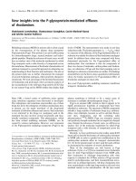
Figure 1 Histology findings in placebo and steroid-induced ANFH rats. (a) Photomicrographs showing histological findings in placebo- (I-IV) and
steroid-treated WKY rats (V-VIII & IX-XII) femoral heads. I-IV: No osteonecrosis, normal osteocytes (arrow), V-VIII: Early stage of osteonecrosis, normal
osteocytes (arrow), empty lacunae (arrow head), IX-XII: Late stage of osteonecrosis, empty lacunae (arrow head), complete necrosis of bone marrow
(asterisk), H&E staining, I, V, IX ×20; II, VI, X ×40; III, VII, XI ×100; IV, VIII, XII ×200, dotted square chosen to be magnified. (b) Photomicrographs showing
apoptosis of osteocytes as a marker of early ANFH. TUNEL staining apoptosis assay counterstained with 0.5% methyl green solution. I-III: Normal fem-
oral head tissues in placebo-treated WKY rats, normal osteocytes (arrow head), normal bone marrow (double asterisk), IV-VI: Early stage of osteone-
crosis in steroid-treated WKY rats, TUNEL positive osteocytes (arrow), empty lacunae (dotted arrow), normal osteocytes (arrow head), normal bone
marrow (double asterisk). VII-IX: Late stage of osteonecrosis in steroid-treated WKY rats, TUNEL positive osteocytes (arrow), empty lacunae (dotted
arrow), complete necrotic bone marrow (asterisk), I, IV, VII ×40; II, V, VIII ×100; III, VI, IX ×200, dotted square chosen to be magnified.
Kerachian et al. Arthritis Research & Therapy 2010, 12:R124
/>Page 4 of 12
stained with 0.5% methyl green solution (0.5 g ethyl violet
(Sigma-Aldrich) in 100 ml sodium acetate buffer, 0.1 M
and pH.4.2), cleared, mounted and evaluated by light
microscopy.
RNA extraction from rat bone specimens
Total RNA was extracted by an innovative method con-
sisting of a combination of TRIzol® Reagent (Invitrogen,
Carlsbad, CA, USA) and RNeasy Mini kit (QIAGEN Inc.)
followed by DNase I treatment (QIAGEN Inc.). Briefly,
femoral head specimens were removed from RNALater
and washed thoroughly with diethyl pyrocarbonate
(DEPC) -treated phosphate buffer solution (PBS). Femo-
ral head specimens were placed in liquid nitrogen. The
specimens were ground to a fine powder with a porcelain
mortar and pestle. TRIzol®
1 ml was then added to each
ground femoral head specimen. After vortexing for one
minute, the homogenized specimen was incubated for
five minutes at room temperature (RT) and 0.2 ml Chlo-
roform (Sigma-Aldrich) was added per 1 ml of TRIzol®.
After vortex use of 15 seconds the samples were incu-
bated for three minutes at room temperature. The sam-
ples were then centrifuged at 12,000 × g for 15 minutes at
4°C. The aqueous phase was removed from each sample
and one volume of ethanol was added to it and mixed
thoroughly. Up to 700 μl of the sample including any pre-
cipitate that may have formed was transferred into an
RNeasy Mini Spin Column. The column was then pro-
cessed according to the RNeasy Mini kit manufacturer
instruction. Any Genomic DNA contamination was
removed by treating the samples with DNase I. The RNA
quality was assessed using RNA 6000 NanoChips with
the Agilent 2100 Bioanalyzer (Agilent, Wilmington, DE,
USA).
Affymetrix exon arrays
Affymetrix GeneChip®
Rat Exon 1.0 ST array interrogat-
ing over 850,000 exon clusters within the known and pre-
dicted transcribed regions of the entire genome and
about one million probe sets was used. Affymetrix exon
array was performed on 15 RNA samples of GC-treated
and non-treated rats divided in three groups based on
histological evaluation: Group 1- Placebo/ANFH(-);
Group 2- GC-treated/ANFH(+) and Group 3- GC-
treated/ANFH(-), each group consisting of five samples.
Biotin-labelled targets for the microarray experiment
were prepared using 1 μg of total RNA. Ribosomal RNA
was removed with the RiboMinus Human/Mouse Tran-
scriptome Isolation Kit (Invitrogen, Eugene, Oregon,
USA) and cDNA was synthesized using the GeneChip
WT (Whole Transcript) Sense Target Labeling and Con-
trol Reagents kit as described by the manufacturer
(Affymetrix, Santa Clara, CA, USA). The sense cDNA
was then fragmented by uracil DNA glycosylase and
apurinic/apyrimidic endonuclease-1 and biotin-labeled
with terminal deoxynucleotidyl transferase using the
GeneChip WT Terminal labeling kit (Affymetrix).
Hybridization was performed using five micrograms of
biotinylated target, which was incubated with the
GeneChip Rat Exon 1.0 ST array (Affymetrix) at 45°C for
16 to 20 h. After hybridization, non-specifically bound
material was removed by washing and specifically bound
target was detected using the GeneChip Hybridization,
Wash and Stain kit, and the GeneChip Fluidics Station
450 (Affymetrix). The arrays were scanned using the
GeneChip Scanner 3000 7G (Affymetrix). We used
Affymetrix Power tools (Affymetrix), R and in-house
built Perl scripts to filter the background noise based on
the detection above background results that is the detec-
tion metric generated by comparing Perfect Match
probes to a distribution of background probes. Rat exon
array data was analyzed by Dr Daniel Bird from Creative
Biomics CD Inc. (Shirley, NY, USA): data were normal-
ized based on the Iter-PLIER algorithm by using Affyme-
trix Power tools, R and in-house built Perl scripts. The
genes with low signal (less than 100) were removed from
the study. The differentially expressed genes were
detected between three groups (G2 vs G1, G3 vs G2, and
G3 vs G1) (P < 0.05, Fold Change (FC) > 1.5) using in
house built R script, infer with t-test and adjusted with
Benjamini and Hochberg FDR method [27].
Real-Time Polymerase Chain Reaction (SybrGreen RT-PCR)
Real-time PCR was carried out according to the protocol
provided by the manufacturer for the QuantiTect
SYBR®Green RT-PCR kit (QIAGEN Inc.). QuantiTect
Primer Assays (Rn_A2m_1_SG, Rn_Col2a1_1_SG,
Rn_Mia1_1_SG, Rn_Actb_1_SG) were provided by QIA-
GEN Inc and a thermal cycler (Prism 7900, Applied Bio-
systems, Foster City, CA, USA) was used. The reaction
was set up in 10 μl final volume applying the following
conditions: cycling 50°C (30 minutes), 95°C (15 minutes)
and for 45 cycles the conditions were 94°C (15 sec), 55°C
(30 sec) and 72°C (30 sec). For the relative quantification
of gene expression, the comparative threshold cycle (ΔCt)
method was employed and normalized against β-Actin
rRNA, which was measured by the same method. All
PCR reactions were performed in triplicate. Control reac-
tions were set up lacking reverse transcriptase to assess
the level of contaminating genomic DNA.
Immunohistochemical (IHC) study
Paraffin-embedded sections were placed at 60°C for 15
minutes, incubated in xylene for 15 minutes, and then
transferred sequentially into 100% ethanol, 95% ethanol,
70% ethanol, and 50% ethanol for five minutes at RT. Sec-
tions were rinsed in deionised water and the endogenous
peroxidase activity was blocked with incubating sections
Kerachian et al. Arthritis Research & Therapy 2010, 12:R124
/>Page 5 of 12
in 3% H
2
O
2
in distilled water for five minutes. The slides
were washed in several changes of distilled water. Antigen
was retrieved by incubating the slides in Digest-All™ 3
(Invitrogen Immunodetection, Carlsbad, CA, USA) for 10
minutes. After several washes with PBS the slides were
stained using R.T.U. Vectastain®
Universal Quick kit (Vec-
tor Laboratories, Inc., Burlingame, CA, USA) according
to the manufacturer's instructions. Several primary anti-
bodies were used: 1:200 dilution of mouse anti-rat α-2-
macroglobulin globulin monoclonal antibody (clone
129736, R&D Systems, Minneapolis, MN, USA); predi-
luted mouse anti-rat collagen type II alpha 1 monoclonal
antibody (Abcam Inc, Cambridge, MA, USA) or 1:50
dilution of rabbit anti-rat melanoma inhibitory activity
(MIA) polyclonal antibody (Santa Cruz Biotechnology,
Santa Cruz, CA, USA). According to the manufacturer's
instructions the secondary antibody is a prediluted bioti-
nylated antibody manufactured in horse, which recog-
nizes rabbit IgG, mouse IgG and goat IgG. The slides
were counterstained with 0.5% methyl green solution as
described before.
Statistical analysis
Data reported on microarray results utilized in-house
Perl scripts with t-test and adjusted with B-H FDR
method to examine differentially expressed genes
between two groups (P < 0.05, Fold Change (FC) > 1.5).
RT-PCR results were given as the mean ± standard error
of the mean (SEM). Comparison between groups was
made with Student's t-test. For small size samples Mann-
Whitney U test was used since normal distribution of
data was not assumed. Differences were considered sig-
nificant at P-values less than 0.05. Principle component
analysis (PCA) was performed using R package to provide
a global view of how the various sample groups were
related.
Results
Histological and apoptosis findings
Histological findings displayed normal, early and late
stages of ANFH based on the presence or absence of
osteocytes in the lacunaes (Figure 1a). The use of the
TUNEL assay to detect apoptosis showed apoptotic
osteocytes were located in the osteonecrotic samples
without features of inflammation and visible necrosis,
such as hyperemia, round cell infiltration, or lipid cyst
formation. There was no appositional bone formation
associated with granulation tissue around dead bone in
keeping with the early stages of ANFH (Figure 1b, IV-X).
When the same TUNEL reaction was performed on con-
trol tissue (without prior digestion with DNase), a fewer
number of cells (one or two) were labeled (Figure 1b, I-
III).
Microarray analysis
In the Affymetrix analysis, G2 replicates were compared
with G1 and G3, separately, and G3 replicates were com-
pared with G1 to generate a list of differentially expressed
genes. The results were analyzed by a defined set of crite-
ria in which the altered expression of a gene must have at
least a change of ± 1.5-fold (FC = fold change) and a P-
value less than 0.05. These criteria resulted in the identifi-
cation of 51 genes with significant modulation in G2
compared with G1 and six genes with significant modula-
tion for G3 to G2 (Tables 2 and 3). They also identified
229 genes in G3 versus G1 (Table 4). In this table, only the
genes with a change of ± 1.8-fold (FC = fold change) are
represented due to the exhaustive list of genes. Although
rat exon array demonstrated a significant upregulation of
51 genes when comparing G2 to G1, alpha-2-macroglob-
ulin gene was particularly found to be overexpressed
when comparing steroid-treated Wistar Kyoto rats which
had developed ANFH (G2) to placebo rats (G1) (FC =
3.52, P = 0.0005). Collagen type II alpha-1 (Col2A1) and
Melanoma Inhibitory Activity-1 (MIA) genes were also
found to be significantly overexpressed by exon array
analysis (FC = 2.52, P = 0.0005 and FC = 2.29, P = 0.0008
respectively). The downregulation of some genes was not
considered significant in terms of fold change compared
to the upregulated genes; therefore we were able to focus
on the genes that were upregulated. Significantly modu-
lated genes were categorized into clusters according to
their biological functions using DAVID, a functional
annotation tool provided by National Institute of Allergy
and Infectious Diseases-NIH. Modulated genes were
grouped mainly into clusters of skeletal development,
ossification and bone remodelling. Other functional
classes significantly represented in the steroid-induced
avascular necrosis included response to steroid stimulus
response, apoptosis, blood vessel morphogenesis, vascu-
lature development, cell growth, proliferation and differ-
entiation associated genes. In comparison of G3 versus
G1, A2M and Col2A1 were not significantly overex-
pressed whereas MIA was found to be the most up-regu-
lated gene in that group comparison (FC = 3.71, P = 0.00).
Real time PCR Verification of GeneChip Data
From the microarray results, the three genes (α-2-macro-
globulin (A2M), collagen type II alpha-1 (Col2A1), mela-
noma inhibitory activity-1 (MIA)) showing the highest
upregulation or fold change were selected for validation
by means of RT-PCR. The directional fold change was
confirmed for all three genes and the correlation with
microarray results was established. Some variations,
however, were noted in the fold-change values demon-
strated by real time PCR compared with values obtained
by GeneChip analysis (for A2M, FC = 3.52 with exon
Kerachian et al. Arthritis Research & Therapy 2010, 12:R124
/>Page 6 of 12
Table 2: Differentially expressed genes from comparing Group 2 (G2) versus Group 1 (G1)
Annotation PV FC
NM_012488 alpha-2-macroglobulin 0.0005 + 3.52
NM_012929 collagen, type II, alpha 1 0.0005 + 2.52
NM_030852 melanoma inhibitory activity 1 0.0008 + 2.29
NM_033499 scrapie responsive gene 1 0.0054 + 2.08
NM_017094 growth hormone receptor 0.0142 + 1.93
NM_053669 SH2B adaptor protein 2 0.0213 + 1.89
NM_080698 fibromodulin 0.0099 + 1.87
NM_133523 matrix metallopeptidase 3 0.0034 + 1.87
NM_012999 Proprotein convertase subtilisin/kexin type 6 0.0117 + 1.80
NM_138889 cadherin 13 0.0049 + 1.77
NM_031808 calpain 6 0.0086 + 1.73
NM_001002826 murinoglobulin 2 0.0022 + 1.72
NM_145776 solute carrier family 38, member 3 0.0040 + 1.71
NM_012846 fibroblast growth factor 1 0.0441 + 1.70
NM_017058 vitamin D receptor 0.0065 + 1.69
NM_001009662 carbonic anhydrase 8 0.0275 + 1.68
NM_031590 WNT1 inducible signaling pathway protein 2 0.0105 + 1.67
NM_012587 integrin binding sialoprotein 0.0276 + 1.66
NM_053816 calcitonin receptor 0.0316 + 1.63
NM_013191 S100 protein, beta polypeptide, neural 0.0123 + 1.62
NM_031828 potassium large conductance calcium-activated channel, subfamily M, alpha
member 1
0.0018 + 1.62
NM_133569 angiopoietin-like 2 0 + 1.62
NM_199398 pannexin 3 0.0032 + 1.62
NM_053605 sphingomyelin phosphodiesterase 3, neutral 0.0126 + 1.62
NM_170668 solute carrier family 13 (sodium-dependent citrate transporter), member 5 0.0167 + 1.60
NM_053977 cadherin 17 0.0233 + 1.60
NM_199407 unc-5 homolog C (C. elegans) 0.0002 + 1.60
NM_012620 serine (or cysteine) peptidase inhibitor, clade E, member 1 (also designated
plasminogen activator inhibitor-1 or PAI-1)
0.0003 + 1.60
NM_022667 solute carrier organic anion transporter family, member 2a1 0.0055 + 1.59
NM_001034009 melanoma cell adhesion molecule 0.0032 + 1.58
NM_053288 orosomucoid 1 0.0236 + 1.57
NM_031131 transforming growth factor, beta 2 0.0015 + 1.57
NM_013059 alkaline phosphatase, liver/bone/kidney 0.0218 + 1.57
NM_133303 basic helix-loop-helix domain containing, class B3 0.0114 + 1.56
NM_198768 immunoglobulin superfamily, member 10 0.0467 + 1.55
NM_001017479 transmembrane protein 100 0.0431 + 1.54
NM_020073 parathyroid hormone receptor 1 0.0370 + 1.54
NM_024400 a disintegrin-like and metallopeptidase (reprolysin type) with thrombospondin
type 1 motif, 1
0.0086 + 1.54
NM_001014043 sphingomyelin synthase 2 0.0131 + 1.53
NM_023970 transient receptor potential cation channel, subfamily V, member 4 0.0219 + 1.52
NM_020656 parvin, alpha 0.0072 + 1.52
Kerachian et al. Arthritis Research & Therapy 2010, 12:R124
/>Page 7 of 12
array and 5.85 with RT-PCR). Variations in fold change
values between GeneChip and real time PCR might have
been due to different methods of normalization and spec-
ificity/sensitivity of each method but the trends were the
same for the two methods (differences with P-values:
0.005 to 0.0009, Table 5).
Immunohistochemistry
We performed immunohistochemistry staining on the
three candidate genes which showed the highest upregu-
lation, A2M, Col2A1 and MIA, when comparing G2 to
G1. Protein expression of A2M was shown to be
increased in rats induced with steroids and developing
ANFH (Group 2) as compared to the placebo rats without
ANFH (Group 1) thus correlating with the mRNA
expression levels from GeneChip analysis and RT-PCR
method (Figure 2). Notably, immunohistochemical find-
ings for the two other genes of interest (COL2A1 and
MIA) failed to show enhanced protein expression.
Discussion
The early events in the pathogenesis of ANFH are incom-
pletely understood due to a typically late diagnosis after
fracture and collapse of the femoral head. Besides bone
marrow changes, evidence has shown that apoptosis is
involved in the early stages of steroid-induced osteone-
crosis [26]. Weinstein et al. reported that the number of
apoptotic bone cells increased significantly in mice after
steroid administration [28]. Recent studies have shown
apoptotic cells in clinical and animal models of GC-
induced ANFH [26,29,30].
In previous studies, we characterized an inbred rat
(WKY) susceptible to develop steroid-induced osteone-
crosis [31]. It is possible that this strain of rats has geneti-
cally predisposing factors to develop ANFH and
additional risk exposures (GC) will facilitate the develop-
ment of the disease. In our animal model, prednisone
administration enhanced the incidence of the disease in
up to 75% (6/8) of the male WKY rats, suggesting it is a
suitable model. In the literature, 5 to 15 week-old rats
have been used to study non-traumatic ANFH [23,26,32].
In the current study, WKY rats started to receive continu-
ous steroid dosage released from the pellets at the age of
five weeks for 25 weeks. Harvest at six months showed
classical histological signs of early ANFH.
For the Affymetrix GeneChip findings, comparison of
G2 versus G1 indicated that multiple pathological reac-
tions occurred. According to the functional annotation
tool (DAVID), modulated genes in the comparison of G2
and G1 (Table 2) were grouped mainly into skeletal devel-
opment, ossification and bone remodelling. Functional
clusters of genes were significantly represented by steroid
NM_175578 regulator of calcineurin 2 0.0390 + 1.52
NM_031655 latexin 0.0080 + 1.52
NM_001013218 receptor accessory protein 6 0.0045 + 1.52
NM_001005562 cAMP responsive element binding protein 3-like 1 0.0376 + 1.50
NM_001017496 chemokine (C-X-C motif) ligand 13 0.0140 - 0.55
ENSRNOT00000060250 similar to T-cell receptor alpha chain precursor V and C regions (TRA29) 0.0154 - 0.64
NM_203410 interferon, alpha-inducible protein 27-like 0.0325 - 0.64
NM_001008836 RT1-CE13//RT1 class I, CE13 0.0157 - 0.64
NM_001002280 MAS-related GPR, member X2 0.0021 - 0.66
NM_001008855 RT1 class Ib gene, H2-TL-like, grc region (N3) 0.0350 - 0.67
(+), positive regulation, (-), negative regulation; FC, fold change; V, P-value.
Table 2: Differentially expressed genes from comparing Group 2 (G2) versus Group 1 (G1) (Continued)
Table 3: Differentially expressed genes from comparing group 3 (G3) versus 2 (G2)
Annotation PV FC
NM_001012357 chemokine (C-C motif) ligand 9 0.0371 + 1.86
NM_013153 hyaluronan synthase 2 0.0103 + 1.70
NM_030852 melanoma inhibitory activity 1 0.0082 + 1.62
NM_001012072 protein phosphatase 1, regulatory (inhibitor) subunit 3C 0.0411 + 1.58
NM_001009639 tubulin polymerization-promoting protein family member 3 0.0243 + 1.56
NM_012497 aldolase C 0.0155 + 1.54
(+), positive regulation; (-), negative regulation; FC, fold change; PV, P-value.
Kerachian et al. Arthritis Research & Therapy 2010, 12:R124
/>Page 8 of 12
Table 4: Differentially expressed genes from comparing Group 3 (G3) versus 1 (G1). Only genes with fold change above 1.8
have been shown
Annotation PV FC
NM_030852 melanoma inhibitory activity 1 0.0000 + 3.71
NM_001002826 murinoglobulin 2 0.0104 + 2.64
NM_031808 calpain 6 0.0002 + 2.62
NM_019189 hyaluronan and proteoglycan link protein 1 0.0000 + 2.61
NM_001002826 murinoglobulin 2 0.0007 + 2.37
NM_001012034 ADP-ribosyltransferase 3 0.0015 + 2.36
NM_057104 ectonucleotide pyrophosphatase/phosphodiesterase 2 0.0091 + 2.25
NM_001009662 carbonic anhydrase 8 0.0010 + 2.24
NM_013191 S100 protein, beta polypeptide, neural 0.0002 + 2.24
NM_138898 phospholipase B 0.0165 + 2.18
NM_134432 angiotensinogen (serpin peptidase inhibitor, clade A, member 8) 0.0294 + 2.16
NM_012620 serine (or cysteine) peptidase inhibitor, clade E, member 1 0.0036 + 2.15
NM_031828 potassium large conductance calcium-activated channel, subfamily M, alpha 1 0.0003 + 2.15
NM_133523 matrix metallopeptidase 3 0.0162 + 2.15
NM_138889 cadherin 13 0.0011 + 2.14
NM_133569 angiopoietin-like 2 0.0000 + 2.14
NM_001012163 LIM and senescent cell antigen like domains 2 0.0048 + 2.13
NM_001013213 integrin beta 3 binding protein (beta3-endonexin) 0.0013 + 2.10
NM_198748 scinderin 0.0017 + 2.09
NM_012497 aldolase C 0.0002 + 2.08
NM_031694 heat shock factor 2 0.0085 + 2.05
NM_198768 immunoglobulin superfamily, member 10 0.0015 + 2.03
NM_053977 cadherin 17 0.0062 + 2.02
NM_001014060 similar to SRY (sex determining region Y)-box 5 isoform a 0.0002 + 1.97
NM_012999 proprotein convertase subtilisin/kexin type 6 0.0025 + 1.96
NM_013080 protein tyrosine phosphatase, receptor-type, Z polypeptide 1 0.0072 + 1.94
NM_001002819 glutamine-fructose-6-phosphate transaminase 2 0.0096 + 1.93
BC079425 hypothetical protein LOC654482 0.0009 + 1.92
NM_031131 transforming growth factor, beta 2 0.0013 + 1.91
NM_022927 midline 1 0.0070 + 1.90
NM_181366 G protein-coupled receptor 64 0.0010 + 1.90
NM_022230 stanniocalcin 2 0.0003 + 1.89
NM_199398 pannexin 3 0.0021 + 1.87
NM_053605 sphingomyelin phosphodiesterase 3, neutral 0.0053 + 1.86
NM_001009647 mitochondrial ribosomal protein L16 0.0008 + 1.85
NM_001077641 phospholipase C, beta 1 0.0116 + 1.85
NM_020073 parathyroid hormone receptor 1 0.0016 + 1.83
NM_017135 adenylate kinase 3-like 1 0.0149 + 1.83
NM_013000 peptidylglycine alpha-amidating monooxygenase 0.0065 + 1.82
NM_001007656 microtubule-associated protein, RP/EB family, member 3 0.0008 + 1.81
NM_031590 WNT1 inducible signaling pathway protein 2 0.0002 + 1.81
NM_022382 phosphodiesterase 4D interacting protein (myomegalin) 0.0127 + 1.80
NM_134327 CD69 antigen 0.0141 - 0.65
Kerachian et al. Arthritis Research & Therapy 2010, 12:R124
/>Page 9 of 12
stimulus response, apoptosis, blood vessel morphogene-
sis, vasculature development, coagulation-related, cell
growth, proliferation and differentiation associated
genes.
The expression of steroid stimulus response genes
(A2M, alkaline phosphatase, tissue-nonspecific, trans-
forming growth factor beta 2 and potassium large conduc-
tance calcium-activated channel, subfamily m, alpha
member 1) were, as predicted, altered significantly. Previ-
ous in vivo and in vitro models as well as clinical studies
showed that steroids induce apoptosis in osteoblasts and
osteocytes [30,33-35]. Amongst the 51 differentially regu-
lated genes identified in our gene array analysis (Table 2),
five genes (S100 protein-beta polypeptide, transforming
growth factor-beta 2, vitamin D receptor, unc-5 homolog c
(C. elegans) and growth hormone receptor) are in fact
components of the apoptosis pathway.
The process of apoptosis can be directly induced by ste-
roids but is also related to thrombosis in the blood vessels
of the femoral head. In fact, the vascular hypothesis
(regional endothelial bed dysfunction) appears to be rele-
vant in the pathogenesis of ANFH. Damage or activation
of femoral head endothelial cells results in abnormal
blood coagulation and thrombi formation [36]. Due to
heterogeneity of the phenotype expression between
endothelial cells in the body, a local endothelial cell dys-
function can occur where the femoral head endothelial
cells react differently to the ANFH risk factors (GCs) than
other endothelial cells in the body. In keeping with the
theory of endothelial cell activation having a role in
ANFH, coagulation-related gene expression in particular
serine (or cysteine) peptidase inhibitor, clade E, member 1
also named plasminogen activator inhibitor 1 (PAI-1), a
serine protease inhibitor that is synthesized and released
by endothelial cells in the blood, was shown to be signifi-
cantly over-expressed in this study. An increase in PAI-1
suppresses the generation of plasmin resulting in hypofi-
brinolysis and a relative hypercoagulable state [1].
Decreased fibrinolytic activity, which may be a conse-
quence of increased PAI-1, has been described in patients
with ANFH [37], although a few studies have reported
that there were no significant differences in the levels of
thrombotic and fibrinolytic factors [18,19].
Similarly, our findings demonstrate that several genes
involved in the dynamic remodelling structure of the
femoral head are also shown to be differentially expressed
in ANFH (Table 2). Clinically this may be relevant in that
if the balance between degradation and repair (bone
remodelling) becomes shifted to degradation and bone
loss by the effect of GC, a failure of structural integrity at
the subchondral region of bone with collapse could occur.
NM_019295 CD5 antigen 0.0114 - 0.65
NM_013121 CD28 antigen 0.0136 - 0.65
NM_031147 cold inducible RNA binding protein 0.0025 - 0.64
NM_001012226 signal transducer and activator of transcription 4 0.0246 - 0.63
NM_001008855 RT1 class Ib gene, H2-TL-like, grc region (N3) 0.0005 - 0.60
NM_001012461 deoxynucleotidyltransferase, terminal 0.0140 - 0.59
NM_173096 myxovirus (influenza virus) resistance 1 0.0186 - 0.59
NM_001009680 2 ' -5 ' oligoadenylate synthetase 1I 0.0039 - 0.58
NM_001008836 RT1 class I, CE13 0.0111 - 0.56
NM_203410 interferon, alpha-inducible protein 27-like 0.0018 - 0.51
(+), positive regulation, (-), negative regulation; FC, fold change; PV, P-value.
Table 4: Differentially expressed genes from comparing Group 3 (G3) versus 1 (G1). Only genes with fold change above 1.8
have been shown (Continued)
Table 5: Correlation of gene expression comparing Groups 2 (G2) and 1 (G1) as assessed by microarray and real time PCR (P
< 0.005 for all genes)
Annotation Fold Change of a signal
Microarray Real time PCR
NM_012488 alpha-2-macroglobulin 3.52 5.85
NM_012929 collagen, type II, alpha 1 2.52 4.42
NM_030852 melanoma inhibitory activity 1 2.29 2.80
Kerachian et al. Arthritis Research & Therapy 2010, 12:R124
/>Page 10 of 12
In the present study, results showed A2M gene expres-
sion to be the most significantly upregulated gene when
comparing G2 to G1. Correlation was obtained at the
microarray, RT-PCR as well as the protein level as dem-
onstrated by IHC study results. Most importantly, A2M
was not significantly upregulated when comparing G3 to
G1. A2M is a plasma-derived matrix metalloproteinase
inhibitor which obstructs cartilage degradation induced
by matrix metalloproteinases [38]. The literature sup-
ports the role of corticosteroids in the modulation of
A2M [39,40]. In both reports, corticosteroids were shown
to enhance A2M levels. A2M is reported as being impli-
cated in cartilage degradation [41], and as an osteogenic
growth peptide (OGP) - binding protein. Activated A2M
may thus participate in the removal of OGP from the sys-
tem [42]. Additional reports suggest inhibition of BMP-1
(bone morphogenic protein-1) by A2M [43]. A2M has
been identified on the luminal surface of endothelial cells
in sections of normal human arteries and veins [44]. A2M
has also been implicated in hemostasis as a regulator of
thrombin [45] and in the development of thromboembo-
lism in children [46]. Together, all these findings suggest
that A2M shares haemostatic, cartilaginous and osteo-
genic properties and may have a potential role in the
development of early steroid-induced ANFH. Determina-
tion of whether A2M over-expression in our study is
either the result or the cause of the apoptosis found in our
rats developing early ANFH following administration of
steroids, will require further study.
Two other genes of interest, Col2A1 and MIA, were also
shown to be over-expressed significantly by microarray
analysis and RT-PCR results but immunohistochemical
study failed to show an increased cell surface expression
of these genes.
Comparing the gene profiling of G3 versus G2, six
genes stood out in our analyses (Table 3). Although G3
animals have not developed ANFH, their gene profile
reflects inhibition of osteoblast proliferation, differentia-
tion and osteoclast activation. Perhaps most osteogenic
cells in this group have not gone through the apoptotic
phase and there are more viable cells expressing these
molecules in comparison to G2. Differences could also be
explained in that gene expression analysis findings are
supportive of a result effect indicating steroid treatment
and a disease effect affecting the apoptotic process are
involved in the early stages of ANFH. Secondly, a genetic
variation based on differences in transcription and trans-
lation could provide an explanation for the phenotypic
differences found in our study. Thirdly, epigenetic varia-
tion, resulting from the interaction between the genotype
and the environment, is also a potential process that
could explain the findings that not all treated animals
developed early ANFH when submitted to the same
experimental conditions. Also, any of the genes listed in
the comparison of G3 to G2 (Table 3) with the exception
of MIA, could have a protective effect against the devel-
opment of steroid-induced early AVN. Similarly, the
absence of A2M over-expression in that same group com-
Figure 2 Upregulation of A2M surface protein expression in steroid-induced early ANFH. Immunohistochemistry comparing the A2M (a, b),
COL2A1 (c, d) and MIA (CD RAP) (e, f) protein expression between G1 (a, c, e) and G2 (b, d, f) WKY rats, showing enhancement of A2M expression in
G2 compared to G1 but no enhancement shown for COL2A1 and MIA genes; brown color demonstrates protein expression and green color displays
intact nucleus of cells, (a-d) ×40, (e, f) ×100.
Kerachian et al. Arthritis Research & Therapy 2010, 12:R124
/>Page 11 of 12
parison G3 to G2, and in group comparison G3 to G1 is
consistent with the phenotypic absence of early ANFH in
rats representing G3.
Conclusions
In summary, it is postulated that multiple pathological
reactions occur during ANFH. Genetic predisposition
contributes to the development of ANFH. There is nor-
mally a balance between degenerative and regenerative
molecules in the bone environment of the femoral head.
GCs may trigger a degenerative process as well as inhibit
the repair. In this study, several molecules are signifi-
cantly upregulated and could be involved in the patho-
genesis of ANFH. However, only A2M gene over-
expression has been consistently found at the microarray,
RT-PCR and protein level for the three genes showing the
most significant upregulation. Besides, A2M was not sig-
nificantly upregulated in rats administered steroids but
without developing the disease. Thus, A2M seems to be a
possible biomarker more of ANFH itself (induced by ste-
roids) than a marker of steroids alone. It remains to be
determined in which specific pathway (although likely in
the endothelial cell activation and/or the apoptosis path-
way) and at which level, the effect of this gene occurs in
corticosteroid-induced ANFH. Identifying its role within
a specific pathway will likely lead to a better understand-
ing of the molecular events that follow the administration
of corticosteroids and subsequent irreversible necrosis
and bone collapse. Obviously, investigation of the use of
A2M as a potential marker for the early warning of ANFH
should be carried out.
ArrayExpress accession code: [E-MEXP-2751].
Abbreviations
A2M: alpha-2-macroglobulin; ANFH: avascular necrosis of the femoral head;
BMP-1: bone morphogenic protein-1; Col2A1: collagen type II alpha-1; ΔCt:
comparative threshold cycle; DAB: 3:3'-diaminobenzidine; DEPC: diethyl pyro-
carbonate; EC: endothelial cells; FC: fold change; G1: group 1; G2: group 2; G3:
group 3; GCs: glucocorticosteroids; IHC: immunohistochemistry; MIA: Mela-
noma Inhibitory Activity-1; OGP: osteogenic growth peptide; PAI-1: plasmino-
gen activator inhibitor 1; PCA: principle component analysis; PBS: phosphate
buffer solution; RT: room temperature; RT-PCR: real-time polymerase chain
reaction; SEM: standard error of the mean; TDT: terminal dexoynucleotidyl
transferase; TUNEL: terminal dexoynucleotidyl transferase mediated deoxyuri-
dine triphosphate biotin nick end labelling; WKY: Wistar Kyoto.
Competing interests
CS has applied for a provisional patent for A2M (α-2-Macroglobulin) as a diag-
nostic assay for Avascular Necrosis of the Femoral Head. The other authors
declare that they have no competing interests.
Authors' contributions
All authors participated in the study. MAK made a major contribution to the
writing of the manuscript's first draft, and conducted the experiments involved
in the study. CS made a major contribution to the design of the study, data
interpretation and scientific revision of the manuscript. DC, EJH and TYC made
equal contributions to data interpretation and scientific revision of the manu-
script. EJH made a major contribution to the editing and grammar of the man-
uscript. LRB and AN made major contributions to the histological experiments
involved in the study. All authors participated in the manuscript preparation
and revision. All authors read and approved the final manuscript.
Acknowledgements
This work has been supported by the Montreal General Hospital Foundation
(CS), by the generous research award from Mr John D. Miller (CS) and support
from FRSQ Chercheur-Boursier Clinicien Senior (EJH). We thank Dr André Pon-
ton at McGill University and Genome Quebec Innovation Centre for perform-
ing the GeneChip technology. We thank Dr Daniel Bird from Creative Biomics
CD Inc. for performing the rat exon array analysis.
Author Details
1
Department of Human Genetics, McGill University Health Center (MUHC),
1650 Cedar Avenue, Montreal, QC H3G 1A4, Canada,
2
Department of
Medicine, Division of Haematology, McGill University Health Center (MUHC),
1650 Cedar Avenue, Montreal, QC H3G 1A4, Canada,
3
Department of
Oncology, McGill University Health Center (MUHC), 1650 Cedar Avenue,
Montreal, QC H3G 1A4, Canada,
4
Division of Orthopaedic Surgery, McGill
University Health Center (MUHC), 1650 Cedar Avenue, Montreal, QC H3G 1A4,
Canada,
5
Division of Anatomic Pathology, Hôpital du Sacré-Coeur de Montréal,
5400 Gouin Blvd, Montreal, QC H4J 1C5, Canada and
6
Department of
Pathology, McGill University Health Center (MUHC), 1650 Cedar Avenue,
Montreal, QC H3G 1A4, Canada
References
1. Aaron RK, Ciombor DM: Coagulopathies and osteonecrosis. Curr Opin
Orthop 2001, 12:378-383.
2. Chandler FA: Coronary disease of the hip. 1949. Clin Orthop Relat Res
2001, 386:7-10.
3. Asano T, Takahashi KA, Fujioka M, Inoue S, Okamoto M, Sugioka N, Nishino
H, Tanaka T, Hirota Y, Kubo T: ABCB1 C3435T and G2677T/A
polymorphism decreased the risk for steroid-induced osteonecrosis of
the femoral head after kidney transplantation. Pharmacogenetics 2003,
13:675-682.
4. Tektonidou MG, Moutsopoulos HM: Immunologic factors in the
pathogenesis of osteonecrosis. Orthop Clin North Am 2004, 35:259-263.
vii
5. Lafforgue P: Pathophysiology and natural history of avascular necrosis
of bone. Joint Bone Spine 2006, 73:500-507.
6. Kerachian MA, Harvey EJ, Cournoyer D, Chow TY, Séguin C: Avascular
necrosis of the femoral head: vascular hypotheses. Endothelium 2006,
13:237-244.
7. Nobillot R, Le Parc JM, Benoit J, Paolaggi JB: Idiopathic osteonecrosis of
the hip in twins. Ann Rheum Dis 1994, 53:702.
8. Miyamoto Y, Matsuda T, Kitoh H, Haga N, Ohashi H, Nishimura G, Ikegawa
S: A recurrent mutation in type II collagen gene causes Legg-Calve-
Perthes disease in a Japanese family. Hum Genet 2007, 121:625-629.
9. Chen WM, Liu YF, Lin MW, Chen IC, Lin PY, Lin GL, Jou YS, Lin YT, Fann CS,
Wu JY, Hsiao KJ, Tsai SF: Autosomal dominant avascular necrosis of the
femoral head in two Thaiwanese pedigress and linkage to
chromosome 12q13. Am J Hum Genet 2004, 75:310-317.
10. Liu YF, Chen WM, Lin YF, Yang RC, Lin MW, Li LH, Chang YH, Jou YS, Lin PY,
Su JS, Huang SF, Hsiao KJ, Fann CS, Hwang HW, Chen YT, Tsai SF: Type II
collagen gene variants and inherited osteonecrosis of the femoral
head. N Engl J Med 2005, 352:2294-2301.
11. Boss JH, Misselevich I: Osteonecrosis of the femoral head of laboratory
animals: the lessons learned from a comparative study of
osteonecrosis in man and experimental animals. Vet Pathol 2003,
40:345-354.
12. Ferrari P, Schroeder V, Anderson S, Kocovic L, Vogt B, Schiesser D, Marti HP,
Ganz R, Frey FJ, Kohler HP: Association of plasminogen activator
inhibitor-1 genotype with avascular osteonecrosis in steroid-treated
renal allograft recipients. Transplantation 2002, 74:1147-1152.
13. Glueck CJ, Fontaine RN, Gruppo R, Stroop D, Sieve-Smith L, Tracy T, Wang
P: The plasminogen activator inhibitor-1 gene, hypofibrinolysis, and
osteonecrosis. Clin Orthop Relat Res 1999, 366:133-146.
14. Jones JP Jr: Coagulopathies and osteonecrosis. Acta Orthop Belg 1999,
65(Suppl 1):5-8.
15. Jones LC, Mont MA, Le TB, Petri M, Hungerford DS, Wang P, Glueck CJ:
Procoagulants and osteonecrosis. J Rheumatol 2003, 30:783.
Received: 12 January 2010 Revised: 16 April 2010
Accepted: 25 June 2010 Published: 25 June 2010
This article is available from: 2010 Amin Kerachian et al.; licensee BioMed Central Ltd. This is an open access article distributed under the terms of the Creative Commons A ttribution License ( which permits unrestricted use, distribution, and reproduction in any medium, provided the original work is properly cited.Arthritis R esearch & Thera py 2010, 12:R124
Kerachian et al. Arthritis Research & Therapy 2010, 12:R124
/>Page 12 of 12
16. Pósán E, Hársfalvi J, Szepesi K, Gáspár L, Batár P, Udvardy M: Increased
platelet activation and decreased fibrinolysis in the pathogenesis of
aseptic necrosis of the femoral head. Platelets 1998, 9:233-235.
17. Hadjigeorgiou G, Dardiotis E, Dardioti M, Karantanas A, Dimitroulias A,
Malizos K: Genetic association studies in osteonecrosis of the femoral
head: mini review of the literature. Skeletal Radiol 2008, 37:1-7.
18. Asano T, Takahashi KA, Fujioka M, Inoue S, Ueshima K, Hirata T, Okamoto
M, Satomi Y, Nishino H, Tanaka T, Hirota Y, Kubo T: Relationship between
postrenal transplant osteonecrosis of the femoral head and gene
polymorphisms related to the coagulation and fibrinolytic systems in
Japanese subjects. Transplantation 2004, 77:220-225.
19. Séguin C, Kassis J, Busque L, Bestawros A, Theodoropoulos J, Alonso ML,
Harvey EJ: Non-traumatic necrosis of bone (osteonecrosis) is associated
with endothelial cell activation but not thrombophilia. Rheumatology
(Oxford) 2008, 47:1151-1155.
20. Schulte CM, Beelen DW: Avascular osteonecrosis after allogeneic
hematopoietic stem-cell transplantation: diagnosis and gender
matter. Transplantation 2004, 78:1055-1063.
21. Assouline-Dayan Y, Chang C, Greenspan A, Shoenfeld Y, Gershwin ME:
Pathogenesis and natural history of osteonecrosis. Semin Arthritis
Rheum 2002, 32:94-124.
22. Kerachian MA, Séguin C, Harvey EJ: Glucocorticoids in osteonecrosis of
the femoral head: a new understanding of the mechanisms of action. J
Steroid Biochem Mol Biol 2009, 114:121-128.
23. Wada M, Kumagai K, Murata M, Yamashita Y, Shindo H: Warfarin reduces
the incidence of osteonecrosis of the femoral head in spontaneously
hypertensive rats. J Orthop Sci 2004, 9:585-590.
24. Arlet J: A Traumatic Necrosis Of The Femoral Head: General Report In Bone
Circulation and Vascularization In Normal And Pathological Conditions New
York: Plenum Press; 1993:235-240.
25. Weinstein RS, Nicholas RW, Manolagas SC: Apoptosis of osteocytes in
glucocorticoid-induced osteonecrosis of the hip. J Clin Endocrinol
Metab 2000, 85:2907-2912.
26. Shibahara M, Nishida K, Asahara H, Yoshikawa T, Mitani S, Kondo Y, Inoue
H: Increased osteocyte apoptosis during the development of femoral
head osteonecrosis in spontaneously hypertensive rats. Acta Med
Okayama 2000, 54:67-74.
27. Benjamini Y, Hochberg Y: Controlling the false discovery rate: a practical
and powerful approach to multiple testing. Journal of Royal Statistical
Society. Series B (Methodological) 1995, 57:289-300.
28. Weinstein RS, Jilka RL, Parfitt AM, Manolagas SC: Inhibition of
osteoblastogenesis and promotion of apoptosis of osteoblasts and
osteocytes by glucocorticoids. Potential mechanisms of their
deleterious effects on bone. J Clin Invest 1998, 102:274-282.
29. Kabata T, Kubo T, Matsumoto T, Nishino M, Tomita K, Katsuda S, Horii T,
Uto N, Kitajima I: Apoptotic cell death in steroid induced osteonecrosis:
an experimental study in rabbits. J Rheumatol 2000, 27:2166-2171.
30. Weinstein RS, Nicholas RW, Manolagas SC: Apoptosis of osteocytes in
glucocorticoid-induced osteonecrosis of the hip. J Clin Endocrinol
Metab 2000, 85:2907-2912.
31. Kerachian MA, Cournoyer D, Nahal A, Harvey EJ, Chow TY, Séguin C:
Apoptotic cell death in steroid-induced bone disease: A pilot study in
rats. Bone 2007, 40:S287.
32. Mihara K, Hirano T: Standing is a causative factor in osteonecrosis of the
femoral head in growing rats. J Pediatr Orthop 1998, 18:665-669.
33. Calder JD, Buttery L, Revell PA, Pearse M, Polak JM: Apoptosis a
significant cause of bone cell death in osteonecrosis of the femoral
head. J Bone Joint Surg Br 2004, 86:1209-1213.
34. O'Brien CA, Jia D, Plotkin LI, Bellido T, Powers CC, Stewart SA, Manolagas
SC, Weinstein RS: Glucocorticoids act directly on osteoblasts and
osteocytes to induce their apoptosis and reduce bone formation and
strength. Endocrinology 2004, 145:1835-1841.
35. Jia D, O'Brien CA, Stewart SA, Manolagas SC, Weinstein RS:
Glucocorticoids act directly on osteoclasts to increase their life span
and reduce bone density. Endocrinology 2006, 147:5592-5599.
36. Kerachian MA, Cournoyer D, Harvey EJ, Chow T, Séguin C: Isolation and
characterization of human bone-derived endothelial cells.
Endothelium 2007, 14:115-121.
37. Glueck CJ, Glueck HI, Mieczkowski L, Tracy T, Speirs J, Stroop D: Familial
high plasminogen activator inhibitor with hypofibrinolysis, a new
pathophysiologic cause of osteonecrosis? Thromb Haemost 1993,
69:460-465.
38. Enghild JJ, Salvesen G, Brew K, Nagase H: Interaction of human
rheumatoid synovial collagenase (matrix metalloproteinase 1) and
stromelysin (matrix metalloproteinase 3) with human alpha 2-
macroglobulin and chicken ovostatin. Binding kinetics and
identification of matrix metalloproteinase cleavage sites. J Biol Chem
1989, 264:8779-8785.
39. Ramadori G, Knittel T, Schwögler S, Bieber F, Rieder H, Meyer zum
Büschenfelde KH: Dexamethasone modulates alpha 2-macroglobulin
and apolipoprotein E gene expression in cultured rat liver fat-storing
(Ito) cells. Hepatology 1991, 14:875-882.
40. Gordon AH, Limaos EA: Effects of bacterial endotoxin and
corticosteroids on plasma concentrations of alpha 2 macroglobulin,
haptoglobin and fibrinogen in rats. Br J Exp Pathol 1979, 60:434-440.
41. Luan Y, Kong L, Howell DR, LLalov K, Fajardo M, Bai XH, Di Cesare PE,
Goldring MB, Abramson SB, Liu CJ: Inhibition of ADAMTS-7 and
ADAMTS-12 degradation of cartilage oligomeric matrix protein by
alpha-2-macroglobulin. Osteoarthritis Cartilage 2008, 16:1413-1420.
42. Gavish H, Bab I, Tartakovsky A, Chorev M, Mansur N, Greenberg Z, Namdar-
Attar M, Muhlrad A: Human alpha 2-macroglobulin is an osteogenic
growth peptide-binding protein. Biochemistry 1997, 36:14883-14888.
43. Zhang Y, Ge G, Greenspan DS: Inhibition of bone morphogenetic
protein 1 by native and altered forms of alpha2-macroglobulin. J Biol
Chem 2006, 281:39096-39104.
44. Becker CG, Harpel PC: alpha2-Macroglobulin on human vascular
endothelium. J Exp Med 1976, 144:1-9.
45. Cvirn G, Gallistl S, Koestenberger M, Kutschera J, Leschnik B, Muntean W:
Alpha 2-macroglobulin enhances prothrombin activation and
thrombin potential by inhibiting the anticoagulant protein C/protein S
system in cord and adult plasma. Thromb Res 2002, 105:433-439.
46. Beheiri A, Langer C, During C, Krumpel A, Thedieck S, Nowak-Göttl U: Role
of elevated alpha2-macroglobulin revisited: results of a case-control
study in children with symptomatic thromboembolism. J Thromb
Haemost 2007, 5:1179-1184.
doi: 10.1186/ar3062
Cite this article as: Kerachian et al., New insights into the pathogenesis of
glucocorticoid-induced avascular necrosis: microarray analysis of gene
expression in a rat model Arthritis Research & Therapy 2010, 12:R124