Báo cáo y học: " Curcumin mediated suppression of nuclear factor-κB promotes chondrogenic differentiation of mesenchymal stem cells in a high-density co-culture microenvironment" pdf
Bạn đang xem bản rút gọn của tài liệu. Xem và tải ngay bản đầy đủ của tài liệu tại đây (2.41 MB, 15 trang )
Buhrmann et al. Arthritis Research & Therapy 2010, 12:R127
/>
Open Access
RESEARCH ARTICLE
Curcumin mediated suppression of nuclear
factor-κB promotes chondrogenic differentiation
of mesenchymal stem cells in a high-density
co-culture microenvironment
Research article
Constanze Buhrmann1, Ali Mobasheri2, Ulrike Matis3 and Mehdi Shakibaei*1
Abstract
Introduction: Osteoarthritis (OA) and rheumatoid arthritis (RA) are characterised by joint inflammation and cartilage
degradation. Although mesenchymal stem cell (MSC)-like progenitors are resident in the superficial zone of articular
cartilage, damaged tissue does not possess the capacity for regeneration. The high levels of pro-inflammatory
cytokines present in OA/RA joints may impede the chondrogenic differentiation of these progenitors. Interleukin (IL)1β activates the transcription factor nuclear factor-κB (NF-κB), which in turn activates proteins involved in matrix
degradation, inflammation and apoptosis. Curcumin is a phytochemical capable of inhibiting IL-1β-induced activation
of NF-κB and expression of apoptotic and pro-inflammatory genes in chondrocytes. Therefore, the aim of the present
study was to evaluate the influence of curcumin on IL-1β-induced NF-κB signalling pathway in MSCs during
chondrogenic differentiation.
Methods: MSCs were either cultured in a ratio of 1:1 with primary chondrocytes in high-density culture or cultured
alone in monolayer with/without curcumin and/or IL-1β.
Results: We demonstrate that although curcumin alone does not have chondrogenic effects on MSCs, it inhibits IL-1βinduced activation of NF-κB, activation of caspase-3 and cyclooxygenase-2 in MSCs time and concentration
dependently, as it does in chondrocytes. In IL-1β stimulated co-cultures, four-hour pre-treatment with curcumin
significantly enhanced the production of collagen type II, cartilage specific proteoglycans (CSPGs), β1-integrin, as well
as activating MAPKinase signaling and suppressing caspase-3 and cyclooxygenase-2.
Conclusions: Curcumin treatment may help establish a microenvironment in which the effects of pro-inflammatory
cytokines are antagonized, thus facilitating chondrogenesis of MSC-like progenitor cells in vivo. This strategy may
support the regeneration of articular cartilage.
Introduction
Osteoarthritis (OA) and rheumatoid arthritis (RA)
involve degenerative changes in the joint, leading to loss
of function, pain and significant disability [1]. OA and RA
are not only common joint diseases in the elderly population but increasingly they affect young individuals. Collectively, they represent a large proportion of orthopaedic
cases [2]. Articular cartilage is an avascular, alymphatic
and aneural tissue with bradytrophic characteristics and a
* Correspondence:
1
Musculoskeletal Research Group, Institute of Anatomy, Ludwig-MaximiliansUniversity Munich, Pettenkoferstrasse 11, D-80336 Munich, Germany
Full list of author information is available at the end of the article
very poor capacity for self-repair and regeneration [3].
Cartilage repair is ineffective and often leads to replacement of the articular cartilage by a mechanically inferior
fibrocartilage tissue thus promoting progressive degeneration and impairment of joint function [4]. This inherent
weakness in cartilage repair highlights the acute need for
novel treatments using tissue engineering and regenerative medicine, and innovative new regenerative strategies
that involve stimulation of articular cartilage repair in
vivo.
OA is characterized by an imbalance between cartilage
anabolism and catabolism. The local production and
© 2010 Buhrmann et al.; licensee BioMed Central Ltd. This is an open access article distributed under the terms of the Creative Commons Attribution License ( which permits unrestricted use, distribution, and reproduction in any medium, provided the original work is properly cited.
Buhrmann et al. Arthritis Research & Therapy 2010, 12:R127
/>
release of pro-inflammatory cytokines (interleukin-1β
(IL-1β), interleukin-6 (IL-6), tumor necrosis factor-α
(TNF-α)) play a central role in the pathogenesis of OA [57]. It is well known that IL-1β and TNF-α production
activates the transcription factor nuclear factor-κB (NFκB) in chondrocytes. Once activated, NF-κB translocates
into the nucleus, where it induces the expression of distinct subsets of genes encoding inflammatory, apoptotic
and extracellular matrix (ECM) degrading enzymes. NFκB activates the expression of matrix degrading enzymes
such as matrix metalloproteinases (MMPs) and enzymes
responsible for production of prostaglandins (that is,
cyclooxygenase-2 (COX-2)) leading to enhanced degradation of the ECM and induction of pain [8]. Additionally, in articular chondrocytes, NF-κB stimulates the
production of pro-inflammatory catabolic cytokines,
which induce apoptosis through activation of the proapoptotic enzyme caspase-3 and cleavage of the DNA
repair enzyme poly(ADP-ribose)polymerase (PARP) [9].
During embryonic development, cartilage develops
from mesenchymal stem cells (MSCs) by condensation
and differentiation. Recent studies have shown that MSClike progenitors also exist in the superficial zone of articular cartilage and that their abundance in arthritic cartilage is elevated [10]. Despite this, cartilage regeneration
in vivo is inefficient and the resulting fibrocartilage is
structurally and functionally inadequate. A possible
explanation for this lack of regeneration is that the ongoing inflammatory processes that occur during the course
of OA/RA result in higher synovial and circulating levels
of pro-inflammatory cytokines, which may in turn
impede the chondrogenic differentiation of cartilage resident progenitors. Therefore, blocking the pro-inflammatory cytokine induced cartilage degeneration and
inflammatory cascades might create a more suitable
microenvironment for the chondrogenesis of MSC-like
progenitors.
In recent years the phytochemical curcumin has been
identified as a potent anti-inflammatory substance in several diseases such as cancer, inflammatory bowel disease,
pancreatitis, chronic anterior uveitis and arthritis [9,1116]. Curcumin is a natural yellow orange dye derived
from the rhizome of Curcuma longa. It is insoluble in
water but is soluble in ethanol, dimethylsulfoxide and
other organic solvents. It has a melting point of 183°C and
a molecular weight of 368.37. Commercial curcumin contains three major components: Diferuloylmethane (82%)
and its derivatives demethoxycurcumin (15%) and bisdemethoxycurcumin (3%), together referred to as curcuminoids [9,11-16], all of which have anti-inflammatory
activity. Curcumin reduces tumor cell survival, tumor
expansion and secondary inflammation via NF-κB inhibition [13,17]. Further, it suppresses constitutive IκBα
phosphorylation through the inhibition of IκB kinase
Page 2 of 15
[13,18]. There is increasing interest in curcumin as a therapeutic option for OA and RA, with evidence that curcumin inhibits the IL-1β-induced activation of NF-κB in
human articular chondrocytes [9,14]. Furthermore, in a
recent study we have demonstrated that curcumin exerts
anti-apoptotic effects on IL-1β stimulated human chondrocytes through inhibition of caspase-3 activation and
PARP cleavage [15].
The aim of the present investigation was to evaluate
whether IL-1β stimulated MSCs (either alone or in a coculture model of OA with primary chondrocytes) pretreated with curcumin may impede the adverse effects of
this pro-inflammatory cytokine and create a more suitable microenvironment for the chondrogenic differentiation of cartilage resident progenitor cells.
Materials and methods
Antibodies and reagents
Polyclonal anti-collagen type II antibody (PAB746),
monoclonal anti-adult cartilage-specific proteoglycan
antibody
(MAB2015),
anti-β1-integrin
antibody
(MAB1965), and alkaline phosphatase linked sheep antimouse (AP303A) and sheep anti-rabbit secondary antibodies (AP304A) for immunoblotting and immuno-electron labelling were purchased from Chemicon
International, Inc. (Temecula, CA, USA). Monoclonal
anti-β-Actin (A4700) was purchased from Sigma, St.
Louis, MO, USA). Monoclonal anti-Sox-9 was purchased
from Acris Antibodies GmbH, Hiddenhausen, Germany.
Monoclonal anti-phospho-p42/p44 ERK1/2 antibody
(610032) and polyclonal anti-Shc antibody (610082) were
purchased from BD (BD Biosciences, Erembodegem, Belgium). Polyclonal anti-active caspase-3 (AF835) was purchased from R&D Systems (Abingdon, UK). Antibodies
against phospho-specific IκBα (Ser 32/36) and against
anti-phospho-specific
p65(NF-κB)/(Ser536)
were
obtained from Cell Technology (Beverly, MA, USA). Curcumin with a purity > 95% was purchased from Indsaff
(Punjab, India). This commercial source of curcumin
contains three major components: Diferuloylmethane
(the most abundant and active component of turmeric)
(82%) and its derivatives demethoxycurcumin (15%) and
bisdemethoxycurcumin (3%), together referred to as curcuminoids [9,11-16]. Curcumin was dissolved in dimethylsulfoxide (DMSO) as a stock concentration of 500 μM
and stored at -80°C. Serial dilutions were prepared in culture medium.
Cell culture
Mesenchymal stem cells (MSCs) were isolated from
canine adipose tissue biopsies and primary canine chondrocytes were isolated from cartilage from the femoral
head. Samples were obtained during total hip replacement surgery with fully-informed owner consent and eth-
Buhrmann et al. Arthritis Research & Therapy 2010, 12:R127
/>
ical project approval from the ethical review committee
of the Ludwig-Maximilians-University, Munich, Germany. Chondrocytes and MSCs used in co-culture experiments were always from the same animal. In total, the
experiments were performed three times and samples
from three different donors were used. Donor ages
ranged from five to seven years.
Briefly, for MSCs isolation, adipose tissue was cut into
small pieces and digested with collagenase 0.2% in Ham'sF12 in a water bath at 37°C for two hours. Digested adipose tissue was centrifuged at 1,000 g for five minutes
and the pellet was resuspended in cell culture medium
consisting of DMEM/Ham's-F12 1:1, 10% FCS, 1% partricin solution, 1% penicillin/streptomycin solution (10 000
IU/10 000 IU), 75 μg/ml ascorbic acid, 1% essential amino
acids and 1% Glutamine, all obtained from Seromed
(Munich, Germany) in a T75 cell culture flask and incubated at 37°C/5%CO2, 95% humidity. After four days,
non-adherent cells were discarded by washing with
Hank's salt solution. The medium was changed three
times per week. Adherent cells were split following formation of fibroblast-like cell colonies and upon reaching
60 to 70% confluence, and were sub-cultured until the
third or fourth passage was achieved. As there are no
definitive MSC specific cellular markers, we identified
them by their ability to adhere to tissue culture plastic in
vitro, through their multilineage differentiation potential
in vitro and through a combination of expression and lack
of defined markers (CD105+, CD90+, CD45-, CD34-) [1922].
For chondrocyte isolation the cartilage sample was
sliced into 1 to 2 mm thick slices and incubated first with
pronase (2%/Hams-F12) (Roche Diagnostics, Mannheim,
Germany), followed by collagenase incubation (0.2%/
Ham's-F12) (Sigma) in a shaking water bath at 37°C. The
digested sample was centrifuged at 1,000 g for five minutes and cells plated at 1 × 10⁶ cells per T75 flask at 37°C/
5%CO2. The first medium change was performed after 24
hours, and the following medium changes three times per
week. Chondrocytes were split at approximately 70% confluency and passaged twice.
High-density culture
Three-dimensional high-density cultures at the air-liquid
interface were prepared as previously described [23].
Cells were centrifuged at 1,000 g for five minutes and
around one million cells (approximately 8 μl) from the
cell pellet were pipetted directly onto a nitrocellulose filter on a steel grid. This model allows the cells to aggregate, forming a distinct pellet, which was examined after
14 days.
High-density culture pellets either consisted only of
MSCs or primary chondrocytes, or a mixture of MSCs
and primary chondrocytes (1:1) (co-culture). In all exper-
Page 3 of 15
iments, cultures and co-cultures were either incubated
with cell culture medium (10% FCS) or with a chondrogenic induction medium as described by Pittenger et al.
[24] consisting of DMEM base medium, D-(+)-glucose
0.35 g/100 ml (Sigma, Cat No. G7021), ITS+ 1 liquid
media supplement (10 μg/ml insulin, 5.5 μg/ml transferrin, 5 ng/ml selenium, 0.5 mg/ml bovine albumin, 4.7 μg/
ml linoleic acid (Sigma, Cat. No. I-2521), 0.1 mM ascorbate-2-phosphate (Sigma, Cat. No. A-8960), 10-7 M dexamethasone (Sigma, Cat. No. D-8893), penicillin/
streptomycin solution (10,000 IU/10,000 IU/100 ml). 10
ng/ml hTGFβ1 (Acris Antibodies GmbH, Hiddenhausen,
Germany) were added fresh to the medium before each
medium change. Furthermore, some cultures and co-cultures were then incubated with one of the following treatments: curcumin only; pre-stimulated with curcumin for
four hours in suspension and then transferred to highdensity culture; 10 ng/ml IL-1β; curcumin and IL-1β; prestimulated with curcumin for four hours in suspension
and then brought into high-density culture and stimulated with IL-1β; or pre-stimulated with curcumin for
four hours in suspension and then brought into high-density culture and stimulated with IL-1β and curcumin.
Medium changes were made every three days.
Time and concentration dependent experiments in
monolayer culture
To examine in more detail the interaction between curcumin and IL-1β in MSCs and the pathological pathways
involved, monolayer cultures of MSCs were evaluated.
First, MSCs were cultured with various concentrations of
curcumin (0, 0.5, 1, 2 and 5 μM) for four hours, followed
by 24 hours 10 ng/ml IL-1β stimulation. Second, MSC
cultures were pre-treated for four hours with 5 μM curcumin followed by one hour 10 ng/ml IL-1β stimulation.
Whole cell lysates, cytoplasmic extracts and nuclear
extracts were taken at various time points and evaluated
with Western blotting.
Electron microscopy
Transmission electron microscopy was performed as previously described [25]. High-density co-cultures were
fixed for one hour in Karnovsky-fixative fixative and
post-fixed in 1% OsO4 solution. After dehydration, pellets
were embedded in Epon, ultrathin cuts were made on a
Reichert-Ultracut E and contrasted with a mixture of 2%
uranyl acetate/lead citrate. A transmission electron
microscope (TEM 10, Zeiss, Jena, Germany) was used to
examine the co-cultures. To quantify apoptosis, the number of cells exhibiting typical morphological features of
apoptotic cell death was determined by scoring 100 cells
from 30 different microscopic fields per culture and the
number of apoptotic cells expressed as an indicator of
MSC culture degradation.
Buhrmann et al. Arthritis Research & Therapy 2010, 12:R127
/>
Western blot analysis
For Western blotting, total cell proteins were either
extracted from the cell cultures with lysis buffer on ice for
30 minutes or nuclear extracts and cytoplasmic extracts
prepared as previously described [9]. Total protein content was measured with the bicinchoninic acid system
(Uptima, France) using bovine serum albumin as a standard. Samples were further reduced with 2-mercaptoethanol and total protein concentrations adjusted. Proteins
(500 ng per lane total protein) were separated with SDSPAGE under reducing conditions on 5%, 7%, 10% or 12%
gels and then blotted onto nitrocellulose membranes
using a trans blot apparatus (Bio-Rad, Munich, Germany). After blocking for two hours in 5% (w/v) skimmed
milk powder in phosphate buffered saline (PBS)/0.1%
Tween 20, membranes were incubated with the primary
antibodies (overnight, 4°C), followed by incubation with
the alkaline phosphatase conjugated secondary antibodies for two hours at room temperature. Finally, specific
antigen-antibody complexes were detected using
nitroblue
tetrazolium
and
5-bromo-4-chloro-3indoylphosphate (p-toluidine salt; Pierce, Rockford, IL,
USA) as substrates for alkaline phosphatase. Specific
binding was quantified by densitometry using "Quantity
One" (Bio-Rad Laboratories Inc. Munich, Germany).
Results
Characterisation of canine adipose tissue derived
mesenchymal stem cells
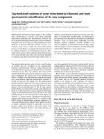
In order to demonstrate that the cells isolated from the
canine adipose tissue (Figure 1A) are indeed mesenchymal stem cells (MSCs) we differentiated them to adipocytes, osteoblasts and chondrocytes (Figure 1B, C). After
Page 4 of 15
three weeks' treatment with the adipogenic induction
medium, the cells contained abundant amounts of vacuoles and Oil Red O staining for fat revealed that these
vacuoles contained neutral lipids (B). After three weeks
culture time with the osteogenic induction medium the
cells changed to a more polygonal appearance, formed
nodules and were stained positive with von Kossa stain
for mineral deposition (C). Alcian blue staining after 14
days in culture revealed a high content of cartilage specific proteoglycans in induced cultures (D). Additionally,
the isolated MSCs showed a strong positive signal for the
stem cell specific markers CD90+ and CD105+ (Figure 1E,
F). In contrast to this they were clearly labelled negative
for the hematopoietic stem cell markers CD45- and
CD34- (Figure 1G, H).
Curcumin alone does not have a chondro-inductive effect
on pure MSC high-density cultures
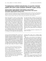
To study the effects of curcumin on MSCs after 14 days of
cultivation in three-dimensional high-density culture,
ultrastructural evaluations were performed (Figure 2A).
In control cultures, MSCs did not survive, but underwent
apoptosis or necrosis and mainly cell debris was observed
(a). Treatment of the cultures with the chondrogenic
induction medium stimulated chondrogenesis (b). The
formation of cartilage nodules consisting of large
rounded viable cells (containing large quantities of endoplasmic reticulum, mitochondria and other cellular
organelles) embedded in a fine structured, highly
organised ECM was observed. Treatment of MSCs with
curcumin alone did not stimulate chondrogenesis and, as
in control cultures, mainly cell debris was observed (c, e).
It did not make a difference whether cultures were either
Figure 1 Characterisation of MSCs. In monolayer culture the adipose derived MSCs (A) assumed a polymorphic, fibroblast-like morphology and
could be differentiated into adipocytes (B; Oil red staining), osteoblasts (C; von Kossa) and chondrocytes (D; alcian blue). The isolated MSCs showed
a strong positive signal for the stem cell specific markers CD90+ (E) and CD105+ (F) and were negative for the hematopoietic stem cell markers CD45(G) and CD34- (H). Magnification: A: 5×; B: 40×; C: 20×; D: 20×; E-H: 40×.
Buhrmann et al. Arthritis Research & Therapy 2010, 12:R127
/>
Page 5 of 15
Figure 2 Curcumin alone does not enhance chondrogenesis in MSCs. A: 14 days high-density culture. Apoptosis or necrosis was observed in MSC
cultures (a), MSC cultures treated with curcumin (c) or MSC cultures pre-stimulated four hours with curcumin, followed by incubation with curcumin
(e). In contrast, chondrogenesis was observed in MSC cultures treated with the chondrogenic induction medium alone (b), in combination with curcumin (d) or a hour-hour pre-stimulation with curcumin, followed by a combination of curcumin and the chondrogenic induction medium (f). Magnification, 6000×; bar, 1 μm; C, chondrocytes; F, fibroblast-like cells; M, ECM; GF, chondrogenic induction medium. B: The ultrastructural findings above
were confirmed by western blotting. Immunoblots of whole cell lysates were probed using antibodies that recognize CSPGs, collagen type II, β1-integrin, Shc, activated-ERK1/2 and Sox-9. Each experiment was performed in triplicate. Expression of the housekeeping gene β-actin was not affected.
GF, chondrogenic induction medium.
Buhrmann et al. Arthritis Research & Therapy 2010, 12:R127
/>
pre-treated for four hours with curcumin (c) or pretreated four hours with curcumin followed by treatment
with curcumin over the entire culture period (e). In contrast, in cultures treated with curcumin and the chondrogenic induction medium (d, f ) chondrogenesis was
observed. Chondrogenesis was similar in cultures that
were either pre-treated for four hours with curcumin (d)
or cultures that were pre-treated four hours with curcumin followed by treatment with curcumin over the
entire culture period (f ).
Western blotting was performed to confirm these
results (Figure 2B). Whole cell lysates were resolved by
SDS-PAGE electrophoresis and blotted onto nitrocellulose. The membranes were probed with antibodies
against cartilage specific proteoglycans (CSPG), collagen
type II, β1-integrins, Shc, activated extracellular regulated kinases 1 and 2 (ERK 1/2) and Sox-9.
In agreement with the ultrastructural findings, MSC
cultures treated with the specific chondrogenic induction
medium alone or in combination with curcumin produced high amounts of CSPGs, collagen type II and β1integrin. Here, four-hour curcumin pre-treatment followed by treatment with the chondrogenic induction
medium was as effective in inducing the production of
CSPGs, collagen type II and β1-integrin as a four-hour
curcumin pre-treatment followed by treatment with curcumin and the chondrogenic induction medium over the
entire culture period. Further, in these cultures the chondrogenic signalling cascade was activated with high
expression of Shc, activated ERK 1/2 and Sox-9. In contrast to this, cartilage specific matrix components and
members of the chondrogenic signalling cascade, were
not expressed in untreated cultures or cultures incubated
only with curcumin. Again, it did not make a difference
whether cultures were pre-treated for four hours with
curcumin or pre-treated four hours with curcumin followed by treatment with curcumin over the entire culture
period.
Curcumin inhibits IL-1β activity in MSCs, enabling growth
factor induced chondrogenesis
It has been reported that IL-1β inhibits chondrocyte proliferation and induces apoptosis [26,27]. We therefore
evaluated the effects of curcumin on MSCs stimulated
with IL-1β and/or the chondrogenic induction medium.
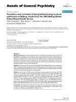
Ultrastructural evaluation revealed that stimulation of
MSCs with IL-1β either alone (Figure 3A-a), in combination with the chondrogenic induction medium (b) or in
combination with four-hour curcumin pre-treatment (c)
resulted in apoptosis. However, treatment with IL-1β,
curcumin and the chondrogenic induction medium lead
to induction of chondrogenesis (d, e) with formation of
cartilage nodules containing viable, rounded cells that
were embedded in a cartilage specific matrix. In these
Page 6 of 15
cultures, a four-hour curcumin pre-treatment (d) was as
effective in inhibiting IL-1β induced apoptosis as a fourhour curcumin pre-treatment followed by treatment with
curcumin over the entire culture period (e).
Apoptosis was further quantified as described in Materials and Methods. The data shown in Figure 3B demonstrate a significantly increased number of apoptotic cells
in cultures stimulated with IL-1β either alone, in combination with the chondrogenic induction medium or in
combination with a four-hour curcumin pre-treatment.
As shown at the ultrastructural level, the number of
apoptotic cells significantly decreased in MSC cultures
treated with IL-1β, curcumin and the chondrogenic
induction medium (Figure 3B). In these cultures the
number of apoptotic cells was similar between cultures
that were either pre-treated for four hours with curcumin
or pre-treated four hours with curcumin followed by curcumin treatment over the entire culture period.
To support these ultrastructural findings, Western blotting was performed by probing whole cell lysates with
antibodies against CSPGs, collagen type II, β1-integrin,
Shc, activated ERK 1/2 and Sox-9 (Figure 3C). Stimulation of MSC cultures with IL-1β either alone, in combination with the chondrogenic induction medium or with a
four-hour curcumin pre-treatment did not lead to production of CSPGs, collagen type II, β1-integrin and activation of Shc, ERK 1/2 and Sox-9. In contrast, production
of CSPGs, collagen type II and β1-integrin was upregulated and Shc, activated ERK 1/2 and the chondrogenic
specific transcription factor Sox-9 was highly expressed
in MSC cultures treated with IL-1β, curcumin and the
chondrogenic induction medium. Underlining the ultrastructural findings, it did not make a difference whether
cultures were either pre-treated for four hours with curcumin or pre-treated four hours with curcumin followed
by curcumin treatment over the entire culture period.
Further, to demonstrate the influence of curcumin on
the induction of the apoptotic signalling cascade by IL-1β
in MSCs, cultures were evaluated for activated caspase-3
and the marker of inflammation and prostaglandin production COX-2 (Figure 3D). Production of activated caspase-3 and COX-2 expression was prominent in all MSC
cultures stimulated with IL-1β alone and was blocked in
all cultures treated with IL-1β and curcumin. Here, a
four-hour pre-treatment with curcumin was as effective
in inhibiting the IL-1β induced apoptotic signalling cascade as a four-hour pre-treatment with curcumin followed by curcumin treatment over the entire culture
period.
Curcumin promotes chondrogenesis in co-cultures
stimulated with IL-1β
As demonstrated above, MSC cultures treated with a
chondro-inductive medium undergo chondrogenesis
Buhrmann et al. Arthritis Research & Therapy 2010, 12:R127
/>
Page 7 of 15
Figure 3 Curcumin inhibits IL-1β activity, enabling growth factor induced chondrogenesis in MSCs. A: Fourteen days high-density culture.
Treatment of MSC cultures with IL-1β alone (a), IL-1β and the chondrogenic induction medium (b) or a four-hour pre-stimulation with curcumin followed by IL-1β incubation (c) resulted in apoptosis or necrosis of the cells. In contrast, a four-hour pre-stimulation of MSCs with curcumin followed
by incubation with IL-1β and the chondrogenic induction medium (d) or followed by incubation with IL-1β, the chondrogenic induction medium
and curcumin (e) resulted in chondrogenesis. Magnification, 6,000×; bar, 1 μm; C, chondrocytes; F, fibroblast-like cells; M, ECM; GF, chondrogenic induction medium. B: Apoptotic cells were counted as an indicator of MSC culture degradation. In cultures stimulated with IL-1β alone, with IL-1β and
the chondrogenic induction medium, or with IL-1β and curcumin the number of apoptotic cells was increased. The number of apoptotic cells remained significantly lower in MSC cultures stimulated with IL-1β, curcumin and the chondrogenic induction medium. Significant values are marked
with (*). C-D: Immunoblots of whole cell lysates were probed using antibodies that recognize CSPGs, collagen type II, β1-integrin, Shc, activated-ERK1/
2, Sox-9, activated-caspase-3 and COX-2. Each experiment was performed in triplicate. Expression of the housekeeping gene β-actin was not affected.
GF, chondrogenic induction medium.
Buhrmann et al. Arthritis Research & Therapy 2010, 12:R127
/>
despite the presence of IL-1β if the apoptotic and inflammatory cascades induced by IL-1β are inhibited by curcumin. Next we evaluated whether the same effect can be
observed in a co-culture model of MSCs and primary
chondrocytes.
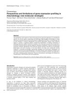
Ultrastructural evaluation demonstrated cellular debris
in untreated MSC cultures (Figure 4A-a), and development of cartilage nodules with rounded, viable cells
embedded in a highly organised, fine structured ECM in
untreated primary chondrocyte cultures (b) and in
untreated co-cultures (c). Stimulation of co-cultures with
IL-1β resulted in cellular degradation (d). Curcumin
treatment of the co-cultures, either as a four-hour pretreatment (e) or over the entire culture period (f ) did not
impede chondrogenesis. Combinational treatment of cocultures with IL-1β and curcumin suppressed the adverse
effects of IL-1β on chondrogenesis (g, h). Here, inhibition
of IL-1β induced apoptosis was as effectively blocked by a
four-hour curcumin treatment (g) as by curcumin treatment for the entire culture period (h).
Western blotting using antibodies against CSPGs, collagen type II, β1-integrin, Shc, activated ERK 1/2 and
Sox-9 was performed to evaluate induction of chondrogenesis in the co-cultures on a molecular level (Figure 4B,
C). Production of cartilage matrix specific markers and of
chondrogenic signalling pathway members was slight in
untreated MSC cultures and high in untreated primary
chondrocyte cultures and untreated co-cultures (Figure
4B, C). Stimulation of co-cultures with IL-1β alone inhibited production and expression of CSPGs, collagen type
II, β1-integrin, Shc, activated ERK 1/2 and Sox-9. In contrast, co-cultures stimulated with IL-1β and curcumin
produced high levels of chondrogenic matrix specific
markers (Figure 4B) and chondrogenic signalling pathway
proteins (Figure 4C). Stimulation of chondrogenesis was
similar between cultures that were either pre-treated for
four hours with curcumin or were pre-treated four hours
with curcumin followed by curcumin treatment over the
entire culture period and comparable to untreated primary chondrocyte cultures and untreated co-cultures.
To further demonstrate the influence of curcumin on
the induction of the apoptotic signalling cascade by IL-1β
in co-cultures, cultures were evaluated for activated caspase-3 and the marker of inflammation and prostaglandin production COX-2 (Figure 4D). Activated caspase-3
and COX-2 were highly expressed in untreated MSC cultures but were not expressed in untreated primary chondrocyte cultures and untreated co-cultures. Treatment of
the co-cultures with IL-1β alone led to high production of
activated caspase-3 and COX-2. In contrast, in co-cultures stimulated with both IL-1β and curcumin neither
activated caspase-3 nor COX-2 was detected. A fourhour pre-treatment of curcumin or curcumin treatment
Page 8 of 15
over the entire culture period both effectively inhibited
IL-1β induced activation of caspase-3 and COX-2 production. These results confirm the ultrastructural findings described above and demonstrate that curcumin
inhibits IL-1β induced apoptotic and inflammatory signalling pathways, promoting co-culture induced chondrogenesis.
Curcumin suppresses IL-1β-induced apoptotic and
inflammatory responses in MSCs in a time and
concentration dependent manner
Further experiments were carried out to evaluate the
interaction between curcumin and IL-1β in MSCs. These
experiments demonstrated that curcumin suppressed IL1β induced activation of apoptotic and inflammatory
pathways in a concentration (Figure 5) and time (Figure
6) dependent manner.
As shown in Figure 5A, treatment with as little as 0.5
μM of curcumin over the entire culture period was sufficient to significantly suppress IL-1β induced activation of
caspase-3 and COX-2 production in MSCs. Pro-caspase3 production remained unaffected.
The IL-1β-activated transcription factor nuclear factorκB (NF-κB) plays an essential role in mediating inflammatory and apoptotic processes in chondrocytes and it is
known that in chondrocytes, curcumin is able to suppress
NF-κB [9,14,28]. To evaluate whether curcumin influences IL-1β-induced NF-κB in MSCs, we investigated the
NF-κB signalling pathway. As demonstrated in Figure 5B,
curcumin suppressed IL-1β-induced activation of Iκ-Bα
in MSCs. This correlated clearly with decreased NF-κB
translocation to the nucleus. Inhibition of Iκ-Bα phosphorylation as well as NF-κB translocation to the nucleus
was evident when curcumin was included at a concentration of 0.5 μM. Higher concentrations of curcumin completely blocked IL-1β-induced activation of Iκ-Bα and
NF-κB translocation to the nucleus (Figure 5B).
Further, pre-treatment of MSC cultures with 5 μM curcumin also inhibited IL-1β-induced activation of caspase3 and COX-2 expression in a time dependent manner
(Figure 6A). After 60 minutes incubation time, activation
of caspase-3 and COX-2 production was completely suppressed. Pre-treatment of MSC cultures with 5 μM curcumin also inhibited activation of Iκ-Bα and NF-κB
translocation to the nucleus in a time dependent fashion
(Figure 6B). In contrast, in IL-1β treated control cultures,
activated caspase-3, higher expression of COX-2, activated Iκ-Bα and higher concentrations of NF-κB in the
nucleus were observed.
Discussion
The aim of this study was to evaluate whether curcumin
has the capacity to modulate inflammatory processes in
Buhrmann et al. Arthritis Research & Therapy 2010, 12:R127
/>
Page 9 of 15
Figure 4 Curcumin inhibits IL-1β activity, enabling co-culture induced chondrogenesis in MSCs. A: Fourteen days high-density culture. Untreated MSC cultures became apoptotic (a). In primary chondrocyte cultures (b), co-cultures (c), co-cultures treated with curcumin (e) or co-cultures
pre-stimulated four hours with curcumin (f), prominent chondrogenesis was observed. Stimulation of the co-culture with IL-1β alone resulted in degeneration of the cell culture (d). In contrast, a four-hour pre-stimulation of the co-culture with curcumin followed by IL-1β incubation (g) or a fourhour pre-stimulation of the co-culture with curcumin followed by IL-1β and curcumin incubation (h) inhibited the adverse effects of IL-1β on the
chondrogenic potential of the co-culture and prominent chondrogenesis was observed. Magnification, 6,000×; bar, 1 μm; C, chondrocytes, F, fibroblast-like cells; M, ECM. B-D: Immunoblots of whole cell lysates were probed with antibodies against CSPGs, collagen type II, β1-integrin, Shc, activated-ERK1/2, Sox-9, activated-caspase-3 and COX-2. In co-cultures pre-stimulated for four hours with curcumin followed either by incubation with IL1β alone or incubation with IL-1β and curcumin, prominent production of chondrogenic matrix and adhesion molecules (B), activation of the chondrogenic signalling pathway (C) and down-regulation of apoptotic and inflammatory markers (D) was observed. Each experiment was performed in
triplicate. Expression of the housekeeping gene β-actin was not affected.
Buhrmann et al. Arthritis Research & Therapy 2010, 12:R127
/>
Page 10 of 15
Figure 5 Curcumin suppresses IL-1β-induced apoptotic and inflammatory responses in monolayers of MSCs in a concentration dependent
manner. Monolayer cultures of MSCs were pre-stimulated for four hours with various concentrations of curcumin (0, 0.5, 1, 2 and 5 μM) followed by
24 h incubation with IL-1β, and Western blotting was performed using whole cell lysates and nuclear extracts. A: A strong dose dependent effect on
IL-1β induced activation of caspase-3 and COX-2 was observed. Curcumin concentrations as low as 0.5 μM suppresses IL-1β induced activation of
caspase-3 and COX-2. Higher concentrations of curcumin completely inhibited IL-1β induced activation of caspase-3 and production of COX-2. This
was confirmed by quantitative densitometry. The mean values and standard deviations from three independent experiments are shown. Expression
of the housekeeping gene β-actin was not affected. B: Curcumin exerts a strong dose dependent effect on IL-1β activated Iκ-Bα in MSCs, by suppressing phosphorylation of Iκ-Bα (which is already fairly robust) and NF-κB nuclear translocation at 0.5 μM curcumin. Higher concentrations of curcumin
blocked IL-1β-induced activation of Iκ-Bα and NF-κB translocation to the nucleus completely. This was confirmed by quantitative densitometry. The
mean values and standard deviations from three independent experiments are shown. Expression of the housekeeping gene β-actin and the DNA
repair enzyme PARP were not affected.
Buhrmann et al. Arthritis Research & Therapy 2010, 12:R127
/>
Page 11 of 15
Figure 6 Curcumin revokes IL-1β-induced apoptotic and inflammatory responses in monolayers of MSCs in a time dependent manner. MSC
Monolayer cultures were pre-stimulated for four hours with 5 μM curcumin followed by 1 h stimulation with IL-1β. Whole cell lysates, cytoplasmic and
nuclear extracts were evaluated by western blotting at various time points. A: A four-hour pre-stimulation with 5 μM curcumin suppressed IL-1β induced activation of caspase-3 and expression of COX-2 in a time dependent manner. This was confirmed by quantitative densitometry. The mean
values and standard deviations from three independent experiments are shown. Expression of the housekeeping gene β-actin was not affected. B:
Western blotting against activated Iκ-Bα and NF-κB demonstrated that in cultures pre-stimulated with 5 μM curcumin for four hours, followed by onehour IL-1β stimulation, neither activation of Iκ-Bα nor translocation of NF-κB to the nucleus can be demonstrated. This was confirmed by quantitative
densitometry. The mean values and standard deviations from three independent experiments are shown. Expression of the housekeeping gene βactin and the DNA repair enzyme PARP were not affected.
Buhrmann et al. Arthritis Research & Therapy 2010, 12:R127
/>
MSCs and thus support chondrogenesis in an in vitro
model of OA incorporating MSCs, primary chondrocytes
and pro-inflammatory cytokines.
Our observations lead to the following conclusions: (1)
Curcumin itself does not have any chondro-inductive
potential in MSCs, and treatment of MSCs with IL-1β
leads to cell apoptosis. (2) Although co-treatment of
MSCs with curcumin and IL-1β does not promote chondrogenesis, it clearly inhibits up-regulation of pro-inflammatory and apoptotic signalling cascades in MSCs; (3) If
MSCs receive a chondrogenic stimulus, curcumin mediated inhibition of IL-1β-induced catabolic signalling cascades enables chondrogenic differentiation. (4) This
effect is observed either by pre-treatment with curcumin
(four hours) or curcumin incubation over the entire culture period. (5) Chondrogenic stimulation can be
achieved either with a chondro-inductive medium or
through direct, close contact co-culture of MSCs with
primary chondrocytes; (6) Similar to chondrocytes, curcumin in MSCs targets the Iκ-Bα cascade, inhibiting IL1β-induced Iκ-Bα phosphorylation and NF-κB nuclear
translocation; (7) The effects of curcumin on the IL-1β
signalling pathway in MSCs are time and concentration
dependent.
OA and RA are characterised by high levels of proinflammatory cytokines in the articular joint. These are
produced by synovial lining cells, macrophages and the
chondrocytes themselves further exacerbating cartilage
degrading and degenerative processes [29,30]. Although,
as in numerous other tissues, MSC-like progenitors are
also resident in adult cartilage tissue [10], these degrading
and degenerative processes gradually lead to an imbalance between cartilage catabolism and anabolism, impeding MSC chondrogenesis. It has been reported that
activation of NF-κB is the key to induction of inflammation during OA and RA, and that NF-κB inhibition might
prove to be a potential concept for arthritis treatment
[31,32].
The polyphenol curcumin, derived from the rhizomes
of Curcuma longa is a promising therapeutic agent for the
treatment of OA and RA as it has pro-apoptotic properties in synovial lining cells [33,34] and has been shown to
have anti-inflammatory and anti-apoptotic effects in
chondrocytes [14,15]. In chondrocytes these effects are
mainly mediated by inhibiting IL-1β-induced activation
of NF-κB and thus suppression of caspase-3 activation, of
production of COX-2 and upregulation of MMPs [9].
In this study we demonstrate that the IL-1β-induced
catabolic signalling cascade is suppressed by curcumin in
MSCs as well as in chondrocytes. Further, we clearly
demonstrate that IL-1β induced Iκ-Bα activation is suppressed by curcumin, resulting in suppression of NF-κB
translocation to the nucleus and attenuated activation of
caspase-3 and COX-2 production. Taken together, our
Page 12 of 15
experiments on MSCs demonstrate that time and concentration dependent effects of curcumin inhibit the
induction of degradative and inflammatory pathways by
IL-1β through revoking activation of caspase-3, production of COX-2, phosphorylation of Iκ-Bα and inhibition
of nuclear translocation of NF-κB.
Interestingly, although we demonstrated that MSCs in
high-density culture cannot survive without a chondrogenic stimulus, stimulating these untreated cultures with
IL-1β and curcumin neither activated caspase-3 nor production of COX-2. This suggests that although MSCs
alone in this high-density model do not survive without a
necessary stimulus, they become necrotic rather than
apoptotic.
We did not observe a chondrogenic effect of curcumin
alone on MSCs, despite demonstrating anti-inflammatory and anti-apoptotic effects of curcumin in MSCs.
However, given the necessary stimulus, chondrogenesis
was observed. This demonstrates that curcumin interferes with the IL-1β induced apoptotic pathways in MSCs
thus providing a suitable microenvironment allowing
MSCs to undergo chondrogenesis even in the presence of
IL-1β, as long as MSCs simultaneously receive the correct
differentiation stimulus. This was confirmed through
high production of ECM and adhesion and signalling
molecules such as β1-Integrin. Similar observations have
been made in chondrocytes [15]. Interestingly, integrins
have already been shown in several tissues to be able to
mediate curcumin action [35,36]. As β1-integrins are
highly expressed in developing and adult cartilage, it is
possible that the mechanism of action of curcumin in
MSCs and chondrocytes might involve the β1-integrin
receptor signalling pathway.
Several studies have suggested that curcumin interacts
with the TGF-β signaling cascade, modulating the action
of TGF-β. For example in renal cells curcumin blocks
multiple sites of the TGF-β signaling [37] or Smad inhibition in human proximal tubule cells [38]. However, no
previously published in vitro studies have explored the
potential interaction between TGF-β and curcumin in
chondrocytes.
In our experiments we did not observe inhibition (or a
major difference in) the amounts of extracellular matrix
production and signaling cascades when co-cultures were
treated with the chondrogenic induction medium and
curcumin or only with curcumin, we assume that here a
possible interaction between curcumin and the TGF-β
signaling pathway does not have an inhibitory effect on
positive chondrogenic signaling. However, interaction of
curcumin and TGF-β signaling in chondrocytes is an
interesting point and will require further detailed investigations.
In the present study we demonstrated that a fairly low
concentration of curcumin (0.5 μM) was sufficient to
Buhrmann et al. Arthritis Research & Therapy 2010, 12:R127
/>
Page 13 of 15
mine bioavailability of curcumin in the synovial fluid following various routes of administration (that is, oral,
intra articular or topical). This is especially interesting as
it may be also possible that in vivo curcumin exerts its
effects via another organ (that is, the liver), which then
leads to positive anabolic signalling in the joint. Therefore, to integrate our in vitro data with results from other
studies, it is important to design and perform further in
vivo studies. Furthermore, our experiments were carried
out using canine derived cells and it may not be possible
to make generalised statements about the activity of curcumin on MSC-like cells in other species.
Figure 7 Schematic demonstrating the inflammatory cycle that
inhibits chondrogenic differentiation of MSCs in OA and the effect of curcumin. Trauma, inflammation or a combination of both (1)
lead to the production and accumulation of high levels of pro-inflammatory cytokines (2) in the joint. These trigger the production of additional pro-inflammatory cytokines which induce genes encoding
prostaglandin synthesizing enzymes (that is, COX-2) and matrix degrading enzymes (3) such as MMPs and aggrecanases. These events
promote cartilage degradation and stimulate further joint inflammation (4) and a self-perpetuating inflammatory and catabolic cascade
develops. As illustrated, cartilage contains chondrocytes and MSC-like
progenitors (5). Chemical or biological agents may help create a suitable microenvironment in order for these progenitor cells to undergo
chondrogenesis and regenerate new cartilage in OA. In this study we
tested the hypothesis that phytochemical modulators of NF-κB can
counteract this inflammatory and catabolic cascade and demonstrated that curcumin (6) has the capacity to block the action of pro-inflammatory cytokines in the joint thus disrupting the inflammatory cycle.
inhibit IL-1β induced activation of degradative pathways
in MSCs. It must be noted that in this study we worked
with low concentrations of curcumin, the highest concentration used being 5 μM. We chose this concentration
based on previous studies in our laboratory demonstrating that canine MSCs do not tolerate curcumin concentrations higher than 5 μM. A recent study by Kim and coworkers demonstrated that chick MSCs treated with 20
μM curcumin become apoptotic and do not undergo
chondrogenesis [39]. This clearly demonstrates that
MSCs can only tolerate lower concentrations of curcumin
in contrast to chondrocytes [40]. However, in vivo administered doses of curcumin in clinical trials differ greatly
from this and have ranged between 2 to 10 g per day [4143]. An explanation for these high concentrations is that
intestinal absorption of curcumin is fairly low, mainly due
to the fact that curcumin is practically insoluble in water,
and that it has a low bio-availability [44,45]. Despite its
low bio-availability, efficacy has been demonstrated in
several in vivo studies [46-48].
In order to develop a therapeutic strategy for OA/RA
treatment with curcumin it would be interesting to deter-
Conclusions
Our results suggest that curcumin, the naturally occurring NF-κB inhibitor, protects MSCs from the deleterious
effects of pro-inflammatory cytokines and thus creates a
suitable microenvironment for MSCs to undergo chondrogenesis and this strategy may help stimulate cartilage
regeneration in vitro (Figure 7). We therefore propose
curcumin as a potential new therapeutic for the prophylactic treatment of OA/RA and for OA/RA cases where
cartilage damage is marginal.
Abbreviations
AP303a: alkaline phosphatase linked sheep anti-mouse; AP304A: sheep antirabbit secondary antibodies; DMSO: dimethylsulfoxide; COX-2: cyclooxygenase-2; CSPG: cartilage specific proteoglycans; ECM: extracellular matrix; ERK 1/
2: extracellular regulated kinases 1 and 2; FCS: fetal calf serum; IKK: IκB kinase; IL:
interleukin; MAB1965: anti-β1-integrin antibody; MAB2015: monoclonal antiadult cartilage-specific proteoglycan antibody; MAPK: mitogen-activated protein kinase; MMP: matrix metalloproteinase; MSC: mesenchymal stem cell; MTT:
3-(4,5-dimethylthiazol-2-yl)-2,5-diphenyltetrazolium bromide; NF: nuclear factor; NF-κB: nuclear factor-κB; OA: osteoarthritis; PAB746: polyclonal anti-collagen type II antibody; PARP: (poly(ADP-Ribose) polymerase); PBS: phosphate
buffered saline; RA: rheumatoid arthritis; Shc: src homology collagen; TNF:
tumor necrosis factor.
Competing interests
The authors declare that they have no competing interests.
Authors' contributions
CB carried out the experimental work, data collection and interpretation, and
manuscript preparation. AM, UM and MS conceived of the study design and
coordinated the studies, data interpretation and manuscript preparation. All
authors have read and approved the final manuscript.
Acknowledgements
Ms. Christina Pfaff and Ms. Ursula Schwikowski are gratefully acknowledged for
their excellent technical assistance.
Author Details
1Musculoskeletal Research Group, Institute of Anatomy, Ludwig-MaximiliansUniversity Munich, Pettenkoferstrasse 11, D-80336 Munich, Germany, 2Division
of Veterinary Medicine, School of Veterinary Medicine and Science, Faculty of
Medicine and Health Sciences, University of Nottingham, Sutton Bonington
Campus, Sutton Bonington LE12 5RD, UK and 3Clinic of Veterinary Surgery,
Ludwig-Maximilians-University Munich, Veterinärstr. 13, 80539 Munich,
Germany
Received: 3 February 2010 Revised: 14 May 2010
Accepted: 1 July 2010 Published: 1 July 2010
ArthritisBuhrmann et al.; licensee BioMedunder the terms of the Creative Commons Attribution License ( which permits unrestricted use, distribution, and reproduction in any medium, provided the original work is properly cited.
© 2010 Research & Therapy 2010, 12:R127Central Ltd.
This article is available article />is an open access from: distributed
Buhrmann et al. Arthritis Research & Therapy 2010, 12:R127
/>
References
1. Sandell LJ, Aigner T: Articular cartilage and changes in arthritis. An
introduction: cell biology of osteoarthritis. Arthritis Res 2001, 3:107-113.
2. Elders MJ: The increasing impact of arthritis on public health. J
Rheumatol Suppl 2000, 60:6-8.
3. Hunziker EB: Articular cartilage repair: are the intrinsic biological
constraints undermining this process insuperable? Osteoarthritis
Cartilage 1999, 7:15-28.
4. Buckwalter JA, Brown TD: Joint injury, repair, and remodeling: roles in
post-traumatic osteoarthritis. Clin Orthop Relat Res 2004:7-16.
5. Heraud F, Heraud A, Harmand MF: Apoptosis in normal and
osteoarthritic human articular cartilage. Ann Rheum Dis 2000,
59:959-965.
6. Todhunter PG, Kincaid SA, Todhunter RJ, Kammermann JR, Johnstone B,
Baird AN, Hanson RR, Wright JM, Lin HC, Purohit RC:
Immunohistochemical analysis of an equine model of synovitisinduced arthritis. Am J Vet Res 1996, 57:1080-1093.
7. Aigner T, Kim HA: Apoptosis and cellular vitality: issues in osteoarthritic
cartilage degeneration. Arthritis Rheum 2002, 46:1986-1996.
8. Ding GJ, Fischer PA, Boltz RC, Schmidt JA, Colaianne JJ, Gough A, Rubin RA,
Miller DK: Characterization and quantitation of NF-kappaB nuclear
translocation induced by interleukin-1 and tumor necrosis factoralpha. Development and use of a high capacity fluorescence
cytometric system. J Biol Chem 1998, 273:28897-28905.
9. Shakibaei M, John T, Schulze-Tanzil G, Lehmann I, Mobasheri A:
Suppression of NF-kappaB activation by curcumin leads to inhibition
of expression of cyclo-oxygenase-2 and matrix metalloproteinase-9 in
human articular chondrocytes: Implications for the treatment of
osteoarthritis. Biochem Pharmacol 2007, 73:1434-1445.
10. Alsalameh S, Amin R, Gemba T, Lotz M: Identification of mesenchymal
progenitor cells in normal and osteoarthritic human articular cartilage.
Arthritis Rheum 2004, 50:1522-1532.
11. Jurenka JS: Anti-inflammatory properties of curcumin, a major
constituent of Curcuma longa: a review of preclinical and clinical
research. Altern Med Rev 2009, 14:141-153.
12. Aggarwal BB, Kumar A, Bharti AC: Anticancer potential of curcumin:
preclinical and clinical studies. Anticancer Res 2003, 23:363-398.
13. Bharti AC, Donato N, Singh S, Aggarwal BB: Curcumin
(diferuloylmethane) down-regulates the constitutive activation of
nuclear factor-kappa B and IkappaBalpha kinase in human multiple
myeloma cells, leading to suppression of proliferation and induction of
apoptosis. Blood 2003, 101:1053-1062.
14. Schulze-Tanzil G, Mobasheri A, Sendzik J, John T, Shakibaei M: Effects of
curcumin (diferuloylmethane) on nuclear factor kappaB signaling in
interleukin-1beta-stimulated chondrocytes. Ann N Y Acad Sci 2004,
1030:578-586.
15. Shakibaei M, Schulze-Tanzil G, John T, Mobasheri A: Curcumin protects
human chondrocytes from IL-l1beta-induced inhibition of collagen
type II and beta1-integrin expression and activation of caspase-3: an
immunomorphological study. Ann Anat 2005, 187:487-497.
16. Hatcher H, Planalp R, Cho J, Torti FM, Torti SV: Curcumin: from ancient
medicine to current clinical trials. Cell Mol Life Sci 2008, 65:1631-1652.
17. Mukhopadhyay A, Bueso-Ramos C, Chatterjee D, Pantazis P, Aggarwal BB:
Curcumin downregulates cell survival mechanisms in human prostate
cancer cell lines. Oncogene 2001, 20:7597-7609.
18. Lin JK, Pan MH, Lin-Shiau SY: Recent studies on the biofunctions and
biotransformations of curcumin. Biofactors 2000, 13:153-158.
19. Conget PA, Minguell JJ: Phenotypical and functional properties of
human bone marrow mesenchymal progenitor cells. J Cell Physiol 1999,
181:67-73.
20. Jaiswal N, Haynesworth SE, Caplan AI, Bruder SP: Osteogenic
differentiation of purified, culture-expanded human mesenchymal
stem cells in vitro. J Cell Biochem 1997, 64:295-312.
21. Dominici M, Le Blanc K, Mueller I, Slaper-Cortenbach I, Marini F, Krause D,
Deans R, Keating A, Prockop D, Horwitz E: Minimal criteria for defining
multipotent mesenchymal stromal cells. The International Society for
Cellular Therapy position statement. Cytotherapy 2006, 8:315-317.
22. Csaki C, Matis U, Mobasheri A, Ye H, Shakibaei M: Chondrogenesis,
osteogenesis and adipogenesis of canine mesenchymal stem cells: a
biochemical, morphological and ultrastructural study. Histochem Cell
Biol 2007, 128:507-520.
Page 14 of 15
23. Shakibaei M, Schroter-Kermani C, Merker HJ: Matrix changes during
long-term cultivation of cartilage (organoid or high-density cultures).
Histol Histopathol 1993, 8:463-470.
24. Pittenger MF, Mackay AM, Beck SC, Jaiswal RK, Douglas R, Mosca JD,
Moorman MA, Simonetti DW, Craig S, Marshak DR: Multilineage potential
of adult human mesenchymal stem cells. Science 1999, 284:143-147.
25. Shakibaei M, De Souza P, Merker HJ: Integrin expression and collagen
type II implicated in maintenance of chondrocyte shape in monolayer
culture: an immunomorphological study. Cell Biol Int 1997, 21:115-125.
26. Csaki C, Keshishzadeh N, Fischer K, Shakibaei M: Regulation of
inflammation signalling by resveratrol in human chondrocytes in vitro.
Biochem Pharmacol 2008, 75:677-687.
27. Shakibaei M, Csaki C, Nebrich S, Mobasheri A: Resveratrol suppresses
interleukin-1beta-induced inflammatory signaling and apoptosis in
human articular chondrocytes: potential for use as a novel
nutraceutical for the treatment of osteoarthritis. Biochem Pharmacol
2008, 76:1426-1439.
28. Csaki C, Mobasheri A, Shakibaei M: Synergistic chondroprotective effects
of curcumin and resveratrol in human articular chondrocytes:
inhibition of interleukin-1beta-induced nuclear factor-kappaBmediated inflammation and apoptosis. Arthritis Res Ther 2009, 11:R165.
29. Fernandes JC, Martel-Pelletier J, Pelletier JP: The role of cytokines in
osteoarthritis pathophysiology. Biorheology 2002, 39:237-246.
30. Feldmann M, Brennan FM, Maini RN: Role of cytokines in rheumatoid
arthritis. Annu Rev Immunol 1996, 14:397-440.
31. Gerlag DM, Ransone L, Tak PP, Han Z, Palanki M, Barbosa MS, Boyle D,
Manning AM, Firestein GS: The effect of a T cell-specific NF-kappa B
inhibitor on in vitro cytokine production and collagen-induced
arthritis. J Immunol 2000, 165:1652-1658.
32. Miagkov AV, Kovalenko DV, Brown CE, Didsbury JR, Cogswell JP, Stimpson
SA, Baldwin AS, Makarov SS: NF-kappaB activation provides the
potential link between inflammation and hyperplasia in the arthritic
joint. Proc Natl Acad Sci USA 1998, 95:13859-13864.
33. Park C, Moon DO, Choi IW, Choi BT, Nam TJ, Rhu CH, Kwon TK, Lee WH,
Kim GY, Choi YH: Curcumin induces apoptosis and inhibits
prostaglandin E(2) production in synovial fibroblasts of patients with
rheumatoid arthritis. Int J Mol Med 2007, 20:365-372.
34. Lev-Ari S, Strier L, Kazanov D, Elkayam O, Lichtenberg D, Caspi D, Arber N:
Curcumin synergistically potentiates the growth-inhibitory and proapoptotic effects of celecoxib in osteoarthritis synovial adherent cells.
Rheumatology (Oxford) 2006, 45:171-177.
35. Binion DG, Heidemann J, Li MS, Nelson VM, Otterson MF, Rafiee P:
Vascular cell adhesion molecule-1 expression in human intestinal
microvascular endothelial cells is regulated by PI 3-kinase/Akt/MAPK/
NF-kappaB: inhibitory role of curcumin. Am J Physiol Gastrointest Liver
Physiol 2009, 297:G259-268.
36. Kim HI, Huang H, Cheepala S, Huang S, Chung J: Curcumin inhibition of
integrin (alpha6beta4)-dependent breast cancer cell motility and
invasion. Cancer Prev Res (Phila Pa) 2008, 1:385-391.
37. Gaedeke J, Noble NA, Border WA: Curcumin blocks multiple sites of the
TGF-beta signaling cascade in renal cells. Kidney Int 2004, 66:112-120.
38. Hu Y, Liang H, Du Y, Zhu Y, Wang X: Curcumin inhibits transforming
growth factor-beta activity via inhibition of Smad signaling in HK-2
cells. Am J Nephrol 2010, 31:332-341.
39. Kim DK, Kim SJ, Kang SS, Jin EJ: Curcumin inhibits cellular condensation
and alters microfilament organization during chondrogenic
differentiation of limb bud mesenchymal cells. Exp Mol Med 2009,
41:656-664.
40. Clutterbuck A, Harris P, Mobasheri A: Comment on: comparison
between chondroprotective effects of glucosamine, curcumin and
diacerein in IL-1beta-stimulated C-28/I2 chondrocytes. Osteoarthritis
Cartilage 2009, 17:135-136. author reply 137.
41. Lao CD, Ruffin MTt, Normolle D, Heath DD, Murray SI, Bailey JM, Boggs ME,
Crowell J, Rock CL, Brenner DE: Dose escalation of a curcuminoid
formulation. BMC Complement Altern Med 2006, 6:10.
42. Cheng AL, Hsu CH, Lin JK, Hsu MM, Ho YF, Shen TS, Ko JY, Lin JT, Lin BR,
Ming-Shiang W, Yu HS, Jee SH, Chen GS, Chen TM, Chen CA, Lai MK, Pu YS,
Pan MH, Wang YJ, Tsai CC, Hsieh CY: Phase I clinical trial of curcumin, a
chemopreventive agent, in patients with high-risk or pre-malignant
lesions. Anticancer Res 2001, 21:2895-2900.
43. Sharma RA, McLelland HR, Hill KA, Ireson CR, Euden SA, Manson MM,
Pirmohamed M, Marnett LJ, Gescher AJ, Steward WP: Pharmacodynamic
Buhrmann et al. Arthritis Research & Therapy 2010, 12:R127
/>
44.
45.
46.
47.
48.
and pharmacokinetic study of oral Curcuma extract in patients with
colorectal cancer. Clin Cancer Res 2001, 7:1894-1900.
Kurien BT, Scofield RH: Increasing aqueous solubility of curcumin for
improving bioavailability. Trends Pharmacol Sci 2009, 30:334-335. author
reply 335.
Wang YJ, Pan MH, Cheng AL, Lin LI, Ho YS, Hsieh CY, Lin JK: Stability of
curcumin in buffer solutions and characterization of its degradation
products. J Pharm Biomed Anal 1997, 15:1867-1876.
Lim GP, Chu T, Yang F, Beech W, Frautschy SA, Cole GM: The curry spice
curcumin reduces oxidative damage and amyloid pathology in an
Alzheimer transgenic mouse. J Neurosci 2001, 21:8370-8377.
Ram A, Das M, Ghosh B: Curcumin attenuates allergen-induced airway
hyperresponsiveness in sensitized guinea pigs. Biol Pharm Bull 2003,
26:1021-1024.
Reyes-Gordillo K, Segovia J, Shibayama M, Vergara P, Moreno MG, Muriel P:
Curcumin protects against acute liver damage in the rat by inhibiting
NF-kappaB, proinflammatory cytokines production and oxidative
stress. Biochim Biophys Acta 2007, 1770:989-996.
doi: 10.1186/ar3065
Cite this article as: Buhrmann et al., Curcumin mediated suppression of
nuclear factor-?B promotes chondrogenic differentiation of mesenchymal
stem cells in a high-density co-culture microenvironment Arthritis Research &
Therapy 2010, 12:R127
Page 15 of 15