Báo cáo y học: "Phenotypic alterations in type II alveolar epithelial cells in cell mediated lung inflammation" pdf
Bạn đang xem bản rút gọn của tài liệu. Xem và tải ngay bản đầy đủ của tài liệu tại đây (1.11 MB, 13 trang )
BioMed Central
Page 1 of 13
(page number not for citation purposes)
Respiratory Research
Open Access
Research
Phenotypic alterations in type II alveolar epithelial cells in CD4
+
T
cell mediated lung inflammation
Marcus Gereke
1
, Lothar Gröbe
2
, Silvia Prettin
2
, Michael Kasper
3
,
Stefanie Deppenmeier
4
, Achim D Gruber
4
, Richard I Enelow
5
, Jan Buer*
2,6
and Dunja Bruder*
1
Address:
1
Immune Regulation Group, Helmholtz Centre for Infection Research, Braunschweig, Germany,
2
Department of Mucosal Immunity,
Helmholtz Centre for Infection Research, Braunschweig, Germany,
3
Institute of Anatomy, Medical Faculty Carl Gustav Carus, Dresden University
of Technology, Dresden, Germany,
4
Department of Veterinary Pathology, Free University Berlin, Berlin, Germany,
5
Departments of Medicine, and
Microbiology/Immunology, Dartmouth Medical School, Lebanon, NH, USA and
6
Department of Medical Microbiology, University Hospital
Essen, Essen, Germany
Email: Marcus Gereke - ; Lothar Gröbe - ;
Silvia Prettin - ; Michael Kasper - ;
Stefanie Deppenmeier - ; Achim D Gruber - ;
Richard I Enelow - ; Jan Buer* - ; Dunja Bruder* -
* Corresponding authors
Abstract
Background: Although the contribution of alveolar type II epithelial cell (AEC II) activities in various aspects of
respiratory immune regulation has become increasingly appreciated, our understanding of the contribution of AEC II
transcriptosome in immunopathologic lung injury remains poorly understood. We have previously established a mouse
model for chronic T cell-mediated pulmonary inflammation in which influenza hemagglutinin (HA) is expressed as a
transgene in AEC II, in mice expressing a transgenic T cell receptor specific for a class II-restricted epitope of HA.
Pulmonary inflammation in these mice occurs as a result of CD4
+
T cell recognition of alveolar antigen. This model was
utilized to assess the profile of inflammatory mediators expressed by alveolar epithelial target cells triggered by antigen-
specific recognition in CD4
+
T cell-mediated lung inflammation.
Methods: We established a method that allows the flow cytometric negative selection and isolation of primary AEC II
of high viability and purity. Genome wide transcriptional profiling was performed on mRNA isolated from AEC II isolated
from healthy mice and from mice with acute and chronic CD4
+
T cell-mediated pulmonary inflammation.
Results: T cell-mediated inflammation was associated with expression of a broad array of cytokine and chemokine genes
by AEC II cell, indicating a potential contribution of epithelial-derived chemoattractants to the inflammatory cell
parenchymal infiltration. Morphologically, there was an increase in the size of activated epithelial cells, and on the
molecular level, comparative transcriptome analyses of AEC II from inflamed versus normal lungs provide a detailed
characterization of the specific inflammatory genes expressed in AEC II induced in the context of CD4
+
T cell-mediated
pneumonitis.
Conclusion: An important contribution of AEC II gene expression to the orchestration and regulation of interstitial
pneumonitis is suggested by the panoply of inflammatory genes expressed by this cell population, and this may provide
insight into the molecular pathogenesis of pulmonary inflammatory states. CD4
+
T cell recognition of antigen presented
by AEC II cells appears to be a potent trigger for activation of the alveolar cell inflammatory transcriptosome.
Published: 4 July 2007
Respiratory Research 2007, 8:47 doi:10.1186/1465-9921-8-47
Received: 20 December 2006
Accepted: 4 July 2007
This article is available from: />© 2007 Gereke et al; licensee BioMed Central Ltd.
This is an Open Access article distributed under the terms of the Creative Commons Attribution License ( />),
which permits unrestricted use, distribution, and reproduction in any medium, provided the original work is properly cited.
Respiratory Research 2007, 8:47 />Page 2 of 13
(page number not for citation purposes)
Background
The epithelium constitutes the interface between the
internal milieu and the external environment, and the res-
piratory epithelium is the initial point of contact for respi-
ratory viruses, airborne allergens and environmental
pollutants [1]. The major function of the respiratory epi-
thelium was at one time felt to be primarily that of a phys-
ical barrier, but recent studies clearly indicate that its cells
are metabolically very active with the capacity to modu-
late a variety of inflammatory processes through the
action of an array of receptor-mediated events. Upon acti-
vation, epithelial cells have the capacity to produce a
number of pro-inflammatory or regulatory mediators,
including arachidonic acid products, nitric oxide,
endothelin-1, transforming growth factor (TGF)-β,
tumour necrosis factor (TNF)-α, and cytokines such as
interleukin (IL)-1, IL-6 and IL-8 [2].
Alveolar type II epithelial cells (AEC II, granular pneumo-
cyte, type II pneumocyte, giant corner cell) represent a
highly specialized subpopulation of the respiratory epi-
thelium. AEC II consist of about 15% of the distal lung
cells and occupy 5% of the alveolar surface [3]. They per-
form a variety of important functions within the lung,
including regulation of surfactant metabolism, ion trans-
port and alveolar repair in response to injury [4-7]. AEC II
synthesize and secrete lung surfactant, a protein-lipid
complex and surface-active material [8]. Ultrastructural
criteria used to identify alveolar type II epithelial cells are
the presence of lamellar bodies, apical microvilli and spe-
cific junctional proteins. AEC II also maintain the integrity
of alveolar epithelium by proliferation (and differentia-
tion to type I cells) in response to injury, and tightly regu-
late alveolar fluid by a variety of mechanisms.
AEC II express a number of molecules necessary for the
transduction as well as the generation of signals involved
in cell-cell as well as in cell-matrix interactions. Cell-cell
interactions may be direct via contact of tight junction
proteins, or indirect via secreted and diffusible signals [9].
Consequently, AEC II have been described as integrative
units of the alveolus [10]. Interactions of AEC II with leu-
kocytes have also been the subject of intense investigation
and there is evidence supporting a role of AEC II in acces-
sory function in T lymphocyte activation [11,12]. Moreo-
ver AEC II chemokine expression is induced upon
antigen-specific CD8
+
T cell recognition and plays a criti-
cal role in the perpetuation of experimental interstitial
pneumonia [13,14].
In order to study the pathophysiology of chronic T cell-
mediated lung injury, we established a novel model in
which a model antigen (influenza A/PR8/34 HA) is
expressed under the control of the SP-C promoter, result-
ing in AEC II cell-specific expression and bred these ani-
mals with mice expressing a transgenic T cell receptor,
specific for a class II-restricted epitope of HA, leading to a
chronic interstitial pneumonitis [15]. Initial characteriza-
tion of these mice focussed on self-antigen specific T cell
function and revealed the induction of peripheral T cell
tolerance at the site of inflammation. In this study we
demonstrate altered AEC II cell morphology in mice with
CD4
+
T cell-mediated pulmonary inflammation suggest-
ing a state of activation that we wanted to explore at a
molecular level. As such, we established a method to iso-
late highly pure primary AEC II for the purpose of per-
forming ex vivo expression profiling in the context of acute
and chronic interstitial pneumonitis. An important role of
AEC II gene expression in the orchestration of inflamma-
tory infiltration of the lung parenchyma is suggested by a
wide array of inflammatory genes and chemoattractants
expressed upon CD4
+
T cell recognition of antigen pre-
sented by the AEC II cells, and this model may prove
extremely useful in dissecting the mechanisms involved in
the perpetuation of chronic autoimmune pulmonary
processes.
Methods
Mice and antibodies
BALB/c mice were obtained from Harlan (Borchen, Ger-
many). TCR-HA transgenic mice expressing a TCR aβ spe-
cific for the I-E
d
-restricted HA-peptide 110–120 from A/
PR8/34 HA have been described previously [16]. SPC-HA
mice expressing the influenza A/PR8/34 HA under the
transcriptional control of the human surfactant protein C
(SP-C) promoter specifically in AEC II have been
described elsewhere [15]. Mice were bred in the animal
facility at the Helmholtz Centre for Infection Research
and were kept under SPF conditions. All mice were rou-
tinely monitored for the absence of bacterial, viral, para-
sitic and fungal infections. Mice aged 10 to 20 weeks were
used for experiments which were all performed according
to national and institutional guidelines. The monoclonal
antibody 6.5 (anti-TCR-HA) was purified from hybrid-
oma supernatants by protein G affinity chromatography.
The antibodies a-CD45 (30-F11), a-CD16/CD32 (2.4G2),
a-CD11b (M1/70) and a-F4/80 were obtained from BD
Biosciences and used either unconjugated or as phyco-
erythrin (PE) conjugates. As secondary polyclonal goat a-
rat IgM/IgG/IgA was used as phycoerythrin (PE) conju-
gate. For specific staining of sorted AEC II, the lectin
Maclura pomifera agglutinin was used. Intracellular stain-
ing for IFN-γ and IL-2 was performed using the antibodies
a-IFN-γ (XMG1.2) and a-IL-2 (JES6-5H4) from BD Bio-
sciences, according to the manufacturer's protocol.
Adoptive transfer of HA-specific CD4
+
T cells
Naïve CD4
+
T cells from the spleens of TCR-HA mice were
isolated by negative selection by AutoMACS using the
CD4
+
T cell isolation kit from Miltenyi Biotec (Bergisch
Respiratory Research 2007, 8:47 />Page 3 of 13
(page number not for citation purposes)
Gladbach, Germany), followed by i.v. injection of 1 × 10
6
antigen-specific CD4
+
T cells into SPC-HA transgenic
mice. At various time points after transfer, animals were
sacrificed and lungs perfused with PBS prior to excision.
The lungs were sectioning for histological analysis and
quantitative morphometry or were used for isolation of
AEC II cells, or infiltrating lymphocytes, as described
below.
Isolation of lymphocytes from the lung
Perfused lungs were excised and finely minced on ice, fol-
lowed by a 60–90 minutes digestion at 37°C with colla-
genase/dispase (0,2 mg/ml each) in IMDM/5% FCS in the
presence of 25 μg/ml DNase. To improve tissue disinte-
gration, lungs were pipeted every 5 min using a Pasteur
pipet. EDTA was added to a final concentration of 5 mM
followed by an additional 5 min incubation at 37°C.
Cells were passed through a 70 μm cell strainer, washed,
and lymphocytes isolated by density centrifugation.
Isolation of alveolar type II epithelial cells
Primary AEC II were prepared using a modified protocol
of a previously published method [17]. Briefly, mice were
anesthetized and exsanguinated by serving the inferior
vena cava and left renal artery. The tracheae was exposed
and cannulated and lungs were perfused with 10 to 20 ml
sterile phosphate buffered saline via the pulmonary artery
until visually free of blood. 2 ml dispase (BD Biosciences,
Heidelberg, Germany) was instilled into lungs via the tra-
cheal catheter followed by instillation of 500 μl 1% low-
melt agarose prior warmed to 45°C. Instilled lungs were
immediately covered with ice and incubated for 2 min to
gel the agarose. Lungs were removed, placed in a culture
tube containing an additional 1 ml of dispase and incu-
bated for 45 min at room temperature. The lungs were
then transferred to a culture dish and 7 ml serum free
DMEM + 25 mM HEPES (GIBCO, Eggenstein, Germany)
containing 100U/ml DNase I (Sigma, Hannover, Ger-
many) was added. The tissue was gently teased away from
the airways using forceps and lungs were carefully dissoci-
ated before agitating the tissue for 10 min on a shaker.
Crude cell suspensions were sequentially filtered through
nylon gauze (100 μm, 45 μm, 30 μm) followed by centrif-
ugation (12 min, 130 × g) to pellet the cells. For fluores-
cence activated cell sorting of alveolar type II epithelial
cells, cells were washed with serum free DMEM + 25 mM
HEPES and subsequently labelled with anti-CD45, anti-
CD32/CD16, anti-CD11b and anti-F4/80 antibodies and
PE-conjugated goat anti rat-IgG as secondary antibody.
After staining the cell suspension was washed with PBS
containing 2% fetal calf serum and 2 mM EDTA and sub-
jected to one-step cell sorting using a MoFlow cell sorter
(Cytomation, Fort Collins, CO). Granular alveolar type II
epithelial cells were identified as SSC
high
population. PE
(CD45/CD32/CD16/CD11b/F4/80)-positive cells were
excited by an argon ion laser emitted at the wavelength of
488 nm and the fluorescence was collected after a 580/
±30 nm band-pass filter. A two parameter sorting window
(side light scattering and PE fluorescent intensity) was
used to identify the PE-negative, side scatter high AEC II
population. Cells were sorted through a flow chamber
with a 100 μm nozzle tip under 25 psi sheath fluid pres-
sure. Using this protocol a purity of 97–99% and viability
of 90% was obtained. Isolated cells were either used for
immunofluorescence staining or RNA preparation.
Histology
Lungs were perfused and fixed with neutral buffered for-
malin, embedded in paraffin, sectioned and stained with
hematoxylin and eosin (H&E).
Immunofluorescence
For immunofluorescence staining sorted AEC II were
mounted onto glass cover slips with a density of 2 × 10
5
cells using a cytospin apparatus and were fixed with meth-
anol-acetone (1:1) mixture at -20°C for 5 min. Rabbit anti
SP-A, SP-B, pro-SPC and SP-D antibodies (Chemicon
Europe, Hampshire, UK) were all diluted 1:100 and incu-
bated with the fixed cells overnight at 4°C. A secondary
FITC conjugated goat anti-rabbit IgG (Dianova, Hamburg,
Germany) was used with a dilution of 1:80 and stained for
30 min at 37°C. All washing steps were performed in PBS
and stained cells were embedded in glycerol-PBS before
microscopic examination.
DNA microarray hybridization and analysis
Total RNA from AEC II sorted from the lung of either
healthy SPC-HA or diseased SPC-HA/TCR-HA mice was
isolated using the RNAeasy kit (Qiagen, Hilden, Ger-
many). Quality and integrity of total RNA isolated from 2
× 10
5
sorted AEC II cells was assessed by running all sam-
ples on an Agilent Technologies 2100 Bioanalyser (Agi-
lent Technologies, Waldbronn, Germany). For RNA
amplification the first round was performed in accordance
with an Affymetrix protocol without biotinylated nucleo-
tides, using the Promega P1300 RiboMax Kit (Promega,
Mannheim, Germany) for T7 amplification. For the sec-
ond round of amplification the precipitated and purified
RNA was converted to cDNA primed with random hexam-
ers (Pharmacia, Freiburg, Germany). Second strand syn-
thesis and probe amplification were done as in the first
round, with two exceptions: incubation with RNase H
preceded the first strand synthesis to digest the aRNA; and
the T7T23V oligonucleotide was used for initiation of the
second strand synthesis. 12.5 μg biotinylated cRNA prep-
aration was fragmented and placed in a hybridization
cocktail containing four biotinylated hybridization con-
trols (BioB, BioC, BioD, and Cre) as recommended by the
manufacturer. Samples were hybridized to an identical lot
of either Affymetrix MOE430A or MOE4302.0 chips for
Respiratory Research 2007, 8:47 />Page 4 of 13
(page number not for citation purposes)
16 hours. After hybridization, GeneChips were washed,
stained with streptavidin-PE and read using an Affymetrix
GeneChip fluidic station scanner. Analysis was done with
gene expression software (GeneChip, MicroDB, and Data
Mining Tool, all Affymetrix).
Real-time RT-PCR
Total RNA was prepared from sorted AEC II cells using the
RNeasy kit (Qiagen, Hilden, Germany) and cDNA synthe-
sis was done using Superscript II Reverse Transcriptase,
Oligo dT and random hexamer primers (Invitrogen).
Quantitative Real-time RT-PCR was performed on an ABI
PRISM cycler (Applied Biosystems) using a SYBR Green
PCR kit from Stratagene and specific primers optimized to
amplify 90–250 bp fragments from the various genes ana-
lyzed. A threshold was set in the linear part of the ampli-
fication curve and the number of cycles needed to reach
this was calculated for every gene. Relative mRNA levels
were determined by using included standard curves for
each individual gene and further normalization to RPS9.
Melting curves were used to establish the purity of the
amplified band.
Results
CD4
+
T cell recognition of epithelial antigen results in
interstitial inflammation accompanied by AEC II
hypertrophy
We have previously shown that HA expressed by AEC II in
SPC-HA transgenic mice results in presentation of a MHC
class II-restricted epitope to CD4
+
T cells and lung pathol-
ogy [15]. Immunopathology, characterized by massive
lymphocytic infiltration of interalveolar septa, was
observed both in SPC-HA mice that were adoptively trans-
ferred with HA-specific CD4
+
T cells as well as in SPC-HA
mice that were crossed with TCR-HA mice to establish
autoimmune conditions (Figure 1A). Interestingly, the
histologic appearance of AEC II cells in acutely inflamed
lungs revealed that they were in close contact with lym-
phocytes and displayed an activated phenotype with cel-
lular hypertrophy, characterized by significantly increased
AEC II surface area and perimeter. This was most promi-
nent during acute inflammation (i.e. shortly after adop-
tive transfer) and was less evident in the chronic
inflammatory state in adult SPC-HA/TCR-HA mice (Fig-
ure 1B and [15]). Accordingly, CD4
+
T cells isolated from
the lung of SPC-HA mice shortly after adoptive transfer
produced elevated levels of the pro-inflammatory
cytokines IL-2 and IFN-γ compared with T cells isolated
from the lungs of SPC-HA/TCR-HA mice at 16–20 weeks
of age (Figure 2).
Isolation of type II alveolar epithelial cells
To assess the contribution of AEC II to the orchestration
and progression of T cell-mediated interstitial pneumoni-
tis in more detail, we established a protocol for isolation
of AEC II from the murine lung entirely by negative selec-
tion. Enzymatic digestion and antibody staining, fol-
lowed by sorting of SSC
high
and CD45/CD32/CD16/
CD11/F4/80
negative
cells, resulted in highly pure and viable
AEC II cells, as indicated by surfactant protein (SP)-A, -B,
-C and -D expression (Figure 3A,B). Identity of sorted cells
as type II pneumocytes was further confirmed by staining
with the lectin Maclura pomifera agglutinin, that specifi-
cally binds to a 185 kDa glycoprotein on AEC II but not
on alveolar type I epithelial cells (AEC I) [18]. As depicted
in Figure 3C, essentially all cells stained positive with the
lectin, demonstrating high purity of AEC II cells obtained
by negative selection cell sorting.
Global changes in AEC II gene expression following CD4
+
T cell recognition of alveolar antigen
To characterize alterations in the transcriptional program
of alveolar epithelial cells in the context of T cell-mediated
interstitial pneumonitis, we performed gene expression
arrays on primary AEC II cells isolated from the lung of
either healthy SPC-HA mice or 16–20 week old SPC-HA/
TCR-HA mice with autoimmune lung inflammation. As
previously mentioned, SPC-HA/TCR-HA mice develop a
spontaneous pneumonitis due to the concomitant expres-
sion of the neo-self antigen influenza HA in AEC II and a
transgenic TCR specifically recognizing an I-E
d
-restricted
epitope from this particular antigen [15]. Thus, lung
inflammation occurs as a consequence of CD4
+
T cell rec-
ognition of a single alveolar epithelial "self antigen".
For gene expression analysis, RNA prepared from AEC II
was subjected to differential gene expression analysis
using oligonucleotide microarrays. An important advan-
tage of this technology is that every analyzed gene is rep-
resented by sixteen independent probe pairs which
together establish the basis for statistical evaluations of
the respective signals. Therefore, only the genes that are
reproducibly regulated are included in the analysis. For
each gene fulfilling these criteria, the average fold change
in expression for AEC II from the inflamed lung of SPC-
HA/TCR-HA and healthy lung of SPC-HA mice was calcu-
lated and the ratio was depicted on a base-2 logarithmic
scale. To establish the basal expression level of analyzed
genes in AEC II under non-pathologic conditions, an
alignment of AEC II derived from the healthy and
inflamed lungs was also performed, in duplicate arrays.
The number of "present calls" (42.1 to 44.7%) as calcu-
lated by the statistical detection algorithm of Affymetrix
was similar to data obtained from analysis of other types
of cells, e.g. T lymphocytes isolated by cell sorting [15].
The purity and integrity of isolated AEC II was examined
using basal gene expression levels of selected genes in AEC
II isolated from the lungs of healthy SPC-HA mice. Con-
sistent with results obtained by immunofluorescence
Respiratory Research 2007, 8:47 />Page 5 of 13
(page number not for citation purposes)
microscopy (Figure 3), sorted AEC II cells showed high
mRNA expression levels for SP-A, SP-B, SP-C and SP-D
(data not shown). Comparison of expression profiles of
AEC II cells from healthy and inflamed lungs revealed 322
genes that exhibited more than a two-fold expression
change. Among these, 288 encode proteins of known or
putative function (depicted in Figure 4), and the remain-
ing 34 genes are currently described as expressed sequence
tags (ESTs) or encoding unknown proteins. The full list of
differentially expressed genes is accessible online at [19].
Regulated genes were grouped into 11 functional classes
by their putative functions (Table 1). Among the genes
most significantly regulated in association with interstitial
inflammation were genes encoding the chemokine
CCL20, matrix metalloproteinases 2 and 3, and tissue
inhibitor of metalloproteinase 1. Also, strong down-regu-
lation of expression of several genes associated with cell
adhesion, including procollagen type XIV, alpha 1,
fibronectin 1 and dermatopontin, was observed in AEC II
cells isolated from the inflamed lung. Interestingly,
CD4
+
T cell recognition of alveolar epithelial antigen results in airway inflammation and AEC II hypertrophyFigure 1
CD4
+
T cell recognition of alveolar epithelial antigen results in airway inflammation and AEC II hypertrophy.
(A) Histological examination of lungs from healthy SPC-HA (a and a'), SPC-HA six days after adoptive transfer of HA-specific
CD4
+
T cells (b, b') and SPC-HA/TCR-HA double transgenic mice (c, c'). Lung sections were stained with H&E. Black arrows
indicate AEC II, red arrows indicate lymphocytes. No lesions were detectable in the lung of SPC-HA mice. Specifically, type II
pneumocytes were completely unchanged (a, a'). A moderate, perivascular and peribronchiolar infiltration with mature lym-
phocytes was detected in the lung of SPC-HA mice after transfer with HA-specific CD4
+
T cells. Adjacent to these infiltrations,
a slight connective tissue edema and a mild infiltration with neutrophils were observed. Type II pneumocytes in the vicinity of
the lymphocytic infiltrations were moderately hypertrophic. A few alveolar macrophages were present in the alveoli (b, b').
Moderate, multifocal, perivascular and peribronchiolar infiltrations with lymphocytes were present in the lung of SPC-HA/
TCR-HA double transgenic mice. Type II pneumocytes close to the lymphocytic infiltrations were mildly activated and hyper-
trophic (c, c'). (B) Histological results were corroborated morphometrically by measuring AEC II surface and perimeter to
quantify the degree of cellular hypertrophy (n = 15, 3 mice with 5 AEC II per mouse; ± standard deviation). AEC II surface:
SPC-HA vs SPC-HA Transfer: P < 0,001), SPC-HA vs SPC-HA/TCR-HA (P < 0,0001), SPC-HA transfer vs SPC-HA/TCR-HA (P
< 0,0001). AEC II perimeter: SPC-HA vs SPC-HA Transfer: P < 0,001), SPC-HA vs SPC-HA/TCR-HA (P < 0,001), SPC-HA
transfer vs SPC-HA/TCR-HA (P < 0,001). All Student's t-test.
a
a´
b b´
c
c´
x40
x400
AB
0
20
40
60
80
100
120
140
160
SPC-HA
SPC-HA/TCR -HA
SPC-HA
Transfer
AEC II surface [µm
2
]
37,40±3,99
90,20±14,9
48,10±5,62
0
5
10
15
20
25
30
35
40
45
50
SPC-HA
SPC-HA/TCR -HA
SPC-HA
Transfer
AEC II
perimeter
[µm]
37,00±3,19
27,70±1,46
24,30±1,55
Respiratory Research 2007, 8:47 />Page 6 of 13
(page number not for citation purposes)
whereas many genes involved in signal transduction (such
as lipoprotein lipase, prosaponin and metallothionein 2)
and cytoskeletal function (such as gelsolin and vimentin)
were down-regulated, genes involved in antigen process-
ing and presentation, such as MHC class II subunits, pro-
teasome subunits and beta-2 microglobulin exhibited
elevated expression in the inflamed lung. These genes
along with other potentially interesting genes differen-
tially expressed in AEC II cells isolated from the inflamed
lung, are listed in Table 1.
The morphology of AEC II differed considerably between
SPC-HA mice that were adoptively transferred with HA-
specific CD4
+
T cells, and analyzed acutely, compared
with those crossed to TCR-HA mice, and analyzed during
a chronic phase (Figure 1), suggesting a more pronounced
pro-inflammatory participation of AEC II during the acute
phase of inflammation. We therefore extended the gene
expression profiling to AEC II isolated 1, 3 or 6 days after
transfer, in order to examine the early activation events in
greater detail. Selected genes including genes associated
with immune responses, proteolysis and peptidolysis,
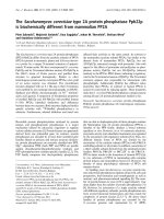
Purification of alveolar type II epithelial cells by fluorescence-activated cell sortingFigure 3
Purification of alveolar type II epithelial cells by fluo-
rescence-activated cell sorting. (A) Cell suspension
obtained by enzymatic tissue disintegration and subsequent
sequential filtration was labelled with antibodies to CD45,
CD16, CD32, CD11b, and F4/80. Antibody negative AEC II
were further distinguished from other cells by size and gran-
ularity. Reanalysis of sorted cells demonstrated an extremely
low frequency of contaminating hematopoetic cells. (B)
Sorted cells express surfactant proteins A, B, C and D. Cyt-
ospins of sorted AEC II cells were stained for the surfactant
proteins A, B, C and D. Almost all cells were found to be
positive for all four surfactant proteins. A, B, C and D repre-
sent phase contrast microscopy, A', B', C', and D' represent
immunohistochemical stainings for the corresponding sur-
factant protein. (C) Staining of sorted AEC II with Maclura
pomifera lectin revealed high purity of isolated cells. Black
histogram indicates staining with the lectin, grey histogram
indicates unstained cells.
PE (CD45, CD16, CD11b, F4/80)
SSC
pre sorting
post sorting
50% 45%
R1 R2
96% 1%
R1 R2
PE (CD45, CD16, CD11b, F4/80)
SSC
pre sorting
post sorting
50% 45%
R1 R2
50% 45%
R1 R2
96% 1%
R1 R2
96% 1%
R1 R2
A
45% 1%
C
SP-A
SP-B
SP-C
SP-D
A
B
C
D
A´
B´
C´
D´
SP-A
SP-B
SP-C
SP-D
SPA
-
SPB
SPC
SPD
AEC II
A
B
C
D
A
B
C
D
A´
B´
C´
D
B´
C´
D´
´
B
Maclura pomifera
98%
Intracellular cytokine staining in CD4
+
T cellsFigure 2
Intracellular cytokine staining in CD4
+
T cells. CD4
+
T
cells from the lung or bronchial lymph nodes (BLN) from
either TCR-HA control mice, SPC-HA/TCR-HA double
transgenic mice or SPC-HA mice adoptively transferred with
HA-specific CD4
+
T cells were analyzed by FACS for the
expression of interleukin 2 and interferon γ.
Interleukin-2
Interferon-Ȗ
TCR-HA
SPC
-HA/TCR-
HA
SPC
-
HA Transfer
BLN
BLN
BLN lung
lung
lung
8,33%
24,77%
5,71%
26,98%
11,00%
17,96%
12,62%
42,19%
93,63%
83,83%
91,41%
91,45%
Interleukin-2
Interferon-
TCR-HA
SPC
SPC
-
HA Transfer
BLN
BLN
BLN lung
lung
lung
8,33%
24,77%
5,71%
26,98%
11,00%
17,96%
12,62%
42,19%
93,63%
83,83%
91,41%
91,45%
Respiratory Research 2007, 8:47 />Page 7 of 13
(page number not for citation purposes)
cytoskeletal function, and antigen presentation and
processing were analyzed for changes in expression over
time (Figure 5). In addition, AEC II expression of selected
chemokines in the acute phase of lung inflammation was
further validated by quantitative real-time RT-PCR analy-
ses (Figure 6). Interestingly, for the majority of genes ana-
lyzed the changes in the expression level observed acutely
mirrored the chronic changes observed in AEC II isolated
from the lung of SPC-HA/TCR-HA mice at 16–20 weeks.
Thus, the alterations of AEC II gene expression profiles
which occurred early after T cell recognition of alveolar
antigen tended to persist into the chronic phase of inflam-
mation. For example, there was a rapid up-regulation of
MHC class II subunit expression, but decreased expression
of cytoskeletal genes both early after T cell transfer as well
as in AEC II isolated from SPC-HA/TCR-HA mice (Table 1
and Figure 4, 5). However, there were notable exceptions
to this pattern, such as was observed with CXCL13 expres-
sion, which was clearly down-regulated in AEC II isolated
from the chronically inflamed lung of SPC-HA/TCR-HA
double transgenic mice but induced acutely in AEC II cells
3 and 6 days after T cell transfer (confirmed by real-time
RT-PCR; Figures 5, 6).
Discussion
A significant number of lung diseases are presumed to be
T cell mediated based in part on the observation of T cell
accumulation at sites of disease activity, particularly the
interstitial lung diseases (ILD). The ILD represent a broad
group of heterogeneous disorders and the participation of
CD4
+
T cells in various forms of ILD has been suggested.
Sarcoidosis, idiopathic interstitial pneumonias, autoim-
mune connective tissue diseases and pulmonary hemor-
rhage syndromes represent some of the major categories
of ILD. Sarcoidosis, for example, appears to be associated
with an exaggerated cellular immune response to an
unknown antigen and CD4
+
Th1 lymphocytes are impor-
tant effectors of pulmonary injury in this disease [20,21].
In addition to ILD, it has been postulated that T cells are
important contributors in other pulmonary disorders
such as chronic obstructive pulmonary disease (COPD)
and asthma [22,23]. In these, it is hypothesized that ciga-
rette smoke or allergen induced immune responses can,
under certain conditions, progress to T cell mediated
autoimmune disease. Recently it has been suggested that
smoking-induced emphysema may represent an autoim-
mune disease of sorts, in which the presence of Th1
responses to a specific lung antigen correlates with
emphysema severity [21]. Furthermore, oligoclonal CD4
+
T cell expansion has been suggested to contribute to the
pathogenesis of obliterative bronchiolitis [24]. Although
there is growing evidence that CD4
+
T cells contribute to
various pulmonary disorders, little is known concerning
the role of AEC II cells in T cell mediated lung injury. To
expand our understanding of the roles of selected cell
types in the induction and progression of inflammatory
pulmonary processes, animal models represent tools of
extraordinary value. To explore the contribution of AEC II
gene expression in T cell mediated lung inflammation, we
made use of a transgenic mouse model of chronic T cell
mediated lung inflammation that mimics some of the fea-
tures of the interstitial lung discussed above, and that was
previously established [15]. We report here the applica-
tion of flow cytometry to efficiently isolate alveolar type II
epithelial cells from mouse lungs by negative selection
followed by whole genome transcriptome analysis. Gene
expression profiling has emerged as an important tool in
the characterization of complex molecular responses in
inflammation and disease. The use of isolated cellular
subpopulations has proven to be more informative than
whole tissues in dissecting the roles of individual cell
types in disease development in general, and immune reg-
ulation in particular. Comparative genetic fingerprinting
of AEC II isolated from healthy mice and mice suffering
from severe lung inflammation promises to be extremely
informative regarding the role of AEC II in the induction
and regulation of pulmonary immunity and inflamma-
tion.
Though confirmation of protein expression is essential,
morphological changes in AEC II phenotype and array
data suggest very active participation of alveolar epithelial
cells in inflammatory processes in the lung. Using Affyme-
trix GeneChip experiments we identified a heterogeneous
set of more than 322 genes differentially expressed in AEC
II under pathophysiologic conditions. Variations in signal
intensities between experimental repetitions may account
for slight differences in the disease progression in individ-
ual pooled mice as well as for differences in cRNA synthe-
sis and hybridization efficiencies between two array
experiments. To exclude as far as possible that changes in
gene expression occur as a consequence of the isolation
procedure, care was taken to purify AEC II from the differ-
ent mouse pools strictly following the described protocol,
i.e. avoiding variations of incubation times or tempera-
ture, etc. Therefore, the influence of cell isolation proce-
dure on gene expression in AEC II cells from healthy
versus inflamed lungs will subtract from each other and
account for changes in the molecular signature of AEC II
as a consequence of CD4
+
T cell mediated lung inflamma-
tion.
The differential expression of several immune modulating
molecules like TGF-β3 or the various chemokines and
chemokine ligands observed, suggests that in an inflamed
environment AEC II may interact with resident and
mobile neighbour cells via secreted and diffusible signals
[9]. Members of the transforming growth factor-beta fam-
ily are linked to proliferation or secretory activities of AEC
II. It has been shown that TGF-β3 production by AEC II is
Respiratory Research 2007, 8:47 />Page 8 of 13
(page number not for citation purposes)
Table 1: Selected genes differentially expressed in AEC II upon airway inflammation
Gene (functional category) Symbol SPC-HA/TCR-HA/SPC-HA Fold change
Array1 Array2 Array1/Array2
Genes associated with cell cycle
cyclin D2 Ccnd2 208/507 250/648 -2,1/-2,1
transforming growth factor, beta 3 Tgfb3 93/311 87/188 -3,0/-2,2
Genes associated with cell adhesion
procollagen, type IV, alpha 5 Col4a5 89/208 72/224 -1,9/-2,8
procollagen, type XIV, alpha 1 Col14a1 194/2003 142/1858 -9,8/-13,1
fibronectin 1 Fn1 252/2564 407/2813 -9,9/-8,6
dermatopontin Dpt 250/5627 277/3997 -11,8/-11,6
claudin 18 Cldn18 592/261 1845/445 2,3/3,9
Genes associated with antigen presentation and processing
major histocompatibility complex, class I, B H2-Q7 1386/85 1666/109 17,6/20,3
major histocompatibility complex, class II, DR alpha H2-Ea 5720/2661 5207/2187 2,2/2,4
major histocompatibility complex, class II, DQ beta 2 H2-Ab1 2217/1008 3971/1286 2,1/2,9
major histocompatibility complex, class II, DQ alpha 1 H2-Aa 4028/2019 6314/1859 1,9/1,8
major histocompatibility complex, class II, DR beta 1 H2-Eb1 2072/1013 2882/1100 1,9/2,3
major histocompatibility complex, class II, DM alpha H2-DMa 406/291 961/293 1,6/3,2
proteasome (prosome, macropain) subunit, beta type, 7 Psmb7 418/222 252/117 2,9/2,2
proteasome (prosome, macropain) subunit, beta type, 8 Psmb8 664/223 634/310 2,5/2,2
proteasome (prosome, macropain) subunit, beta type, 9 Psmb9 317/122 528/244 2,8/2,5
beta-2-microglobulin B2m 8579/4177 8784/3119 2,1/2,9
transporter 1 ATP-binding cassette, sub-family B (MDR/TAP) Tap1 277/107 283/120 2,4/3,0
Genes associated with transport
potassium inwardly-rectifying channel, subfamily J, member 15 Kcnj15 946/253 1160/231 4,0/4,9
lipocalin 2 Lcn2 11034/3130 13952/1966 3,6/7,4
sodium channel, nonvoltage-gated, type I, alpha polypeptide Scnn1a 405/292 448/225 2,1/2,4
Genes associated with immune response
Chemokine (C-X-C motif) ligand 1 CXCL1 313/96 235/64 2,5/3,1
Chemokine (C-X-C motif) ligand 13 CXCL13 128/556 100/634 -4,5/-5,9
Chemokine (C-C motif) ligand 12 CXCL12 253/1827 211/1541 -6,7/-7,4
Chemokine (C-X-C motif) ligand 20 CCL20 188/11 141/10 17,1/11,5
chemokine (C-C motif) ligand 11 CCL11 39/302 30/162 -8,5/-4,1
Genes associated with proteolysis and peptidolysis
Matrix metalloproteinase 2 MMP2 154/1788 116/1504 -10,8/-10,3
Matrix metalloproteinase 3 MMP3 51/599 67/547 -10,8/-10,9
Matrix metalloproteinase 23 MMP23 102/685 143/568 -6,2/-3,8
Tissue inhibitor of metalloproteinase 1 TIMP1 54/842 70/569 -11,1/-8,6
Tissue inhibitor of metalloproteinase 2 TIMP2 313/2265 388/2576 -8,6/-8,5
Tissue inhibitor of metalloproteinase 3 TIMP3 623/2935 434/3363 -3,0/-6,0
Genes associated with cytoskelett
elastin Eln 150/524 177/398 -4,1/-2,5
gelsolin Gsn 1438/16701 1620/15697 -8,1/-9,7
vimentin Vim 204/1974 308/2043 -9,6/-6,5
tubulin, alpha 1 Tuba1 1285/6486 1145/6076 -4,7/-5,3
Respiratory Research 2007, 8:47 />Page 9 of 13
(page number not for citation purposes)
Genes associated with metabolism
vanin 1 Vnn1 1752/200 993/181 9,6/6,3
5,10-methylenetetrahydrofolate reductase Mthfr 141/300 114/276 -2,0/-2,2
paraoxonase 1 Pon1 460/901 334/774 -2,3/-2,4
hexosaminidase B Hexb 94/303 93/228 -2,5/-2,2
Genes associated with signal transduction
insulin-like growth factor binding protein 7 Igfbp7 1356/4715 1849/5414 -3,8/-2,52
lipoprotein lipase Lpl 504/1542 228/1495 -3,3/-5,5
prosaposin Psap 236/761 319/963 -3,9/-3,0
fibroblast growth factor receptor 3 Fgfr3 160/345 165/304 -2,0/-3,2
interleukin 11 receptor, alpha chain 1 Il11ra1 88/428 146/350 -2,7/-2,7
Genes associated with signal transduction
annexin A1 Anxa11 1230/2328 866/1949 -1,8/-2,4
metallothionein 2 Mt2 118/737 153/758 -5,8/-7,1
Genes associated with transcription
thyrotroph embryonic factor Tef 350/189 429/203 2,2/2,3
CREBBP/EP300 inhibitory protein 1 Cri1 179/352 116/324 -2,2/-3,1
transcription factor 4 Tcf4 92/462 142/519 -5,0/-4,3
necdin Ndn 216/1425 152/1895 -5,5/-8,7
Genes associated with development
smoothened homolog (Drosophila) Smo 148/382 115/335 -2,6/-2,7
four and a half LIM domains 1 Fhl1 704/3478 454/3561 -4,7/-6,4
Differential gene expression was investigated by Affimetrix Gene Chip technology in AEC II from diseased SPC-HA/TCR-HA and healthy SPC-HA
mice (n = 3). For each population two independent experiments were performed and data obtained from individual experiments are depicted. The
table represents a compilation of regulated genes.
Table 1: Selected genes differentially expressed in AEC II upon airway inflammation (Continued)
dynamically down-regulated during the proliferative
phase of recovery from acute hyperoxic injury [25]. Con-
sistent with this, TGF-β3 expression was down-regulated
in AEC II from the inflamed lung, and since AEC II repre-
sent the stem cells for alveolar type I epithelial cells (AEC
I), this suggests a role of the TGF-β family in AEC II prolif-
erative responses and/or the cellular hypertrophy of AEC
II observed in the inflamed lung.
In addition to TGF-β3, the CXC chemokines CXCL2,
CXCL13 and CXCL12 were also differentially expressed in
AEC II from inflamed compared to healthy lungs (Figure
4, 5, 6, Table 1). These chemokines praticipate in the proc-
ess of attracting various cell populations into the lung.
CXCL12 and CXCL13 bind to CXCR4 and CXCR5, which
are primarily expressed on T lymphocytes or on circulat-
ing fibrocytes [26]. Interestingly, CXCL12 and CXCL13
expression was induced shortly after T cell recognition of
epithelial antigen (Figure 5, 6 and data not shown) and
massive lymphocytic infiltrates were observed shortly
after T cell transfer (data not shown). Furthermore, down-
regulation of T cell chemoattractants was evident at later
stages of inflammation (Figure 4 and Table 1) and could
contribute to a more controlled infiltration of specific T
cells into the lung. Accordingly it has been shown that
CXCL13 plays an important role in the development of
inducible bronchus associated lymphoid tissue (iBALT) in
respiratory immunity [27] by attracting T lymphocytes. It
has been suggested that infection or inflammation triggers
the organization of lymphoid structures in the lung of
both mice and humans [28,29], though this is somewhat
controversial. These structures do not fit the classical defi-
nition of BALT, as they are not formed independently of
antigen [30,31]. Because the iBALT appears in the lung
only after infection or inflammation, it is generally
assumed that iBALT is simply an accumulation of effector
cells that were initially primed in conventional lymphoid
organs. The neo-formation of iBALT is caused by inflam-
matory responses which directly promote the recruitment,
priming and expansion of antigen-specific lymphocytes
Respiratory Research 2007, 8:47 />Page 10 of 13
(page number not for citation purposes)
Heat map including genes differentially expressed in AEC II cells isolated from lungs of diseased SPC-HA/TCR-HA as well as healthy SPC-HA miceFigure 4
Heat map including genes differentially expressed in AEC II cells isolated from lungs of diseased SPC-HA/TCR-
HA as well as healthy SPC-HA mice. Red indicates induction of gene expression, green indicates repression (+2: bright
red; -2: bright green). Black indicates no changes. Blue squares indicate genes further highlighted in Table 1. Genes were con-
sidered to be regulated whose expression was at least twofold increased or decreased.
Respiratory Research 2007, 8:47 />Page 11 of 13
(page number not for citation purposes)
[27]. It is interesting to speculate that AEC II in SPC-HA/
TCR-HA double transgenic mice, after the initial inflam-
matory responses, down-regulate CXCL13 expression in
order to counteract new formation of iBALT and infiltra-
tion of specific T cells.
The chemokine CXCL2 is involved in attraction of poly-
morphonuclear granulocytes to sites of infection [32].
These neutrophils play an important role as regulators of
immune responses through release of cytokines such as
IL-1, IL-3, IL-6, IL-12, tumor necrosis factor-α (TNF-α) or
TGF-β as well as chemokines such as CCL2 (MCP-1) or
CCL20 (MIP-3α) [33,34].
Elevated expression of CCL20 by AEC II has been shown
to attract other pro-inflammatory cells [34,35]. CCL20,
which was dramatically up-regulated in the inflamed lung
(Figure 4, 5, 6, Table 1), has been shown to be constitu-
tively produced by AEC II cells and can attract immature
dendritic cells (imDC) to the lung [36,37]. Immature den-
dritic cells are known to exert immune modulatory func-
tions and may contribute to the establishment of a
controlled immune response in SPC-HA/TCR-HA double
transgenic mice. In contrast to CCL20, CCL11 (eotaxin),
an eosinophil chemoattractant, was dramatically down-
regulated in AEC II from the inflamed lung (Figure 4 and
6, Table 1). Not surprisingly, anti-CCL11 reduced eosi-
nophils infiltration of the lungs of RSV-infected mice. In
Chemokine expression in AEC II after adoptive CD4
+
T cell transfer into SPC-HA miceFigure 6
Chemokine expression in AEC II after adoptive CD4
+
T cell transfer into SPC-HA mice. AEC II cells were iso-
lated from the lung of SPC-HA mice one (n = 3), three (n =
3) and six (n = 3) days after adoptive transfer of HA-specific
CD4
+
T cells. Cells were subjected to quantitative real-time
RT-PCR analyses. mRNA expression levels of CXCL1,
CCL20, CXCL13, CCL11, CXCL2, and RPS9 (as internal
control) were analyzed in real-time RT-PCR assays. Relative
mRNA amounts were normalized with respect to expression
levels in AEC II cells isolated from SPC-HA mice not receiv-
ing CD4
+
T cell transfer (fold change = 1).
SPC-HA
1 d
4 d
7 d
010
5
15
20
CCL20
0
1
2
3
4
56
7
CXCL1
SPC-HA
1 d
4 d
7 d
0
1
2
-2
-1
-3-4-5
CCL11
25
SPC-HA
1 d
4 d
7 d
02
4
6
8
10
12
14 16
18
Fold change
CXCL13
Fold change
-2
6012345-1
CXCL2
SPC-HA
1 d
4 d
7 d
SPC-HA
1 d
4 d
7 d
010
5
15
20
CCL20
0
1
2
3
4
56
7
CXCL1
SPC-HA
1 d
4 d
7 d
SPC-HA
1 d
4 d
7 d
0
1
2
-2
-1
-3-4-5
CCL11
25
SPC-HA
1 d
4 d
7 d
SPC-HA
1 d
4 d
7 d
02
4
6
8
10
12
14 16
18
Fold change
CXCL13
Fold change
-2
6012345-1
CXCL2
Time course of gene expression in AEC II after adoptive CD4
+
T cell transfer into SPC-HA miceFigure 5
Time course of gene expression in AEC II after adop-
tive CD4
+
T cell transfer into SPC-HA mice. AEC II
cells were isolated from the lung of SPC-HA mice one (n =
3), three (n = 3) and six (n = 3) days after adoptive transfer
of HA-specific CD4
+
T cells. Cells were subjected to micro-
array analysis and the level of gene expression over time is
depicted for selected genes. Data obtained from two differ-
ent experiments are represented.
Signal intensity
Genes associated with immune response
Genes associated with antigen presentation and processing
Signal intensity
Genes associated with proteolysis and peptidolysis
Signal intensity
Genes associated with cytoskelett
H2-Ea Array 1
Signal intensity
0
1000
2000
3000
4000
5000
6000
7000
8000
9000
10000
1 day 3 day 6 day
H2-Ea Array 2 H2-Eb1 Array 1
H2-Eb1 Array 2
Psmb8 Array 1
Psmb8 Array 2
0
200
400
600
800
1000
1200
1400
1600
1800
1 day 3 day 6 day
MMP2 Array 1 MMP2 Array 2 TIMP2 Array 1
TIMP2 Array 2
TIMP3 Array 1 TIMP3 Array 2
0
1000
2000
3000
4000
5000
6000
1 day 3 day 6 day
Gelsolin Array 1 Gelsolin Array 2 Tubulin Array 1
Tubulin Array 2 Vimentin Array 1 Vimentin Array 2
0
100
200
300
400
500
600
700
800
900
1000
1 day 3 day 6 day
CXCL1 Array 1 CXCL1 Array 2 CXCL12 Array 1
CXCL12 Array 2 CXCL13 Array 1 CXCL13 Array 2
Respiratory Research 2007, 8:47 />Page 12 of 13
(page number not for citation purposes)
addition, however, anti-CCL11 also caused inhibited
CD4-T-cell influx [38]. Together, these data indicate an
active immune regulatory function of AEC II in inflamma-
tory pneumonitis involving the expression and secretion
of soluble mediators that may affect other immune cells
with regulatory features which may amplify, or interfere
with, inflammatory responses in the lung.
Although gene expression data provide evidence that AEC
II may (either directly or indirectly) exhibit immune regu-
latory functions, we also identified genes involved in the
induction of T cell mediated immunity. In this context it
is interesting to note that the expression levels for mole-
cules involved in antigen processing and presentation
were up-regulated in AEC II obtained from diseased mice.
For instance, increased expression of molecules needed
for the MHC class-II restricted antigen presentation, like
H2-Ea and H2-Ab1, but also invariant chain (CD74), was
observed. Furthermore, expression of genes encoding for
the transporter associated with antigen processing (TAP1)
and various proteasomal subunits, all related with MHC
class I presentation, were increased (Figure 4, 5, 6, Table
1). This effect was observed both in SPC-HA/TCR-HA
mice that exhibit chronic inflammation as well as in AEC
II from SPC-HA mice shortly after T cell transfer. Up-regu-
lation of MHC encoded genes is likely the result of inter-
feron (IFN)-γ production by the CD4
+
T cells, and is well
known to induce the transcription of genes encoded
within the MHC region. Based on our previous observa-
tion in an adoptive transfer model for CD8
+
T cell medi-
ated pulmonary inflammation, as well as in cell culture
experiments [13,14,39], we have strong evidence that T
cell antigen recognition triggers inflammatory gene
expression in AEC II cells, a significant portion of which is
IFN-γ dependent. Although CD4
+
T cell derived IL-2 and
IFN-γ are likely pro-inflammatory mediators that trigger
AEC II gene expression in SPC-HA/TCR-HA mice or SPC-
HA mice after adoptive T cell transfer, it is possible that
other T cell derived factors contribute to the observed
changes in AEC II gene expression, such as TNF-α.
Further genes differentially expressed in AEC II upon air-
way inflammation are cyclin A2 and cyclin D2, both
involved in cell cycle regulation [40,41] and several
matrix metalloproteinases (MMP) and tissue inhibitor
metalloproteinases (TIMP), all of which are critical in
repair and remodelling in response to injury [42,43]. In
addition to these, genes with roles in adhesion, cytoskele-
tal function, transport, metabolism, signal transduction,
transcription and development suggest that AEC II are
active participants in all aspects of immune regulation,
inflammation and responses to injury. The impact of
these gene products on the ethiopathogenesis of pulmo-
nary inflammation remain to be elucidated in further
detail.
Conclusion
We have developed a new AEC II isolation protocol based
on flow cytometric negative selection for the isolation of
cell populations of high purity and viability. Employing
this technique, we determined the genome-wide profile of
gene expression in response to T cell-mediated interstitial
pneumonitis. Overall, these results provide a detailed
description of AEC II gene expression under pathophysio-
logic, autoimmune conditions. Differentially expressed
genes of diverse molecular functions have been identified
that may be critical for numerous physiologic activities,
some of which may be currently unappreciated. Data
obtained by such analysis will help to understand the
function of these important immune cells in the respira-
tory system and may point out strategies for intervention
in the progression of chronic inflammatory processes in
the lung.
Competing interests
The author(s) declare that they have no competing inter-
ests.
Authors' contribution
MG carried out all experiments except for immunofluo-
rescence stainings, was involved in the interpretation of
data, designed figures and tables. LG performed cell sort-
ing. SP was involved in mice genotyping and assisted with
most of the experiments. MK performed immunoflures-
cence staining and interpreted this set of data. SD and
ADG performed histological examination and scoring.
RIE provided basic protocols, contributed to the concep-
tion of the study and critically revised the manuscript. JB
has substantially contributed to the overall study design
and also revised the manuscript. DB is primary investiga-
tor, who conceived the study, helped to prepare figures
and wrote the manuscript. All authors have read and
approved the final manuscript.
Acknowledgements
We thank Tanja Toepfer (HZI) for expert technical assistance and Andreas
Schmiedel (MHH) for providing Maclura pomifera lectin. This work was
supported by grants from the Deutsche Forschungsgemeinschaft (SFB587)
to D.B. and J.B.
References
1. Folkerts G, Nijkamp FP: Airway epithelium: more than just a
barrier! Trends Pharmacol Sci 1998, 19:334-341.
2. Knight DA, Holgate ST: The airway epithelium: structural and
functional properties in health and disease. Respirology 2003,
8:432-446.
3. Crapo JD, Barry BE, Gehr P, Bachofen M, Weibel ER: Cell number
and cell characteristics of the normal human lung. Am Rev
Respir Dis 1982, 126:332-337.
4. Kalina M, Mason RJ, Shannon JM: Surfactant protein C is
expressed in alveolar type II cells but not in Clara cells of rat
lung. Am J Respir Cell Mol Biol 1992, 6:594-600.
5. Lesur O, Arsalane K, Lane D: Lung alveolar epithelial cell migra-
tion in vitro: modulators and regulation processes. Am J Phys-
iol 1996, 270:L311-L319.
Publish with BioMed Central and every
scientist can read your work free of charge
"BioMed Central will be the most significant development for
disseminating the results of biomedical researc h in our lifetime."
Sir Paul Nurse, Cancer Research UK
Your research papers will be:
available free of charge to the entire biomedical community
peer reviewed and published immediately upon acceptance
cited in PubMed and archived on PubMed Central
yours — you keep the copyright
Submit your manuscript here:
/>BioMedcentral
Respiratory Research 2007, 8:47 />Page 13 of 13
(page number not for citation purposes)
6. Lubman RL, Danto SI, Crandall ED: Evidence for active H+ secre-
tion by rat alveolar epithelial cells. Am J Physiol 1989,
257:L438-L445.
7. Lubman RL, Crandall ED: Regulation of intracellular pH in alve-
olar epithelial cells. Am J Physiol 1992, 262:L1-14.
8. Griese M: Pulmonary surfactant in health and human lung dis-
eases: state of the art. Eur Respir J 1999, 13:1455-1476.
9. Fehrenbach H: Alveolar epithelial type II cell: defender of the
alveolus revisited. Respir Res 2001, 2:33-46.
10. Mason RJ, Williams MC: Type II alveolar cell. Defender of the
alveolus. Am Rev Respir Dis 1977, 115:81-91.
11. Schneeberger EE, DeFerrari M, Skoskiewicz MJ, Russell PS, Colvin RB:
Induction of MHC-determined antigens in the lung by inter-
feron-gamma. Lab Invest 1986, 55:138-144.
12. Zissel G, Ernst M, Rabe K, Papadopoulos T, Magnussen H, Schlaak M,
Muller-Quernheim J: Human alveolar epithelial cells type II are
capable of regulating T-cell activity. J Investig Med 2000,
48:66-75.
13. Zhao MQ, Foley MP, Stoler MH, Enelow RI: Alveolar epithelial cell
chemokine expression induced by specific antiviral CD8+ T-
cell recognition plays a critical role in the perpetuation of
experimental interstitial pneumonia. Chest 2001, 120:11S-13S.
14. Zhao MQ, Amir MK, Rice WR, Enelow RI: Type II pneumocyte-
CD8+ T-cell interactions. Relationship between target cell
cytotoxicity and activation. Am J Respir Cell Mol Biol 2001,
25:362-369.
15. Bruder D, Westendorf AM, Geffers R, Gruber AD, Gereke M,
Enelow RI, Buer J: CD4 T Lymphocyte-mediated lung disease:
steady state between pathological and tolerogenic immune
reactions. Am J Respir Crit Care Med 2004, 170:1145-1152.
16. Kirberg J, Baron A, Jakob S, Rolink A, Karjalainen K, von BH: Thymic
selection of CD8+ single positive cells with a class II major
histocompatibility complex-restricted receptor. J Exp Med
1994, 180:25-34.
17. Corti M, Brody AR, Harrison JH: Isolation and primary culture of
murine alveolar type II cells. Am J Respir Cell Mol Biol 1996,
14:309-315.
18. Weller NK, Karnovsky MJ: Identification of a 185 kd Maclura
pomifera agglutinin binding glycoprotein as a candidate for a
differentiation marker for alveolar type II cells in adult rat
lung. Am J Pathol 1989, 134:277-285.
19. Website title [ />]. [NCBI:
GSE5300]
20. Baumer I, Zissel G, Schlaak M, Muller-Quernheim J: Th1/Th2 cell
distribution in pulmonary sarcoidosis. Am J Respir Cell Mol Biol
1997, 16:171-177.
21. Semenzato G, Bortoli M, Agostini C: Applied clinical immunology
in sarcoidosis. Curr Opin Pulm Med 2002, 8:441-444.
22. Cosio MG, Majo J, Cosio MG: Inflammation of the airways and
lung parenchyma in COPD: role of T cells. Chest 2002,
121:160S-165S.
23. Larche M, Robinson DS, Kay AB: The role of T lymphocytes in
the pathogenesis of asthma. J Allergy Clin Immunol 2003,
111:450-463.
24. Duncan SR, Leonard C, Theodore J, Lega M, Girgis RE, Rosen GD,
Theofilopoulos AN: Oligoclonal CD4(+) T cell expansions in
lung transplant recipients with obliterative bronchiolitis. Am
J Respir Crit Care Med 2002, 165:1439-1444.
25. Buckley S, Bui KC, Hussain M, Warburton D: Dynamics of TGF-
beta 3 peptide activity during rat alveolar epithelial cell pro-
liferative recovery from acute hyperoxia. Am J Physiol 1996,
271:L54-L60.
26. Ebert LM, Schaerli P, Moser B: Chemokine-mediated control of
T cell traffic in lymphoid and peripheral tissues. Mol Immunol
2005, 42:799-809.
27. Moyron-Quiroz JE, Rangel-Moreno J, Kusser K, Hartson L, Sprague F,
Goodrich S, Woodland DL, Lund FE, Randall TD: Role of inducible
bronchus associated lymphoid tissue (iBALT) in respiratory
immunity. Nat Med 2004, 10:927-934.
28. Chvatchko Y, Kosco-Vilbois MH, Herren S, Lefort J, Bonnefoy JY:
Germinal center formation and local immunoglobulin E
(IgE) production in the lung after an airway antigenic chal-
lenge. J Exp Med 1996, 184:2353-2360.
29. Tschernig T, Pabst R: Bronchus-associated lymphoid tissue
(BALT) is not present in the normal adult lung but in differ-
ent diseases. Pathobiology 2000, 68:1-8.
30. Bienenstock J, Johnston N: A morphologic study of rabbit bron-
chial lymphoid aggregates and lymphoepithelium.
Lab Invest
1976, 35:343-348.
31. Plesch BE, Gamelkoorn GJ, van de EM: Development of bronchus
associated lymphoid tissue (BALT) in the rat, with special
reference to T- and B-cells. Dev Comp Immunol 1983, 7:179-188.
32. Matzer SP, Rodel F, Strieter RM, Rollinghoff M, Beuscher HU: Con-
stitutive expression of CXCL2/MIP-2 is restricted to a Gr-
1high, CD11b+, CD62Lhigh subset of bone marrow derived
granulocytes. Int Immunol 2004, 16:1675-1683.
33. Cassatella MA, Gasperini S, Calzetti F, McDonald PP, Trinchieri G:
Lipopolysaccharide-induced interleukin-8 gene expression in
human granulocytes: transcriptional inhibition by inter-
feron-gamma. Biochem J 1995, 310(Pt 3):751-755.
34. Galligan C, Yoshimura T: Phenotypic and functional changes of
cytokine-activated neutrophils. Chem Immunol Allergy 2003,
83:24-44.
35. Maurer M, von SE: Macrophage inflammatory protein-1. Int J
Biochem Cell Biol 2004, 36:1882-1886.
36. Dieu-Nosjean MC, Massacrier C, Homey B, Vanbervliet B, Pin JJ,
Vicari A, Lebecque S, zutter-Dambuyant C, Schmitt D, Zlotnik A,
Caux C: Macrophage inflammatory protein 3alpha is
expressed at inflamed epithelial surfaces and is the most
potent chemokine known in attracting Langerhans cell pre-
cursors. J Exp Med 2000, 192:705-718.
37. Reibman J, Hsu Y, Chen LC, Bleck B, Gordon T: Airway epithelial
cells release MIP-3alpha/CCL20 in response to cytokines and
ambient particulate matter. Am J Respir Cell Mol Biol 2003,
28:648-654.
38. Matthews SP, Tregoning JS, Coyle AJ, Hussell T, Openshaw PJ: Role
of CCL11 in eosinophilic lung disease during respiratory syn-
cytial virus infection. J Virol 2005, 79:2050-2057.
39. Ramana CV, Chintapalli J, Xu L, Alia C, Zhou J, Bruder D, Enelow RI:
Lung epithelial NF-kappaB and Stat1 signaling in response to
CD8+ T cell antigen recognition. J Interferon Cytokine Res 2006,
26:318-327.
40. Bui KC, Wu F, Buckley S, Wu L, Williams R, Carbonaro-Hall D, Hall
FL, Warburton D: Cyclin A expression in normal and trans-
formed alveolar epithelial cells. Am J Respir Cell Mol Biol 1993,
9:115-125.
41. Wu F, Buckley S, Bui KC, Warburton D: Differential expression of
cyclin D2 and cdc2 genes in proliferating and nonproliferat-
ing alveolar epithelial cells. Am J Respir Cell Mol Biol 1995,
12:95-103.
42. Chakrabarti S, Patel KD: Matrix metalloproteinase-2 (MMP-2)
and MMP-9 in pulmonary pathology. Exp Lung Res 2005,
31:599-621.
43. Hayashi T, Stetler-Stevenson WG, Fleming MV, Fishback N, Koss MN,
Liotta LA, Ferrans VJ, Travis WD: Immunohistochemical study of
metalloproteinases and their tissue inhibitors in the lungs of
patients with diffuse alveolar damage and idiopathic pulmo-
nary fibrosis. Am J Pathol 1996, 149:1241-1256.