Báo cáo y học: " Epigallocatechin-3-gallate (EGCG) inhibits the migratory behavior of tumor bronchial epithelial cells" docx
Bạn đang xem bản rút gọn của tài liệu. Xem và tải ngay bản đầy đủ của tài liệu tại đây (1.34 MB, 13 trang )
BioMed Central
Page 1 of 13
(page number not for citation purposes)
Respiratory Research
Open Access
Research
Epigallocatechin-3-gallate (EGCG) inhibits the migratory behavior
of tumor bronchial epithelial cells
Salma Hazgui
1
, Arnaud Bonnomet
1
, Béatrice Nawrocki-Raby
1
,
Magali Milliot
1
, Christine Terryn
2
, Jérôme Cutrona
1,2
, Myriam Polette
1,3
,
Philippe Birembaut
1,3
and Jean-Marie Zahm*
1
Address:
1
INSERM, UMRS903, Reims, F-51092 France,
2
Univ Reims Champagne Ardenne, IFR53, Reims, F-51100 France and
3
CHU Reims,
Hôpital Maison Blanche, Laboratoire Pol Bouin, Reims, F-51092 France
Email: Salma Hazgui - ; Arnaud Bonnomet - ; Béatrice Nawrocki-
Raby - ; Magali Milliot - ; Christine Terryn - ;
Jérôme Cutrona - ; Myriam Polette - ; Philippe Birembaut - pbirembaut@chu-
reims.fr; Jean-Marie Zahm* -
* Corresponding author
Abstract
Background: Many studies associated the main polyphenolic constituent of green tea, (-)-
Epigallocatechin-3-gallate (EGCG), with inhibition of cancers, invasion and metastasis. To date,
most of the studies have focused on the effect of EGCG on cell proliferation or death. Since cell
migration is an important mechanism involved in tumor invasion, the aim of the present work was
to target another approach of the therapeutic effect of EGCG, by investigating its effect on the cell
migratory behavior.
Methods: The effect of EGCG (at concentrations lower than 10 μg/ml) on the migration speed of
invasive cells was assessed by using 2D and 3D models of cell culture. We also studied the effects
of EGCG on proteinases expression by RT-PCR analysis. By immunocytochemistry, we analyzed
alterations of vimentin organization in presence of different concentrations of EGCG.
Results: We observed that EGCG had an inhibitory effect of cell migration in 2D and 3D cell
culture models. EGCG also inhibited MMP-2 mRNA and protein expression and altered the
intermediate filaments of vimentin.
Conclusion: Taken together, our results demonstrate that EGCG is able to inhibit the migration
of bronchial tumor cells and could therefore be an attractive candidate to treat tumor invasion and
cell migration.
Background
Cell migration is a prerequisite for cancer invasion and
metastasis. Much of the focus on the therapeutic treat-
ment of cancer has involved compounds that target cell
proliferation and subsequent cell death. However, target-
ing migration is another approach that has not been
extensively pursued but holds promise for alternative
means of therapy [1].
Published: 21 April 2008
Respiratory Research 2008, 9:33 doi:10.1186/1465-9921-9-33
Received: 24 July 2007
Accepted: 21 April 2008
This article is available from: />© 2008 Hazgui et al; licensee BioMed Central Ltd.
This is an Open Access article distributed under the terms of the Creative Commons Attribution License ( />),
which permits unrestricted use, distribution, and reproduction in any medium, provided the original work is properly cited.
Respiratory Research 2008, 9:33 />Page 2 of 13
(page number not for citation purposes)
Tea (Camellia sinensis) is a popular beverage worldwide. (-
)-Epigallocatechin-3-gallate (EGCG), the main polyphe-
nolic constituent of green tea, has been shown to have
association with prevention of cancer development,
metastasis, invasion and angiogenesis [2]. To date, most
of the studies have focused on the effect of EGCG on cell
proliferation or death. EGCG has been shown to induce
apoptosis in many human cell lines: human lymphoid
leukemia cells [3], prostate cancer cell lines [4], human
epidermoid carcinoma A431 cells [5], breast carcinoma
MCF-7 cells [6], melanoma cells [7] and pancreatic cancer
cells [8]. Previous studies demonstrated that it has a selec-
tive apoptotic effect in tumor cells compared with normal
cells [9]. This polyphenolic component has also an inhib-
itory effect on angiogenesis that is an important process in
tumor growth [10].
The acquisition of an invasive phenotype by epithelial
cells implicates a series of changes altering their differen-
tiation [11]. Components of the extracellular matrix play
a fundamental role in the process of tumor invasion.
Extensive studies in the last decade have revealed that
matrix metalloproteases (MMP) are frequently overex-
pressed in most forms of human tumor [12,13] and are
implicated in the destruction of the extracellular matrix,
thus facilitating tumor invasion [14,14,15]. EGCG has
inhibitory effects on MMP-2 and MT1-MMP in glioblast-
oma cells [16], reduces MT1-MMP activity in an invasive
human fibrosarcoma cell line [17] and induces repression
of MMP-9 expression in lung carcinoma cell invasion
[18]. It reduces cancer cell proliferation and migration by
a combination with ascorbic acid [19], by reducing VEGF
production [20]. EGCG also downregulates ephrin-A1-
mediated endothelial cell migration [21] and melanoma
and pancreatic cancer growth and metastasis [22,23].
Using a wound healing assay, Siddiqui et al [24] demon-
strated that co-treatment of prostate carcinoma cells with
EGCG and TNF-related apoptosis-inducing ligand led to a
decrease in cell migration. However, the studies dealing
with cell migration were mostly performed by using in
vitro models by which cell migration was evaluated by
using the Boyden chamber technique, or referred to qual-
itative rather than quantitative data. Our aim was to use in
vitro models of cell migration and to study the EGCG
effects on cell movement by analyzing the dynamic cell
behavior of a tumor epithelial bronchial cell line. We used
a two-dimensional (2D) model of cell dispersion [25] and
a three-dimensional (3D) model of cell migration to
mimic conditions similar to those observed in vivo during
tumor invasion [26]. In parallel we analyzed the effect of
EGCG on protease expression and vimentin organization.
Methods
Cell lines
The BZR human bronchial cell line used in our study [27]
was derived from normal human bronchial cells immor-
talized after transfection with the SV40 large T-antigen
gene and infected with the v-Ha-ras oncogene. This cell
line displays an invasive potential in vitro and tumori-
genicity and metastatic ability in athymic nude mice. Cells
were cultured in a 5% CO
2
fully humidified atmosphere at
37°C in Dulbecco modified Eagle's medium (DMEM)
(Gibco BRL, Grand Island, USA) supplemented with pen-
icillin, streptomycin (Eurobio, les Ulis, France) and 10%
fetal calf serum (Gibco BRL). Human epithelial MCF10A
cells were obtained from the American Type culture col-
lection and cultured in HAM F12 and DMEM (1:3 v/v)
supplemented with 20 μg/ml of adenine, 5 μg/ml of insu-
lin, 0.5 μg/ml of hydrocortisone, 2 ng/ml of EGF, 5 μg/ml
of transferrin, 1.5 ng/ml of triiodothyronine and 10%
fetal calf serum. EGCG was purchased from Sigma Aldrich
(Saint-Quentin Fallavier, France) and stored at 4°C.
Effect of EGCG on cell death
The BZR cell line was plated at 1 × 10
5
cells/ml and after 2
days of culture, the medium was removed from the cul-
ture plates and replaced with serum free medium with 5,
10, or 20 μg/ml of EGCG. After 18 h of cell interaction
with EGCG, the fluorescent probe propidium iodide (Inv-
itrogen, Cergy Pontoise, France), diluted at 20 mM in the
culture medium, was used to visualize the cell death. Flu-
orescent images were recorded using an inverted micro-
scope (Zeiss Axiovert 200, Le Pecq, France). From the
fluorescent images, the mean grey level, proportional to
the number of dead cells, was measured and reported as
cell death index.
2D cell Migration Assay
The BZR cell line was plated at 10
3
cells/ml and after 2
days of culture, the medium was removed from the cul-
ture plates and replaced with serum free medium with 5
μg/ml or 7.5 μg/ml of EGCG. Cell migration experiments
were performed using an inverted microscope (Axiovert
200, Zeiss, Le Pecq, France) equipped with a small trans-
parent environmental chamber (Climabox, Zeiss) with
5% CO2 in air at 37°C. The microscope was driven by the
Metamorph software (Roper Scientific, Evry, France) and
images of the cells were recorded every 15 min for 18
hours with a CCD camera (Coolsnap, Roper Scientific) at
20× magnification The migration speed of BZR cell line
was determined as previously described by Zahm et al
[28].
3D cell migration assay
Type I collagen gel was extracted from rat tails according
to the method described by Chambard et al [29]. To visu-
alize cells in a 3D model, we have developed a microenvi-
Respiratory Research 2008, 9:33 />Page 3 of 13
(page number not for citation purposes)
ronment model that consists of a two-layer type I collagen
gel (figure 1). The first collagen gel layer was prepared by
mixing 400 μl type I collagen at 2 mg/ml with 150 μl
RPMI 5×, 15 μl NaOH 1 N and 100 μl DMEM with 10%
fetal calf serum. To form the first layer of the microenvi-
ronment, 150 μl of this mixture was deposited on the
membrane of a double compartment chamber (Tran-
swell, Corning, Acton, MA) and polymerized for 30 min-
utes at 37°C. A second collagen gel layer was formed by
mixing 400 μl type I collagen at 2 mg/ml with 150 μl
RPMI 5×, 15 μl NaOH 1 N, 100 μl DMEM with 10% fetal
calf serum and BZR cell suspension at 13 × 10
4
cells/ml.
150 μl of this mixture was added over the first layer and
1.5 ml of DMEM was placed into the basal compartment
of the chamber that was thereafter maintained for 24
hours at 37°C. To test the effect of EGCG, the serum free
DMEM medium in the baso-lateral compartment was
complemented with EGCG at 5 μg/ml or 7.5 μg/ml
3D time-lapse videomicroscopy
Using the same microscope as for the 2D migration assay,
image sequences of the cells within the collagen gel were
recorded every hour at 110 depth levels (3 μm between
each depth level) at 20× magnification. To quantify cell
migration, we performed interactive tracking of cell posi-
tions in a four-dimensional dataset, as previously
described [26]. Once the coordinates (x
ij
, y
ij
, z
ij
, t
j
) of every
cell i at each j time setting are recorded in a data file, all
the trajectories are known and parameters can be
deduced. We measured the cell trajectory length in the
horizontal plane (xy), in the vertical direction (z) and the
total length of the trajectory (l). It was also useful to visu-
alize these trajectories in the corresponding 3D space (X,
Y, Z).
RT-PCR Analysis
Total RNA extraction from subconfluent BZR cells was
performed with the High Pure RNA isolation kit (Roche
Diagnostics, Meylan, France). RT-PCR was performed
with 4 ng/μl of total RNA using the GeneAmp Thermosta-
ble RNA PCR kit (Perkin-Elmer, Foster City, CA) and pairs
of primers for human MMP-2, MMP-9, MT1-MMP, u-PA
and for 28S rRNA (Eurogentec, Seraing, Belgium). For-
ward and reverse primers for human MMP-2, MMP9,
MT1-MMP, u-PA and 28S were designed as follows:
MMP-2 primers (forward 5'-GGCTGGTCAGTGGCTT-
GGGGTA-3', reverse5'-AGATCTTCTTCTTC AAGGACCG-
GTT-3'),
MMP9 primers (forward 5'-GCGGAGATTGGGAAC-
CAGCTGTA-3', reverse 5'-GACGCGCCTGTGTACAC-
CCAACA-3'),
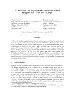
Representation of the 3D culture modelFigure 1
Representation of the 3D culture model. A collagen gel layer was prepared by mixing 400 μl type I collagen at 2 mg/ml
with 150 μl RPMI culture medium 5 fold concentrated, 15 μl NaOH 1 N and 100 μl DMEM with 10% fetal calf serum. To form
the first layer of the microenvironment, 150 μl of this mixture was deposited on the membrane of a double compartment
chamber (Transwell) and polymerized for 30 minutes at 37°C. A second collagen gel layer was formed by mixing 400 μl type I
collagen at 2 mg/ml with 150 μl RPMI 5×, 15 μl NaOH 1N, 100 μl DMEM with 10% fetal calf serum and BZR cell suspension at
13 × 10
4
cells/ml. 150 μl of this mixture was added over the first layer and 1.5 ml of DMEM was placed into the basal compart-
ment of the chamber which was thereafter maintained for 24 hours at 37°C.
Upper collagen gel
with cells
Lower collagen
gel
Culture medium
3D culture in a double compartment chamber
Respiratory Research 2008, 9:33 />Page 4 of 13
(page number not for citation purposes)
MT1-MMP primers (forward 5'-GGATACCCAAT-
GCCCATTGGCCA-3', reverse 5'-CCATTGGGCATCCA-
GAAGAGAGC-3'),
u-PA primers (forward 5'-CTGTAATACGACTCACTATAG-
GGGGCACCGG-3', reverse 5'-TCCGGATAGAGAT-
AGTCGGTGTGGTGAGCAAG-3'),
28S primers (forward 5'-GTTCACCCACTAATAG-
GGAACGTGA-3', reverse 5'-GGATTCTGACTTAGAG-
GCGTTCAGT-3').
Reverse transcription was performed at 70°C for 15 min-
utes. Amplification cycles were as follows: 15 seconds at
94°C, 20 seconds at 68°C, and 10 seconds at 72°C.
Twenty one cycles were allowed for MMP-2 amplification,
30 cycles for MMP9 amplification, 20 cycles for MT1-
MMP amplification, 26 cycles for u-PA amplification, 12
cycles for 28S amplification. Products were separated on
acrylamide gels, stained with SYBR Gold (Invitrogen,
Cergy Pontoise, France), and images were recorded by
fluorimetric scanning (LAS-1000, Fuji, Stamford, CT).
Zymography analysis
The BZR cell line was cultured in 12-well plates (10
4
cells
per well). After 48 h of incubation, the medium was
changed to serum-free medium and EGCG was added at
different concentrations: 0, 5 or 7.5 μg/ml. After 18 h of
incubation, the medium conditioned by the BZR was cen-
trifuged. Samples were separated on a 10% polyacryla-
mide SDS gel containing 0.1% (w/v) gelatine (Sigma
Aldrich, Saint-Quentin Fallavier, France). Electrophoresis
was carried out at the constant current of 40 mA. The gel
was washed for 1 hour at room temperature in a 2% (v/v)
Triton X-100 solution, transferred to a 50 mmol/L Tris-
HCl/10 mmol/L CaCl
2
(pH 7.6) buffer and incubated
overnight at 37°C. The gel was stained for 30 minutes in
a 0.1% (w/v) Coomassie blue (G250)/45% (v/v) metha-
nol/10% (v/v) acetic acid solution and de-stained in 10%
(v/v) acetic acid/20% (v/v) methanol. Proteolytic activity
was semi-quantified by densitometric scanning of the
bands (LAS-1000, Fuji).
Effect of EGCG on vimentin
We used the human breast cell line MCF10A in an in vitro
model of cell migration. This model consisted in plating 5
× 10
4
cells inside a 6-mm glass ring placed in the middle
of a collagen-coated coverslip [30]. Twenty four hours
after plating, the glass ring was removed and the cells were
covered with growth medium. The cells at the periphery of
the culture were left to migrate for 24 h, then they were
incubated with EGCG at 0, 5, or 7.5 μg/ml for another 24
h period. The migratory speeds were measured for 1 h as
previously described and the cells were fixed in cold meth-
anol for 10 min at -20°C. The coverslips were then satu-
rated for 30 min with 3% bovine serum albumin in PBS.
After intermediate washes in PBS, monolayers were suc-
cessively incubated for 1 h with a monoclonal antibody to
vimentin (clone Vim 3B4; Dako, Glostrup, Denmark),
with biotinylated sheep anti-mouse antibody and with
Alexa Fluor
®
488-conjugated streptavidin (Dako). Cover-
slips were mounted with aqua polymount antifading
solution (Polysciences, Warrington, PA) onto glass slides
and observed under a fluorescence microscope at x10 or
x63 magnification (AxioImager, Zeiss, Le Pecq, France).
Data analysis
Values were reported as mean ± SD from at least 3 differ-
ent experiments. Student's t-test was used for comparisons
between groups and differences were considered to be sta-
tistically significant with P values less than 0.05.
Results
Effect of EGCG on cell death
To visualize the effect of EGCG on cell death, we used the
fluorescent probe propidium iodide that specifically tags
the nucleus of necrotic cells. Typical images are shown in
figure 2. Cells were incubated without EGCG (figure 2A),
or with EGCG at 5 μg/ml (figure 2B), 10 μg/ml (figure
2C), or 20 μg/ml (figure 2D). We observed a dose-
dependent increase in the number of positive cell nuclei
and this increase became significant (p < 0.01) in presence
of 20 μg/ml of EGCG (figure 2E).
2D analysis of BZR trajectories in relation with cell
migration speed
Observation of time lapse movies built from the phase
contrast images recorded every 15 minutes for 18 hours
showed an increasing inhibition of BZR migration in par-
allel with the increase of EGCG concentration. Time-lapse
images recorded every 15 min showed that BZR cells in
absence of EGCG continuously modified their shape and
acquired an elongated morphology corresponding to a
migratory phenotype (figure 3A, B, C). At the opposite,
the incubation of BZR cells with EGCG at 5 μg/ml (figure
3D, E, F) or 7.5 μg/ml (figure 3G, H, I) induced the inhi-
bition of cell shape modifications. From these time-lapse
sequences, we quantified the cell migration speed and the
results in figure 4 display the cell trajectories computed
after 18 hours for the control, (figure 4A), with 5 μg/ml
EGCG (figure 4B) and with 7.5 μg/ml EGCG (figure 4C).
Quantification of the migration speed showed a signifi-
cant (p < 0.01) and progressive decrease in presence of
EGCG at 5 μg/ml and 7.5 μg/ml. This decrease reached
40% with 5 μg/ml and 68% with 7.5 μg/ml of EGCG as
compared with the control (figure 4D).
3D analysis of BZR trajectories
Videomicroscopy and computational techniques were
used to analyze the migratory behavior of cells and the
Respiratory Research 2008, 9:33 />Page 5 of 13
(page number not for citation purposes)
Effect of EGCG on cell deathFigure 2
Effect of EGCG on cell death. Fluorescent images representing the effect of increasing concentrations of EGCG on cell
death. The fluorescent probe propidium iodide was used to visualize dead cells. Compared to control (A) or to 5 μg/ml (B) and
10 μg/ml (C) of EGCG, we observed that 20 μg/ml (D) of EGCG induced a significant (p < 0.01) increase in the dead cell
number (E).
Figur2 6
EGCG concentration (μg/ml)
Cell death index
**
A
E
D
C
B
Respiratory Research 2008, 9:33 />Page 6 of 13
(page number not for citation purposes)
effect of EGCG on 3D cell migration. Figure 5A displays
the 3D trajectories of control BZR cells over 18 h of obser-
vation. The trajectories obtained under the same condi-
tions with the BZR cells incubated with 7.5 μg/ml of
EGCG are presented in figure 5B. We observed a higher
trajectory length for control BZR cells compared to BZR
cells incubated with EGCG. The migration parameters
computed from these trajectories are summarized in Fig-
ure 5C: a significant decrease (p < 0.05) of the migration
speed along the XY horizontal plane was observed for BZR
cells in presence of EGCG at 5 μg/ml compared with con-
trol BZR cells. When incubated with EGCG at 7.5 μg/ml, a
significantly (p < 0.01) higher decrease in the migration
speed of BZR cells was observed along the XY horizontal
plane, the Z plane and in the XYZ volume, compared with
control BZR cells.
RT-PCR and zymography analysis
To evaluate the effect of EGCG on protease gene expres-
sion, we analyzed the mRNA amount of MMP-2, MMP-9,
Phase contrast images of BZR cellsFigure 3
Phase contrast images of BZR cells. Phase contrast images of BZR cells in control medium or in medium with 5 or 7.5 μg/
ml of EGCG. The images were recorded every 15 min. In absence of EGCG, evident alterations of the cell morphology were
observed in parallel with the cell displacement (A, B, C). Cell shape modifications and cell movements were less important in
presence of 5 μg/ml of EGCG (D, E, F) and were almost completely inhibited in presence of 7.5 μg/ml of EGCG. Scale bar = 50
μm.
A
t t+15 mn t+30 mn
A
B
D
C
G
E
I
H
F
Respiratory Research 2008, 9:33 />Page 7 of 13
(page number not for citation purposes)
MT1-MMP and u-PA using semi-quantitative RT-PCR (fig-
ure 6). We observed a significant (p < 0.05) decrease of the
MMP-2 transcript expression in a dose-dependent manner
after 18 h of treatment with EGCG but we did not observe
any significant change of the MT1-MMP and u-PA tran-
script expression. The level of MMP9 transcript expression
was not detectable.
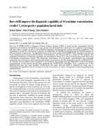
Zymography analysis shows a significant dose-dependent
decrease (p < 0.05) of the active and latent form of MMP-
2 after 18 hours of incubation with EGCG in comparison
with the control (Figure 7). No enzymatic activity corre-
sponding to MMP-9 was observed.
Effect of EGCG on vimentin expression
To examine the potential effect of EGCG on vimentin-
dependent migration, we used the ring culture system that
allowed the MCF10A cell line to specifically express
vimentin in migratory cells at the periphery of the culture
[30]. As shown in figure 8G, we observed that the incuba-
tion of migrating MCF10A cells with increasing concentra-
tions of EGCG significantly decreased the cell migration
speed. In parallel with the decrease in cell migration
speed, we noticed different patterns of vimentin expres-
sion. In control experiments, most of the cells at the
periphery of the ring culture system express vimentin (fig-
ure 8A). When the cells were incubated with increasing
concentrations of EGCG, the number of cells expressing
vimentin progressively decreased (figure 8B, C). Changes
in vimentin organization induced by EGCG are shown in
figure 8D, E, F. Untreated Cells were characterized by an
homogeneous network of vimentin (figure 8D). Within
18 h of incubation with 5 μg/ml of EGCG, we observed
alterations of the vimentin network that was less
expressed and more condensed (figure 8E). In presence of
7.5 μg/ml of EGCG, cell shape changes were observed, in
parallel with vimentin disorganization (figure 8F).
Discussion
Previous studies have shown that EGCG had beneficial
effects on cancer prevention and inhibition and that these
effects were associated to a large number of mechanisms
[10,16,31-33]. In most studies, the concentrations needed
to observe these effects typically range from 0.5 to 50 μg/
ml. EGCG represents approximately 200 mg in a brewed
cup of tea, and in mice, for reaching a plasma concentra-
tion near to 5 μg/ml, the ingestion of 2000 mg/kg EGCG
is necessary. EGCG delivered in the form of capsules (200
mg) has been reported to be effective in the patients with
human papilloma virus-infected cervical lesions [34].
EGCG has been reported to inhibit cell migration or inva-
sion in liver cancer cells [5], glioblastoma cells [3], vascu-
lar smooth muscle cells [35,36], pancreatic stellate cells
[37] or during angiogenesis [38]. However, these latter
studies were performed by using in vitro models similar to
the well-known Boyden chamber assay by which the cell
migration was evaluated by counting the number of cells
present on the basal side of a porous membrane. To date,
the effect of EGCG on the migration speed of tumor cells
has not been investigated. We therefore used in vitro mod-
els of cell migration associated to computational tech-
niques for studying the effect of EGCG on the migration
of invasive cell lines. The effect of EGCG on cell migration
was higher when cells were cultured in 2D systems (65%
decrease in presence of 7.5 μg/ml of EGCG) compared
with a 3D environment (25% decrease of the total dis-
tance in presence of 7.5 μg/ml of EGCG). This difference
in EGCG effect on cell migration speed according to the
culture model could be related to the cell-EGCG interac-
tion that could be less effective in the collagen-rich envi-
ronment used in the 3D culture model. To confirm that
the EGCG acted exclusively on cell migration, in prelimi-
nary experiments, we also tested the apoptotic effect of
EGCG on the BZR cell line and we did not observe signif-
icant cell death at EGCG concentrations lower than 10 μg/
Two-dimensional representation of the cell trajectoriesFigure 4
Two-dimensional representation of the cell trajecto-
ries. Trajectories of control BZR cells (A), BZR cells incu-
bated with 5 μg/ml (B) or with 7.5 μg/ml (C) of EGCG for a
18 h migratory period. A significant (p < 0.01) decrease of
the migration speed was observed when BZR cells were
incubated with increasing concentrations of EGCG, com-
pared with control BZR cells (D). Scale bar = 10 μm.
A
B
C
0
2
4
6
8
10
0
5
7.5
EGCG concentration (μg/ml)
migration speed (μm/h)
D
**
**
Respiratory Research 2008, 9:33 />Page 8 of 13
(page number not for citation purposes)
ml. This emphasizes the inhibitory effect of EGCG on cell
migration.
In parallel with the decrease of the migration of cells incu-
bated with EGCG, we observed alterations of the vimentin
cytoskeleton network. Vimentin expression has been
described in epithelial cells to be involved in pathological
or physiological processes that require epithelial cell
migration. In addition, data from Gilles et al [39] clearly
demonstrated that vimentin expression was related to the
migratory status of cells, suggesting that vimentin may
play a fundamental role in cell migration. Moreover,
vimentin expression was only found in human epithelial
tumor cells lines displaying high invasive abilities. The
Three-dimensional representation of the cell trajectoriesFigure 5
Three-dimensional representation of the cell trajectories. Trajectories of control BZR cells (A) and BZR cells incu-
bated with 7.5 μg/ml of EGCG (B) for 18 h. Each color on the figure corresponds to different cells. A longer distance of migra-
tion was observed for control BZR cells compared with BZR cells treated with EGCG. A significantly lower (p < 0.05)
migration speed along the xy direction was observed for BZR cells in presence of EGCG at 5 μg/ml. The presence of EGCG at
7.5 μg/ml in the lower compartment of the cell culture chamber significantly decreased (p < 0.01) BZR cell migration speed
along the xy, z and xyz directions, compared with BZR cell migration speed in absence of EGCG (C).
0
2
4
6
8
10
12
XY plane Z plane
XYZ
Control
EGCG 5 μg/ml
EGCG 7.5 μg/ml
*
**
**
**
Migration speed (μm/h)
A
B
C
Respiratory Research 2008, 9:33 />Page 9 of 13
(page number not for citation purposes)
mRNA expressionFigure 6
mRNA expression. mRNA expression for MMP2, MMP9, MT1-MMP and u-PA by BZR cells incubated with increasing con-
centrations of EGCG. A progressive inhibition of the mRNA level for MMP2 and no changes in MT1-MMP and u-PA mRNA
level were observed in parallel with the increase of EGCG concentration. MMP9 expression was not detectable.
0 5 7.5
*
*
EGCG057.5 μ
μμ
μg/ml
0
1
2
3
4
5
6
EGCG concentration (μg/ml)
MMP-2/28S Ratio
MMP-2
MMP-9
MT1-MMP
u-PA
Respiratory Research 2008, 9:33 />Page 10 of 13
(page number not for citation purposes)
BZR cells used in the present study constitutively expresses
vimentin independently from their migratory status. To
provide a direct link between the inhibitory effect of
EGCG on vimentin and migration, we used the MCF10A
cell line that has been reported to specifically express
vimentin during migration [30]. We observed that the
decreased cell migration induced by EGCG was accompa-
nied by a decrease in vimentin expression and organiza-
tion. We hypothesize that the alterations of the vimentin
network induced by EGCG likely led to the decrease of cell
migration. Our results are emphasized by those previ-
ously reported by Ermakova et al [40] who demonstrated
that vimentin is a target for EGCG by inhibiting phospho-
rylation of vimentin. Although for most motile cells, cell
movement is clearly dependent upon the dynamics of an
actin microfilament system, intermediate filaments such
as vimentin are also important in cell movement because
they act to stiffen the internal cytoskeleton and thereby
organize actin networks from which filopodia or lamel-
lipodia polymerize outward [41].
Beside the effect of EGCG on vimentin organization, we
observed an important inhibition of the expression and
the gelatinolytic activity of matrix metalloproteases such
as MMP-2 during the incubation with EGCG, but no
change was observed concerning MT1-MMP expression.
MMP-2 has been shown to be involved in tumor invasion
in vitro. Indeed, MMP-2 overexpression has been associ-
ated not only with the invasive potential of many tumor
cell lines in vitro [11,27,42] but also with the malignant
zymography of the gelatinolytic activities of MMPsFigure 7
zymography of the gelatinolytic activities of MMPs. Analysis of the gelatinolytic activities of MMP9 (92 kDa), pro-MMP2
form (72 kDa) and MMP2 active form (62 kDa) of BZR cells incubated with different concentrations of EGCG (0, 5 and 7.5 μg/
ml). A significant EGCG dose-dependent decrease (p < 0.05) of pro-MMP2 form was observed compared to control. Neither
active MMP2 form, nor gelatinolytic activity for MMP9, were observed in presence of EGCG.
72 kDa
62 kDa
MMP-2 gelatinolytic activity
0 5 7.5
*
*
EGCG 0 5 7.5 μg/ml
0
200
400
600
800
EGCG concentration (μg/ml)
92 kDa
Respiratory Research 2008, 9:33 />Page 11 of 13
(page number not for citation purposes)
phenotype in vivo [43,44]. Furthermore, many reports
have indicated that increased MMP-2 activity was
observed in human tumor cell lines displaying an invasive
phenotype and was associated with the metastatic poten-
tial of breast and colon carcinomas, supporting the essen-
tial role of MMP-2 in tumor invasion. We demonstrated,
in this study, that EGCG treatment inhibits the activation
of MMP-2 associated with a decreased of migratory and
invasive capacities of human bronchial tumor cells. We
did not observed EGCG-induced variations in MT1-MMP
mRNA level. These results are similar to those reported by
El Bedoui et al [45] who demonstrated inhibition of MT1-
MMP activity by green tea extracts rather than changes in
MT1-MMP mRNA and protein expression. Accordingly, it
has been previously demonstrated that the activity of
MT1-MMP and of the active form of MMP-2 in the
Effect of EGCG on vimentinFigure 8
Effect of EGCG on vimentin. Immunolocalization of vimentin in control MCF10A cells (A, D) or MCF10A cells incubated
with 5 (B, E) and 7.5 μg/ml (C, F) of EGCG. In presence of EGCG at 5 (B) or 7.5 μg/ml (C), we observed a decrease in the
number of cells expressing vimentin. At higher magnification (D, E, F), alterations of the vimentin network were observed: less
expression and more condensed (E, F) compared to control (D). Scale bar = 50 μm (A, B, C) or 20 μm (D, E F).
Figur8 6
A
F
E
D
CB
0
2
4
6
8
10
12
14
16
057.5
***
***
Cell migration (μm/h)
EGCG concentration (μg/ml)
G
Respiratory Research 2008, 9:33 />Page 12 of 13
(page number not for citation purposes)
medium of human endothelial cells was decreased in
presence of EGCG [46] and that the consequence of the
inhibitory activity of metalloproteases was a blocking of
tumor cell invasion [2]. EGCG has been shown to affect
MMPs both directly and indirectly. Recently, EGCG has
been reported to inhibit activating protein-1 (AP-1) that
regulates MMP expression. In another way, EGCG could
also inhibit the proMMP-2 protein secretion by perturb-
ing the general intracellular vesicular trafficking [16]. A
contradictory result is observed for MMP9 which is not
expressed under the present experimental conditions, but
has been reported in previous experiments [47] to be
expressed by BZR cells. This apparent discrepancy in the
results could be related to differences in culture condi-
tions. In the present work we used cultures at 50 to 60%
of confluency (which is a necessary condition for accurate
measurement of cell migration), whereas the cultures
were subconfluent in the previous experiments. In the
same manner, we did not detect any variation in u-PA.
These results are apparently contradictory with those
reported by Jankun et al [48] who noticed an inhibitory
effect on u-PA at EGCG concentrations ranging between 1
mM to 10 mM, which are much higher than the concen-
trations used in the present study (5 to 15 μM).
Conclusion
Taken together our results demonstrate that beside their
well-known antiproliferative effects, green tea catechins
are also able to inhibit the migration of bronchial tumor
cells and could therefore be attractive candidates to treat
tumor invasion.
Competing interests
The authors declare that they have no competing interest.
Authors' contributions
SH carried out the BZR cell cultures, videomicroscopic
recordings, quantification and drafted the manuscript. AB
performed MCF10 cell cultures and migration experi-
ments. BNR participated in RT-PCR, zymography and
helped to draft the manuscript. MM participated in RT-
PCR and performed immunofluorescence. CT partici-
pated in the development of images analysis techniques.
JC developed images analysis techniques. MP participated
in RT-PCR, zymography and helped to draft the manu-
script. PB and JMZ conceived the study, participated in its
design, coordination and helped to draft the manuscript.
All authors read and approved the final manuscript.
Acknowledgements
SH was supported by Association de Recherche contre le Cancer (ARC)
and Société de Pneumologie de Langue Française (SPLF). AB was supported
by Région Champagne Ardenne and Ligue Contre le Cancer. MM was sup-
ported by Institut National du Cancer (INCa).
References
1. Levin EG: Cancer therapy through control of cell migration.
Curr Cancer Drug Targets 2005, 5:505-518.
2. Garbisa S, Sartor L, Biggin S, Salvato B, Benelli R, Albini A: Tumor
gelatinases and invasion inhibited by the green tea flavanol
epigallocatechin-3-gallate. Cancer 2001, 91:822-832.
3. Hibasami H, Achiwa Y, Fujikawa T, Komiya T: Induction of pro-
grammed cell death (apoptosis) in human lymphoid leuke-
mia cells by catechin compounds. Anticancer Res 1996,
16:1943-1946.
4. Paschka AG, Butler R, Young CY: Induction of apoptosis in pros-
tate cancer cell lines by the green tea component, (-)-epigal-
locatechin-3-gallate. Cancer Lett 1998, 130:1-7.
5. Gupta S, Hastak K, Afaq F, Ahmad N, Mukhtar H: Essential role of
caspases in epigallocatechin-3-gallate-mediated inhibition of
nuclear factor kappa B and induction of apoptosis. Oncogene
2004, 23:2507-2522.
6. Mittal A, Pate MS, Wylie RC, Tollefsbol TO, Katiyar SK: EGCG
down-regulates telomerase in human breast carcinoma
MCF-7 cells, leading to suppression of cell viability and induc-
tion of apoptosis. Int J Oncol 2004, 24:703-710.
7. Nihal M, Ahmad N, Mukhtar H, Wood GS: Anti-proliferative and
proapoptotic effects of (-)-epigallocatechin-3-gallate on
human melanoma: possible implications for the chemopre-
vention of melanoma. Int J Cancer 2005, 114:513-521.
8. Qanungo S, Das M, Haldar S, Basu A: Epigallocatechin-3-gallate
induces mitochondrial membrane depolarization and cas-
pase-dependent apoptosis in pancreatic cancer cells. Carcino-
genesis 2005, 26:958-967.
9. Yang GY, Liao J, Li C, Chung J, Yurkow EJ, Ho CT, Yang CS: Effect
of black and green tea polyphenols on c-jun phosphorylation
and H(2)O(2) production in transformed and non-trans-
formed human bronchial cell lines: possible mechanisms of
cell growth inhibition and apoptosis induction. Carcinogenesis
2000, 21:2035-2039.
10. Kojima-Yuasa A, Hua JJ, Kennedy DO, Matsui-Yuasa I: Green tea
extract inhibits angiogenesis of human umbilical vein
endothelial cells through reduction of expression of VEGF
receptors. Life Sci 2003, 73:1299-1313.
11. Polette M, Gilles C, de Bentzmann S, Gruenert D, Tournier JM,
Birembaut P: Association of fibroblastoid features with the
invasive phenotype in human bronchial cancer cell lines. Clin
Exp Metastasis 1998, 16:105-112.
12. Edwards DR, Murphy G: Cancer. Proteases invasion and more.
Nature 1998, 394:527-528.
13. Egeblad M, Werb Z: New functions for the matrix metallopro-
teinases in cancer progression. Nat Rev Cancer 2002, 2:161-174.
14. Yamamura T, Nakanishi K, Hiroi S, Kumaki F, Sato H, Aida S, Kawai
T: Expression of membrane-type-1-matrix metalloprotein-
ase and metalloproteinase-2 in nonsmall cell lung carcino-
mas. Lung Cancer 2002, 35:249-255.
15. Liotta LA, Stetler-Stevenson WG: Tumor invasion and metasta-
sis: an imbalance of positive and negative regulation. Cancer
Res 1991, 51:5054s-5059s.
16. Annabi B, Lachambre MP, Bousquet-Gagnon N, Page M, Gingras D,
Beliveau R: Green tea polyphenol (-)-epigallocatechin 3-gal-
late inhibits MMP-2 secretion and MT1-MMP-driven migra-
tion in glioblastoma cells. Biochim Biophys Acta 2002,
1542:209-220.
17. Dell'Aica I, Dona M, Sartor L, Pezzato E, Garbisa S: (-)Epigallocate-
chin-3-gallate directly inhibits MT1-MMP activity, leading to
accumulation of nonactivated MMP-2 at the cell surface. Lab
Invest 2002, 82:1685-1693.
18. Yang J, Wei D, Liu J: Repressions of MMP-9 expression and NF-
kappa B localization are involved in inhibition of lung carci-
noma 95-D cell invasion by (-)-epigallocatechin-3-gallate.
Biomed Pharmacother 2005, 59:98-103.
19. Wei DZ, Yang JY, Liu JW, Tong WY: Inhibition of liver cancer cell
proliferation and migration by a combination of (-)-epigallo-
catechin-3-gallate and ascorbic acid. J Chemother 2003,
15:591-595.
20. Zhu BH, Zhan WH, Li ZR, Wang Z, He YL, Peng JS, Cai SR, Ma JP,
Zhang CH: (-)-Epigallocatechin-3-gallate inhibits growth of
gastric cancer by reducing VEGF production and angiogen-
esis.
World J Gastroenterol 2007, 13:1162-1169.
Publish with BioMed Central and every
scientist can read your work free of charge
"BioMed Central will be the most significant development for
disseminating the results of biomedical research in our lifetime."
Sir Paul Nurse, Cancer Research UK
Your research papers will be:
available free of charge to the entire biomedical community
peer reviewed and published immediately upon acceptance
cited in PubMed and archived on PubMed Central
yours — you keep the copyright
Submit your manuscript here:
/>BioMedcentral
Respiratory Research 2008, 9:33 />Page 13 of 13
(page number not for citation purposes)
21. Tang FY, Chiang EP, Shih CJ: Green tea catechin inhibits ephrin-
A1-mediated cell migration and angiogenesis of human
umbilical vein endothelial cells. J Nutr Biochem 2007, 18:391-399.
22. Liu JD, Chen SH, Lin CL, Tsai SH, Liang YC: Inhibition of
melanoma growth and metastasis by combination with (-)-
epigallocatechin-3-gallate and dacarbazine in mice. J Cell Bio-
chem 2001, 83:631-642.
23. Shankar S, Ganapathy S, Hingorani SR, Srivastava RK: EGCG inhibits
growth, invasion, angiogenesis and metastasis of pancreatic
cancer. Front Biosci 2008, 13:440-452.
24. Siddiqui IA, Malik A, Adhami VM, Asim M, Hafeez BB, Sarfaraz S,
Mukhtar H: Green tea polyphenol EGCG sensitizes human
prostate carcinoma LNCaP cells to TRAIL-mediated apop-
tosis and synergistically inhibits biomarkers associated with
angiogenesis and metastasis. Oncogene 2007.
25. Nawrocki RB, Polette M, Gilles C, Clavel C, Strumane K, Matos M,
Zahm JM, Van Roy F, Bonnet N, Birembaut P: Quantitative cell dis-
persion analysis: new test to measure tumor cell aggressive-
ness. Int J Cancer 2001, 93:644-652.
26. Hazgui S, Bonnet N, Cutrona J, Nawrocki-Raby B, Polette M,
Chouchane L, Birembaut P, Zahm JM: 3D culture model and com-
puter-assisted videomicroscopy to analyze migratory behav-
ior of noninvasive and invasive bronchial epithelial cells. Am
J Physiol Cell Physiol 2005, 289:C1547-C1552.
27. Ura H, Bonfil RD, Reich R, Reddel R, Pfeifer A, Harris CC, Klein-
Szanto AJ: Expression of type IV collagenase and procollagen
genes and its correlation with the tumorigenic, invasive, and
metastatic abilities of oncogene-transformed human bron-
chial epithelial cells. Cancer Res 1989, 49:4615-4621.
28. Zahm JM, Kaplan H, Herard AL, Doriot F, Pierrot D, Somelette P,
Puchelle E: Cell migration and proliferation during the in vitro
wound repair of the respiratory epithelium. Cell Motil Cytoskel-
eton 1997, 37:33-43.
29. Chambard M, Gabrion J, Mauchamp J: Influence of collagen gel on
the orientation of epithelial cell polarity: follicle formation
from isolated thyroid cells and from preformed monolayers.
J Cell Biol 1981,
91:157-166.
30. Gilles C, Polette M, Zahm JM, Tournier JM, Volders L, Foidart JM,
Birembaut P: Vimentin contributes to human mammary epi-
thelial cell migration. J Cell Sci 1999, 112 ( Pt 24):4615-4625.
31. Borska S, Gebarowska E, Wysocka T, Drag-Zalesinska M, Zabel M:
Induction of apoptosis by EGCG in selected tumour cell lines
in vitro. Folia Histochem Cytobiol 2003, 41:229-232.
32. Lee YK, Bone ND, Strege AK, Shanafelt TD, Jelinek DF, Kay NE:
VEGF receptor phosphorylation status and apoptosis is
modulated by a green tea component, epigallocatechin-3-
gallate (EGCG), in B-cell chronic lymphocytic leukemia.
Blood 2004, 104:788-794.
33. Yokoyama M, Noguchi M, Nakao Y, Pater A, Iwasaka T: The tea
polyphenol, (-)-epigallocatechin gallate effects on growth,
apoptosis, and telomerase activity in cervical cell lines. Gyne-
col Oncol 2004, 92:197-204.
34. Ahn WS, Yoo J, Huh SW, Kim CK, Lee JM, Namkoong SE, Bae SM,
Lee IP: Protective effects of green tea extracts (polyphenon E
and EGCG) on human cervical lesions. Eur J Cancer Prev 2003,
12:383-390.
35. Cheng XW, Kuzuya M, Nakamura K, Liu Z, Di Q, Hasegawa J, Iwata
M, Murohara T, Yokota M, Iguchi A: Mechanisms of the inhibitory
effect of epigallocatechin-3-gallate on cultured human vascu-
lar smooth muscle cell invasion. Arterioscler Thromb Vasc Biol
2005, 25:1864-1870.
36. Maeda K, Kuzuya M, Cheng XW, Asai T, Kanda S, Tamaya-Mori N,
Sasaki T, Shibata T, Iguchi A: Green tea catechins inhibit the cul-
tured smooth muscle cell invasion through the basement
barrier. Atherosclerosis 2003, 166:23-30.
37. Masamune A, Kikuta K, Satoh M, Suzuki N, Shimosegawa T: Green
tea polyphenol epigallocatechin-3-gallate blocks PDGF-
induced proliferation and migration of rat pancreatic stellate
cells. World J Gastroenterol 2005, 11:3368-3374.
38. Tang FY, Nguyen N, Meydani M: Green tea catechins inhibit
VEGF-induced angiogenesis in vitro through suppression of
VE-cadherin phosphorylation and inactivation of Akt mole-
cule. Int J Cancer 2003, 106:871-878.
39. Gilles C, Polette M, Piette J, Birembaut P, Foidart JM: Epithelial-to-
mesenchymal transition in HPV-33-transfected cervical
keratinocytes is associated with increased invasiveness and
expression of gelatinase A. Int J Cancer 1994, 59:661-666.
40. Ermakova S, Choi BY, Choi HS, Kang BS, Bode AM, Dong Z: The
intermediate filament protein vimentin is a new target for
epigallocatechin gallate. J Biol Chem 2005, 280:16882-16890.
41. Eckes B, Dogic D, Colucci-Guyon E, Wang N, Maniotis A, Ingber D,
Merckling A, Langa F, Aumailley M, Delouvee A, Koteliansky V, Babi-
net C, Krieg T: Impaired mechanical stability, migration and
contractile capacity in vimentin-deficient fibroblasts. J Cell Sci
1998, 111 ( Pt 13):1897-1907.
42. Gilles C, Polette M, Seiki M, Birembaut P, Thompson EW: Implica-
tion of collagen type I-induced membrane-type 1-matrix
metalloproteinase expression and matrix metalloprotein-
ase-2 activation in the metastatic progression of breast car-
cinoma. Lab Invest 1997, 76:651-660.
43. Curran S, Murray GI: Matrix metalloproteinases in tumour
invasion and metastasis. J Pathol 1999, 189:300-308.
44. Nawrocki B, Polette M, Marchand V, Monteau M, Gillery P, Tournier
JM, Birembaut P: Expression of matrix metalloproteinases and
their inhibitors in human bronchopulmonary carcinomas:
quantificative and morphological analyses. Int J Cancer 1997,
72:556-564.
45. El Bedoui J, Oak MH, Anglard P, Schini-Kerth VB: Catechins pre-
vent vascular smooth muscle cell invasion by inhibiting MT1-
MMP activity and MMP-2 expression. Cardiovasc Res 2005,
67:317-325.
46. Oku N, Matsukawa M, Yamakawa S, Asai T, Yahara S, Hashimoto F,
Akizawa T: Inhibitory effect of green tea polyphenols on mem-
brane-type 1 matrix metalloproteinase, MT1-MMP. Biol
Pharm Bull 2003, 26:1235-1238.
47. Nawrocki-Raby B, Gilles C, Polette M, Martinella-Catusse C, Bonnet
N, Puchelle E, Foidart JM, Van Roy F, Birembaut P: E-Cadherin
mediates MMP down-regulation in highly invasive bronchial
tumor cells. Am J Pathol 2003, 163:653-661.
48. Jankun J, Selman SH, Swiercz R, Skrzypczak-Jankun E: Why drinking
green tea could prevent cancer. Nature 1997, 387:561.