Báo cáo y học: " Detection of epithelial to mesenchymal transition in airways of a bleomycin induced pulmonary fibrosis model derived from an α-smooth muscle actin-Cre transgenic mouse" potx
Bạn đang xem bản rút gọn của tài liệu. Xem và tải ngay bản đầy đủ của tài liệu tại đây (2.22 MB, 11 trang )
BioMed Central
Page 1 of 11
(page number not for citation purposes)
Respiratory Research
Open Access
Research
Detection of epithelial to mesenchymal transition in airways of a
bleomycin induced pulmonary fibrosis model derived from an
α-smooth muscle actin-Cre transgenic mouse
Zhuang Wu
†1
, Leilei Yang
†2
, Lin Cai
1
, Min Zhang
1
, Xuan Cheng
2
,
Xiao Yang*
2
and Jun Xu*
1
Address:
1
Guangzhou Institute of Respiratory Disease, First Affiliated Hospital of Guangzhou Medical College, Guangzhou, 510120, P. R. China
and
2
Genetic Laboratory of Development and Diseases, Institute of Biotechnology, 20 Fengtai Eastern Street, Beijing, 100071, P.R.China
Email: Zhuang Wu - ; Leilei Yang - ; Lin Cai - ;
Min Zhang - ; Xuan Cheng - ; Xiao Yang* - ;
Jun Xu* -
* Corresponding authors †Equal contributors
Abstract
Background: Epithelial to mesenchymal transition (EMT) in alveolar epithelial cells (AECs) has been widely
observed in patients suffering interstitial pulmonary fibrosis. In vitro studies have also demonstrated that AECs
could convert into myofibroblasts following exposure to TGF-β1. In this study, we examined whether EMT occurs
in bleomycin (BLM) induced pulmonary fibrosis, and the involvement of bronchial epithelial cells (BECs) in the
EMT. Using an α-smooth muscle actin-Cre transgenic mouse (α-SMA-Cre/R26R) strain, we labelled
myofibroblasts in vivo. We also performed a phenotypic analysis of human BEC lines during TGF-β1 stimulation
in vitro.
Methods: We generated the α-SMA-Cre mouse strain by pronuclear microinjection with a Cre recombinase
cDNA driven by the mouse α-smooth muscle actin (α-SMA) promoter. α-SMA-Cre mice were crossed with the
Cre-dependent LacZ expressing strain R26R to produce the double transgenic strain α-SMA-Cre/R26R. β-
galactosidase (βgal) staining, α-SMA and smooth muscle myosin heavy chains immunostaining were carried out
simultaneously to confirm the specificity of expression of the transgenic reporter within smooth muscle cells
(SMCs) under physiological conditions. BLM-induced peribronchial fibrosis in α-SMA-Cre/R26R mice was
examined by pulmonary βgal staining and α-SMA immunofluorescence staining. To confirm in vivo observations
of BECs undergoing EMT, we stimulated human BEC line 16HBE with TGF-β1 and examined the localization of
the myofibroblast markers α-SMA and F-actin, and the epithelial marker E-cadherin by immunofluorescence.
Results: βgal staining in organs of healthy α-SMA-Cre/R26R mice corresponded with the distribution of SMCs,
as confirmed by α-SMA and SM-MHC immunostaining. BLM-treated mice showed significantly enhanced βgal
staining in subepithelial areas in bronchi, terminal bronchioles and walls of pulmonary vessels. Some AECs in
certain peribronchial areas or even a small subset of BECs were also positively stained, as confirmed by α-SMA
immunostaining. In vitro, addition of TGF-β1 to 16HBE cells could also stimulate the expression of α-SMA and F-
actin, while E-cadherin was decreased, consistent with an EMT.
Conclusion: We observed airway EMT in BLM-induced peribronchial fibrosis mice. BECs, like AECs, have the
capacity to undergo EMT and to contribute to mesenchymal expansion in pulmonary fibrosis.
Published: 07 January 2007
Respiratory Research 2007, 8:1 doi:10.1186/1465-9921-8-1
Received: 02 September 2006
Accepted: 07 January 2007
This article is available from: />© 2007 Wu et al; licensee BioMed Central Ltd.
This is an Open Access article distributed under the terms of the Creative Commons Attribution License ( />),
which permits unrestricted use, distribution, and reproduction in any medium, provided the original work is properly cited.
Respiratory Research 2007, 8:1 />Page 2 of 11
(page number not for citation purposes)
Background
Myofibroblast cells, an intermediate cell type between
fibroblasts and smooth muscle cells (SMCs), have been
suggested to play an important role in the development of
interstitial pulmonary fibrosis (IPF), which produces
excessive amounts of extracellular matrix (ECM), leading
to formation of fibroblastic foci [1-3]. However, much is
still unknown regarding the origin of myofibroblasts and
the process resulting in devastating airway aggravation.
Previously, it was suggested that peribronchiolar and
perivascular fibroblasts transdifferentiate into myofibrob-
lasts following exposure to profibrotic mediators such as
TGF-β1 [4]. Alternatively, airway SMCs might dedifferen-
tiate into myofibroblasts, but this possibility has been
ruled out by several studies suggesting that ultrastructural
features and ECM expression profiles of myofibroblasts
are more similar to fibroblasts than to SMCs [1,5].
Recently, fibrocytes originating in the bone marrow have
been proposed to be recruited into the lung after bleomy-
cin (BLM) administration and to act as myofibroblast pro-
genitors [6]. More recently, alveolar epithelial cells (AECs)
have been shown to undergo epithelial to mesenchymal
transition (EMT) to produce myofibroblasts in IPF
patients and following TGF-β1 treatment in vitro [7-9].
Moreover, EMT in AECs has been demonstrated in a
mouse pulmonary fibrosis model [10]. The BLM induced
peribronchial fibrosis mouse model largely recapitulates
histological features of human pulmonary fibrosis [11],
and thus provides a convenient and powerful in vivo tool
that has been the most widely used animal model to study
the pathogenetic mechanisms of pulmonary fibrosis.
However, the common BLM-induced pulmonary fibrotic
model is derived from wild mouse and thus is unsuitable
for tracking the origin of active myofibroblasts in the
development of pulmonary fibrosis, due to their great
"plasticity" and tendency to switch to other phenotypes
[12].
In the present study, we employed the Cre/LoxP recombi-
nase system, using the α-smooth muscle actin (α-SMA)
promoter to drive Cre-dependent recombination in pre-
sumptive myofibroblast cells as well as SMCs. We then
generated an α-SMA-Cre/R26R transgenic mouse strain
that allows permanent β-galactosidase (βgal) labeling in
airway SMCs and the other structural cells undergoing
transdifferentiation into myofibroblasts. Since the recom-
bination is achieved by Cre-dependent removal of the
transcriptional stop sequence between the two LoxP sites
upstream of the lacZ gene in R26R mice, lacZ expression
will permanently label Cre-expressing cells [13,14]. As
expected, our transgenic mouse model accurately labeled
the distribution of SMCs in various organs under physio-
logical conditions; cumulatively recorded the activation
of myofibroblasts in the lung under BLM induced fibrotic
conditions and revealed EMT occurring in AECs and even
in BECs. Moreover, to verify the occurrence of EMT in
BECs in vitro, we treated the human BEC cell line 16HBE
with TGF-β1, which was also capable of inducing EMT.
Methods
Reagents
For histological immunofluorescent staining, anti-α-SMA
monoclonal antibody (mAb) was purchased from Sigma
(reactive with human and mouse α-SMA, Cat A2547);
anti-bovine smooth muscle myosin heavy chains (SM-
MHC) polyclonal antibody (pAb) was kindly provided by
Professor Mary Anne (NIH/NHLBI, US); rabbit anti-
human E-cadherin pAb was purchased from Santa Cruz
Biotechnology (Cat sc-7870), rabbit anti-mouse/human
E-cadherin pAb was purchased from Boster Company
(Cat BA0475). GAPDH mAb was purchased from Chemi-
con (Cat CB1001). Secondary antibodies of goat anti-rab-
bit pAb conjugated with FITC and goat anti-mouse pAb
conjugated with TRITC were purchased from Bethyl(Cat
A120-201F) and Open Biosystems (Cat SAB1428), respec-
tively. Goat anti-mouse pAb conjugated to HRP was pur-
chased from Santa Cruz Biotechnology (Cat sc-2005).
Bleomycin (BLM) used for establishing the peribronchial
fibrosis model was purchased from Nipponkayaku
(Tokyo, Japan). Primers were synthesized in Sangon
(Shanghai, China). All chemicals for βgal staining were
purchased from Jingmei Company (Shenzhen, China).
Generation of the -SMA-Cre/R26R transgenic mouse
strain
The Cre recombinase cDNA was PCR amplified from the
pMCI-13Cre plasmid (a gift from Professor F. Costantini,
Department of Genetics, Columbia University, NY, USA)
using the following primers: forward 5'-
GAAGATCTATGCCCAAGAAGAAGAGGAAGGTGTC-
CAATTTACTGAC-3' and reverse 5'-CGGAATTCT-
GAACAAACGACCCAAC-3'. The PCR product was then
sub-cloned into the BamHI-EcoRI site of the VSMP8 plas-
mid (a gift of Professor Art Strauch, Dorothy M. Davis
Heart and Lung Research Institute, Columbus, OH, USA)
which contains the mouse αSMA promoter fragment -
1070~+2582, including the first exon and part of first
intron (GenBank: U63129
and M57409). The α-SMA pro-
moter-Cre fragment was released from the construct using
Sphl and EcoRI for transgenic microinjection (Fig. 1).
Transgenic α-SMA-Cre mice were produced by pronuclear
injection of the recombinant DNA fragment into fertilized
F2 eggs of CBA mice using standard microinjection tech-
niques. Offspring from an α-SMA-Cre-carrying transgenic
founder mouse were selected and crossed to the Cre
dependent conditional reporter strain R26R+/+ (Rosa26,
Soriano P)[15].
α
Respiratory Research 2007, 8:1 />Page 3 of 11
(page number not for citation purposes)
Generation of the BLM-induced pulmonary fibrosis mouse
model
5–6 wk old SMA-Cre/R26R mice were endotracheally
injected with 80 μl BLM (3 mg/kg in PBS) or with 80 μl
PBS (n = 4 for each group). These mice were sacrificed 20
days later for western blot analysis, βgal and immunoflu-
orescent staining.
Tissue
β
-galactosidase (
β
gal) staining
Organs were dissected from BLM or PBS treated transgenic
mice and subjected to βgal staining. Briefly, organs were
fixed in 0.1 M PBS, pH7.3 containing 0.25% glutaralde-
hyde, 2 mM MgCl2, 5 mM EGTA at 4°C for 1–2 hrs. Left
lung lobes were perfused with 1 ml fixing solution by
endotracheal injection and right lobes were ligated and
removed. Tissues were then incubated in wash buffer (0.1
M PBS, pH7.3 with 2 mM MgCl2, 0.01% deoxycholate,
0.02%NP-40) 3 times for 30 min each, and then in stain-
ing buffer (0.1 M PBS, pH7.3 with 1 mg/ml βgal, 2 mM
MgCl2, 0.01% deoxycholate, 5 mM K3Fe(CN)6, 6 mM
K4Fe(CN)6, 0.02% NP-40) at 37°C overnight. Following
staining, wholemount tissues were observed under XTL-
3400 Zoom Stereo Microscope (CANY, Shanghai, China)
or processed by dehydrating, wax embedding, sectioning
at 8 μm intervals and counterstaining with Carmine
Alum. Microscopic analyses were performed with a Leica
DM LB2 microscope equipped with a digital camera.
Lung histology and immunohistochemistry
After sacrificing α-SMA-Cre/R26R mice, right lung lobes
(upper and middle lobes) were dissected and fixed in for-
malin and processed by conventional histological proce-
dures. After sectioning at 4 μm intervals, sections were
dewaxed, rehydrated, blocked with 10% goat serum for 60
min at room temperature and immunofluorescently
stained with α-SMA, SM-MHC or E-cadherin. Sections
were incubated with anti-α-SMA mAb (1:400), anti-
bovine SM-MHC pAb (1:400) or co-incubated with E-cad-
herin pAb (1:100) overnight at 4°C and subsequently
incubated with goat anti-mouse IgG-TRITC (1:800) pAb
or goat anti-rabbit IgG-FITC (1:400) pAb for 1 hour. DAPI
was used to stain nuclei (500 ng/ml in 95% ethanol) for
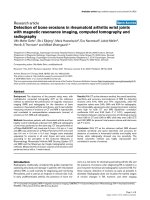
Transgene construction and βgal staining of lung lobesFigure 1
Transgene construction and βgal staining of lung lobes. A. Transgene fragment for microinjection. The Cre cDNA and
a Neo polyadenylylation signal were placed under the control of the mouse α-SMA promoter (-1070 to +2582, including the
first exon and part of the first intron). B. Comparison of βgal staining in the bronchi of R26R and α-SMA-Cre/R26R mice. Pos-
itive βgal staining (blue color) is observed in the bronchi of α-SMA-Cre/R26R mice (a, 20× magnification), but not in R26R mice
(b, 20× magnification).
a
b
R26R
SMA-Cre/R26R
Respiratory Research 2007, 8:1 />Page 4 of 11
(page number not for citation purposes)
20 sec, and coverslips were mounted with 80% glycerol.
Slides were examined using a Leica DC 500-fluorescence
microscope equipped with a digital camera.
Alternatively, lung sections were processed for Masson's
trichrome staining to detect collagen and elastin. The
staining was carried out using Masson trichrome staining
Kit (Maxim-Bio, Fuzhou, China) according to the manu-
facturer's instruction.
Western Blot
α-SMA protein levels in lungs were evaluated by western
blot as previously described [16]. After cytoplasmic pro-
tein extraction from the lower lobe of right lung of PBS or
BLM-injected mice, protein was quantified using a BCA
assay kit (Pierce, USA) and 20 μg was used for SDS-PAGE
electrophoresis. Following electrophoretic transfer, mem-
branes were incubated with anti-α-SMA mAb (1:1000) in
TBS/T buffer at 4°C overnight. Membranes were incu-
bated with anti-mouse IgG secondary antibody conju-
gated to HRP (1:1000), followed by exposure to ECL
chemiluminescent substrate (Amersham, UK) and digital
scanning in Image station 2000 (Kodak, US). Following
α-SMA blotting, films were placed in stripping buffer (50
mM DTT, 50 mM Tris. HCI,2%SDS) at 50°C for 30 min-
utes, washed 5 times, reblocked and reprobed with
GAPDH mAb (1:800) and HRP conjugated secondary
antibody. Then the membranes went through chemilumi-
nescence as discribed above to detect GAPDH protein in
the same film. α-SMA protein levels were measured by
densitometry, and expressed relative to GAPDH. Dupli-
cate samples were analyzed for each mouse.
Cell culture and immunofluorescent staining
The human bronchial epithelial cell line 16HBE-14o
(16HBE), a generous gift from Professor S. Holgate
(Southampton University, UK) was routinely maintained
in growth medium consisting of MEM (Life Technologies,
USA) and 10% FCS (Shijiqing Co, China). Cells were
seeded into sterile round coverslips placed inside 12-well
plates. On reaching 70% confluence, medium was
changed to FCS-free MEM, and rhTGF-β1 (R&D company,
US) was added to a subset of wells to a final concentration
of 10 μg/L. 72 hours later, all wells were washed twice
with cold PBS and a subset of wells were fixed with cold
methanol:acetone (1:1) at -20°C for 10 min. Coverslips
were removed from the wells and placed on glass slides,
blocked with 10% goat serum for 60 min. Cells on cover-
slips were incubated with anti α-SMA mAb (1:400) or rab-
bit anti human E-cadherin (1:50) overnight at 4°C and
subsequently incubated with goat anti-mouse IgG-TRITC
(1:800) or goat anti-rabbit IgG-FITC (1:400) for 1 hour.
Another subset of wells were fixed with PFA at RT for 20
min and treated with 0.1% TritonX-100 for 5 min. Cover-
slips were removed from wells, placed on slides, blocked
with 10% goat serum for 30 min and incubated with 100
μl Alex 594 phalloidin (1:500) for 20 min at RT. DAPI was
used to stain nuclei (500 ng/ml in 95% ethanol) for 20
sec, and coverslips were mounted with 80% glycerol.
Slides were examined using a Leica DC 500-fluorescence
microscope equipped with a digital camera.
Results
Generation of
α
-SMA-Cre/R26R transgenic mice
To permanently label myofibroblasts, we firstly generated
transgenic mice bearing an α-SMA promoter driven Cre.
Ten pseudopregnant mice were implanted oviductally
with fertilized eggs injected with the construct, yielding 19
offspring, 4 of which were identified to carry the ran-
domly integrated transgene. Two founder mice were
selected and used to produce inbred strains. We then
crossed an α-SMA-Cre transgenic strain to reporter strain
R26R+/+ whereby Cre-specific recombination at the
ROSA26 locus allows expression of β-galactosidase in
smooth muscle cells and myofibroblasts.
β
gal staining corresponds with distribution of the smooth
muscles of the
α
-SMA-Cre R26R strain
βgal staining was performed on offspring of the α-SMA-
Cre/R26R and the R26R mice respectively. Positive βgal
staining was observed in the trachea of the α-SMA-Cre/
R26R strain (Fig. 1b), but not in that of the R26R mouse
(Fig. 1a) under anatomy microscopy, confirming that the
βgal staining resulted from α-SMA-driven Cre-mediated
recombination.
As expected, βgal staining was highly restricted to SMCs in
smooth muscle-rich organs isolated from the α-SMA-Cre/
R26R mice (data not shown). In pulmonary arteries and
veins, βgal staining was consistent with the natural distri-
bution of smooth muscle tissue at these sites (Fig. 2a, b).
In the intrapulmonary bronchus (Fig. 2d, g), the staining
was precisely localized to the muscularis layer of the air-
way, which paralleled the immunofluorescent staining
pattern of α-SMA (Fig. 2e, h) and SM-MHC (Fig. 2f, i).
Neither the terminal bronchioles, which lack SMC (Fig.
2c), nor the bronchial epithelia (Fig. 2a,b,c,d,g) showed
positive βgal staining.
A great enhancement of
β
gal staining in the lungs of the
double transgenic mice after Bleomycin treatment
The double transgenic mouse strain described above pro-
vides a simple means to follow expression of α-SMA, and
thus the regulation of myofibroblast development during
pulmonary fibrosis. We next endotracheally adminis-
trated BLM in the transgenic mice for induction of lung
injury and fibrosis. As shown in figure 3A~b, in the whole-
mount lung preparations from BLM-treated α-SMA-Cre/
R26R transgenic mice, βgal staining is easily observed. In
contrast, except for the helium area, the staining is not
Respiratory Research 2007, 8:1 />Page 5 of 11
(page number not for citation purposes)
observed in preparations from PBS-treated transgenic
mice (Fig. 3A~a).
Histological observations reveal a significantly increased
number of βgal staining- positive cells located at subepi-
thelial areas of bronchioles, terminal bronchioles (Fig.
3A~d,f) and tunica media of pulmonary vessels, particu-
larly pulmonary veins (Fig. 3A~f, h arrowheads) in BLM
treated mice. At these sites, distribution of collagen is vis-
ualized by Masson's trichrome staining, showing
increased collagen deposition around the walls of small
veins and terminal respiratory bronchioles and in certain
parenchymal areas (Fig. 3A~j arrowheads). In contrast,
there is only minimal βgal staining (Fig. 3A~c, e, g) and
Masson trichrome staining (Fig. 3A~i) in the lung of con-
trol mice.
Correspondingly, western blot analysis revealed an over-
all increase in lung α-SMA protein in the BLM treated
transgenic mice, compared with the control mice (Fig. 3B)
βgal and immunofluorescent staining in lung tissues of α-SMA-Cre/R26R miceFigure 2
βgal and immunofluorescent staining in lung tissues of α-SMA-Cre/R26R mice. In βgal stained sections (a, b, c, d, g),
intrapulmonary veins were homogeneously stained (a, b) and pulmonary arteries were heterogeneously stained (a). In the main
bronchus of pulmonary hilum, unstained ciliated epithelia were surrounded by a βgal stained muscular layer (a, arrowhead),
βgal staining was not detected in terminal bronchioles, although small veins were positively stained (c). The thin layer of βgal
staining was observed in the sub-epithelial areas of small and medium bronchi, respectively (arrowheads in b, d, g). The βgal
stained areas of bronchus (d, g) paralleled the staining pattern for α-SMA (TRITC-labeled, arrowheads in e, h) and SM-MHC
(FITC-labeled, arrowheads in f, i) (a-c, 100×, d-i, 400× magnification).
a
b
c
d
e
f
g
h
i
Respiratory Research 2007, 8:1 />Page 6 of 11
(page number not for citation purposes)
Effects of BLM on lung α-SMA protein levels, ECM deposition and βgal stainingFigure 3
Effects of BLM on lung α-SMA protein levels, ECM deposition and βgal staining. Panel A: βgal and Masson's tri-
chrome-staining in sections of lung tissue shows βgal staining to wholemount left lung lobes of the PBS-treated (a) and the
BLM-treated mice (b) (a, b 10× magnification). In moderate bronchi, thickened bronchial wall with homogeneous βgal stained
fusiform cells was observed in the BLM-treated lungs (d), and not in the PBS-treated mouse (c), Arrowheads indicate that a few
cells in alveolar wall were positively stained (d). In pulmonary bronchioles and vessels, BLM treated lung demonstrated
enhanced βgal expression (f, h), compared with that of PBS treated lung (e, g). βgal stained venous wall was thickened (arrow-
head in f) and the positively stained cells infiltrated outwards (arrowhead in h). In the Masson's trichrome-stained lung sections
(i, j), extensive collagen staining (Blue color) was seen in BLM treated lung (arrowheads in j), but not in the control with PBS (i).
(c-j 400× magnification). Panel B: Western blot analysis of protein extracts from lower right lobes of the BLM treated mice (2)
and control mice (1).
A
a
b
c
d
e
f
g
h
i
j
B
1 2
Respiratory Research 2007, 8:1 />Page 7 of 11
(page number not for citation purposes)
Detection of EMT in bronchial epithelial cells of the
α
-
SMA-Cre/R26R mice during BLM-induced pulmonary
fibrosis
As shown in Fig. 4, we also detected a few βgal positive
cells, with basal or columnar epithelial cell morphology,
existing in epithelia lining the bronchioles (Fig. 4b, c
arrowheads) and air-sacs (Fig. 3A~d arrowheads), in the
BLM-treated transgenic mice. This was not observed in the
control mice (Fig. 3A~c,e; Fig. 4a). Using double immun-
ofluorescent staining, with antibodies against α-SMA and
E-cadherin, we further demonstrated that certain cells
located at bronchiolar epithelium of BLM treated mice
were simultaneously stained with these epithelial and
mesenchymal markers (Fig. 4g,h,i arrowheads), indicat-
ing their undergoing of EMT. No common-staining was
found in control mice (Fig. 4d,e,f). We have also found
similar results from lung tissues after BLM treatment at
day 7, 14, 20 and 28.
In vitro phenotype analysis of 16HBE following exposure
of TGF-
β
1
In vitro immunofluorescent staining of 16HBE cells
(human bronchial epithelial cell line) demonstrates that
exposure to TGF-β1 results in an apparent reduction of E-
cadherin staining, an epithelial marker, concomitant with
its redistribution from intercellular junction areas into the
cytoplasm (Fig. 5a, b). In contrast, the mesenchymal
marker F-actin, whose expression was detectable only at
the cellular margin before the exposure, shows an
increased level in the epithelial cells where it is diffusely
distributed throughout the cytoplasm after stimulation
with TGF-β1 (Fig. 5c, d). Meanwhile, positive α-SMA
immunofluorescent staining, which was undetectable
prior exposure to TGF-β1, appeared in the cytoplasm in a
small number of the 16HBE cells (Fig. 5e, f).
Discussion
Using the Cre/Loxp system, we generated a transgenic
mouse strain that expressed lacZ specifically in SMCs and
myofibroblasts containing tissues. The βgal expression
pattern in the α-SMA-Cre/R26R transgenic model closely
resembled the expression of endogenous α-SMA in the
airways, and that in the gastrointestinal channel, vessels
and genitourinary tract under normal physiological con-
ditions. These data suggest that the SMP8 promoter region
of the α-SMA gene, including the first exon and part of the
first intron (-1070 to +2582 of the mouse α-SMA pro-
moter), is sufficient to recapitulate endogenous α-SMA
expression patterns, in concordance with previous studies
[17-19].
Smooth muscle-targeted Cre recombinase mice that have
previously been generated by others for study of diseases,
including SM22-CreER and SMMHC-Cre strains in which
Cre is driven by the promoter of SM22 gene or SM-MHC
gene, respectively. Feil and colleagues have generated the
SM22-CreER transgenic mice to the effect that the expres-
sion of the transgene is confined to smooth muscle cells
for studying vascular and gastrointestinal diseases [20].
However, gene knockout studies suggest that SM22 is not
required for vascular and visceral SMC homeostatic func-
tions in the developing mouse [21], and there are no data
demonstrating that SM22 expression signifies myofibrob-
last activation. With regards to the SMMHC-Cre strain that
has also been used to study vascular development and dis-
eases [22], it has been documented that SM-MHC is sel-
dom expressed in non-SMC cells such as myofibroblasts
[23,24]. In contrast, our α-SMA-Cre/R26R strain appears
to be sensitive to myofibroblast activation after BLM expo-
sure, as Cre-mediated recombination is controlled by the
promoter of the gene encoding α-SMA, a marker of myofi-
broblast transition.
Additionally, for the reason that in vivo recombination in
Cre/Loxp system is irreversible, βgal staining in the lung of
our transgenic strain could reflect past and present myofi-
broblast transition events post BLM treatment. This may
assist discovery of the cellular source of the active myofi-
broblasts in the development of pulmonary fibrosis. In
the chronic progression of fibrosis, multiple cycles of
injury and repair may occur repeatedly with a broad time
period and range of sites. Activation of the α-SMA pro-
moter may be a transient event and limited to a subgroup
of cells at a given time point [25]. So the α-SMA-Cre
mouse strain is likely to be highly relevant for studies of
fibrotic diseases and activation of myofibroblasts, and
trace the source of myofibroblasts.
In the BLM-treated α-SMA-Cre/R26R mice, we observed a
number of βgal staining positive cells emerging in the sub-
epithelial areas of bronchioles and terminal bronchioles
and in the ectoblast of vessels, concomitant with extensive
Masson Trichrome-stained extracellular matrix. In com-
parison, in the PBS-treated α-SMA-Cre/R26R mice, βgal
positive cells were seldom seen, suggesting that not only
can the reporter mice demonstrate the inherent distribu-
tion of pulmonary SMCs under physiologic condition,
but also have the capability to sensitively record the trail
of myofibroblast transition in the lung of the mice follow-
ing pathologic stimulation. We demonstrate herein that
increased βgal expression in the lungs of the BLM-treated
mice is mainly to be due to the appearance of myofibrob-
lasts in the subepithelial areas of bronchiole and terminal
bronchiole. Previous studies had shown that airway BLM
administration does not result in remarkable morpholog-
ical changes in the SMC layer [26].
There have been prior suggestions that EMT occurs in the
lung during fibrogenesis, but these suggestions derive
largely from studies of transformed cells or primary AECs
Respiratory Research 2007, 8:1 />Page 8 of 11
(page number not for citation purposes)
cultured on plastic, the in vivo significance of which is
unclear [7,9]. It has recently been reported from IPF lung
biopsies that epithelial cells had acquired mesenchymal
features, raising the possibility of EMT during fibrogenesis
[8]. More recently, Kim and colleagues developed a trans-
genic mouse reporter strain in which lung epithelial cells
were genetically altered to permanently express βgal, and
their fates are followed in an established model of pulmo-
nary fibrosis induced by intranasal Adeno-TGF-β1. They
showed that βgal-positive cells expressing mesenchymal
markers accumulated within 3 weeks of in vivo TGF-β1
expression, demonstrating that EMT occurs in vivo in an
animal model [10]. As shown at figure 3, we also observed
the occurrence of EMT in parenchymal alveloar areas fol-
lowing BLM stimulation in our α-SMA-Cre/R26R reporter
mice where a few βgal-positive cells located in alveolar
wall demonstrated that the cells were undergoing EMT.
Alternatively, the βgal-stained epithelial cells may simply
βgal and αSMA positively stained bronchial epithelial cells in the α-SMA-Cre R26R mice treated with BLMFigure 4
βgal and αSMA positively stained bronchial epithelial cells in the α-SMA-Cre R26R mice treated with BLM. βgal
stained lung sections of α-SMA-Cre/R26R mice without and with BLM treatment (a-c). The section from BLM treated mice
showed a few βgal stained bronchial epithelial cells (arrowheads in b and c), but not from PBS treated mice (a). Double immun-
ofluorescent staining for α-SMA and E-Cadherin was performed on the sections from PBS (d-f) or BLM (g-i) treated mice. d, g:
FITC-labeled E-cadherin; e, h: TRITC labeled α-SMA. Positive double immunofluorescent staining (g, h, i) was observed in the
bronchial epithelial cells lining the bronchioles of the BLM-treated lung where βgal staining was detected as above, but not in
the control (d, e, f). The images of (d) and (e), or (g) and (h) were merged into (f) and (i). The red fluorescence (h arrowhead)
indicated the positive α-SMA staining and yellow fluorescent staining (i arrowhead) indicated that the epithelial cells positively
co-stained with α-SMA and E-Cadherin. (all images are 400× magnification).
ab
def
c
ghi
Respiratory Research 2007, 8:1 />Page 9 of 11
(page number not for citation purposes)
Phenotypic analysis of the human bronchial epithelial cell line (16HBE) following exposure to TGF-β1Figure 5
Phenotypic analysis of the human bronchial epithelial cell line (16HBE) following exposure to TGF-β1. Immun-
ofluorescent staining for E-cadherin (a, b) showed that exposure to TGF-β1 (b) resulted in an apparent reduction and redistri-
bution of E-cadherin from intercellular junction areas into cytoplasm, compared to control (a). Mesenchymal marker F-actin,
was faintly stained at the cell margin in the control (c), whereas the staining was substantially enhanced and abundantly located
throughout cytoplasm after TGF-β1 stimulation (d). Immunofluorescent staining for (-SMA was not detected in the cells under
basal conditions (e), but was observable in a few cells after TGF-β1 exposure (f).
d
e
a
b
c
f
Respiratory Research 2007, 8:1 />Page 10 of 11
(page number not for citation purposes)
demonstrate transcriptional activation of α-SMA gene in
these cells.
Additionally, βgal was stained positively in a few basal
epithelial cells and columnar epithelial cells lining the
bronchiole during bleomycin-induced lung fibrosis in the
reporter mice. The immunofluorescent co-staining of the
both E-cadherin and α-SMA confirmed further that the
BECs were undergoing EMT. When we focused on the BEC
cell line 16HBE in vitro, we found that exposure to TGF-
β1 led to a remarkable myofibroblast cell-like phenotype,
marked by expression of α-SMA and F-actin and the
reduction of the epithelial-specific junction localization
of E-cadherin. Taken together, these observations suggest
that BECs might also be capable of undergoing EMT and
thereby provide another cellular source for the parenchy-
mal aggregation of myofibroblasts during fibrosis.
As mentioned above, however, the present histological
observations in the reporter mice do not support the
rationale that EMT exerts a critical influence on the pro-
gression of pulmonary fibrosis, because EMT indicated by
βgal staining and α-SMA immunostaining was rarely
detected in BECs and AECs of BLM-treated mice. For the
predominant activation of sub-epithelial myofibroblasts
in the development of BLM-treated lung fibrosis, fibrob-
lasts or other sources of progenitors may play more essen-
tial roles which contribute to the pool of expanded
myofibroblasts after lung injury. To what extent does EMT
contribute to the aggravation of fibrosis, whether similar
EMT in BECs occur in IPF patients are all interesting ques-
tions for future studies.
Additionally, the α-SMA-Cre single transgenic strain bear-
ing the α-SMA driven Cre is sufficiently sensitive to test
the function of a candidate gene in SMCs or myofibrob-
lasts on the development of pulmonary fibrosis. Tissue-
specific gene knockout or knock-in can be accomplished
via crossing the α-SMA-Cre mouse to a strain containing a
loxP site flanked sequence of interest.
Conclusion
In conclusion, we have developed a double transgenic
reporter mouse strain to map the natural distribution of
α-SMA-expressing cells in vivo under basal physiological
condition. Moreover, lung cells that do not express α-SMA
under normal conditions may permanently express βgal
via α-SMA activation in response to pathologic stimula-
tion, thus allowing tracking of the cellular source of
myofibroblasts and to definitively test whether EMT
occurs in vivo.
Competing interests
The author(s) declare that they have no competing inter-
ests.
Authors' contributions
ZW carried out the transgene construction, transgenic
screening and breeding, histological works and drafted
the manuscript. LLY carried out the microinjections and
transgenic screening and breeding. LC participated in the
histological work and mice screening and breeding. MZ
participated in the in vitro immunostaining. XC partici-
pated in the microinjections. XY guided the microinjec-
tion and animal breeding. JX design the study, technical
support the research, revise the manuscript and give final
approval of the version to be published. All authors read
and approved the final manuscript.
Acknowledgements
We thank Professor F. Costantini (Department of genetics, Columbia Uni-
versity) and Professor Art Strauch (Department of Physiology and Cell
Biology, Dorothy M. Davis Heart and Lung Research Institute, OH, USA)
for providing the Cre and Smp8 containing plasmids. We thank Professor
Conti, Mary Anne (NIH/NHLBI, USA) for providing SM-MHC polyclonal
antibody. We also thank Professor Xiaoping Jian (Guangdong Teacher Col-
lege of Foreign language and art, China) and Dr Elaina Collie-Dugui
(Department of Medicine and Therapeutics, University of Aberdeen, Scot-
land) for critically reading the manuscript. This research was supported by
the National Natural Science Foundation of China (NO. 30230180). This
funding covered all the cost in study design; in the collection, analysis, and
interpretation of data; in the writing of the manuscript and in the decision
to submit the manuscript for publication.
References
1. Kuhn C, McDonald JA: The roles of the myofibroblast in idio-
pathic pulmonary fibrosis. Ultrastructural and immunohis-
tochemical features of sites of active extracellular matrix
synthesis. Am J Pathol 1991, 138(5):1257-1265.
2. Schissel SL, Layne MD: Telomerase, myofibroblasts, and pulmo-
nary fibrosis. Am J Respir Cell Mol Biol 2006, 34(5):520-522.
3. White ES, Atrasz RG, Hu B, Phan SH, Stambolic V, Mak TW, Hoga-
boam CM, Flaherty KR, Martinez FJ, Kontos CD, et al.: Negative
regulation of myofibroblast differentiation by PTEN (Phos-
phatase and Tensin Homolog Deleted on chromosome 10).
Am J Respir Crit Care Med 2006, 173(1):112-121.
4. Chambers RC, Leoni P, Kaminski N, Laurent GJ, Heller RA: Global
expression profiling of fibroblast responses to transforming
growth factor-betal reveals the induction of inhibitor of dif-
ferentiation-1 and provides evidence of smooth muscle cell
phenotypic switching. Am J Pathol 2003, 162(2):533-546.
5. Oda D, Gown AM, Vande Berg JS, Stern R: The fibroblast-like
nature of myofibroblasts. Exp Mol Pathol 1988, 49(3):316-329.
6. Hashimoto N, Jin H, Liu T, Chensue SW, Phan SH: Bone marrow-
derived progenitor cells in pulmonary fibrosis. J Clin Invest
2004, 113(2):243-252.
7. Kasai H, Allen JT, Mason RM, Kamimura T, Zhang Z: TGF-betal
induces human alveolar epithelial to mesenchymal cell tran-
sition (EMT). Respir Res 2005, 6:56.
8. Willis BC, Liebler JM, Luby-Phelps K, Nicholson AG, Crandall ED, du
Bois RM, Borok Z: Induction of epithelial-mesenchymal transi-
tion in alveolar epithelial cells by transforming growth fac-
tor-betal: potential role in idiopathic pulmonary fibrosis. Am
J Pathol 2005, 166(5):1321-1332.
9. Yao HW, Xie QM, Chen JQ, Deng YM, Tang HF: TGF-beta lin-
duces alveolar epithelial to mesenchymal transition in vitro.
Life Sci 2004, 76(1):29-37.
10. Kim KK, Kugler MC, Wolters PJ, Robillard L, Galvez MG, Brumwell
AN, Sheppard D, Chapman HA: Alveolar epithelial cellmesen-
chymal transition develops in vivo during pulmonary fibrosis
andis regulated by the extracellular matrix. Proc Natl Acad Sci
USA 2006.
Publish with BioMed Central and every
scientist can read your work free of charge
"BioMed Central will be the most significant development for
disseminating the results of biomedical research in our lifetime."
Sir Paul Nurse, Cancer Research UK
Your research papers will be:
available free of charge to the entire biomedical community
peer reviewed and published immediately upon acceptance
cited in PubMed and archived on PubMed Central
yours — you keep the copyright
Submit your manuscript here:
/>BioMedcentral
Respiratory Research 2007, 8:1 />Page 11 of 11
(page number not for citation purposes)
11. Snider GL, Hayes JA, Korthy AL: Chronic interstitial pulmonary
fibrosis produced in hamsters by endotracheal bleomycin:
pathology and stereology. Am Rev Respir Dis 1978,
117(6):1099-1108.
12. Hinz B, Mastrangelo D, Iselin CE, Chaponnier C, Gabbiani G:
Mechanical tension controls granulation tissue contractile
activity and myofibroblast differentiation. Am J Pathol 2001,
159(3):1009-1020.
13. Hamilton DL, Abremski K: Site-specific recombination by the
bacteriophage P1 lox-Cre system. Cre-mediated synapsis of
two lox sites. J Mol Biol 1984, 178(2):481-486.
14. Metzger D, Clifford J, Chiba H, Chambon P: Conditional site-spe-
cific recombination in mammalian cells using a ligand-
dependent chimeric Cre recombinase. Proc Natl Acad Sci USA
1995, 92(15):6991-6995.
15. Soriano P: Generalized lacZ expression with the ROSA26 Cre
reporter strain. Nat Genet 1999, 21(1):70-71.
16. Hinz B, Celetta G, Tomasek JJ, Gabbiani G, Chaponnier C: Alpha-
smooth muscle actin expression upregulates fibroblast con-
tractile activity. Mol Biol Cell 2001, 12(9):2730-2741.
17. Zhang M, Smith EP, Kuroda H, Banach W, Chernausek SD, Fagin JA:
Targeted expression of a protease-resistant IGFBP-4
mutant in smooth muscle of transgenic mice results in
IGFBP-4 stabilization and smooth muscle hypotrophy. J Biol
Chem 2002, 277(24):21285-21290.
18. Wang J, Niu W, Nikiforov Y, Naito S, Chernausek S, Witte D,
LeRoith D, Strauch A, Fagin JA: Targeted overexpression of IGF-
I evokes distinct patterns of organ remodeling in smooth
muscle cell tissue beds of transgenic mice. J Clin Invest 1997,
100(6):1425-1439.
19. Zadelaar SM, Boesten LS, Pires NM, van Nieuwkoop A, Biessen EA,
Jukema W, Havekes LM, van Vlijmen BJ, Willems van Dijk K: Local
cre-mediated gene recombination in vascular smooth mus-
cle cells in mice. Transgenic Res 2006, 15(1):31-36.
20. Kuhbandner S, Brummer S, Metzger D, Chambon P, Hofmann F, Feil
R: Temporally controlled somatic mutagenesis in smooth
muscle. Genesis 2000, 28(1):15-22.
21. Zhang JC, Kim S, Helmke BP, Yu WW, Du KL, Lu MM, Strobeck M,
Yu Q, Parmacek MS: Analysis of SM22alpha-deficient mice
reveals unanticipated insights into smooth muscle cell differ-
entiation and function. Mol Cell Biol 2001, 21(4):1336-1344.
22. Regan CP, Manabe I, Owens GK: Development of a smooth mus-
cle-targeted cre recombinase mouse reveals novel insights
regarding smooth muscle myosin heavy chain promoter reg-
ulation. Circ Res 2000, 87(5):363-369.
23. Miano JM, Cserjesi P, Ligon KL, Periasamy M, Olson EN: Smooth
muscle myosin heavy chain exclusively marks the smooth
muscle lineage during mouse embryogenesis. Circ Res 1994,
75(5):803-812.
24. Owens GK, Kumar MS, Wamhoff BR: Molecular regulation of
vascular smooth muscle cell differentiation in development
and disease. Physiol Rev 2004, 84(3):767-801.
25. Grinnell F, Ho CH: Transforming growth factor beta stimu-
lates fibroblast-collagen matrix contraction by different
mechanisms in mechanically loaded and unloaded matrices.
Exp Cell Res 2002, 273(2):248-255.
26. Borzone G, Moreno R, Urrea R, Meneses M, Oyarzun M, Lisboa C:
Bleomycin-induced chronic lung damage does not resemble
human idiopathic pulmonary fibrosis. Am J Respir Crit Care Med
2001, 163(7):1648-1653.