Saladin Anatomy and Physiology The Unity of Form and Function Episode 7 potx
Bạn đang xem bản rút gọn của tài liệu. Xem và tải ngay bản đầy đủ của tài liệu tại đây (4.93 MB, 70 trang )
Saladin: Anatomy &
Physiology: The Unity of
Form and Function, Third
Edition
Atlas B Surface Anatomy Text
© The McGraw−Hill
Companies, 2003
Atlas B
Tibia
Soleus
Tibialis anterior
Medial malleolus
Head of metatarsal I
Hallux (great toe)
(a)
Lateral malleolus
Site for palpating dorsal pedal artery
Extensor digitorum longus tendons
Extensor hallucis longus tendon
IIIIIIIVV
(b)
Lateral longitudinal arch
Lateral malleolus
Transverse arch
Digits (I–V)
Medial longitudinal arch
Calcaneus
Head of metatarsal I
Head of metatarsal V
Abductor digiti minimi
Abductor hallucis
Hallux (great toe)
I
II
III
IV
V
Figure B.14 The Right Foot. (a) Dorsal aspect, (b) plantar aspect.
405
Saladin: Anatomy &
Physiology: The Unity of
Form and Function, Third
Edition
Atlas B Surface Anatomy Text
© The McGraw−Hill
Companies, 2003
Atlas B
406 Part Two Support and Movement
8
9
10
11
12
13
14
15
16
5
3
2
1
4
6
7
(a)
Figure B.15 Muscle Test. To test your knowledge of muscle anatomy, match the 30 labeled muscles on these photographs to the alphabetical list
of muscles below. Answer as many as possible without referring to the previous illustrations. Some of these names will be used more than once, since the
same muscle may be shown from different perspectives, and some of these names will not be used at all. The answers are in appendix B.
(b)
27
26
25
24
28
29
30
23
17
18
19
20
21
22
a. biceps brachii
b. brachioradialis
c. deltoid
d. erector spinae
e. external abdominal oblique
f. flexor carpi ulnaris
g. gastrocnemius
h. gracilis
i. hamstrings
j. infraspinatus
k. latissimus dorsi
l. pectineus
m. pectoralis major
n. rectus abdominis
o. rectus femoris
p. serratus anterior
q. soleus
r. splenius capitis
s. sternocleidomastoid
t. subscapularis
u. teres major
v. tibialis anterior
w. transversus abdominis
x. trapezius
y. triceps brachii
z. vastus lateralis
Saladin: Anatomy &
Physiology: The Unity of
Form and Function, Third
Edition
11. Muscular Tissue Text
© The McGraw−Hill
Companies, 2003
Types and Characteristics of Muscular
Tissue 408
• Universal Characteristics of Muscle 408
• Skeletal Muscle 408
Microscopic Anatomy of Skeletal Muscle 409
• The Muscle Fiber 409
• Myofilaments 409
• Striations 411
The Nerve-Muscle Relationship 412
• Motor Neurons 412
• The Motor Unit 412
• The Neuromuscular Junction 413
• Electrically Excitable Cells 415
Behavior of Skeletal Muscle Fibers 416
• Excitation 417
• Excitation-Contraction Coupling 417
• Contraction 417
• Relaxation 422
• The Length-Tension Relationship and Muscle
Tone 422
Behavior of Whole Muscles 423
• Threshold, Latent Period, and Twitch 423
• Contraction Strength of Twitches 424
• Isometric and Isotonic Contraction 425
Muscle Metabolism 427
• ATP Sources 427
• Fatigue and Endurance 428
• Oxygen Debt 429
• Physiological Classes of Muscle Fibers 429
• Muscular Strength and Conditioning 431
Cardiac and Smooth Muscle 432
• Cardiac Muscle 432
• Smooth Muscle 433
Chapter Review 438
INSIGHTS
11.1 Clinical Application:
Neuromuscular Toxins and
Paralysis 414
11.2 Clinical Application: Rigor
Mortis 422
11.3 Medical History: Galvani, Volta,
and Animal Electricity 424
11.4 Clinical Application: Muscular
Dystrophy and Myasthenia
Gravis 437
11
CHAPTER
Muscular Tissue
Neuromuscular junctions (SEM)
CHAPTER OUTLINE
Brushing Up
To understand this chapter, it is important that you understand or
brush up on the following concepts:
• Aerobic and anaerobic metabolism (p. 86)
• The functions of membrane proteins, especially receptors and
ion gates (p. 100)
• Structure of a neuron (p. 175)
• General histology of the three types of muscle (p. 176)
• Desmosomes and gap junctions (p. 179)
• Connective tissues of a muscle (p. 326)
407
Saladin: Anatomy &
Physiology: The Unity of
Form and Function, Third
Edition
11. Muscular Tissue Text
© The McGraw−Hill
Companies, 2003
Chapter 11
M
ovement is a fundamental characteristic of all living things,
but reaches its highest development in animals because of
their muscular tissue. Muscular tissue is composed of elongated
cells that contract when stimulated. A muscle cell is essentially a
device for converting the chemical energy of ATP into the mechan-
ical energy of contraction. This chapter discusses contraction at the
cellular and molecular levels and explains the basis of such aspects
of muscle performance as warm-up, strength, endurance, and
fatigue. These phenomena have obvious relevance to athletic per-
formance, and they become very important when old age or lack of
physical conditioning interferes with a person’s ability to carry out
everyday motor tasks. The effects of old age on the muscular sys-
tem are discussed in chapter 29.
The three types of muscle tissue—skeletal, cardiac, and
smooth—were described and compared in chapter 5. The expres-
sion “muscular system” refers only to skeletal muscle. This chap-
ter is concerned primarily with the microscopic anatomy and
physiology of skeletal muscle. Cardiac and smooth muscle are dis-
cussed more briefly to compare their properties and functions
with skeletal muscle. Cardiac muscle is discussed more extensively
in chapter 19.
Types and Characteristics
of Muscular Tissue
Objectives
When you have completed this section, you should be able to
• describe the physiological properties that all muscle types
have in common;
• list the defining characteristics of skeletal muscle; and
• describe the elastic functions of the connective tissue
components of a muscle.
Universal Characteristics of Muscle
The functions of muscular tissue were detailed in the pre-
ceding chapter: movement, stability, communication, con-
trol of body openings and passages, and heat production.
To carry out those functions, all muscular tissue has the
following characteristics:
• Responsiveness (excitability). Responsiveness is a
property of all living cells, but muscle and nerve cells
have developed this property to the highest degree.
When stimulated by chemical signals
(neurotransmitters), stretch, and other stimuli, muscle
cells respond with electrical changes across the
plasma membrane.
• Conductivity. Stimulation of a muscle fiber produces
more than a local effect. The local electrical change
triggers a wave of excitation that travels rapidly along
the muscle fiber and initiates processes leading to
muscle contraction.
• Contractility. Muscle fibers are unique in their ability
to shorten substantially when stimulated. This enables
them to pull on bones and other tissues and create
movement of the body and its parts.
• Extensibility. In order to contract, a muscle cell must
also be extensible—able to stretch again between
contractions. Most cells rupture if they are stretched
even a little, but skeletal muscle fibers can stretch to
as much as three times their contracted length.
• Elasticity. When a muscle cell is stretched and the
tension is then released, it recoils to its original resting
length. Elasticity, commonly misunderstood as the
ability to stretch, refers to this tendency of a muscle
cell (or other structures) to return to the original
length when tension is released.
Skeletal Muscle
Skeletal muscle may be defined as voluntary striated mus-
cle that is usually attached to one or more bones. A typi-
cal skeletal muscle cell is about 100 m in diameter and 3
cm long; some are as thick as 500 m and as long as 30 cm.
Because of their extraordinary length, skeletal muscle
cells are usually called muscle fibers or myofibers. A
skeletal muscle fiber exhibits alternating light and dark
transverse bands, or striations, that reflect the overlapping
arrangement of the internal contractile proteins (fig. 11.1).
Skeletal muscle is called voluntary because it is usually
subject to conscious control. The other types of muscle are
involuntary (not usually under conscious control), and
they are never attached to bones.
Recall from chapter 10 that a skeletal muscle is com-
posed not only of muscular tissue, but also of fibrous con-
nective tissue: the endomysium that surrounds each mus-
cle fiber, the perimysium that bundles muscle fibers
together into fascicles, and the epimysium that encloses
the entire muscle. These connective tissues are continu-
ous with the collagen fibers of tendons and those, in turn,
408
Part Two Support and Movement
Nucleus
Muscle fiber
Endomysium
Striations
Figure 11.1 Skeletal Muscle Fibers. Note the striations.
Saladin: Anatomy &
Physiology: The Unity of
Form and Function, Third
Edition
11. Muscular Tissue Text
© The McGraw−Hill
Companies, 2003
Chapter 11
Chapter 11 Muscular Tissue 409
with the collagen of the bone matrix. Thus, when a mus-
cle fiber contracts, it pulls on these collagen fibers and
moves a bone.
Collagen is not excitable or contractile, but it is some-
what extensible and elastic. It stretches slightly under ten-
sion and recoils when released. Because of this elasticity and
because the connective tissue components are connected to
each other in a linear series, the connective tissues are called
the series-elastic components of a muscle. Their elasticity
helps to return muscles to their resting lengths when con-
traction ceases. Elastic recoil of the tendons adds signifi-
cantly to the power output and efficiency of the muscles.
Before You Go On
Answer the following questions to test your understanding of the
preceding section:
1. Define responsiveness, conductivity, contractility, extensibility,
and elasticity. State why each of these properties is necessary for
muscle function.
2. How is skeletal muscle different from the other types of muscle?
3. Why would the skeletal muscles perform poorly without their
series-elastic components?
Microscopic Anatomy
of Skeletal Muscle
Objectives
When you have completed this section, you should be able to
• describe the structural components of a muscle fiber;
• relate the striations of a muscle fiber to the overlapping
arrangement of its protein filaments; and
• name the major proteins of a muscle fiber and state the
function of each.
The Muscle Fiber
In order to understand muscle function, you must know
how the organelles and macromolecules of a muscle fiber
are arranged. Perhaps more than any other cell, a muscle
fiber exemplifies the adage, Form follows function. It has
a complex, tightly organized internal structure in which
even the spatial arrangement of protein molecules is
closely tied to its contractile function.
Muscle fibers have multiple flattened or sausage-
shaped nuclei pressed against the inside of the plasma mem-
brane. This unusual condition results from their embryonic
development—several stem cells called myoblasts
1
fuse to
produce each muscle fiber, with each myoblast contributing
a nucleus to the mature fiber. Some myoblasts remain as
unspecialized satellite cells between the muscle fiber and
endomysium. When a muscle is injured, satellite cells can
multiply and produce new muscle fibers to some degree.
Most muscle repair, however, is by fibrosis rather than
regeneration of functional muscle.
The plasma membrane, called the sarcolemma,
2
has
tunnel-like infoldings called transverse (T) tubules that
penetrate through the fiber and emerge on the other side.
The function of a T tubule is to carry an electrical current
from the surface of the cell to the interior when the cell is
stimulated. The cytoplasm, called sarcoplasm, is occu-
pied mainly by long protein bundles called myofibrils
about 1 m in diameter (fig. 11.2). Most other organelles of
the cell, such as mitochondria and smooth endoplasmic
reticulum (ER), are located between adjacent myofibrils.
The sarcoplasm also contains an abundance of glycogen,
which provides stored energy for the muscle to use during
exercise, and a red pigment called myoglobin, which
binds oxygen until it is needed for muscular activity.
The smooth ER of a muscle fiber is called sarcoplas-
mic reticulum (SR). It forms a network around each
myofibril, and alongside the T tubules it exhibits dilated
sacs called terminal cisternae. The SR is a reservoir for
calcium ions; it has gated channels in its membrane that
can release a flood of calcium into the cytosol, where the
calcium activates the muscle contraction process.
Myofilaments
Let’s return to the myofibrils just mentioned—the long
protein cords that fill most of the muscle cell—and look at
their structure at a finer, molecular level. It is here that the
key to muscle contraction lies. Each myofibril is a bundle
of parallel protein microfilaments called myofilaments.
There are three kinds of myofilaments:
1. Thick filaments (fig. 11.3a, b) are about 15 nm in
diameter. Each is made of several hundred
molecules of a protein called myosin. A myosin
molecule is shaped like a golf club, with two
polypeptides intertwined to form a shaftlike tail
and a double globular head, or cross-bridge,
projecting from it at an angle. A thick filament may
be likened to a bundle of 200 to 500 such “golf
clubs,” with their heads directed outward in a
spiral array around the bundle. The heads on one
half of the thick filament angle to the left, and the
heads on the other half angle to the right; in the
middle is a bare zone with no heads.
2. Thin filaments (fig. 11.3c, d), 7 nm in diameter, are
composed primarily of two intertwined strands of a
protein called fibrous (F) actin. Each F actin is like
1
myo ϭ muscle ϩ blast ϭ precursor
2
sarco ϭ flesh, muscle ϩ lemma ϭ husk
Saladin: Anatomy &
Physiology: The Unity of
Form and Function, Third
Edition
11. Muscular Tissue Text
© The McGraw−Hill
Companies, 2003
Chapter 11
a bead necklace—a string of subunits called
globular (G) actin. Each G actin has an active site
that can bind to the head of a myosin molecule.
A thin filament also has 40 to 60 molecules of
yet another protein called tropomyosin. When a
muscle fiber is relaxed, tropomyosin blocks the
active sites of six or seven G actins, and prevents
myosin cross-bridges from binding to them. Each
tropomyosin molecule, in turn, has a smaller
calcium-binding protein called troponin bound to it.
3. Elastic filaments (fig. 11.4b, c), 1 nm in diameter,
are made of a huge springy protein called titin
3
(connectin). They run through the core of a thick
filament, emerge from the end of it, and connect it
to a structure called the Z disc, explained shortly.
They help to keep thick and thin filaments aligned
with each other, resist overstretching of a muscle,
and help the cell recoil to resting length after it is
stretched.
Myosin and actin are called the contractile proteins of
muscle because they do the work of shortening the muscle
fiber. Tropomyosin and troponin are called the regulatory
proteins because they act like a switch to determine when
it can contract and when it cannot. Several clues as to how
they do this may be apparent from what has already been
said—calcium ions are released into the sarcoplasm to acti-
vate contraction; calcium binds to troponin; troponin is
410
Part Two Support and Movement
Sarcoplasm
Sarcolemma
Openings into
transverse tubules
Sarcoplasmic
reticulum
Mitochondria
Myofibrils
A band
I band
Z disc
Nucleus
Triad
Terminal cisternae
Transverse tubule
Figure 11.2 Structure of a Skeletal Muscle Fiber. This is a single cell containing 11 myofibrils (9 shown at the left end and 2 cut off at midfiber).
3
tit ϭ giant ϩ in ϭ protein
Saladin: Anatomy &
Physiology: The Unity of
Form and Function, Third
Edition
11. Muscular Tissue Text
© The McGraw−Hill
Companies, 2003
Chapter 11
Chapter 11 Muscular Tissue 411
also bound to tropomyosin; and tropomyosin blocks the
active sites of actin, so that myosin cannot bind to it when
the muscle is not stimulated. Perhaps you are already form-
ing some idea of the contraction mechanism to be explained
shortly.
Striations
Myosin and actin are not unique to muscle; these proteins
occur in all cells, where they function in cellular motility,
mitosis, and transport of intracellular materials. In skele-
tal and cardiac muscle they are especially abundant, how-
ever, and are organized in a precise array that accounts for
the striations of these two muscle types (fig. 11.4).
Striated muscle has dark A bands alternating with
lighter I bands. (A stands for anisotropic and I for isotropic,
which refers to the way these bands affect polarized light.
To help remember which band is which, think “dArk” and
“lIght.”) Each A band consists of thick filaments lying side
by side. Part of the A band, where thick and thin filaments
overlap, is especially dark. In this region, each thick fila-
ment is surrounded by thin filaments. In the middle of the
A band, there is a lighter region called the H band,
4
into
which the thin filaments do not reach.
Each light I band is bisected by a dark narrow Z disc
5
(Z line) composed of the protein connectin. The Z disc
provides anchorage for the thin filaments and elastic fila-
ments. Each segment of a myofibril from one Z disc to the
next is called a sarcomere
6
(SAR-co-meer), the functional
contractile unit of the muscle fiber. A muscle shortens
because its individual sarcomeres shorten and pull the Z
discs closer to each other, and the Z discs are connected to
the sarcolemma by way of the cytoskeleton. As the Z discs
are pulled closer together during contraction, they pull on
the sarcolemma to achieve overall shortening of the cell.
The terminology of muscle fiber structure is reviewed
in table 11.1; this table may be a useful reference as you
study the mechanism of contraction.
Before You Go On
Answer the following questions to test your understanding of the
preceding section:
4. What special terms are given to the plasma membrane,
cytoplasm, and smooth ER of a muscle cell?
5. What is the difference between a myofilament and a myofibril?
6. List five proteins of the myofilaments and describe their physical
arrangement.
7. Sketch the overlapping pattern of myofilaments to explain how
they account for the A bands, I bands, H bands, and Z discs.
Myosin molecule
Thick filament
Thin filament
Portion of a sarcomere showing the
overlap of thick and thin filaments
Bare zone
Tail
Thin filament
Thick filament
Troponin complex
Heads
G actin
Tropomyosin
Myosin head
(d)
(a)
(b)
(c)
Figure 11.3 Molecular Structure of Thick and Thin
Filaments. (a) A single myosin molecule consists of two intertwined
polypeptides forming a filamentous tail and a double globular head. (b)A
thick filament consists of 200 to 500 myosin molecules bundled together
with the heads projecting outward in a spiral array. (c) A thin filament
consists of two intertwined chains of G actin molecules, smaller
filamentous tropomyosin molecules, and a three-part protein called
troponin associated with the tropomyosin. (d) A region of overlap
between the thick and thin filaments.
4
H ϭ helle ϭ bright
5
Z ϭ Zwichenscheibe ϭ “between disc”
6
sarco ϭ muscle ϩ mere ϭ part, segment
Saladin: Anatomy &
Physiology: The Unity of
Form and Function, Third
Edition
11. Muscular Tissue Text
© The McGraw−Hill
Companies, 2003
Chapter 11
The Nerve-Muscle Relationship
Objectives
When you have completed this section, you should be able to
• explain what a motor unit is and how it relates to muscle
contraction;
• describe the structure of a junction where a nerve fiber
meets a muscle fiber; and
• explain why a cell has an electrical charge difference across
its plasma membrane and, in general terms, how this relates
to muscle contraction.
Skeletal muscle never contracts unless it is stimulated
by a nerve (or artificially with electrodes). If its nerve
connections are severed or poisoned, a muscle is para-
lyzed. If innervation is not restored, the paralyzed mus-
cle undergoes a shrinkage called denervation atrophy.
Thus, muscle contraction cannot be understood without
first understanding the relationship between nerve and
muscle cells.
Motor Neurons
Skeletal muscles are innervated by somatic motor neu-
rons. The cell bodies of these neurons are in the brainstem
and spinal cord. Their axons, called somatic motor fibers,
lead to the skeletal muscles. At its distal end, each somatic
motor fiber branches about 200 times, with each branch
leading to a different muscle fiber (fig. 11.5). Each muscle
fiber is innervated by only one motor neuron.
The Motor Unit
When a nerve signal approaches the end of an axon, it
spreads out over all of its terminal branches and stimu-
lates all the muscle fibers supplied by them. Thus, these
muscle fibers contract in unison. Since they behave as a
single functional unit, one nerve fiber and all the muscle
fibers innervated by it are called a motor unit. The muscle
fibers of a single motor unit are not all clustered together
but are dispersed throughout a muscle (fig. 11.6). Thus,
when they are stimulated, they cause a weak contraction
over a wide area—not just a localized twitch in one small
region.
Earlier it was stated that a motor nerve fiber supplies
about 200 muscle fibers, but this is just a representative
number. Where fine control is needed, we have small
motor units. In the muscles of eye movement, for example,
there are only 3 to 6 muscle fibers per nerve fiber. Small
motor units are not very strong, but they provide the fine
degree of control needed for subtle movements. They also
have small neurons that are easily stimulated. Where
strength is more important than fine control, we have large
motor units. The gastrocnemius muscle of the calf, for
example, has about 1,000 muscle fibers per nerve fiber.
412
Part Two Support and Movement
Individual myofibrils
123 4 5
Sarcomere
I band
A band
H band
(a)
Z disc
Nucleus
Figure 11.4 Muscle Striations and Their Molecular Basis.
(a) Five myofibrils of a single muscle fiber, showing the striations in
the relaxed state. (b) The overlapping pattern of thick and thin
myofilaments that accounts for the striations seen in figure a.
(c) The pattern of myofilaments in a contracting muscle fiber.
Note that all myofilaments are the same length as before, but they
overlap to a greater extent.
Which band narrows or disappears when muscle contracts?
Elastic
filament
Thin
filament
Thick
filament
Sarcomere
H
ZZ
IIA
(b)
(c)
Saladin: Anatomy &
Physiology: The Unity of
Form and Function, Third
Edition
11. Muscular Tissue Text
© The McGraw−Hill
Companies, 2003
Chapter 11
Chapter 11 Muscular Tissue 413
Large motor units are much stronger, but have larger neu-
rons that are harder to stimulate, and they do not produce
such fine control.
One advantage of having multiple motor units in a
muscle is that they are able to “work in shifts.” Muscle
fibers fatigue when subjected to continual stimulation. If
all of the fibers in one of your postural muscles fatigued at
once, for example, you might collapse. To prevent this,
other motor units take over while the fatigued ones rest,
and the muscle as a whole can sustain long-term contrac-
tion. The role of motor units in muscular strength is dis-
cussed later in the chapter.
The Neuromuscular Junction
The functional connection between a nerve fiber and its tar-
get cell is called a synapse (SIN-aps). When the second cell
is a muscle fiber, the synapse is called a neuromuscular
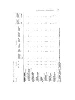
Table 11.1 Structural Components of a Muscle Fiber
Term Definition
General Structure and Contents of the Muscle Fiber
Sarcolemma The plasma membrane of a muscle fiber
Sarcoplasm The cytoplasm of a muscle fiber
Glycogen An energy-storage polysaccharide abundant in muscle
Myoglobin An oxygen-storing red pigment of muscle
T tubule A tunnel-like extension of the sarcolemma extending from one side of the muscle fiber to the other; conveys electrical signals
from the cell surface to its interior
Sarcoplasmic reticulum The smooth ER of a muscle fiber; a Ca
2ϩ
reservoir
Terminal cisternae The dilated ends of sarcoplasmic reticulum adjacent to a T tubule
Myofibrils
Myofibril A bundle of protein microfilaments (myofilaments)
Myofilament A threadlike complex of several hundred contractile protein molecules
Thick filament A myofilament about 11 nm in diameter composed of bundled myosin molecules
Elastic filament A myofilament about 1 nm in diameter composed of a giant protein, titin, that emerges from the core of a thick filament and
links it to a Z disc
Thin filament A myofilament about 5 to 6 nm in diameter composed of actin, troponin, and tropomyosin
Myosin A protein with a long shaftlike tail and a globular head; constitutes the thick myofilament
F actin A fibrous protein made of a long chain of G actin molecules twisted into a helix; main protein of the thin myofilament
G actin A globular subunit of F actin with an active site for binding a myosin head
Regulatory proteins Troponin and tropomyosin, proteins that do not directly engage in the sliding filament process of muscle contraction but
regulate myosin-actin binding
Tropomyosin A regulatory protein that lies in the groove of F actin and, in relaxed muscle, blocks the myosin-binding active sites
Troponin A regulatory protein associated with tropomyosin that acts as a calcium receptor
Titin A springy protein that forms the elastic filaments and Z discs
Striations and Sarcomeres
Striations Alternating light and dark transverse bands across a myofibril
A band Dark band formed by parallel thick filaments that partly overlap the thin filaments
H band A lighter region in the middle of an A band that contains thick filaments only; thin filaments do not reach this far into the A
band in relaxed muscle
I band A light band composed of thin filaments only
Z disc A protein disc to which thin filaments and elastic filaments are anchored at each end of a sarcomere; appears as a narrow
dark line in the middle of the I band
Sarcomere The distance from one Z disc to the next; the contractile unit of a muscle fiber
Saladin: Anatomy &
Physiology: The Unity of
Form and Function, Third
Edition
11. Muscular Tissue Text
© The McGraw−Hill
Companies, 2003
Chapter 11
junction (fig. 11.7). Each branch of a motor nerve fiber ends
in a bulbous swelling called a synaptic (sih-NAP-tic) knob,
which is nestled in a depression on the sarcolemma called
the motor end plate. The two cells do not actually touch
each other but are separated by a tiny gap, the synaptic cleft,
about 60 to 100 nm wide. A third cell, called a Schwann
cell, envelops the entire neuromuscular junction and iso-
lates it from the surrounding tissue fluid.
The electrical signal (nerve impulse) traveling down
a nerve fiber cannot cross the synaptic cleft like a spark
jumping between two electrodes—rather, it causes the
nerve fiber to release a neurotransmitter that stimulates the
next cell. Although many chemicals function as neuro-
transmitters, the one released at the neuromuscular junc-
tion is acetylcholine (ASS-eh-till-CO-leen) (ACh). ACh is
stored in spherical organelles called synaptic vesicles.
Directly across from the synaptic vesicles, the sar-
colemma of the muscle cell exhibits infoldings called
junctional folds, about 1 m deep. The muscle fiber has
about 50 million membrane proteins called ACh recep-
tors, which bind the acetylcholine release by the nerve
fiber. Most ACh receptors are concentrated in and near
these junctional folds. Very few ACh receptors are found
anywhere else on a muscle fiber. Junctional folds increase
the surface area for receptor sites and ensure a more effec-
tive response to ACh. The muscle nuclei beneath the junc-
tional folds are specifically dedicated to the synthesis of
ACh receptors and other proteins of the motor end plate.
A deficiency of ACh receptors leads to muscle paralysis in
the disease myasthenia gravis (see insight 11.4, p. 437).
The entire muscle fiber is surrounded by a basal lam-
ina that passes through the synaptic cleft and virtually fills
it. Both the sarcolemma and that part of the basal lamina in
the cleft contain an enzyme called acetylcholinesterase
(ASS-eh-till-CO-lin-ESS-ter-ase) (AChE), which breaks
down ACh, shuts down the stimulation of muscle fibers,
and allows a muscle to relax (see insight 11.1).
Insight 11.1 Clinical Application
Neuromuscular Toxins and Paralysis
Toxins that interfere with synaptic function can paralyze the muscles.
Some pesticides, for example, contain cholinesterase inhibitors that
bind to acetylcholinesterase and prevent it from degrading ACh. This
causes spastic paralysis, a state of continual contraction of the mus-
cle that poses the danger of suffocation if the laryngeal and respira-
tory muscles are affected. A person poisoned by a cholinesterase
inhibitor must be kept lying down and calm, and sudden noises or
other disturbances must be avoided. A minor startle response can esca-
late to dangerous muscle spasms in a poisoned individual.
Tetanus (“lockjaw”) is a form of spastic paralysis caused by a toxin
from the bacterium Clostridium tetani. In the spinal cord, an inhibitory
neurotransmitter called glycine stops motor neurons from producing
unwanted muscle contractions. The tetanus toxin blocks glycine
release and thus allows overstimulation of the muscles. (At the cost of
414 Part Two Support and Movement
Neuromuscular junction
Motor nerve fibers
Muscle fibers
Figure 11.5 Innervation of Skeletal Muscle.
Motor unit
Muscle fiber
nucleus
Neuromuscular
junctions
Skeletal muscle
fibers
Motor nerve fiber
Figure 11.6 A Motor Unit. The motor nerve fiber shown here
branches to supply those muscle fibers shown in color. The other muscle
fibers (gray) belong to other motor units.
Saladin: Anatomy &
Physiology: The Unity of
Form and Function, Third
Edition
11. Muscular Tissue Text
© The McGraw−Hill
Companies, 2003
Chapter 11
Chapter 11 Muscular Tissue 415
some confusion, the word tetanus also refers to a completely different
and normal muscle phenomenon discussed later in this chapter.)
Flaccid paralysis is a state in which the muscles are limp and can-
not contract. It can cause respiratory arrest when it affects the thoracic
muscles. Flaccid paralysis can be caused by poisons such as curare (cue-
RAH-ree) that compete with ACh for receptor sites but do not stimu-
late the muscle. Curare is extracted from certain plants and used by
some South American natives to poison blowgun darts. It has been
used to treat muscle spasms in some neurological disorders and to relax
abdominal muscles for surgery, but other muscle relaxants have now
replaced curare for most purposes.
You must be very familiar with the foregoing terms to
understand how a nerve stimulates a muscle fiber and
how the fiber contracts. They are summarized in table 11.2
for your later reference.
Electrically Excitable Cells
Muscle fibers and neurons are regarded as electrically
excitable cells because their plasma membranes exhibit
voltage changes in response to stimulation. The study of
the electrical activity of cells, called electrophysiology, is
a key to understanding nervous activity, muscle contrac-
tion, the heartbeat, and other physiological phenomena.
The details of electrophysiology are presented in chapter
12, but a few fundamental principles must be introduced
here so you can understand muscle excitation.
In an unstimulated (resting) cell, there are more
anions (negative ions) on the inside of the plasma mem-
brane than on the outside. Thus, the plasma membrane is
electrically polarized, or charged, like a little battery. In a
resting muscle cell, there is an excess of sodium ions (Na
ϩ
)
in the extracellular fluid (ECF) outside the cell and an
excess of potassium ions (K
ϩ
) in the intracellular fluid
(ICF) within the cell. Also in the ICF, and unable to pene-
trate the plasma membrane, are anions such as proteins,
nucleic acids, and phosphates. These anions make the
inside of the plasma membrane negatively charged by
comparison to its outer surface.
A difference in electrical charge from one point to
another is called an electrical potential, or voltage. The
difference is typically 12 volts (V) for a car battery and 1.5 V
Myelin
Motor nerve fiber
Axon terminal
Schwann cell
Synaptic vesicles
(containing ACh)
Basal lamina
(containing AChE)
Sarcolemma
Region of
sarcolemma
with ACh receptors
Junctional folds
Nucleus of muscle fiber
Synaptic cleft
Figure 11.7 A Neuromuscular Junction.
Saladin: Anatomy &
Physiology: The Unity of
Form and Function, Third
Edition
11. Muscular Tissue Text
© The McGraw−Hill
Companies, 2003
Chapter 11
for a flashlight battery, for example. On a sarcolemma of a
muscle cell, the voltage is much smaller, about Ϫ90 milli-
volts (mV), but critically important to life. (The negative
sign refers to the relative charge on the intracellular side
of the membrane.) This voltage is called the resting mem-
brane potential (RMP). It is maintained by the sodium-
potassium pump, as explained in chapter 3.
When a nerve or muscle cell is stimulated, dramatic
things happen electrically, as we shall soon see in our
study of the excitation of muscle. Ion gates in the plasma
membrane open and Na
ϩ
instantly diffuses down its con-
centration gradient into the cell. These cations override
the negative charges in the ICF, so the inside of the plasma
membrane briefly becomes positive. Immediately, Na
ϩ
gates close and K
ϩ
gates open. K
ϩ
rushes out of the cell,
partly because it is repelled by the positive sodium charge
and partly because it is more concentrated in the ICF than
in the ECF, so it diffuses down its concentration gradient
when it has the opportunity. The loss of positive potas-
sium ions from the cell turns the inside of the membrane
negative again. This quick up-and-down voltage shift,
from the negative RMP to a positive value and then back
to a negative value again, is called an action potential. The
RMP is a stable voltage seen in a “waiting” cell, whereas
the action potential is a quickly fluctuating voltage seen in
an active, stimulated cell.
Action potentials have a way of perpetuating
themselves—an action potential at one point on a plasma
membrane causes another one to happen immediately in
front of it, which triggers another one a little farther along,
and so forth. A wave of action potentials spreading along
a nerve fiber like this is called a nerve impulse or nerve sig-
nal. Such signals also travel along the sarcolemma of a
muscle fiber. We will see shortly how this leads to muscle
contraction. Chapter 12 explains the mechanism of action
potentials more fully.
Before You Go On
Answer the following questions to test your understanding of the
preceding section:
8. What differences would you expect to see between one motor
unit where muscular strength is more important than fine control
and another motor unit where fine control is more important?
9. Distinguish between acetylcholine, an acetylcholine receptor,
and acetylcholinesterase. State where each is found and describe
the function it serves.
10. What accounts for the resting membrane potential seen in
unstimulated nerve and muscle cells?
11. What is the difference between a resting membrane potential
and an action potential?
Behavior of Skeletal
Muscle Fibers
Objectives
When you have completed this section, you should be able to
• explain how a nerve fiber stimulates a skeletal muscle fiber;
• explain how stimulation of a muscle fiber activates its
contractile mechanism;
• explain the mechanism of muscle contraction;
• explain how a muscle fiber relaxes; and
• explain why the force of a muscle contraction depends on its
length prior to stimulation.
416
Part Two Support and Movement
Table 11.2 Components of the Neuromuscular Junction
Term Definition
Neuromuscular junction A functional connection between the distal end of a nerve fiber and the middle of a muscle fiber; consists of a
synaptic knob and motor end plate
Synaptic knob The dilated tip of a nerve fiber that contains synaptic vesicles
Motor end plate A depression in the sarcolemma, near the middle of the muscle fiber, that receives the synaptic knob; contains
acetylcholine receptors
Synaptic cleft A gap of about 60 to 100 nm between the synaptic knob and motor end plate
Synaptic vesicle A secretory vesicle in the synaptic knob that contains acetylcholine
Junctional folds Invaginations of the membrane of the motor end plate where ACh receptors are especially concentrated; located
across from the active zones
Acetylcholine (ACh) The neurotransmitter released by a somatic motor fiber that stimulates a skeletal muscle fiber (also used elsewhere
in the nervous system)
ACh receptor An integral protein in the sarcolemma of the motor end plate that binds to ACh
Acetylcholinesterase (AChE) An enzyme in the sarcolemma and basal lamina of the muscle fiber in the synaptic region; responsible for degrading
ACh and stopping the stimulation of the muscle fiber
Saladin: Anatomy &
Physiology: The Unity of
Form and Function, Third
Edition
11. Muscular Tissue Text
© The McGraw−Hill
Companies, 2003
Chapter 11
Chapter 11 Muscular Tissue 417
The process of muscle contraction and relaxation can be
viewed as occurring in four major phases: (1) excitation,
(2) excitation-contraction coupling, (3) contraction, and
(4) relaxation. Each phase occurs in several smaller steps,
which we now examine in detail. The steps are numbered
in the following descriptions to correspond to those in fig-
ures 11.8 to 11.11.
Excitation
Excitation is the process in which action potentials in the
nerve fiber lead to action potentials in the muscle fiber.
The steps in excitation are shown in figure 11.8.
1. A nerve signal arrives at the synaptic knob and
stimulates voltage-gated calcium channels to open.
Calcium ions enter the synaptic knob.
2. Calcium ions stimulate exocytosis of the synaptic
vesicles, which release acetylcholine (ACh) into the
synaptic cleft. One action potential causes
exocytosis of about 60 synaptic vesicles, and each
vesicle releases about 10,000 molecules of ACh.
3. ACh diffuses across the synaptic cleft and binds to
receptor proteins on the sarcolemma.
4. These receptors are ligand-gated ion channels.
When ACh (the ligand) binds to them, they change
shape and open an ion channel through the middle
of the receptor protein. Each channel allows Na
ϩ
to
diffuse quickly into the cell and K
ϩ
to diffuse
outward. As a result of these ion movements, the
sarcolemma reverses polarity—its voltage quickly
jumps from the RMP of Ϫ90 mV to a peak of ϩ75
mV as Na
ϩ
enters, and then falls back to a level
close to the RMP as K
ϩ
diffuses out. This rapid
fluctuation in membrane voltage at the motor end
plate is called the end-plate potential (EPP).
5. Areas of sarcolemma next to the end plate have
voltage-gated ion channels that open in response to
the EPP. Some of the voltage-gated channels are
specific for Na
ϩ
and admit it to the cell, while
others are specific for K
ϩ
and allow it to leave.
These ion movements create an action potential.
The muscle fiber is now excited.
Think About It
An impulse begins at the middle of a 100-mm-long
muscle fiber and travels 5 m/sec. How long would it
take to reach the ends of the muscle fiber?
Excitation-Contraction Coupling
Excitation-contraction coupling refers to the events that
link the action potentials on the sarcolemma to activation
of the myofilaments, thereby preparing them to contract.
The steps in the coupling process are shown in figure 11.9.
6. A wave of action potentials spreads from the end
plate in all directions, like ripples on a pond. When
this wave of excitation reaches the T tubules, it
continues down them into the sarcoplasm.
7. Action potentials open voltage-regulated ion gates in
the T tubules. These are physically linked to
calcium channels in the terminal cisternae of the
sarcoplasmic reticulum (SR), so gates in the SR open
as well and calcium ions diffuse out of the SR, down
their concentration gradient and into the cytosol.
8. The calcium ions bind to the troponin of the thin
filaments.
9. The troponin-tropomyosin complex changes shape
and shifts to a new position. This exposes the active
sites on the actin filaments and makes them
available for binding to myosin heads.
Contraction
Contraction is the step in which the muscle fiber develops
tension and may shorten. (Muscles often “contract,” or
develop tension, without shortening, as we see later.) How
a muscle fiber shortens remained a mystery until sophisti-
cated techniques in electron microscopy enabled cytolo-
gists to see the molecular organization of muscle fibers. In
1954, two researchers at the Massachusetts Institute of
Technology, Jean Hanson and Hugh Huxley, found evi-
dence for a model now called the sliding filament theory.
This theory holds that the thin filaments slide over the
thick ones and pull the Z discs behind them, causing the
cell as a whole to shorten. The individual steps in this
mechanism are shown in figure 11.10.
10. The myosin head must have an ATP molecule
bound to it to initiate the contraction process.
Myosin ATPase, an enzyme in the head, hydrolyzes
this ATP. The energy released by this process
activates the head, which “cocks” into an extended,
high-energy position. The head temporarily keeps
the ADP and phosphate group bound to it.
11. The cocked myosin binds to an active site on the
thin filament.
12. Myosin releases the ADP and phosphate and flexes
into a bent, low-energy position, tugging the thin
filament along with it. This is called the power
stroke. The head remains bound to actin until it
binds a new ATP.
13. Upon binding more ATP, myosin releases the actin.
It is now prepared to repeat the whole process—it
will hydrolyze the ATP, recock (the recovery
stroke), attach to a new active site farther down the
thin filament, and produce another power stroke.
It might seem as if releasing the thin filament at step
13 would simply allow it to slide back to its previous posi-
tion, so that nothing would have been accomplished.
Think of the sliding filament mechanism, however, as
Saladin: Anatomy &
Physiology: The Unity of
Form and Function, Third
Edition
11. Muscular Tissue Text
© The McGraw−Hill
Companies, 2003
Chapter 11
418
K
+
Na
+
Ca
2+
K
+
Na
+
Motor
neuron
Sarcolemma
Sarcolemma
Sarcolemma
ACh
ACh receptor
Synaptic
knob
Synaptic
vesicles
Motor end plate
Motor nerve
fiber
2. Acetylcholine (ACh) release
3. Binding of ACh to receptors
4. Opening of ligand-gated ion channel;
creation of end-plate potential
5. Opening of voltage-gated ion channels;
creation of action potentials
1. Arrival of nerve signal
Figure 11.8 Excitation of a Muscle Fiber. These events link action potentials in a nerve fiber to the generation of action potentials in the muscle fiber.
Saladin: Anatomy &
Physiology: The Unity of
Form and Function, Third
Edition
11. Muscular Tissue Text
© The McGraw−Hill
Companies, 2003
Chapter 11
Chapter 11 Muscular Tissue 419
being similar to the way you would pull in a boat anchor
hand over hand. When the myosin head cocks, it is like
your hand reaching out to grasp the anchor rope. When it
flexes back into the low-energy position, it is like your
elbow flexing to pull on the rope and draw the anchor up
a little bit. When you let go of the rope with one hand, you
hold onto it with the other, alternating hands until the
anchor is pulled in. Similarly, when one myosin head
releases the actin in preparation for the recovery stroke,
there are many other heads on the same thick filament
holding onto the thin filament so that it doesn’t slide back.
At any given moment during contraction, about half of the
heads are bound to the thin filament and the other half are
extending forward to grasp the filament farther down.
That is, the myosin heads of a thick filament do not all
stroke at once but contract sequentially.
As another analogy, consider a millipede—a little
wormlike animal with a few hundred tiny legs. Each leg
Actin
Tropomyosin
Active sites
Troponin
7. Calcium release
Ca
2+
Ca
2+
8. Binding of calcium to troponin
9. Shifting of tropomyosin; exposure
of active sites on actin
6. Action potentials propagated
down T tubules
Ca
2+
Ca
2+
Myosin
Figure 11.9 Excitation-Contraction Coupling. These events link action potentials in the muscle fiber to the release and binding of calcium ions.
The numbered steps in this figure begin where the previous figure left off.
Saladin: Anatomy &
Physiology: The Unity of
Form and Function, Third
Edition
11. Muscular Tissue Text
© The McGraw−Hill
Companies, 2003
Chapter 11
420 Part Two Support and Movement
ADP
P
i
ADP
P
i
ATP
10. Activation and cocking of myosin head
11. Formation of myosin-actin cross-bridge
12. Power stroke; sliding of thin
filament over thick filament
13. Binding of new ATP; breaking of cross-bridge
Troponin
Tropomyosin
Figure 11.10 The Sliding Filament Mechanism of Contraction. This is a cycle of repetitive events that cause a thin filament to slide over a
thick filament and generate tension in the muscle. The numbered steps in this figure begin where the previous figure left off.
Saladin: Anatomy &
Physiology: The Unity of
Form and Function, Third
Edition
11. Muscular Tissue Text
© The McGraw−Hill
Companies, 2003
Chapter 11
Chapter 11 Muscular Tissue 421
Ca
2+
Ca
2+
15. ACh breakdown by
acetylcholinesterase (AChE)
16. Reabsorption of calcium ions by
sarcoplasmic reticulum
18. Return of tropomyosin to position
blocking active sites of actin
17. Loss of calcium ions from troponin
14. Cessation of nervous stimulation
and ACh release
AChE
Ca
2+
Ca
2+
Figure 11.11 Relaxation of a Muscle Fiber. These events lead from the cessation of a nerve signal to the release of thin filaments by myosin.
The numbered steps in this figure begin where the previous figure left off.
Saladin: Anatomy &
Physiology: The Unity of
Form and Function, Third
Edition
11. Muscular Tissue Text
© The McGraw−Hill
Companies, 2003
Chapter 11
takes individual jerky steps, but all the legs working
together produce smooth, steady movement—just as all
the heads of a thick filament collectively produce a
smooth, steady pull on the thin filament. Note that even
though the muscle fiber contracts, the myofilaments do
not become shorter any more than a rope becomes shorter
as you pull in an anchor. The thin filaments slide over the
thick ones, as the name of the theory implies.
A single cycle of power and recovery strokes by all
the myosin heads in a muscle fiber would shorten the
fiber by about 1%. A fiber, however, may shorten by as
much as 40% of its resting length, so obviously the cycle
of power and recovery must be repeated many times by
each myosin head. Each head carries out about five
strokes per second, and each stroke consumes one mole-
cule of ATP.
Relaxation
When its work is done, a muscle fiber relaxes and returns
to its resting length. This is achieved by the steps shown
in figure 11.11.
14. Nerve signals stop arriving at the neuromuscular
junction, so the synaptic knob stops
releasing ACh.
15. As ACh dissociates (separates) from its receptor,
acetylcholinesterase breaks it down into fragments
that cannot stimulate the muscle. The synaptic
knob reabsorbs these fragments for recycling. All of
this happens continually while the muscle is being
stimulated, too; but when nerve signals stop, no
new ACh is released to replace that which is broken
down. Therefore, stimulation of the muscle fiber by
ACh ceases.
16. Active transport pumps in the sarcoplasmic
reticulum (SR) begin to pump Ca
2ϩ
from the cytosol
back into the cisternae. Here, the calcium binds to a
protein called calsequestrin (CAL-see-QUES-trin)
and is stored until the fiber is stimulated again.
Since active transport requires ATP, you can see
that ATP is needed for muscle relaxation as well as
for muscle contraction (see insight 11.2).
17. As calcium ions dissociate from troponin, they are
pumped into the SR and are not replaced.
18. Tropomyosin moves back into the position where it
blocks the active sites of the actin filament. Myosin
can no longer bind to actin, and the muscle fiber
ceases to produce or maintain tension.
A muscle returns to its resting length with the aid of
two forces: (1) like a recoiling rubber band, the series-elastic
components stretch it; and (2) since muscles often occur in
antagonistic pairs, the contraction of an antagonist length-
ens the relaxed muscle. Contraction of the triceps brachii,
for example, extends the elbow and lengthens the biceps
brachii.
Insight 11.2 Clinical Application
Rigor Mortis
Rigor mortis
7
is the hardening of the muscles and stiffening of the
body that begins 3 to 4 hours after death. It occurs partly because
the deteriorating sarcoplasmic reticulum releases calcium ions into
the cytosol, and the deteriorating sarcolemma admits more calcium
ions from the extracellular fluid. The calcium ions activate myosin-
actin cross bridging and muscle contraction. Furthermore, the mus-
cle cannot relax without ATP, and ATP is no longer produced after
death. Thus, the fibers remain contracted until the myofilaments
begin to decay. Rigor mortis peaks about 12 hours after death and
then diminishes over the next 48 to 60 hours.
7
rigor ϭ rigidity ϩ mortis ϭ of death
The Length-Tension Relationship
and Muscle Tone
The amount of tension generated by a muscle, and therefore
the force of its contraction, depends on how stretched or
contracted it was before it was stimulated, among other
422
Part Two Support and Movement
0.0
0.5
1.0
Tension (g) generated
upon stimulation
1.0 2.0
Sarcomere length (µm) before stimulation
3.0 4.0
zz
zz
zz
Overly contracted
Overly stretched
Optimum resting length
Figure 11.12 The Length-Tension Relationship. Center: In a
resting muscle fiber, the sarcomeres are usually 2.0 to 2.25 m long, the
optimum length for producing maximum tension when the muscle is
stimulated to contract. Note how this relates to the degree of overlap
between the thick and thin filaments. Left: If the muscle is overly
contracted, the thick filaments butt against the Z discs and the fiber
cannot contract very much more when it is stimulated. Right: If the muscle
is overly stretched, there is so little overlap between the thick and thin
filaments that few cross-bridges can form between myosin and actin.
Saladin: Anatomy &
Physiology: The Unity of
Form and Function, Third
Edition
11. Muscular Tissue Text
© The McGraw−Hill
Companies, 2003
Chapter 11
Chapter 11 Muscular Tissue 423
factors. This principle is called the length-tension rela-
tionship. The reasons for it can be seen in figure 11.12. If a
fiber is overly contracted at rest, its thick filaments are
rather close to the Z discs. The stimulated muscle may con-
tract a little, but then the thick filaments butt up against the
Z discs and can go no farther. The contraction is therefore a
weak one. On the other hand, if a muscle fiber is too
stretched before it is stimulated, there is relatively little
overlap between its thick and thin filaments. When the mus-
cle is stimulated, its myosin heads cannot “get a good grip”
on the thin filaments, and again the contraction is weak. (As
mentioned in chapter 10, this is one reason you should not
bend at the waist to pick up a heavy object. Muscles of the
back become overly stretched and cannot contract effec-
tively to straighten your spine against a heavy resistance.)
Between these extremes, there is an optimum resting
length at which a muscle produces the greatest force when
it contracts. The central nervous system continually mon-
itors and adjusts the length of a resting muscle, maintain-
ing a state of partial contraction called muscle tone. This
maintains optimum length and makes the muscles ideally
ready for action. The elastic filaments of the sarcomere
also help to maintain enough myofilament overlap to
ensure an effective contraction when the muscle is called
into action.
Before You Go On
Answer the following questions to test your understanding of the
preceding section:
12. What change does ACh cause in an ACh receptor? How does this
electrically affect the muscle fiber?
13. How do troponin and tropomyosin regulate the interaction
between myosin and actin?
14. Describe the roles played by ATP in the power and recovery
strokes of myosin.
15. What steps are necessary for a contracted muscle to return to its
resting length?
Behavior of Whole Muscles
Objectives
When you have completed this section, you should be able to
• describe the stages of a muscle twitch;
• describe treppe and explain how it relates to muscle warm-up;
• explain how muscle twitches add up to produce stronger
muscle contractions;
• distinguish between isometric and isotonic contraction; and
• distinguish between concentric and eccentric contractions.
Now you know how an individual muscle cell shortens.
Our next objective is to move up to the organ grade of con-
struction and consider how this relates to the action of the
muscle as a whole.
Threshold, Latent Period, and Twitch
Muscle contraction has often been studied and demon-
strated using the gastrocnemius (calf) muscle of a frog,
which can easily be isolated from the leg along with its con-
nected sciatic nerve (see insight 11.3). This nerve-muscle
preparation can be attached to stimulating electrodes and to
a recording device that produces a myogram, a chart of the
timing and strength of the muscle’s contraction.
A sufficiently weak electrical stimulus to a muscle
causes no contraction. By gradually increasing the voltage
and stimulating the muscle again, we can determine the
threshold, or minimum voltage necessary to generate an
action potential in the muscle fiber and produce a con-
traction. The action potential triggers the release of a pulse
of Ca
2ϩ
into the cytoplasm and activates the sliding fila-
ment mechanism. At threshold or higher, a stimulus thus
causes a quick cycle of contraction and relaxation called a
twitch (fig. 11.13).
There is a delay, or latent period, of about 2 mil-
liseconds (msec) between the onset of the stimulus and the
onset of the twitch. This is the time required for excitation,
excitation-contraction coupling, and tensing of the series-
elastic components of the muscle. The force generated
during this time is called internal tension. It is not visible
on the myogram because it causes no shortening of the
muscle.
Once the series-elastic components are taut, the mus-
cle begins to produce external tension and move a resist-
ing object, or load. This is called the contraction phase of
the twitch. In the frog gastrocnemius preparation, the load
is the sensor of the recording apparatus; in the body, it is
usually a bone. By analogy, imagine lifting a weight from
a table with a rubber band. At first, internal tension would
Contraction
phase
Relaxation
phase
Time
Latent
period
Time of
stimulation
Muscle tension
Figure 11.13 A Muscle Twitch.
What role does ATP play during the relaxation phase?
Saladin: Anatomy &
Physiology: The Unity of
Form and Function, Third
Edition
11. Muscular Tissue Text
© The McGraw−Hill
Companies, 2003
Chapter 11
stretch the rubber band. Then as the rubber band became
taut, external tension would lift the weight.
The contraction phase is short-lived, because the
sacroplasmic reticulum quickly pumps Ca
2ϩ
back into
itself before the muscle develops maximal force. As the
Ca
2ϩ
level in the cytoplasm falls, myosin releases the thin
filaments and muscle tension declines. This is seen in the
myogram as the relaxation phase. The entire twitch lasts
from about 7 to 100 msec.
Insight 11.3 Medical History
Galvani, Volta, and Animal Electricity
The invention of modern dry cells can be traced to studies of frog mus-
cle by Italian anatomist Luigi Galvani (1737–98). He suspended isolated
frog legs from a copper hook and noticed that they twitched when
touched with an iron scalpel. He attributed this to “animal electricity”
in the legs. The physicist Alessandro Volta (1745–1827) investigated
Galvani’s discovery further. He concluded that when two different
metals (such as the copper hook and iron scalpel) are separated by an
electrolyte solution (a frog’s tissue fluids), a chemical reaction occurs
that produces an electrical current. This current had stimulated the
muscle in the legs of Galvani’s frogs and caused the twitch. Based on
this principle, Volta invented the first simple voltaic cell, the forerun-
ner of today’s dry cells.
Contraction Strength of Twitches
As long as the voltage of an artificial stimulus delivered
directly to a muscle is at threshold or higher, a muscle gives
a complete twitch. Increasing the voltage still more does
not cause the twitches to become any stronger. There are
other factors, however, that can produce stronger twitches.
Indeed, an individual twitch is not strong enough to do any
useful work. Muscles must be able to contract with variable
strength—differently in lifting a glass of champagne than
in lifting a heavy barbell, for example.
If we stimulate the nerve rather than the muscle,
higher voltages produce stronger muscle contractions
because they excite more nerve fibers and therefore more
motor units. The more motor units that contract, the more
strongly the muscle as a whole contracts (fig. 11.14). The
process of bringing more motor units into play is called
recruitment, or multiple motor unit (MMU) summation. It
is seen not just in artificial stimulation but is part of the
way the nervous system behaves normally to produce vari-
able muscle contractions.
Another way to produce a stronger muscle contrac-
tion is to stimulate the muscle at a higher frequency. Even
when voltage remains the same, high-frequency stimula-
tion causes stronger contractions than low-frequency
stimulation. In figure 11.15a, we see that when a muscle is
stimulated at a low frequency (up to 10 stimuli/sec in this
example), it produces an identical twitch for each stimu-
lus and fully recovers between twitches.
Between 10 and 20 stimuli per second, the muscle still
recovers fully between twitches, but each twitch develops
more tension than the one before. This pattern of increasing
tension with repetitive stimulation is called treppe
8
(TREP-
eh), or the staircase phenomenon, after the appearance of
the myogram (fig. 11.15b). One cause of treppe is that when
stimuli arrive so rapidly, the sarcoplasmic reticulum does
not have time between stimuli to completely reabsorb all
the calcium that it released. Thus, the calcium concentra-
tion in the cytosol rises higher and higher with each stimu-
lus and causes subsequent twitches to be stronger. Another
factor is that the heat released by each twitch causes mus-
cle enzymes such as myosin ATPase to work more effi-
ciently and produce stronger twitches as the muscle warms
up. One purpose of warm-up exercises before athletic com-
petition is to induce treppe, so that the muscle contracts
more effectively when the competition begins.
At a still higher stimulus frequency (20–40 stimuli/
sec in fig. 11.15c), each new stimulus arrives before the
previous twitch is over. Each new twitch “rides piggy-
back” on the previous one and generates higher tension.
424
Part Two Support and Movement
123 45 67 8 9
12345 678 9
Threshold
Stimulus voltage
Stimuli to nerve
Tension
Proportion
of nerve
fibers
excited
Maximum contraction
Figure 11.14 The Relationship Between Stimulus Intensity
(voltage) and Muscle Tension. Weak stimuli (1–2) fail to stimulate
any nerve fibers and therefore produce no muscle contraction. When
stimuli reach or exceed threshold (3–7), they excite more and more nerve
fibers and motor units and produce stronger and stronger contractions.
This is multiple motor unit summation (recruitment). Once all of the
nerve fibers are stimulated (7–9), further increases in stimulus strength
produce no further increase in muscle tension.
8
treppe ϭ staircase
Saladin: Anatomy &
Physiology: The Unity of
Form and Function, Third
Edition
11. Muscular Tissue Text
© The McGraw−Hill
Companies, 2003
Chapter 11
Chapter 11 Muscular Tissue 425
This phenomenon goes by two names: temporal
9
summa-
tion, because it results from two stimuli arriving close
together, or wave summation, because it results from one
wave of contraction added to another. Wave is added upon
wave, so each twitch reaches a higher level of tension than
the one before, and the muscle relaxes only partially
between stimuli. This effect produces a state of sustained
fluttering contraction called incomplete tetanus.
At a still higher frequency, such as 40 to 50 stimuli
per second, the muscle has no time to relax at all between
stimuli, and the twitches fuse into a smooth, prolonged
contraction called complete tetanus. A muscle in com-
plete tetanus produces about four times as much tension
as a single twitch (fig. 11.15d). This type of tetanus should
not be confused with the disease of the same name caused
by the tetanus toxin, explained in insight 11.1.
Complete tetanus is a phenomenon seen in artificial
stimulation of a muscle, however, and rarely if ever occurs
in the body. Even during the most intense muscle contrac-
tions, the frequency of stimulation by a motor neuron
rarely exceeds 25/sec, which is far from sufficient to pro-
duce complete tetanus. The reason for the smoothness of
muscle contractions is that motor units function asynchro-
nously; when one motor unit relaxes, another contracts
and “takes over” so that the muscle does not lose tension.
Isometric and Isotonic Contraction
In muscle physiology, “contraction” does not always mean
the shortening of a muscle—it may mean only that the
muscle is producing internal tension while an external
resistance causes it to stay the same length or even to
become longer. Thus, physiologists speak of different
kinds of muscle contraction as isometric versus isotonic
and concentric versus eccentric.
(a) (b)
(c) (d)
Twitch
Incomplete tetanus
Treppe
Complete tetanus
Fatigue
Figure 11.15 The Relationship Between Stimulus Frequency and Muscle Tension. (a) Twitch: At low frequency, the muscle relaxes
completely between stimuli and shows twitches of uniform strength. (b) Treppe: At a moderate frequency of stimulation, the muscle relaxes fully between
contractions, but successive twitches are stronger. (c) Wave summation and incomplete tetanus: At still higher stimulus frequency, the muscle does not
have time to relax completely between twitches, and the force of each twitch builds on the previous one. (d) Complete tetanus: At high stimulus
frequency, the muscle does not have time to relax at all between stimuli and exhibits a state of continual contraction with about four times as much
tension as a single twitch. Tension declines as the muscle fatigues.
9
tempor ϭ time
Saladin: Anatomy &
Physiology: The Unity of
Form and Function, Third
Edition
11. Muscular Tissue Text
© The McGraw−Hill
Companies, 2003
Chapter 11
Suppose you lift a heavy box of books from a table.
When you first contract the muscles of your arms, you can
feel the tension building in them even though the box is
not yet moving. At this point, your muscles are contract-
ing at a cellular level, but their tension is being absorbed
by the series-elastic components and is resisted by the
weight of the load; the muscle as a whole is not producing
any external movement. This phase is called isometric
10
contraction—contraction without a change in length
(fig. 11.16a). Isotonic
11
contraction—contraction with a
change in length but no change in tension—begins when
internal tension builds to the point that it overcomes the
resistance. The muscle now shortens, moves the load,
and maintains essentially the same tension from then on
(fig. 11.16b). Isometric and isotonic contraction are both
phases of normal muscular action (fig. 11.17).
There are two forms of isotonic contraction—
concentric and eccentric. In concentric contraction, a
muscle shortens as it maintains tension—for example,
when the biceps brachii contracts and flexes the elbow.
In an eccentric contraction, a muscle lengthens as it
maintains tension. If you set that box of books down again
(fig. 11.16c), your biceps brachii lengthens as you extend
your elbow, but it maintains tension to act as a brake and
keep you from simply dropping the box. A weight lifter
426
Part Two Support and Movement
Muscle shortens,
tension remains
constant
Movement
Movement
Muscle develops
tension but does
not shorten
No movement
Muscle lengthens
while maintaining
tension
(
a
)
Isometric contraction
(
b
)
Isotonic, concentric contraction
(
c
)
Isotonic, eccentric contraction
Figure 11.16 Isometric and Isotonic Contraction. (a) Isometric contraction, in which a muscle develops tension but does not shorten. This
occurs at the beginning of any muscle contraction but is prolonged in actions such as lifting heavy weights. (b) Isotonic concentric contraction, in which
the muscle shortens while maintaining a constant degree of tension. In this phase, the muscle moves a load. (c) Isotonic eccentric contraction, in which
the muscle maintains tension while it lengthens, allowing a muscle to relax without going suddenly limp.
Name a muscle that undergoes eccentric contraction as you sit down in a chair.
Time
Muscle
tension
Muscle
length
Isometric
phase
Isotonic
phase
Figure 11.17 Isometric and Isotonic Phases of Contraction.
At the beginning of a contraction (isometric phase), muscle tension
rises but the length remains constant (the muscle does not shorten).
When tension overcomes the resistance of the load, the tension levels
off and the muscle begins to shorten and move the load (isotonic
phase).
How would you extend this graph in order to show eccentric
contraction?
10
iso ϭ same, uniform ϩ metr ϭ length
11
iso ϭ same, uniform ϩ ton ϭ tension
Saladin: Anatomy &
Physiology: The Unity of
Form and Function, Third
Edition
11. Muscular Tissue Text
© The McGraw−Hill
Companies, 2003
Chapter 11
Chapter 11 Muscular Tissue 427
uses concentric contraction when lifting a barbell and
eccentric contraction when lowering it to the floor.
In summary, during isometric contraction, a muscle
develops tension without changing length, and in isotonic
contraction, it changes length while maintaining constant
tension. In concentric contraction, a muscle maintains
tension as it shortens, and in eccentric contraction, it
maintains tension while it is lengthening.
Before You Go On
Answer the following questions to test your understanding of the
preceding section:
16. Explain how warm-up is related to treppe and why it improves
athletic performance.
17. Explain the role of tetanus in normal muscle action.
18. Describe an everyday activity not involving the arms in which
your muscles would switch from isometric to isotonic
contraction.
19. Describe an everyday activity not involving the arms that would
involve concentric contraction and one that would involve
eccentric contraction.
Muscle Metabolism
Objectives
When you have completed this section, you should be able to
• explain how skeletal muscle meets its energy demands during
rest and exercise;
• explain the basis of muscle fatigue and soreness;
• define oxygen debt and explain why extra oxygen is needed
even after an exercise has ended;
• distinguish between two physiological types of muscle fibers,
and explain the functional roles of these two types;
• discuss the factors that affect muscular strength; and
• discuss the effects of resistance and endurance exercises on
muscle.
ATP Sources
All muscle contraction depends on ATP; no other energy
source can serve in its place. The supply of ATP depends,
in turn, on the availability of oxygen and organic energy
sources such as glucose and fatty acids. To understand
how muscle manages its ATP budget, you must be famil-
iar with the two main pathways of ATP synthesis—anaer-
obic fermentation and aerobic respiration (see fig. 2.31,
p. 86). Each of these has advantages and disadvantages.
Anaerobic fermentation enables a cell to produce ATP in
the absence of oxygen, but the ATP yield is very limited
and the process produces a toxic end product, lactic acid,
which is a major factor in muscle fatigue. By contrast, aer-
obic respiration produces far more ATP and less toxic end
products (carbon dioxide and water), but it requires a con-
tinual supply of oxygen. Although aerobic respiration is
best known as a pathway for glucose oxidation, it is also
used to extract energy from other organic compounds. In a
resting muscle, most ATP is generated by the aerobic res-
piration of fatty acids.
During the course of exercise, different mechanisms
of ATP synthesis are used depending on the exercise dura-
tion. We will view these mechanisms from the standpoint
of immediate, short-term, and long-term energy, but it
must be stressed that muscle does not make sudden shifts
from one mechanism to another like an automobile trans-
mission shifting gears. Rather, these mechanisms blend
and overlap as the exercise continues (fig. 11.18).
Immediate Energy
In a short, intense exercise such as a 100 m dash, the re-
spiratory and cardiovascular systems cannot deliver oxy-
gen to the muscles quickly enough for aerobic respiration
to meet the increased ATP demand. The myoglobin in a
muscle fiber supplies oxygen for a limited amount of aer-
obic respiration, but in brief exercises a muscle meets
most of its ATP demand by borrowing phosphate (P
i
)
groups from other molecules and transferring them to
ADP. Two enzyme systems control these phosphate trans-
fers (fig. 11.19):
1. Myokinase (MY-oh-KY-nase) transfers P
i
groups from
one ADP to another, converting the latter to ATP.
2. Creatine kinase (CREE-uh-tin KY-nase) obtains P
i
groups from an energy-storage molecule, creatine
phosphate (CP), and donates them to ADP to make
ATP. This is a fast-acting system that helps to
maintain the ATP level while other ATP-generating
mechanisms are being activated.
ATP and CP, collectively called the phosphagen sys-
tem, provide nearly all the energy used for short bursts of
intense activity. Muscle contains about 5 millimoles of
ATP and 15 millimoles of CP per kilogram of tissue, which
is enough to power about 1 minute of brisk walking or 6
seconds of sprinting or fast swimming. The phosphagen
system is especially important in activities requiring brief
but maximal effort, such as football, baseball, and weight
lifting.
Short-Term Energy
As the phosphagen system is exhausted, the muscles shift
to anaerobic fermentation to “buy time” until cardiopul-
monary function can catch up with the muscle’s oxygen
demand. During this period, the muscles obtain glucose
from the blood and their own stored glycogen. The pathway
from glycogen to lactic acid, called the glycogen–lactic acid
Saladin: Anatomy &
Physiology: The Unity of
Form and Function, Third
Edition
11. Muscular Tissue Text
© The McGraw−Hill
Companies, 2003
Chapter 11
system, produces enough ATP for 30 to 40 seconds of max-
imum activity. To play basketball or to run completely
around a baseball diamond, for example, depends heavily
on this energy-transfer system.
Long-Term Energy
After 40 seconds or so, the respiratory and cardiovascular
systems “catch up” and deliver oxygen to the muscles fast
enough for aerobic respiration to meet most of the ATP
demand. One’s rate of oxygen consumption rises for 3 to 4
minutes and then levels off at a steady state in which aer-
obic ATP production keeps pace with the demand. In exer-
cises lasting more than 10 minutes, more than 90% of the
ATP is produced aerobically.
Little lactic acid accumulates under steady state con-
ditions, but this does not mean that aerobic exercise can
continue indefinitely or that it is limited only by a per-
son’s willpower. The depletion of glycogen and blood glu-
cose, together with the loss of fluid and electrolytes
through sweating, set limits to endurance and perform-
ance even when lactic acid does not.
Fatigue and Endurance
Muscle fatigue is the progressive weakness and loss of
contractility that results from prolonged use of the mus-
cles. For example, if you hold this book at arm’s length for
a minute, you will feel your muscles growing weaker and
428
Part Two Support and Movement
Aerobic respiration
using oxygen from
myoglobin
Glycogen—
lactic acid
system
(anaerobic
fermentation)
Phosphagen
system
Duration of exercise
0 10 seconds 40 seconds
Aerobic
respiration
Repayment of
oxygen debt
ATP synthesis
Figure 11.18 Phases of ATP Production During Exercise.
Creatine
phosphate
Creatine
ADP
AT P
Creatine
kinase
P
i
ADP
AMP
ADP
AT P
Myokinase
P
i
Figure 11.19 The Phosphagen System. Two enzymes, myokinase
and creatine kinase, generate ATP in the absence of oxygen. Myokinase
borrows phosphate groups from ADP, and creatine kinase borrows them
from creatine phosphate, to convert an ADP to ATP.