Saladin Anatomy and Physiology The Unity of Form and Function Episode 5 ppt
Bạn đang xem bản rút gọn của tài liệu. Xem và tải ngay bản đầy đủ của tài liệu tại đây (2.58 MB, 70 trang )
Saladin: Anatomy &
Physiology: The Unity of
Form and Function, Third
Edition
8. The Skeletal System Text
© The McGraw−Hill
Companies, 2003
Chapter 8
Chapter 8 The Skeletal System 265
Excessive stress can crack the annulus and cause the
nucleus to ooze out. This is called a herniated disc (“rup-
tured” or “slipped” disc in lay terms) and may put painful
pressure on the spinal cord or a spinal nerve. To relieve
the pressure, a procedure called a laminectomy may be
performed—each lamina is cut and the laminae and spin-
ous processes are removed. This procedure is also used to
expose the spinal cord for anatomical study or surgery.
Regional Characteristics of Vertebrae
We are now prepared to consider how vertebrae differ
from one region of the vertebral column to another and
from the generalized anatomy just described. Knowing
these variations will enable you to identify the region of
the spine from which an isolated vertebra was taken. More
importantly, these modifications in form reflect functional
differences among the vertebrae.
Cervical Vertebrae
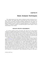
The cervical vertebrae (C1–C7) are the smallest and
lightest ones other than the coccygeals. The first two (C1
and C2) have unique structures that allow for head
movements (fig. 8.24). Vertebra C1 is called the atlas
because it supports the head in a manner reminiscent of
the Titan of Greek mythology who was condemned by
Zeus to carry the world on his shoulders. It scarcely
resembles the typical vertebra; it is little more than a del-
icate ring surrounding a large vertebral foramen. On
each side is a lateral mass with a deeply concave supe-
rior articular facet that articulates with the occipital
condyle of the skull. A nodding motion of the skull, as
in gesturing “yes,” causes the occipital condyles to rock
back and forth on these facets. The inferior articular
facets, which are comparatively flat or only slightly con-
cave, articulate with C2. The lateral masses are con-
nected by an anterior arch and a posterior arch, which
bear slight protuberances called the anterior and poste-
rior tubercle, respectively.
Vertebra C2, the axis, allows rotation of the head as
in gesturing “no.” Its most distinctive feature is a promi-
nent knob called the dens (denz), or odontoid
36
process,
on its anterosuperior side. No other vertebra has a dens. It
begins to form as an independent ossification center dur-
ing the first year of life and fuses with the axis by the age
of 3 to 6 years. It projects into the vertebral foramen of the
atlas, where it is nestled in a facet and held in place by a
transverse ligament (fig. 8.24c). A heavy blow to the top of
the head can cause a fatal injury in which the dens is
driven through the foramen magnum into the brainstem.
The articulation between the atlas and the cranium is
called the atlanto-occipital joint; the one between the atlas
and axis is called the atlantoaxial joint.
The axis is the first vertebra that exhibits a spinous
process. In vertebrae C2 to C6, the process is forked, or
bifid,
37
at its tip (fig. 8.25a). This fork provides attachment
for the nuchal ligament of the back of the neck. All seven
cervical vertebrae have a prominent round transverse
foramen in each transverse process. These foramina pro-
vide passage and protection for the vertebral arteries,
which supply blood to the brain. Transverse foramina
occur in no other vertebrae and thus provide an easy
means of recognizing a cervical vertebra.
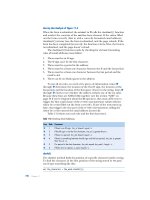
Transverse
process
L3
L4
Centrum (body)
Intervertebral disc
Inferior articular
process of L3
Superior articular
process of L4
Lamina
Inferior vertebral
notch of L1
Superior vertebral
notch of L2
L1
L2
L3
Superior articular
process of L1
Inferior articular
process of L3
(a)
Intervertebral disc
Spinous process
Intervertebral
foramen
(b)
Figure 8.23 Articulated Vertebrae. (a) Dorsal view of vertebrae L3 to L4. (b) Left lateral view of vertebrae L1 to L3.
36
dens ϭ odont ϭ tooth ϩ oid ϭ resembling
37
bifid ϭ cleft into two parts
Saladin: Anatomy &
Physiology: The Unity of
Form and Function, Third
Edition
8. The Skeletal System Text
© The McGraw−Hill
Companies, 2003
Chapter 8
Think About It
How would head movements be affected if
vertebrae C1 and C2 had the same structure as C3?
What is the functional advantage of the lack of a
spinous process in C1?
Cervical vertebrae C3 to C6 are similar to the typical
vertebra described earlier, with the addition of the trans-
verse foramina and bifid spinous processes. Vertebra C7 is
a little different—its spinous process is not bifid, but it is
especially long and forms a prominent bump on the lower
back of the neck. This feature is a convenient landmark for
counting vertebrae. Because it is so conspicuous, C7 is
sometimes called the vertebra prominens.
Thoracic Vertebrae
There are 12 thoracic vertebrae (T1–T12), corresponding
to the 12 pairs of ribs attached to them. They lack the
transverse foramina and bifid processes that distinguish
the cervicals, but possess the following distinctive fea-
tures of their own (fig. 8.25b):
• The spinous processes are relatively pointed and angle
sharply downward.
• The body is somewhat heart-shaped, more massive
than in the cervical vertebrae but less than in the
lumbar vertebrae.
• The body has small, smooth, slightly concave spots
called costal facets (to be described shortly) for
attachment of the ribs.
• Vertebrae T1 to T10 have a shallow, cuplike transverse
costal
38
facet at the end of each transverse process.
These provide a second point of articulation for ribs 1
to 10. There are no transverse costal facets on T11 and
T12 because ribs 11 and 12 attach only to the bodies of
the vertebrae.
No other vertebrae have ribs articulating with them.
Thoracic vertebrae vary among themselves mainly
because of variations in the way the ribs articulate. In most
cases, a rib inserts between two vertebrae, so each vertebra
contributes one-half of the articular surface. A rib articu-
lates with the inferior costal facet (FASS-et) of the upper
vertebra and the superior costal facet of the vertebra
below that. This terminology may be a little confusing, but
note that the superior and inferior facets are named for
266
Part Two Support and Movement
Anterior tubercle
Anterior arch
Superior articular
facet
Transverse
foramen
Transverse foramen
Transverse process
Inferior articular process
Spinous process
Lamina
(b)
Posterior arch
Posterior tubercle
Dens (odontoid process)
Superior articular facet
Body
Atlas
Axis
Pedicle
Lateral
masses
Dens
Axis of rotation
Transverse
ligament
Atlas
Axis
(a)
(c)
Figure 8.24 The Atlas and Axis, Cervical Vertebrae C1 and C2. (a) The atlas, superior view. (b) The axis, posterosuperior view. (c) Articulation
of the atlas and axis and rotation of the atlas. This movement turns the head from side to side, as in gesturing “no.” Note the transverse ligament holding
the dens of the axis in place.
38
costa ϭ rib ϩ al ϭ pertaining to
Saladin: Anatomy &
Physiology: The Unity of
Form and Function, Third
Edition
8. The Skeletal System Text
© The McGraw−Hill
Companies, 2003
Chapter 8
Chapter 8 The Skeletal System 267
their position on the vertebral body, not for which part of
the rib’s articulation they provide. Vertebrae T1 and T10 to
T12, however, have complete costal facets on the bodies
for ribs 1 and 10 to 12, which articulate on the vertebral
body instead of between vertebrae. Vertebrae T11 and T12,
as noted, have no transverse costal facets. These variations
will be more functionally understandable after you have
studied the anatomy of the ribs, so we will return then to
the details of these articular surfaces.
Lumbar Vertebrae
There are five lumbar vertebrae (L1–L5). Their most dis-
tinctive features are a thick, stout body and a blunt, squar-
ish spinous process (fig. 8.25c). In addition, their articular
processes are oriented differently than on other vertebrae.
In thoracic vertebrae, the superior processes face forward
and the inferior processes face to the rear. In lumbar ver-
tebrae, the superior processes face medially (like the
palms of your hands about to clap), and the inferior
processes face laterally, toward the superior processes of
the next vertebra. This arrangement makes the lumbar
region of the spine especially resistant to twisting. These
differences are best observed on an articulated skeleton.
Vertebra L1 is an exception to this pattern, as it represents
a transition between the thoracic and lumbar pattern. Its
superior articular surfaces face dorsally to meet the infe-
rior processes of T12, while its inferior articular surfaces
face laterally like those of the rest of the lumbar vertebrae.
Sacrum
The sacrum is a bony plate that forms the dorsal wall of
the pelvic cavity (fig. 8.26). It is named for the fact that it
Transverse foramen
Transverse process
Transverse process
Superior costal facet
Inferior costal facet
Transverse process
Pedicle
Body
Body
Transverse costal facet
Spinous process
Lamina
Spinous process
Spinous process
Spinous process
Inferior articular process
Inferior articular facet
Inferior articular facet
Transverse costal facet
Spinous process
Spinous process
Lamina
Superior articular facet Superior articular process
Superior articular facet
Superior articular facet
Body
(a) Cervical vertebrae
(b) Thoracic vertebrae
(c) Lumbar vertebrae
Figure 8.25 Typical Cervical, Thoracic, and Lumbar Vertebrae. The left-hand figures are superior views, and the right-hand figures are left
lateral views.
List all the features that distinguish vertebra L1 from T12.
Saladin: Anatomy &
Physiology: The Unity of
Form and Function, Third
Edition
8. The Skeletal System Text
© The McGraw−Hill
Companies, 2003
Chapter 8
was once considered the seat of the soul.
39
In children,
there are five separate sacral vertebrae (S1–S5). They
begin to fuse around age 16 and are fully fused by age 26.
The anterior surface of the sacrum is relatively
smooth and concave and has four transverse lines that
indicate where the five vertebrae have fused. This surface
exhibits four pairs of large anterior sacral (pelvic) foram-
ina, which allow for passage of nerves and arteries to the
pelvic organs. The dorsal surface of the sacrum is very
rough. The spinous processes of the vertebrae fuse into a
dorsal ridge called the median sacral crest. The transverse
processes fuse into a less prominent lateral sacral crest on
each side of the median crest. Again on the dorsal side of
the sacrum, there are four pairs of openings for spinal
nerves, the posterior sacral foramina. The nerves that
emerge here supply the gluteal region and lower limb.
A sacral canal runs through the sacrum and ends in
an inferior opening called the sacral hiatus (hy-AY-tus).
This canal contains spinal nerve roots in life. On each side
of the sacrum is an ear-shaped region called the auricular
40
(aw-RIC-you-lur) surface. This articulates with a similarly
shaped surface on the os coxae and forms the strong, nearly
immovable sacroiliac (SAY-cro-ILL-ee-ac) joint. At the
superior end of the sacrum, lateral to the median crest, are
a pair of superior articular processes that articulate with
vertebra L5. Lateral to these are a pair of large, rough, wing-
like extensions called the alae
41
(AIL-ee).
Coccyx
The coccyx
42
(fig. 8.26) usually consists of four (some-
times five) small vertebrae, Co1 to Co4, which fuse by
the age of 20 to 30 into a single triangular bone. Vertebra
Co1 has a pair of hornlike projections, the cornua,
which serve as attachment points for ligaments that
bind the coccyx to the sacrum. The coccyx can be frac-
tured by a difficult childbirth or a hard fall to the but-
tocks. Although it is the vestige of a tail, it is not entirely
useless; it provides attachment for muscles of the pelvic
floor.
The Thoracic Cage
The thoracic cage (fig. 8.27) consists of the thoracic verte-
brae, sternum, and ribs. It forms a more or less conical
enclosure for the lungs and heart and provides attachment
for the pectoral girdle and upper limb. It has a broad base
and a somewhat narrower superior apex; it is rhythmically
expanded by the respiratory muscles to create a vacuum
that draws air into the lungs. The inferior border of the
thoracic cage is formed by a downward arc of the ribs
called the costal margin. The ribs protect not only the tho-
racic organs but also the spleen, most of the liver, and to
some extent the kidneys.
268
Part Two Support and Movement
Anterior sacral
foramina
Superior articular
process
Transverse
process
Transverse
lines
Coccyx
Cornu of
coccyx
Sacral hiatus
Median
sacral crest
Coccyx
Posterior sacral
foramina
Ala
Sacral canal
Lateral sacral
crest
Auricular
surface
S1
S2
S3
S4
S5
Co1
Co2
Co3
Co4
(a)
(b)
Figure 8.26 The Sacrum and Coccyx. (a) Anterior surface, which faces the viscera of the pelvic cavity. (b) Posterior surface. The processes of this
surface can be palpated in the sacral region.
39
sacr ϭ sacred
40
auri ϭ ear ϩ cul ϭ little ϩ ar ϭ pertaining to
41
alae ϭ wings
42
coccyx ϭ cuckoo (named for resemblance to a cuckoo’s beak)
Saladin: Anatomy &
Physiology: The Unity of
Form and Function, Third
Edition
8. The Skeletal System Text
© The McGraw−Hill
Companies, 2003
Chapter 8
Chapter 8 The Skeletal System 269
Sternum
The sternum (breastbone) is a bony plate anterior to the
heart. It is subdivided into three regions: the manubrium,
body, and xiphoid process. The manubrium
43
(ma-NOO-
bree-um) is the broad superior portion. It has a superome-
dial suprasternal notch (jugular notch), which you can
easily palpate between your clavicles (collarbones), and
right and left clavicular notches, where it articulates with
the clavicles. The body, or gladiolus,
44
is the longest part
of the sternum. It joins the manubrium at the sternal
angle, which can be palpated as a transverse ridge at the
point where the sternum projects farthest forward. In some
people, however, it is rounded or concave. The second rib
attaches here, making the sternal angle a useful landmark
for counting ribs in a physical examination. The
manubrium and body have scalloped lateral margins
where cartilages of the ribs are attached. At the inferior
end of the sternum is a small, pointed xiphoid
45
(ZIF-oyd)
process that provides attachment for some of the abdomi-
nal muscles.
Ribs
There are 12 pairs of ribs, with no difference between the
sexes. Each is attached at its posterior (proximal) end to
the vertebral column. A strip of hyaline cartilage called the
costal cartilage extends from the anterior (distal) ends of
ribs 1 to 7 to the sternum. Ribs 1 to 7 are thus called true
ribs. Ribs 8 to 10 attach to the costal cartilage of rib 7, and
ribs 11 and 12 do not attach to anything at the distal end but
are embedded in thoracic muscle. Ribs 8 to 12 are therefore
called false ribs, and ribs 11 and 12 are also called floating
ribs for lack of any connection to the sternum.
Ribs 1 to 10 each have a proximal head and tubercle,
connected by a narrow neck; ribs 11 and 12 have a head
only (fig. 8.28). Ribs 2 to 9 have beveled heads that come
to a point between a superior articular facet above and an
inferior articular facet below. Rib 1, unlike the others, is a
flat horizontal plate. Ribs 2 to 10 have a sharp turn called
the angle, distal to the tubercle, and the remainder con-
sists of a flat blade called the shaft. Along the inferior mar-
gin of the shaft is a costal groove that marks the path of the
intercostal blood vessels and nerve.
Variations in rib anatomy relate to the way different
ribs articulate with the vertebrae. Once you observe these
articulations on an intact skeleton, you will be better able
to understand the anatomy of isolated ribs and vertebrae.
Sternoclavicular joint
Acromioclavicular joint
Clavicle
Scapula
Suprasternal notch
Clavicular notch
Manubrium
Angle
Body
Xiphoid process
Costal cartilages
Costal margin
True ribs (1–7)
1
2
3
4
5
6
7
8
9
10
11
12
T12
L1
False ribs (8–12)
Floating ribs
(11–12)
Sternum
Pectoral
girdle
Figure 8.27 The Thoracic Cage and Pectoral Girdle, Anterior View.
43
manubrium ϭ handle
44
gladiolus ϭ sword
45
xipho ϭ sword ϩ oid ϭ resembling
Saladin: Anatomy &
Physiology: The Unity of
Form and Function, Third
Edition
8. The Skeletal System Text
© The McGraw−Hill
Companies, 2003
Chapter 8
Vertebra T1 has a complete superior costal facet on the body
that articulates with rib 1, as well as a small inferior costal
facet that provides half of the articulation with rib 2. Ribs 2
through 9 all articulate between two vertebrae, so these ver-
tebrae have both superior and inferior costal facets on the
respective margins of the body. The inferior costal facet of
each vertebra articulates with the superior articular facet of
the rib, and the superior costal facet of the next vertebra
articulates with the inferior articular facet of the same rib
(fig. 8.29a). Ribs 10 through 12 each articulate with a single
costal facet on the bodies of the respective vertebrae.
Ribs 1 to 10 each have a second point of attachment
to the vertebrae: the tubercle of the rib articulates with the
costal facet of the same-numbered vertebra (fig. 8.29b).
Ribs 11 and 12 articulate only with the vertebral bodies;
they do not have tubercles and vertebrae T11 and T12 do
not have costal facets.
Table 8.5 summarizes these variations. Table 8.6 pro-
vides a checklist that you can use to review your knowl-
edge of the vertebral column and thoracic cage.
270
Part Two Support and Movement
Head
Neck
Neck
Tubercle
Shaft
Head
Superior
Articular facets
for vertebral bodies
Articular facet
for transverse
process
Costal groove
Inferior
Tubercle
Angle
(a)
(b)
(c)
Figure 8.28 Anatomy of the Ribs. (a) Rib 1 is an atypical flat
plate. (b) Typical features of ribs 2 to 10. (c) Appearance of the floating
ribs, 11 and 12.
Superior
costal
facet
for rib 6
Superior
articular
facet
Transverse
costal facet
for rib 6
Head
Neck
Tubercle
Rib 6
T6
(
b
)
Figure 8.29 Articulation of Rib 6 with Vertebrae T5 and T6.
(a) Anterior view. Note the relationships of the articular facets of the rib
with the costal facets of the two vertebrae. (b) Superior view. Note that
the rib articulates with a vertebra at two points: the costal facet on the
vertebral body and the transverse costal facet on the transverse process.
Inferior costal facet of T5
Superior articular facet of rib 6
Inferior articular facet of rib 6
Superior costal facet of T6
(a)
Vertebral
body T6
Vertebral
body T5
Rib 6
Before You Go On
Answer the following questions to test your understanding of the
preceding section:
10. Make a table with three columns headed “cervical,” “thoracic,”
and “lumbar.” In each column, list the identifying characteristics
of each type of vertebra.
11. Describe how rib 5 articulates with the spine. How do ribs 1 and 12
differ from this and from each other in their modes of
articulation?
12. Distinguish between true, false, and floating ribs. State which
ribs fall into each category.
13. Name the three divisions of the sternum and list the sternal
features that can be palpated on a living person.
The Pectoral Girdle
and Upper Limb
Objective
When you have completed this section, you should be able to
• identify and describe the features of the clavicle, scapula,
humerus, radius, ulna, and bones of the wrist and hand.
Saladin: Anatomy &
Physiology: The Unity of
Form and Function, Third
Edition
8. The Skeletal System Text
© The McGraw−Hill
Companies, 2003
Chapter 8
Chapter 8 The Skeletal System 271
Table 8.5 Articulations of the Ribs
Articulating Articulating with a
Rib Type Costal Cartilage Vertebral Bodies Transverse Costal Facet? Rib Tubercle
1 True Individual T1 Yes Present
2 True Individual T1 and T2 Yes Present
3 True Individual T2 and T3 Yes Present
4 True Individual T3 and T4 Yes Present
5 True Individual T4 and T5 Yes Present
6 True Individual T5 and T6 Yes Present
7 True Individual T6 and T7 Yes Present
8 False Shared with rib 7 T7 and T8 Yes Present
9 False Shared with rib 7 T8 and T9 Yes Present
10 False Shared with rib 7 T10 Yes Present
11 False, floating None T11 No Absent
12 False, floating None T12 No Absent
Pectoral Girdle
The pectoral girdle (shoulder girdle) supports the arm.
It consists of two bones on each side of the body: the
clavicle (collarbone) and the scapula (shoulder blade).
The medial end of the clavicle articulates with the ster-
num at the sternoclavicular joint, and its lateral end
articulates with the scapula at the acromioclavicular
joint (see fig. 8.27). The scapula also articulates with the
humerus at the humeroscapular joint. These are loose
attachments that result in a shoulder far more flexible
than that of most other mammals, but they also make the
shoulder joint easy to dislocate.
Think About It
How is the unusual flexibility of the human shoulder
joint related to the habitat of our primate
ancestors?
Clavicle
The clavicle
46
(fig. 8.30) is a slightly S-shaped bone, some-
what flattened dorsoventrally and easily seen and pal-
pated on the upper thorax (see fig. B.1b in atlas B). The
superior surface is relatively smooth, whereas the inferior
surface is marked by grooves and ridges for muscle attach-
ment. The medial sternal end has a rounded, hammerlike
head, and the lateral acromial end is markedly flattened.
Near the acromial end is a rough tuberosity called the
conoid tubercle—a ligament attachment that faces toward
the rear and slightly downward. The clavicle braces the
shoulder and is thickened in people who do heavy man-
ual labor. Without it, the pectoralis major muscles would
pull the shoulders forward and medially, as occurs when
a clavicle is fractured. Indeed, the clavicle is the most
commonly fractured bone in the body because it is so close
to the surface and because people often reach out with
their arms to break a fall.
Scapula
The scapula (fig. 8.31) is a triangular plate that dorsally
overlies ribs 2 to 7. The three sides of the triangle are
called the superior, medial (vertebral), and lateral (axil-
lary) borders, and its three angles are the superior, infe-
rior, and lateral angles. A conspicuous suprascapular
notch in the superior border provides passage for a
nerve. The broad anterior surface of the scapula, called
the subscapular fossa, is slightly concave and relatively
featureless. The posterior surface has a transverse ridge
called the spine, a deep indentation superior to the spine
called the supraspinous fossa, and a broad surface infe-
rior to it called the infraspinous fossa.
47
The scapula is
held in place by numerous muscles attached to these
three fossae.
The most complex region of the scapula is its lateral
angle, which has three main features:
1. The acromion
48
(ah-CRO-me-on) is a platelike
extension of the scapular spine that forms the apex
46
clav ϭ hammer, club, key ϩ icle ϭ little
47
supra ϭ above; infra ϭ below
48
acr ϭ extremity, point ϩ omi ϭ shoulder
Saladin: Anatomy &
Physiology: The Unity of
Form and Function, Third
Edition
8. The Skeletal System Text
© The McGraw−Hill
Companies, 2003
Chapter 8
272 Part Two Support and Movement
Table 8.6 Anatomical Checklist for the Vertebral Column and Thoracic Cage
Vertebral Column
Spinal Curvatures (fig. 8.19)
Cervical curvature
Thoracic curvature
Lumbar curvature
Pelvic curvature
General Vertebral Structure (figs. 8.22 and 8.23)
Body (centrum)
Vertebral foramen
Vertebral canal
Vertebral arch
Pedicle
Lamina
Spinous process
Transverse process
Superior articular process
Inferior articular process
Intervertebral foramen
Inferior vertebral notch
Superior vertebral notch
Intervertebral Discs (fig. 8.22)
Annulus fibrosus
Nucleus pulposus
Cervical Vertebrae (figs. 8.24 and 8.25a)
Transverse foramina
Bifid spinous process
Atlas
Anterior arch
Anterior tubercle
Posterior arch
Thoracic Cage
Sternum (fig. 8.27)
Manubrium
Suprasternal notch
Clavicular notch
Sternal angle
Body (gladiolus)
Xiphoid process
Rib Types (fig. 8.27)
True ribs (ribs 1–7)
False ribs (ribs 8–12)
Floating ribs (ribs 11 and 12)
Cervical Vertebrae (figs. 8.24 and 8.25a)—(Cont.)
Posterior tubercle
Lateral mass
Superior articular facet
Inferior articular facet
Transverse ligament
Axis
Dens (odontoid process)
Thoracic Vertebrae (fig. 8.25b)
Superior costal facet
Inferior costal facet
Transverse costal facet
Lumbar Vertebrae (fig. 8.25c)
Sacral Vertebrae (fig. 8.26)
Sacrum
Anterior sacral foramina
Posterior sacral foramina
Median sacral crest
Lateral sacral crest
Sacral canal
Sacral hiatus
Auricular surface
Superior articular process
Alae
Coccygeal Vertebrae (fig. 8.26)
Coccyx
Cornu
Rib Features (fig. 8.28)
Head
Superior articular facet
Inferior articular facet
Neck
Tubercle
Angle
Shaft
Costal groove
Costal cartilage
Saladin: Anatomy &
Physiology: The Unity of
Form and Function, Third
Edition
8. The Skeletal System Text
© The McGraw−Hill
Companies, 2003
Chapter 8
Chapter 8 The Skeletal System 273
of the shoulder. It articulates with the clavicle—the
sole point of attachment of the arm and scapula to
the rest of the skeleton.
2. The coracoid
49
(COR-uh-coyd) process is shaped
like a finger but named for a vague resemblance to a
crow’s beak; it provides attachment for the biceps
brachii and other muscles of the arm.
3. The glenoid
50
(GLEN-oyd) cavity is a shallow
socket that articulates with the head of the
humerus.
Upper Limb
The upper limb is divided into four regions containing a
total of 30 bones per limb:
1. The brachium
51
(BRAY-kee-um), or arm proper,
extends from shoulder to elbow. It contains only
one bone, the humerus.
2. The antebrachium,
52
or forearm, extends from
elbow to wrist and contains two bones—the radius
and ulna. In anatomical position, these bones are
parallel and the radius is lateral to the ulna.
3. The carpus,
53
or wrist, contains eight small bones
arranged in two rows.
4. The manus,
54
or hand, contains 19 bones in two
groups—5 metacarpals in the palm and 14
phalanges in the fingers.
Conoid tubercle
Acromial end
Sternal end
Conoid tubercle
(a)
(b)
Figure 8.30 The Right Clavicle (collarbone). (a) Superior view;
(b) inferior view.
Superior angle
Acromion
Suprascapular
notch
Coracoid
process
Glenoid
cavity
Subscapular
fossa
Lateral
border
Spine
Medial
border
Supraspinous
fossa
Infraspinous
fossa
Superior
border
Acromion
Lateral
angle
Inferior angle
(a)
(b)
Figure 8.31 The Right Scapula. (a) Anterior view; (b) posterior view.
50
glen ϭ pit, socket
51
brachi ϭ arm
52
ante ϭ before
53
carp ϭ wrist
54
man ϭ hand
49
corac ϭ crow ϩ oid ϭ resembling
Saladin: Anatomy &
Physiology: The Unity of
Form and Function, Third
Edition
8. The Skeletal System Text
© The McGraw−Hill
Companies, 2003
Chapter 8
Humerus
The humerus has a hemispherical head that articulates
with the glenoid cavity of the scapula (fig. 8.32). The
smooth surface of the head (covered with articular carti-
lage in life) is bordered by a groove called the anatomical
neck. Other prominent features of the proximal end are
muscle attachments called the greater and lesser tuber-
cles and an intertubercular sulcus between them that
accommodates a tendon of the biceps muscle. The surgi-
cal neck, a common fracture site, is a narrowing of the
bone just distal to the tubercles, at the transition from the
head to the shaft.
The shaft has a rough area called the deltoid
tuberosity on its lateral surface. This is an insertion for
the deltoid muscle of the shoulder. The distal end of the
humerus has two smooth condyles. The lateral one,
called the capitulum
55
(ca-PIT-you-lum), is shaped some-
what like a fat tire and articulates with the radius. The
medial one, called the trochlea
56
(TROCK-lee-uh), is
pulleylike and articulates with the ulna. Immediately
proximal to these condyles, the humerus flares out to
form two bony processes, the lateral and medial epi-
condyles. The medial epicondyle protects the ulnar
nerve, which passes close to the surface across the back
of the elbow. This epicondyle is popularly known as the
“funny bone” because striking the elbow on the edge of
a table stimulates the ulnar nerve and produces a sharp
tingling sensation.
The distal end of the humerus also shows three deep
pits—two anterior and one posterior. The posterior pit,
called the olecranon (oh-LEC-ruh-non) fossa, accommo-
dates the olecranon of the ulna when the arm is extended.
On the anterior surface, a medial pit called the coronoid
fossa accommodates the coronoid process of the ulna
when the arm is flexed. The lateral pit is the radial fossa,
named for the nearby head of the radius.
Radius
The proximal head of the radius (fig. 8.33) is a distinctive
disc that rotates freely on the humerus when the palm is
turned forward and back. It articulates with the capitulum of
the humerus and radial notch of the ulna. On the shaft,
immediately distal to the head, is a medial rough tuberosity,
which is the insertion of the biceps muscle. The distal end of
the radius has the following features, from lateral to medial:
1. a bony point, the styloid process, which can be
palpated proximal to the thumb;
2. two shallow depressions (articular facets) that
articulate with the scaphoid and lunate bones of the
wrist; and
3. the ulnar notch, which articulates with the end of
the ulna.
Ulna
At the proximal end of the ulna (fig. 8.33) is a deep, C-
shaped trochlear notch that wraps around the trochlea of
the humerus. The posterior side of this notch is formed by
a prominent olecranon—the bony point where you rest
your elbow on a table. The anterior side is formed by a less
prominent coronoid process. Medially, the head of the
ulna has a less conspicuous radial notch, which accom-
modates the head of the radius.
At the distal end of the ulna is a medial styloid
process. The bony lumps you can palpate on each side of
your wrist are the styloid processes of the radius and ulna.
The radius and ulna are attached along their shafts by a lig-
ament called the interosseous (IN-tur-OSS-ee-us) mem-
brane, which is attached to an angular ridge called the
interosseous margin on the medial side of each bone.
274
Part Two Support and Movement
Greater
tubercle
Greater
tubercle
Lesser
tubercle
Intertubercular
groove
Deltoid
tuberosity
Deltoid
tuberosity
Radial
fossa
Coronoid
fossa
Olecranon
fossa
Lateral
epicondyle
Capitulum
Anatomical
neck
Lateral
epicondyle
Nutrient
foramen
Head
Surgical
neck
Anterior surface Posterior surface
Trochlea
Medial
epicondyle
(a) (b)
Figure 8.32 The Right Humerus. (a) Anterior view; (b) posterior
view.
55
capit ϭ head ϩ ulum ϭ little
56
troch ϭ wheel, pulley
Saladin: Anatomy &
Physiology: The Unity of
Form and Function, Third
Edition
8. The Skeletal System Text
© The McGraw−Hill
Companies, 2003
Chapter 8
Chapter 8 The Skeletal System 275
Carpal Bones
The carpal bones, which form the wrist, are arranged in
two rows of four bones each (fig. 8.34). These short bones
allow movements of the wrist from side to side and up and
down. The carpal bones of the proximal row, starting at the
lateral (thumb) side, are the scaphoid (navicular), lunate,
triquetrum (tri-QUEE-trum), and pisiform—Latin for boat-,
moon-, triangle-, and pea-shaped, respectively. Unlike the
other carpal bones, the pisiform is a sesamoid bone; it
develops within the tendon of the flexor carpi ulnaris
muscle.
The bones of the distal row, again starting on the lat-
eral side, are the trapezium,
57
trapezoid, capitate,
58
and
hamate.
59
The hamate can be recognized by a prominent
hook on the palmar side.
Metacarpal Bones
Bones of the palm are called metacarpals.
60
Metacarpal I
is located at the base of the thumb and metacarpal V at the
base of the little finger. On a skeleton, the metacarpals look
like extensions of the fingers, so that the fingers seem
much longer than they really are. The proximal end of a
metacarpal bone is called the base, the shaft is called the
body, and the distal end is called the head. The heads of
the metacarpals form knuckles when you clench your fist.
Phalanges
The bones of the fingers are called phalanges (fah-LAN-
jeez); in the singular, phalanx (FAY-lanks). There are two
phalanges in the pollex (thumb) and three in each of the
other digits. Phalanges are identified by Roman numerals
preceded by proximal, middle, and distal. For example,
proximal phalanx I is in the basal segment of the thumb
Olecranon
Olecranon
Anterior surface Posterior surface
Radial notch
of ulna
Head of
radius
Head of
radius
Neck of
radius
Styloid
process
Neck of
radius
Tuberosity of
radius
Styloid
process
(a) (b)
Styloid process
Articular facets
Head of ulna
Ulnar notch
of radius
Interosseous
margins
Interosseous
membrane
Ulna
Radius
Tuberosity of ulna
Coronoid process
Trochlear notch
Figure 8.33 The Right Radius and Ulna. (a) Anterior view; (b) posterior view.
57
trapez ϭ table, grinding surface
58
capit ϭ head ϩ ate ϭ possessing
59
ham ϭ hook ϩ ate ϭ possessing
60
meta ϭ beyond ϩ carp ϭ wrist
Saladin: Anatomy &
Physiology: The Unity of
Form and Function, Third
Edition
8. The Skeletal System Text
© The McGraw−Hill
Companies, 2003
Chapter 8
Phalanges
I
IIIII
IV
V
Metacarpus
Carpus
(a)
Head
Body
Base
Head
Body
Base
Hamulus of hamate
Hamate
Pisiform
Triquetrum
Lunate
Capitate
Trapezium
Trapezoid
First
metacarpal
Proximal
phalanx
Proximal
Distal
phalanx
Distal
Middle
Scaphoid
Proximal bones of carpus
Distal bones of carpus
Sesamoid bone
(b)
Figure 8.34 The Right Wrist and Hand, Anterior (palmar) View. (a) Carpal bones are color-coded to distinguish the proximal and distal
rows. Some people remember the names of the carpal bones with the mnemonic, “Sally left the party to take Charlie home.” The first letters of these
words correspond to the first letters of the carpal bones, from lateral to medial, proximal row first. (b) X ray of an adult hand. Identify the unlabeled
bones in the X ray by comparing it to the drawing.
How does figure
b
differ from the X ray of a child’s hand, figure 7.11?
Saladin: Anatomy &
Physiology: The Unity of
Form and Function, Third
Edition
8. The Skeletal System Text
© The McGraw−Hill
Companies, 2003
Chapter 8
Chapter 8 The Skeletal System 277
(the first segment beyond the web between the thumb and
palm); the left proximal phalanx IV is where people usu-
ally wear wedding rings; and distal phalanx V forms the
tip of the little finger. The three parts of a phalanx are the
same as in a metacarpal: base, body, and head. The ventral
surface of a phalanx is slightly concave from end to end
and flattened from side to side; the dorsal surface is
rounder and slightly convex.
Table 8.7 summarizes the bones of the pectoral girdle
and upper limb.
Before You Go On
Answer the following questions to test your understanding of the
preceding section:
14. Describe how to distinguish the medial and lateral ends of the
clavicle from each other, and how to distinguish its superior and
inferior surfaces.
15. Name the three fossae of the scapula and describe the location
of each.
16. What three bones meet at the elbow? Identify the fossae,
articular surfaces, and processes of this joint and state to which
bone each of these features belongs.
17. Name the four bones of the proximal row of the carpus from
lateral to medial, and then the four bones of the distal row in
the same order.
18. Name the four bones from the tip of the little finger to the base
of the hand on that side.
The Pelvic Girdle
and Lower Limb
Objectives
When you have completed this section, you should be able to
• identify and describe the features of the pelvic girdle, femur,
patella, tibia, fibula, and bones of the foot; and
• compare the anatomy of the male and female pelvis and
explain the functional significance of the differences.
Pelvic Girdle
The adult pelvic
61
girdle is composed of four bones: a right
and left os coxae (plural, ossa coxae), the sacrum, and the
coccyx (fig. 8.35). Another term for the os coxae—arguably
the most self-contradictory term in anatomy—is the
innominate
62
(ih-NOM-ih-nate) bone, “the bone with no
name.” The pelvic girdle supports the trunk on the legs and
encloses and protects viscera of the pelvic cavity—mainly
the lower colon, urinary bladder, and reproductive organs.
Each os coxae is joined to the vertebral column at one
point, the sacroiliac joint, where its auricular surface
matches the one on the sacrum. On the anterior side of the
pelvis is the pubic symphysis,
63
the point where the right
and left pubic bones are joined by a pad of fibrocartilage
(the interpubic disc). The symphysis can be palpated
immediately above the genitalia.
The pelvic girdle has a bowl-like shape with the
broad greater (false) pelvis between the flare of the hips
and the narrower lesser (true) pelvis below. The two are
separated by a somewhat round margin called the pelvic
brim. The opening circumscribed by the brim is called the
pelvic inlet—an entry into the lesser pelvis through which
an infant’s head passes during birth. The lower margin of
the lesser pelvis is called the pelvic outlet.
The os coxae has three distinctive features that will
serve as landmarks for further description. These are the
iliac
64
crest (superior crest of the hip); acetabulum
65
(ASS-eh-TAB-you-lum) (the hip socket—named for its
resemblance to vinegar cups used in ancient Rome); and
obturator
66
foramen (a large round-to-triangular hole
below the acetabulum, closed by a ligament called the
obturator membrane in life).
The adult os coxae forms by the fusion of three child-
hood bones called the ilium (ILL-ee-um), ischium (ISS-
kee-um), and pubis (PEW-biss), identified by color in fig-
ure 8.36. The largest of these is the ilium, which extends
from the iliac crest to the superior wall of the acetabulum.
The iliac crest extends from a point or angle on the ante-
rior side, called the anterior superior spine, to a sharp
posterior angle, called the posterior superior spine. In a
lean person, the anterior superior spines form visible ante-
rior protrusions, and the posterior superior spines are
sometimes marked by dimples above the buttocks where
connective tissue attached to the spines pulls inward on
the skin.
Below the superior spines are the anterior and pos-
terior inferior spines. Below the posterior inferior spine is
a deep greater sciatic (sy-AT-ic) notch, named for the sci-
atic nerve that passes through it and continues down the
posterior side of the thigh.
The posterolateral surface of the ilium is relatively
rough-textured because it serves for attachment of several
muscles of the buttocks and thighs. The anteromedial sur-
face, by contrast, is the smooth, slightly concave iliac
fossa, covered in life by the broad iliacus muscle. Medi-
ally, the ilium exhibits an auricular surface that matches
the one on the sacrum, so that the two bones form the
sacroiliac joint.
The ischium forms the inferoposterior portion of the
os coxae. Its heavy body is marked with a prominent
spine. Inferior to the spine is a slight indentation, the
63
sym ϭ together ϩ physis ϭ growth
64
ili ϭ flank, loin ϩ ac ϭ pertaining to
65
acetabulum ϭ vinegar cup
66
obtur ϭ to close, stop up ϩ ator ϭ that which
61
pelv ϭ basin, bowl
62
in ϭ without ϩ nomin ϭ name ϩ ate ϭ having
Saladin: Anatomy &
Physiology: The Unity of
Form and Function, Third
Edition
8. The Skeletal System Text
© The McGraw−Hill
Companies, 2003
Chapter 8
278 Part Two Support and Movement
Table 8.7 Anatomical Checklist for the Pectoral Girdle and Upper Limb
Pectoral Girdle
Clavicle (fig. 8.30)
Sternal end
Acromial end
Conoid tubercle
Scapula (fig. 8.31)
Borders
Superior border
Medial (vertebral) border
Lateral (axillary) border
Angles
Superior angle
Inferior angle
Lateral angle
Upper Limb
Humerus (fig. 8.32)
Proximal end
Head
Anatomical neck
Surgical neck
Greater tubercle
Lesser tubercle
Intertubercular sulcus
Shaft
Deltoid tuberosity
Distal end
Capitulum
Trochlea
Lateral epicondyle
Medial epicondyle
Olecranon fossa
Coronoid fossa
Radial fossa
Radius (fig. 8.33)
Head
Tuberosity
Styloid process
Articular facets
Ulnar notch
Ulna (fig. 8.33)
Trochlear notch
Coronoid process
Scapula (fig. 8.31)—(Cont.)
Suprascapular notch
Spine
Fossae
Subscapular fossa
Supraspinous fossa
Infraspinous fossa
Acromion
Coracoid process
Glenoid cavity
Olecranon
Ulna (fig. 8.33)—(Cont.)
Radial notch
Styloid process
Interosseous border
Interosseous membrane
Carpal Bones (fig. 8.34)
Proximal group
Scaphoid
Lunate
Triquetrum
Pisiform
Distal group
Trapezium
Trapezoid
Capitate
Hamate
Bones of the Hand (fig. 8.34)
Metacarpal bones I–V
Base
Body
Head
Phalanges I–V
Proximal phalanx
Middle phalanx
Distal phalanx
Saladin: Anatomy &
Physiology: The Unity of
Form and Function, Third
Edition
8. The Skeletal System Text
© The McGraw−Hill
Companies, 2003
Chapter 8
Chapter 8 The Skeletal System 279
Fossa
Ilium
Anterior
superior
spine
Anterior inferior
spine
Ischium
Coccyx
Body
Ramus
Pubis
Symphysis
Acetabulum
Spine
Pelvic inlet
Sacroiliac joint
Base of
sacrum
Pelvic surface
of sacrum
Crest
Obturator
foramen
Body
Superior ramus
Inferior ramus
Figure 8.35 The Pelvic Girdle, Anterosuperior View. The pelvic girdle consists of the os coxae, sacrum, and coccyx.
Ilium Ischium Pubis
Posterior superior
spine of ilium
Anterior superior
spine of ilium
Posterior inferior
spine of ilium
Greater sciatic notch
Inferior gluteal
line
Anterior gluteal
line
Posterior gluteal
line
Spine of ischium
Acetabulum
Ischial tuberosity
Body of ischium
Lesser sciatic notch
Iliac crest
Anterior inferior
spine of ilium
Body of ilium
Body of pubis
Inferior ramus
of pubis
Obturator foramen
Ramus of ischium
Superior ramus
of pubis
Figure 8.36 The Right Os Coxae, Lateral View. The three childhood bones that fuse to form the adult os coxae are identified by color.
Saladin: Anatomy &
Physiology: The Unity of
Form and Function, Third
Edition
8. The Skeletal System Text
© The McGraw−Hill
Companies, 2003
Chapter 8
280 Part Two Support and Movement
Table 8.8 Comparison of the Male and Female Pelves
Male Female
General Appearance
Tilt
Ilium, greater pelvis
Lesser Pelvis
Sacrum
Coccyx
Width of Greater Pelvis
Pelvic Inlet
Pelvic Outlet
Greater Sciatic Notch
Obturator Foramen
Acetabulum
Pubic arch
More massive; rougher; heavier processes
Upper end of pelvis relatively vertical
Deeper; projects farther above sacroiliac joint
Narrower and deeper
Narrower and longer
Less movable; more vertical
Anterior superior spines closer together, hips less flared
Heart-shaped
Smaller
Narrower
Round
Faces more laterally, larger
Usually 90° or less
Less massive; smoother; more delicate processes
Upper end of pelvis tilted forward
Shallower; does not project as far above sacroiliac joint
Wider and shallower
Shorter and wider
More movable; tilted dorsally
Anterior superior spines farther apart; hips more flared
Round or oval
Larger
Wider
Triangular to oval
Faces slightly ventrally, smaller
Usually greater than 100°
Pubic arch
Obturator foramen
Pelvic inlet
Pelvic brim
Male
Female
Figure 8.37 Comparison of the Male and Female Pelvic Girdles.
lesser sciatic notch, and then the thick, rough-surfaced
ischial tuberosity, which supports your body when you
are sitting. The tuberosity can be palpated by sitting on
your fingers. The ramus of the ischium joins the inferior
ramus of the pubis anteriorly.
The pubis (pubic bone) is the most anterior portion
of the os coxae. It has a superior and inferior ramus and a
triangular body. The body of one pubis meets the body of
the other at the pubic symphysis. The pubis and ischium
encircle the obturator foramen.
The female pelvis is adapted to the needs of preg-
nancy and childbirth. Some of the differences between the
male and female pelves are described in table 8.8 and
illustrated in figure 8.37.
Lower Limb
The number and arrangement of bones in the lower limb
are similar to those of the upper limb. In the lower limb,
however, they are adapted for weight-bearing and locomo-
tion and are therefore shaped and articulated differently.
The lower limb is divided into four regions containing a
total of 30 bones per limb:
1. The femoral region, or thigh, extends from hip to
knee and contains the femur (the longest bone in
the body). The patella (kneecap) is a sesamoid
bone at the junction of the femoral and crural
regions.
Saladin: Anatomy &
Physiology: The Unity of
Form and Function, Third
Edition
8. The Skeletal System Text
© The McGraw−Hill
Companies, 2003
Chapter 8
Chapter 8 The Skeletal System 281
2. The crural (CROO-rul) region, or leg proper,
extends from knee to ankle and contains two bones,
the medial tibia and lateral fibula.
3. The tarsal region (tarsus), or ankle, is the union of
the crural region with the foot. The tarsal bones are
treated as part of the foot.
4. The pedal region (pes), or foot, is composed of 7
tarsal bones, 5 metatarsals, and 14 phalanges in
the toes.
Femur
The femur (FEE-mur) (fig. 8.38) has a nearly spherical head
that articulates with the acetabulum of the pelvis, forming
a quintessential ball-and-socket joint. A ligament extends
from the acetabulum to a pit, the fovea capitis
67
(FOE-vee-
uh CAP-ih-tiss), in the head of the femur. Distal to the head
is a constricted neck and then two massive, rough processes
called the greater and lesser trochanters (tro-CAN-turs),
which are insertions for the powerful muscles of the hip.
They are connected on the posterior side by a thick oblique
ridge of bone, the intertrochanteric crest, and on the ante-
rior side by a more delicate intertrochanteric line.
The primary feature of the shaft is a posterior ridge
called the linea aspera
68
(LIN-ee-uh ASS-peh-ruh) at its
midpoint. It branches into less conspicuous lateral and
medial ridges at its inferior and superior ends.
The distal end of the femur flares into medial and lat-
eral epicondyles, which serve as sites of muscle and liga-
ment attachment. Distal to these are two smooth round
surfaces of the knee joint, the medial and lateral condyles,
separated by a groove called the intercondylar (IN-tur-
CON-dih-lur) fossa. On the anterior side of the femur, a
smooth medial depression called the patellar surface
articulates with the patella.
Patella
The patella,
69
or kneecap (fig. 8.38), is a roughly triangular
sesamoid bone that forms within the tendon of the knee as a
child begins to walk. It has a broad superior base, a pointed
inferior apex, and a pair of shallow articular facets on its
posterior surface where it articulates with the femur. The lat-
eral facet is usually larger than the medial. The quadriceps
femoris tendon extends from the anterior muscle of the thigh
(the quadriceps femoris) to the patella, and it continues as
the patellar ligament from the patella to the tibia.
Tibia
The leg has two bones—a thick, strong tibia (TIB-ee-uh)
and a slender, lateral fibula (FIB-you-luh) (fig. 8.39). The
tibia, on the medial side, is the only weight-bearing bone
of the crural region. Its broad superior head has two fairly
flat articular surfaces, the medial and lateral condyles,
separated by a ridge called the intercondylar eminence.
The condyles of the tibia articulate with those of the
femur. The rough anterior surface of the tibia, the tibial
tuberosity, can be palpated just below the patella. This is
where the patellar ligament inserts and the thigh muscles
exert their pull when they extend the leg. Distal to this,
the shaft has a sharply angular anterior crest, which can
be palpated in the shin. At the ankle, just above the rim of
a standard dress shoe, you can palpate a prominent bony
knob on each side. These are the medial and lateral
malleoli
70
(MAL-ee-OH-lie). The medial malleolus is part
of the tibia, and the lateral malleolus is the part of the
fibula.
Fibula
The fibula (fig. 8.39) is a slender lateral strut that helps to
stabilize the ankle. It does not bear any of the body’s
weight; indeed, orthopedic surgeons sometimes remove
the fibula and use it to replace damaged or missing bone
elsewhere in the body. The fibula is somewhat thicker and
broader at its proximal end, the head, than at the distal
end. The point of the head is called the apex. The distal
expansion is the lateral malleolus.
Like the radius and ulna, the tibia and fibula are
joined by an interosseous membrane along their shafts.
The Ankle and Foot
The tarsal bones of the ankle are arranged in proximal and
distal groups somewhat like the carpal bones of the wrist
(fig. 8.40). Because of the load-bearing role of the ankle,
however, their shapes and arrangement are conspicuously
different from those of the carpal bones, and they are thor-
oughly integrated into the structure of the foot. The largest
tarsal bone is the calcaneus
71
(cal-CAY-nee-us), which
forms the heel. Its posterior end is the point of attachment
for the calcaneal (Achilles) tendon from the calf muscles.
The second-largest tarsal bone, and the most superior, is
the talus. It has three articular surfaces: an inferoposterior
one that articulates with the calcaneus, a superior
trochlear surface that articulates with the tibia, and an
anterior surface that articulates with a short, wide tarsal
bone called the navicular. The talus, calcaneus, and nav-
icular are considered the proximal row of tarsal bones.
(Navicular is also used as a synonym for the scaphoid
bone of the wrist.)
The distal group forms a row of four bones. Proceed-
ing from the medial side to the lateral, these are the medial,
67
fovea ϭ pit ϩ capitis ϭ of the head
68
linea ϭ line ϩ asper ϭ rough
69
pat ϭ pan ϩ ella ϭ little
70
malle ϭ hammer ϩ olus ϭ little
71
calc ϭ stone, chalk
Saladin: Anatomy &
Physiology: The Unity of
Form and Function, Third
Edition
8. The Skeletal System Text
© The McGraw−Hill
Companies, 2003
Chapter 8
intermediate, and lateral cuneiforms
72
(cue-NEE-ih-
forms) and the cuboid. The cuboid is the largest.
The remaining bones of the foot are similar in
arrangement and name to those of the hand. The proximal
metatarsals
73
are similar to the metacarpals. They are
metatarsals I to V from medial to lateral, metatarsal I being
proximal to the great toe. (Note that Roman numeral I rep-
resents the medial group of bones in the foot but the lateral
group in the hand. In both cases, however, Roman numeral
I refers to the largest digit of the limb.) Metatarsals I to III
articulate with the first through third cuneiforms;
metatarsals IV and V both articulate with the cuboid.
Bones of the toes, like those of the fingers, are called
phalanges. The great toe is the hallux and contains only two
bones, the proximal and distal phalanx I. The other toes
each contain a proximal, middle, and distal phalanx. The
metatarsal and phalangeal bones each have a base, body,
and head, like the bones of the hand. All of them, especially
the phalanges, are slightly concave on the ventral side.
The sole of the foot normally does not rest flat on the
ground; rather, it has three springy arches that absorb the
stress of walking (fig. 8.41). The medial longitudinal arch,
which essentially extends from heel to hallux, is formed
from the calcaneus, talus, navicular, cuneiforms, and
metatarsals I to III. The lateral longitudinal arch extends
from heel to little toe and includes the calcaneus, cuboid,
and metatarsals IV and V. The transverse arch includes
the cuboid, cuneiforms, and proximal heads of the
282
Part Two Support and Movement
Greater trochanter
Intertrochanteric line
Lateral epicondyle
Patellar surface
(a) (b)
Lateral epicondyle
Lateral condyle
Linea aspera
Intertrochanteric crest
Greater trochanter
Head
Fovea capitis
Neck
Lesser trochanter
Shaft
Medial epicondyle
Medial condyle
Intercondylar fossa
Base of patella
Articular facets
Apex of patella
Figure 8.38 The Right Femur and Patella. (a) Anterior view; (b) posterior view.
72
cunei ϭ wedge ϩ form ϭ in the shape of
73
meta ϭ beyond ϩ tars ϭ ankle
Saladin: Anatomy &
Physiology: The Unity of
Form and Function, Third
Edition
8. The Skeletal System Text
© The McGraw−Hill
Companies, 2003
Chapter 8
Chapter 8 The Skeletal System 283
Lateral condyle
Apex
Head of fibula
Intercondylar eminence
Proximal tibiofibular
joint
Lateral surface
Fibula
Distal tibiofibular joint
Fibula
Lateral malleolus
Tibia
Anterior crest
Tibial
tuberosity
Lateral malleolus
(a) (b)
Medial
malleolus
Medial
condyle
Figure 8.39 The Right Tibia and Fibula. (a) Anterior view; (b) posterior view.
Why is the distal end of the tibia broader than that of the fibula?
Distal phalanx
Proximal phalanx
First metatarsal
Medial cuneiform
Intermediate cuneiform
Lateral cuneiform
Navicular
Talus
(a) (b)
Tuberosity of calcaneus
Calcaneus
Trochlear surface
of talus
Cuboid
Fifth metatarsal
Head
Shaft
Base
Proximal phalanx
Middle phalanx
Distal phalanx
Phalanges
Metatarsals
Tarsals
Figure 8.40 The Right Foot. (a) Superior view; (b) inferior view.
Saladin: Anatomy &
Physiology: The Unity of
Form and Function, Third
Edition
8. The Skeletal System Text
© The McGraw−Hill
Companies, 2003
Chapter 8
metatarsals. These arches are held together by short,
strong ligaments. Excessive weight, repetitious stress, or
congenital weakness of these ligaments can stretch them,
resulting in pes planis (commonly called flat feet or fallen
arches). This condition makes a person less tolerant of
prolonged standing and walking. A comparison of the flat-
footed apes with humans underscores the significance of
the human foot arches (see insight 8.5, p. 286).
Table 8.9 summarizes the pelvic girdle and lower limb.
284
Part Two Support and Movement
Fibula
(b)
Tibia
Talus
Calcaneus
Navicular
Cuneiform
Cuboid
Metatarsal I
Proximal phalanx I
Distal phalanx I
Figure 8.41 Arches of the Foot. (a) Inferior view of the right foot. (b) X ray of the right foot, lateral view, showing the lateral longitudinal arch.
Transverse
arch
Medial
longitudinal arch
Lateral
longitudinal
arch
(a)
Before You Go On
Answer the following questions to test your understanding of the
preceding section:
19. Name the bones of the adult pelvic girdle. What three bones of a
child fuse to form the os coxae of an adult?
20. Name any four structures of the pelvis that you can palpate and
describe where to palpate them.
21. What parts of the femur are involved in the hip joint? What
parts are involved in the knee joint?
22. Name the prominent knobs on each side of your ankle. What
bones contribute to these structures?
23. Name all the bones that articulate with the talus and describe
the location of each.
Saladin: Anatomy &
Physiology: The Unity of
Form and Function, Third
Edition
8. The Skeletal System Text
© The McGraw−Hill
Companies, 2003
Chapter 8
Chapter 8 The Skeletal System 285
Table 8.9 Anatomical Checklist for the Pelvic Girdle and Lower Limb
Pelvic Girdle
Os Coxae (figs. 8.35 and 8.36)
Pubic symphysis
Greater (false) pelvis
Lesser (true) pelvis
Pelvic brim
Pelvic inlet
Pelvic outlet
Acetabulum
Obturator foramen
Ilium
Iliac crest
Anterior superior spine
Anterior inferior spine
Posterior superior spine
Posterior inferior spine
Lower Limb
Femur (fig. 8.38)
Proximal end
Head
Fovea capitis
Neck
Greater trochanter
Lesser trochanter
Intertrochanteric crest
Intertrochanteric line
Shaft
Linea aspera
Distal end
Medial condyle
Lateral condyle
Intercondylar fossa
Medial epicondyle
Lateral epicondyle
Patellar surface
Patella (fig. 8.38)
Base
Apex
Articular facets
Tibia (fig. 8.39)
Medial condyle
Lateral condyle
Intercondylar eminence
Tibial tuberosity
Ilium—(Cont.)
Greater sciatic notch
Iliac pillar
Iliac fossa
Auricular surface
Ischium
Body
Ischial spine
Lesser sciatic notch
Ischial tuberosity
Ramus
Pubis
Superior ramus
Inferior ramus
Body
Tibia (fig. 8.39) —(Cont.)
Anterior crest
Medial malleolus
Fibula (fig. 8.39)
Head
Apex (styloid process)
Lateral malleolus
Tarsal Bones (fig. 8.40)
Proximal group
Calcaneus
Talus
Navicular
Distal group
Medial cuneiform
Intermediate cuneiform
Lateral cuneiform
Cuboid
Bones of the Foot (figs. 8.40 and 8.41)
Metatarsal bones I–V
Phalanges
Proximal phalanx
Middle phalanx
Distal phalanx
Arches of the foot
Medial longitudinal arch
Lateral longitudinal arch
Transverse arch
Saladin: Anatomy &
Physiology: The Unity of
Form and Function, Third
Edition
8. The Skeletal System Text
© The McGraw−Hill
Companies, 2003
Chapter 8
286 Part Two Support and Movement
Insight 8.5 Evolutionary Medicine
Skeletal Adaptations for Bipedalism
Some mammals can stand, hop, or walk briefly on their hind legs, but
humans are the only mammals that are habitually bipedal. Footprints
preserved in a layer of volcanic ash in Tanzania indicate that hominids
walked upright as early as 3.6 million years ago. This bipedal locomotion
is possible only because of several adaptations of the human feet, legs,
spine, and skull (fig. 8.42). These features are so distinctive that pale-
oanthropologists (those who study human fossil remains) can tell with
considerable certainty whether a fossil species was able to walk upright.
As important as the hand has been to human evolution, the foot
may be an even more significant adaptation. Unlike other mammals,
humans support their entire body weight on two feet. While apes are
flat-footed, humans have strong, springy foot arches that absorb shock
as the body jostles up and down during walking and running. The tarsal
bones are tightly articulated with each other, and the calcaneus is
strongly developed. The hallux (great toe) is not opposable as it is in
most Old World monkeys and apes, but it is highly developed so that it
provides the “toe-off” that pushes the body forward in the last phase
of the stride. For this reason, loss of the hallux has a more crippling
effect than the loss of any other toe.
While the femurs of apes are nearly vertical, in humans they angle
medially from the hip to the knee. This places our knees closer together,
beneath the body’s center of gravity. We lock our knees when stand-
ing, allowing us to maintain an erect posture with little muscular
effort. Apes cannot do this, and they cannot stand on two legs for very
long without tiring—much as you would if you tried to maintain an
erect posture with your knees slightly bent.
In apes and other quadrupedal (four-legged) mammals, the abdom-
inal viscera are supported by the muscular wall of the abdomen. In
humans, the viscera bear down on the floor of the pelvic cavity, and
a bowl-shaped pelvis is necessary to support their weight. This has
resulted in a narrower pelvic outlet—a condition quite incompatible
with the fact that we, including our infants, are such a large-brained
species. The pain of childbirth is unique to humans and, one might
say, a price we must pay for having both a large brain and a bipedal
stance.
The largest muscle of the buttock, the gluteus maximus, serves in
apes primarily as an abductor of the thigh—that is, it moves the leg lat-
(a)
Chimp
Chimp
Human Human
Chimp
Human(c)
(b)
Figure 8.42 Skeletal Adaptations for Bipedalism. These adaptations are best understood by comparison to our close living relative, the
chimpanzee, which is not adapted for a comfortable or sustained erect stance. (a) The great toe (hallux) is adapted for grasping in apes and for striding
and “toe-off” in humans. (b) The femur is nearly vertical in apes but angles medially in humans, which places the knees under the center of gravity.
(c) The os coxae is shortened and more bowl-like in humans than in apes. The iliac crest is expanded posteriorly and the sciatic notch is deeper in humans.
(continued)
Saladin: Anatomy &
Physiology: The Unity of
Form and Function, Third
Edition
8. The Skeletal System Text
© The McGraw−Hill
Companies, 2003
Chapter 8
Chapter 8 The Skeletal System 287
erally. In humans, however, the ilium has expanded posteriorly, so the
gluteus maximus originates behind the hip joint. This changes the
function of the muscle—instead of abducting the thigh, it pulls the
thigh back in the second half of a stride (pulling back on your right
thigh, for example, when your left foot is off the ground and swinging
forward). This action accounts for the smooth, efficient stride of a
human as compared to the awkward, shuffling gait of a chimpanzee or
gorilla when it is walking upright. The posterior growth of the ilium is
the reason the greater sciatic notch is so deeply concave.
The lumbar curvature of the human spine allows for efficient
bipedalism by shifting the body’s center of gravity to the rear, above
and slightly behind the hip joint. Because of their C-shaped spines,
chimpanzees cannot stand as easily. Their center of gravity is anterior
to the hip joint when they stand; they must exert a continual muscu-
lar effort to keep from falling forward, and fatigue sets in relatively
quickly. Humans, by contrast, require little muscular effort to keep
their balance. Our australopithecine ancestors probably could travel all
day with relatively little fatigue.
The human head is balanced on the vertebral column with the gaze
directed forward. The cervical curvature of the spine and remodeling
of the skull have made this possible. The foramen magnum has moved
to a more inferior location, and the face is much flatter than in an ape,
so there is less weight anterior to the occipital condyles. Being bal-
anced on the spine, the head does not require strong muscular attach-
ments to hold it erect. Apes have prominent supraorbital ridges for the
attachment of muscles that pull back on the skull. In humans these
ridges are much lighter and the muscles of the forehead serve only for
facial expression, not to hold the head up.
The forelimbs of apes are longer than the hindlimbs; indeed, some
species such as the orangutan and gibbons hold their long forelimbs
over their heads when they walk on their hind legs. By contrast, our
arms are shorter than our legs and far less muscular than the forelimbs
of apes. No longer needed for locomotion, our forelimbs have become
better adapted for carrying objects, holding things closer to the eyes,
and manipulating them more precisely.
(d) Chimp
(e)
(f)
Human
Chimp Human
Chimp Human
Pivot
Pivot
Nuchal
line
Nuchal
line
Foramen
magnum
Figure 8.42 Skeletal Adaptations for Bipedalism (continued). (d ) In humans, the gluteus medius and minimus help to balance the body
weight over one leg when the other leg is lifted from the ground. (e) The curvature of the human spine centers the body’s weight over the pelvis, so
humans can stand more effortlessly than apes. (f ) The foramen magnum is shifted ventrally and the face is flatter in humans; thus the skull is balanced
on the vertebral column and the gaze is directed forward when a person is standing.
Saladin: Anatomy &
Physiology: The Unity of
Form and Function, Third
Edition
8. The Skeletal System Text
© The McGraw−Hill
Companies, 2003
Chapter 8
288 Part Two Support and Movement
Integumentary System
Bones lying close to body surfaces shape the skin
Initiates synthesis of vitamin D needed for bone deposition
Muscular System
Bones provide leverage and sites of attachment for muscles;
provide calcium needed for muscle contraction
Muscles move bones; stress produced by muscles affects patterns
of ossification and remodeling, as well as shape of mature bones
Nervous System
Cranium and vertebral column protect brain and spinal cord;
bones provide calcium needed for neural function
Sensory receptors provide sensations of body position and pain
from bones and joints
Endocrine System
Bones protect endocrine organs in head, chest, and pelvis
Hormones regulate mineral deposition and resorption, bone
growth, and skeletal mass and density
Circulatory System
Myeloid tissue forms blood cells; bone matrix stores calcium
needed for cardiac muscle activity
Delivers O
2
, nutrients, and hormones to bone tissue and carries
away wastes; delivers blood cells to marrow
Lymphatic/Immune Systems
Most types of blood cells produced in myeloid tissue function as
part of immune system
Maintains balance of interstitial fluid in bones; lymphocytes assist
in defense and repair of bones
Respiratory System
Bones form respiratory passageway through nasal cavity; protect
lungs and aid in ventilation
Provides O
2
and removes CO
2
Urinary System
Skeleton physically supports and protects organs of urinary system
Kidneys activate vitamin D and regulate calcium and phosphate
excretion
Digestive System
Skeleton provides bony protection for digestive organs
Provides nutrients needed for bone growth and maintenance
Reproductive System
Skeleton protects some reproductive organs
Gonads produce hormones that affect bone growth and closure of
epiphyseal plates
Interactions Between the SKELETAL SYSTEM and Other Organ Systems
indicates ways in which this system affects other systems
indicates ways in which other systems affect this one