Báo cáo sinh học: "Alterations in the transcriptome and antibiotic susceptibility of Staphylococcus aureus grown in the presence of diclofenac" pps
Bạn đang xem bản rút gọn của tài liệu. Xem và tải ngay bản đầy đủ của tài liệu tại đây (544.79 KB, 11 trang )
Riordan et al. Annals of Clinical Microbiology and Antimicrobials 2011, 10:30
/>
RESEARCH
Open Access
Alterations in the transcriptome and antibiotic
susceptibility of Staphylococcus aureus grown in
the presence of diclofenac
James T Riordan1*, JoAnne M Dupre2, Stephanie A Cantore-Matyi2, Atul Kumar-Singh3, Yang Song3,
Shahrear Zaman2, Sonia Horan2, Nada S Helal1, Vijayaraj Nagarajan4,5, Mohamed O Elasri4, Brian J Wilkinson3 and
John E Gustafson2
Abstract
Background: Diclofenac is a non-steroidal anti-inflammatory drug (NSAID) which has been shown to increase the
susceptibility of various bacteria to antimicrobials and demonstrated to have broad antimicrobial activity. This study
describes transcriptome alterations in S. aureus strain COL grown with diclofenac and characterizes the effects of this
NSAID on antibiotic susceptibility in laboratory, clinical and diclofenac reduced-susceptibility (DcRS) S. aureus strains.
Methods: Transcriptional alterations in response to growth with diclofenac were measured using S. aureus gene
expression microarrays and quantitative real-time PCR. Antimicrobial susceptibility was determined by agar diffusion
MICs and gradient plate analysis. Ciprofloxacin accumulation was measured by fluorescence spectrophotometry.
Results: Growth of S. aureus strain COL with 80 μg/ml (0.2 × MIC) of diclofenac resulted in the significant alteration by
≥2-fold of 458 genes. These represented genes encoding proteins for transport and binding, protein and DNA
synthesis, and the cell envelope. Notable alterations included the strong down-regulation of antimicrobial efflux pumps
including mepRAB and a putative emrAB/qacA-family pump. Diclofenac up-regulated sigB (sB), encoding an alternative
sigma factor which has been shown to be important for antimicrobial resistance. Staphylococcus aureus microarray
metadatabase (SAMMD) analysis further revealed that 46% of genes differentially-expressed with diclofenac are also sBregulated. Diclofenac altered S. aureus susceptibility to multiple antibiotics in a strain-dependent manner. Susceptibility
increased for ciprofloxacin, ofloxacin and norfloxacin, decreased for oxacillin and vancomycin, and did not change for
tetracycline or chloramphenicol. Mutation to DcRS did not affect susceptibility to the above antibiotics. Reduced
ciprofloxacin MICs with diclofenac in strain BB255, were not associated with increased drug accumulation.
Conclusions: The results of this study suggest that diclofenac influences antibiotic susceptibility in S. aureus, in
part, by altering the expression of regulatory and structural genes associated with cell wall biosynthesis/turnover
and transport.
Keywords: Diclofenac, S. aureus, antibiotic resistance, non-steroidal anti-inflammatory drugs (NSAIDs)
Background
Staphylococcus aureus is a human pathogen associated
with integumental infections and life-threatening systemic diseases, such as sepsis and endocarditis. The tendency of S. aureus to acquire antibiotic resistance has led
to the global dissemination of clones expressing multiple
* Correspondence:
1
Department of Cell Biology, Microbiology and Molecular Biology, University
of South Florida, Tampa, FL 33620, USA
Full list of author information is available at the end of the article
antimicrobial resistance including some that express
intermediate or full resistance to the glycopeptide vancomycin [1-3]. Intrinsic mechanisms of antibiotic resistance
(i.e. those not acquired by mutation or lateral genetic
transfer) in S. aureus, might facilitate the acquisition of
clinical resistance by allowing for protracted survival in
the presence of subinhibitory drug concentrations [4,5].
This could, in part, be achieved by reducing the intracellular concentration of antibiotics due to the up-regulation of drug efflux systems and alterations in membrane
© 2011 Riordan et al; licensee BioMed Central Ltd. This is an Open Access article distributed under the terms of the Creative Commons
Attribution License ( which permits unrestricted use, distribution, and reproduction in
any medium, provided the original work is properly cited.
Riordan et al. Annals of Clinical Microbiology and Antimicrobials 2011, 10:30
/>
permeability [6]. Intrinsic resistance mechanisms can be
induced upon exposure to antibiotics, as well as chemical
repellants, such as the non-steroidal anti-inflammatory
drug (NSAID) salicylate [7]. Salicylate, the principal pharmacoactive metabolite of aspirin, has been shown to
induce reduced susceptibility to mechanistically-unrelated antimicrobials by both efflux-dependent and -independent mechanisms in S. aureus [8-12], and in various
Gram-negative bacteria [7]. Salicylates have also been
shown to inhibit growth of staphylococci at therapeutically-relevant concentrations [13-15].
The NSAID diclofenac is antibacterial in vitro, and
administration to mice or rats infected with Listeria monocytogenes, Salmonella typhimurium, Mycobacterium tuberculosis or S. aureus has been reported to significantly
reduce bacterial pathogen cell counts in blood and in
organ homogenates [16-18]. Growth of E. coli with inhibitory concentrations (2 × MIC or 100 μg/ml) of diclofenac
was shown to reduce the rate of Ci (3H) deoxythymidine
incorporation into DNA, indicating that diclofenac may
target DNA biosynthesis [19]. As for salicylate and other
NSAIDs, diclofenac probably acts on multiple targets in
the cell. For example, the antibacterial effects of salicylate
have been attributed to the down-regulation of adhesins
and toxin production [20,21], the alteration of central and
energy metabolism [8,22,23], and physiochemical effects
on internal pH and membrane potential [24].
Diclofenac has been shown to increase the susceptibility
of bacteria in vitro to streptomycin and to act synergistically with streptomycin, other aminoglycosides, and cephalosporins to reduce bacterial pathogen counts in animals
[25-27]. This could result from any combination of diclofenac-inducible host- or bacteria-specific effects, or through
chemical interactions between diclofenac and antibiotics.
For example, diclofenac stimulates pro-inflammatory cytokines such as TNF-a and IFN-g in BALB/c mice [28], and
has been observed to improve the pharmacokinetic properties of ceftriaxone and cefotiam in a rabbit model of experimental E. coli endocarditis [26]. Diclofenac may also alter
the expression of bacterial antibiotic resistance genes, as
has been shown for salicylate [7]. Salicylate is a ligand for
transcriptional regulators of multidrug resistance, such as
the multiple antibiotic resistance regulator (MarR) protein
of E. coli [29], and alters the expression of MarR-family
genes such as sarA, sarR, and mgrA in S. aureus [8,9].
The effect of diclofenac on antimicrobial resistance has
thus far been determined for drugs which have limited
therapeutic value for S. aureus infections. This includes
the psychotropic drug trifluoperazine [30], and the aminoglycoside, streptomycin [25]. In addition, the changes in
bacterial gene expression which occur in response to
diclofenac have not been reported. The present study
describes transcriptome alterations in the methicillin-resistant S. aureus (MRSA) strain COL when grown with
Page 2 of 11
diclofenac. Furthermore, the effect of diclofenac on the
susceptibility of laboratory, and antibiotic-resistant clinical
strains to several classes of antibiotics was determined.
Methods
Strains, chemicals and growth conditions
For a complete list of S. aureus strains used in this study
see Table 1. Strains were stocked in glycerol (20% vol/vol)
at -80°C. Working cultures were grown on Mueller Hinton
agar (MHA) or tryptic soy agar (TSA) and maintained at
4°C. Overnight cultures (18 h, 37°C, 200 RPM) were prepared by inoculating single colonies into MHB, TSB or
Luria Bertani broth (LB). All NSAIDs and antibiotics were
purchased from Sigma Chemical Co. (St. Louis, MO),
except when indicated. Stocks of ciprofloxacin (kind gift of
Bayer Corporation, West Haven CT), ofloxacin, oxacillin,
and vancomycin were prepared in double-distilled water,
and stocks of chloramphenicol, norfloxacin, and tetracycline were prepared in 100% ethanol. Antibiotic stock
solutions (25 mg/ml) were filter-sterilized (0.2 μm) and
stored at -20°C. NSAID stock solutions of acetaminophen
(0.5 M), acetylsalicylic acid (0.5 M) and ibuprofen (0.4 M)
were made-up in 100% ethanol; sodium diclofenac
(0.15 M) was made up in methanol, and sodium benzoate
(1 M) and sodium salicylate (0.5 M) stocks were prepared
in distilled water. The effect of diclofenac on growth in
TSB was measured for SH1000, COL and diclofenac
reduced-susceptibility (DcRS) mutants by measuring optical density at 580 nm (OD580) every hour for 8 h. For transcriptional analysis, fresh TSB cultures of strain COL were
prepared by inoculating at 1:100 (vol/vol) from overnight
TSB cultures. Cultures (biological replicates: N = 4 arrays;
N = 3 qRT-PCR) were then grown to exponential phase
(OD580 = 0.5) before the addition of diclofenac (80 μg/ml
final concentration), or an equal volume of sterile methanol (0.16% vol/vol) for microarrays or sterile water for
qRT-PCR as controls, and incubated for an additional
15 min before sampling. There was no significant difference in the expression patterns of genes between controls
(see results for qRT-PCR validation of microarray genes).
Table 1 Strains used in this study
Strain name
SH1000
SC1
Relevant strain characteristics
Reference
Derivative of 8325-4, rsbU+
Derivative of SH1000, DcRS
[85]
This study
COL
mec+, OxaR
SC4
Derivative of COL, DcRS, OxaR
BB255
[86]
This study
Derivative of NCTC 8325, rsbU
[87]
WBG8287
Clinical isolate, mec+, OxaR
[12]
WBG9312
Clinical isolate, CipR
SA1199B
CipR
[12]
This study
Riordan et al. Annals of Clinical Microbiology and Antimicrobials 2011, 10:30
/>
RNA purification and cDNA synthesis
Purification of RNA and the synthesis of cDNA for
microarrays and quantitative real-time PCR (qRT-PCR)
followed previously described methods [8,31]. Briefly,
samples were added to RNA Protect (Qiagen, Valencia,
CA) and processed according to the manufacturer’s
instructions. Cells were harvested by centrifugation
(8,000 × g, 20 min, 4°C) and then resuspended in 1 ml
Trizol (Invitrogen, Carlsbad, CA) and processed in an
FP120 FastPrep cell disruptor (MP Biomedicals, Irvine,
CA). Chloroform was subsequently added to the lysates,
followed by centrifugation (16,000 × g, 15 min, 4°C) and
RNA was precipitated 1:1 (vol/vol) in 100% ethyl alcohol. The RNA was then purified using the RNeasy™ kit
(Qiagen) according to the manufacturer’s instructions.
Contaminating DNA was removed from purified RNA
using DNAfree (Ambion, Austin, TX). For microarrays,
cDNA was produced using SuperScript II Reverse Transcriptase (Invitrogen) from 2 μg of total RNA combined
with random hexamers, 0.25 mM deoxynucleoside triphosphate, and 0.25 mM aminoallyl-dUTP. For qRTPCR, cDNA was prepared as above with the exclusion
of aminoallyl-dUTP.
S. aureus DNA microarray hybridization and analysis
Hybridization of synthesized cDNAs to S. aureus DNA
microarrays TIGR slides ver. 6 ( />php/microarray/array_description/staphylococcus_aureus/
version6.html) followed previously described protocols
[8,31]. Hybridized arrays were scanned with a GenePix
4000B Microarray Scanner (Axon Instruments, Union
City, CA) and LOWESS normalized TIFF images were
analyzed using Spotfinder ver. 3.2.1 (JCVI). Statistical analysis was performed using a Significance Analysis of
Microarrays (SAM) [32] unpaired contrast, available
through the TM4 software package (JCVI). A false discovery rate of 0.05 and at least a 2-fold upregulation or downregulation in expression levels was used to assign a critical
cutoff for significance. Microarray data was also compared
to published S. aureus gene expression microarray datasets
using the Staphylococcus aureus Microarray Metadatabase
(SAMMD) as described [33]. Microarray intensity data
files have been deposited in NCBI Gene Expression Omnibus (series accession number GSE30724) (http://www.
ncbi.nlm.nih.gov/geo/).
Quantitative real-time PCR
Quantitative real-time PCR (qRT-PCR) was used to validate microarray data as described [8]. Control (uninduced)
and diclofenac-induced cDNAs were used in qRT-PCR
with an iCycler iQ Real-Time PCR Detection System (BioRad, Hercules, CA) and SYBR Green Supermix (Bio-Rad).
Gene-specific primers are listed in Additional File 1. Critical threshold values were normalized using the 23S rRNA
Page 3 of 11
gene rrlA and the average (N = 3 biological replicates; N =
2 technical replicates) relative change in gene expression
was reported using the method of Pfaffl [34].
Agar diffusion MICs, and the gradient plate technique
For agar diffusion minimum-inhibitory concentration
(MIC) determination, overnight S. aureus MHB cultures
were diluted to an OD625 nm = 0.01 in fresh MHB. Two
microliters of each diluted culture was then plated onto
MHA plates containing increasing concentrations of antibiotic with 0 μg/ml (control), 32 μg/ml or 64 μg/ml of
diclofenac, or diclofenac alone (control). Plates were
allowed to air-dry (approx. 15 min), and were then
inverted and incubated at 37°C for 24 h. The MIC was
determined as the lowest concentration of antibiotic (with
and without diclofenac) at which there was no visible
growth. Gradient plates were utilized to determine the
effect of diclofenac on antibiotic and NSAID susceptibility
as described [35]. Differences in average (N = 3) MICs or
distance (mm) grown into gradient plates were analyzed
statistically by analysis of variance.
Ciprofloxacin accumulation assay
Ciprofloxacin accumulation assays were performed using a
Hitachi F2000 Fluorescent Spectrophotometer (Hitachi
High Technologies America, Inc., Schaumburg, Ill) as
described [10,36], and using exponential (OD580 = 0.5)
cultures of strain BB255 grown in LB (control) or LB containing 32 μg/ml diclofenac. Differences in ciprofloxacin
accumulation (ng antibiotic/mg dry cell weight) were analyzed using a Student’s t-test, N = 6.
Results
The transcriptome of S. aureus grown in the presence of
diclofenac
Gene expression microarray analysis was used to measure
transcriptome alterations in response to growth in the presence of a subinhibitory concentration of diclofenac. The
addition of 80 μg/ml diclofenac to exponential cultures of
S. aureus strain COL resulted in the significant alteration
in expression by ≥2-fold of 458 genes, representing 16.8%
(458/2723) of COL genome ORFs (GenBank:CP000046);
226 of which were up-regulated, and 232 down-regulated
(Additional File 2). The prevailing ontology of altered
genes included those involved in transport and binding
(61/459), protein synthesis (32/459) and the cell envelope
(24/459). In addition, genes encoding hypothetical proteins
represented 33.1% (152/459) of those significantly altered
(Additional File 3).
Genes involved with resistance to antibiotics, disinfectants, and antimicrobial peptides were altered during growth with diclofenac. Many of these were downregulated. For example, mepR, encoding a multiple
antibiotic resistance regulator (MarR)-family protein
Riordan et al. Annals of Clinical Microbiology and Antimicrobials 2011, 10:30
/>
was down- regulated -2.8-fold. MarR is a transcriptional repressor of the marRAB operon in E. coli. The
expression of marRAB is important for E. coli multidrug resistance, and has been shown to be induced by
salicylate [27,29,37]. Kaatz et al. [38] reported an
increase in expression of mepR in multidrug-resistant
S. aureus, in addition to two genes directly downstream and contiguous with mepR, which together
constitute the mepRAB operon. The mepA gene
encodes a multidrug and toxin family extrusion
(MATE) efflux pump, and mepB encodes a hypothetical protein of unknown function. MepRAB confers
reduced susceptibility to fluoroquinolones, tigecycline,
and various biocides [39,40]. Importantly, diclofenac
induction also led to the down-regulation of mepA
(-9.2-fold) and mepB (-2.8-fold), revealing that the
mepRAB operon is being repressed in its presence.
Growth with diclofenac also led to the down-regulation
(-24.2-fold) of a TetR-family regulator, SACOL2593.
TetR-family proteins are broadly distributed among bacteria, and have been shown to reduce expression of antimicrobial resistance through negative regulation of drug
transporters [41]. For example, the S. aureus TetR regulator QacR represses transcription of qacA, encoding a
major facilitator superfamily (MFS) drug transporter
important for resistance to antiseptics [42,43]. TetRfamily proteins also control genes involved in metabolism
and in adaptation to changing environments or stressors
[41]. SACOL2593 shares only 14% amino acid identity
with QacR, and is similarly limited in homology with
other characterized TetR-family regulators, but it is conserved among sequenced S. aureus strains in GenBank.
Four genes encoding putative MFS drug transporters
were altered in response to diclofenac. Only one of
these, SACOL0086, was up-regulated (3-fold) and its function is unknown. SACOL0086 shares 69% amino acid identity with the putative EmrB/QacA drug transporter
SACOL1475, and 59% and 36% identity with the MFS
transporters SACOL2449 and SACOL026, respectively.
Down-regulated MFS transporters included SACOL2347
(-12.8-fold) and SACOL2348 (-40.7-fold), encoding an
EmrB/QacA- and an EmrA-family drug efflux system,
respectively. The E. coli multidrug efflux system (emrRAB)
confers resistance to various antimicrobials, including quinolone antibiotics [44,45]. EmrR is a MarR-family repressor
of emrAB, and like marRAB, the emr operon is inducible by
salicylate [45]. Interestingly, Delgado et al. [31] observed a
17-fold up-regulation of SACOL2347 in the presence of
fusidic acid, indicating that the expression of this putative
efflux system is sensitive to both NSAIDs and antibiotics.
Immediately downstream of SACOL2347-2348 is the divergently-transcribed gene SACOL2349, which encodes a
conserved but uncharacterized TetR/AraC-family regulator;
this gene was not, however, significantly altered in
Page 4 of 11
expression. Also down-regulated was the uncharacterized
MFS drug transporter, SACOL2159 (-2-fold), and a multiple resistance and pH adaptation (MRP)-type transporter
SACOL2156 (-2.2-fold).
Several cell envelope genes linked to antibiotic resistance
were altered in response to diclofenac. This included the
down-regulation of penicillin-binding protein genes pbpB
(-3-fold) and pbp4 (-2.3-fold), which are involved in peptidoglycan biosynthesis and cell growth. Mutations which
inactivate pbp4 have been identified in vancomycin resistant strains selected in the laboratory [46]. In addition, the
dlt operon genes dltAB, encoding proteins involved in
D-alanine metabolism were also down-regulated. Mutations in this operon have been shown to increase the
sensitivity of S. aureus to antimicrobial peptides [47].
Diclofenac induction was observed to up-regulate sigB
(2-fold) encoding sB, an alternative sigma factor which
directs the transcription of more than one hundred genes
in response to stressors [48,49]. An intact sigB has been
determined to be important for intrinsic antimicrobial
resistance in S. aureus [35], and sigB is up-regulated by
salicylate [9]. Diclofenac was also found to up-regulate
rsbW by 2.3-fold. This gene encodes an anti-sB protein
that sequesters cytosolic sB and interferes with its ability
to associate with RNA polymerase [50]. sB is largely regulated at the post-translational level, and induction of sB
upon exposure to stress is through the phosphatase activity of RsbV on RsbW, which results in the dissociation of
s B and RsbW [51]. Thus alterations in sigB transcript
levels may not correlate with altered sB activity. However,
in support of sB up-regulation, comparison of diclofenacinduced microarray data with publicly available microarray
datasets using SAMMD [33] revealed that 46% of the
genes which are regulated by sB are also altered in expression upon exposure to diclofenac. This included a 6-fold
increase in asp23, encoding alkaline shock protein, and
shown to be an indicator of s B -directed transcription
[50,52,53].
Genes encoding virulence-associated proteins were significantly altered by diclofenac. For example, the staphylococcal respiratory response genes srrA and srrB were upregulated 4.9- and 3.1-fold, respectively. When overexpressed, srrAB down-regulates virulence factors such as
agr RNAIII, tsst-1 and spa, and leads to a reduced virulence in a rabbit model of endocarditis [54-56]. The srrAB
system is also up-regulated under conditions of anaerobic
growth [57]. The sensory histidine kinase gene saeS was
down-regulated -2.8-fold in the presence of diclofenac.
Rogasch et al. [58] have shown that the loss of saeS and
the response regulator saeR, results in reduced expression
of extracellular and cell surface-associated virulence factors. In agreement with saeS down-regulation, cap genes
encoding capsular polysaccharide serotype 5 (CP5) were
shown to be up-regulated by diclofenac; an saeS mutant
Riordan et al. Annals of Clinical Microbiology and Antimicrobials 2011, 10:30
/>
demonstrates increased cap gene expression and CP5 production [59]. Down-regulated CP5 genes included those
involved in chain-length determination (cap5A and cap5B)
by -20.1- and -8.3-fold, as well as O-acetylation (cap5H) by
-3.3-fold, respectively. Importantly, CP5 is one of the most
prevalent S. aureus capsule serotypes among human clinical isolates [60], and strains null for CP5 production are
more susceptible to phagocytosis, and are less virulent in a
model of murine bacteremia [61-63].
Genes involved in central and energy metabolism, as
well as in the metabolism of amino and fatty acids, DNA,
and metabolic cofactors accounted for >30% of those significantly altered in response to diclofenac. This included
the up-regulation of genes important for anaerobic
growth, such as srrAB (above). In addition, the nitrate/
nitrite respiration genes nitrate reductase (narG) and
nitrite reductase (nirB) were strongly up-regulated 12.1and 20.4-fold, and the nitrite transporter, narK was upregulated 31-fold, respectively. Nitrate can be used by
staphylococci as an alternative electron acceptor to drive
oxidative phosphorylation, reducing nitrate to nitrite via
nitrate reductase A (NarGHI) [64,65]. Nitrite can then be
extruded from the cell via NarK, or it can be further
reduced to ammonia by NirB. Nitrate reduction can also
be coupled to the fermentation of organic acids such as
formate to allow for survival in the presence of stressors
which dissipate proton-motive force (PMF) [66,67].
Importantly, NSAIDs such as salicylate have been shown
to uncouple oxidative phosphorylation and deplete PMF
in mitochondria (reviewed in [68]). In support of organic
acid fermentation in the presence of diclofenac, both formate (SACOL0301) and lactate (SACOL2363) transporters were strongly up-regulated 16.1- and 25.9-fold.
Finally, genes of the urease operon (ureABCEF and ureD)
were shown to be down-regulated (-3.5- to -11-fold) by
diclofenac. These genes encode the urease enzyme
(UreABC) or are accessory to its formation, and catalyze
the conversion of urea to ammonia and carbon dioxide.
Diclofenac altered the expression of genes involved in
DNA stability and repair. This included the down-regulation of radA, SACOL1154, recU, topA, parC, xerD and nfo
(-2.0- to -3.7-fold). These encode a DNA repair protein, a
DNA strand exchange inhibitor, an endonuclease, topoisomerase I and the A subunit of topoisomerase IV, a tyrosine recombinase, and endonuclease IV, respectively.
Up-regulated DNA repair genes included lexA (2.6-fold),
hexA (2-fold), SACOL0751 (2.6-fold), encoding the repressor of the global SOS DNA repair system, a mismatchrepair protein, and a putative photolyase, respectively.
Genes of the pyrimidine DNA biosynthesis pyr operon
were also strongly down-regulated (2.9- to 14.2-fold). This
finding is concordant with a previous study demonstrating
impaired DNA biosynthesis in response to growth of
E. coli with diclofenac [19].
Page 5 of 11
Quantitative real-time PCR (qRT-PCR) validation of
microarray genes
Ten genes which were altered in expression as determined
by microarray analysis were validated using qRT-PCR.
This included genes with roles in antimicrobial resistance
(mepR, mepA, SACOL2347), virulence (cap5A, srrA, sigB)
metabolism (nirB, SACOL0301) and with other functions.
The expression ratios of these genes were shown to be in
strong agreement by correlation analysis (r 2 = 0.92)
between both approaches (Additional File 2).
Diclofenac induced alterations in susceptibility to
antibiotics
Diclofenac down-regulated structural and regulatory
genes of drug transport systems and other mechanisms,
which may lead to alterations in phenotypic resistance to
antimicrobials. To examine this possibility, the susceptibility of lab and clinical strains to seven antibiotics was
examined by determining agar diffusion minimum inhibitory concentrations (MICs) and by drug gradient plate
analysis. MIC and gradient plate experiments revealed
diclofenac to significantly increase susceptibility of
S. aureus to three fluoroquinolone antibiotics in a concentration- and strain-dependent manner. For example,
addition of 32 μg/ml diclofenac reduced MICs for ciprofloxacin and norfloxacin in all strains (Table 2) (P <
0.05). MICs were reduced 2-fold in strains SH1000, COL,
BB255 and SA1199A, and were reduced by 4- and 8-fold
in WBG8287 and WGB9312, respectively. Increasing
diclofenac to 64 μg/ml further reduced ciprofloxacin
MICs only for SH1000, but had no further impact on
norfloxacin MICs. Interestingly, 32 μg/ml diclofenac did
not alter ofloxacin MICs for strains SH1000 and COL
(MIC = 1 μg/ml) or for BB255 and WGB8287 (MIC = 0.5
μg/ml), but did decrease MICs for strains SA1199B and
WGB9312 (P < 0.05) (Table 2). Increasing diclofenac to
64 μg/ml further decreased ofloxacin MICs for SA1199B,
but not for WGB9312. Gradient plate analysis for fluoroquinolones supported MIC data, where growth into
plates containing 32 μg/ml diclofenac was significantly
reduced for SH1000 by 2.8-fold (ciprofloxacin) and 26fold (norfloxacin) and for COL by 1.5-fold (ciprofloxacin)
and 2.2-fold (norfloxacin), but not for ofloxacin for either
strain (P < 0.05) (data not shown). Addition of 32 μg/ml
and 64 μg/ml diclofenac did not significantly alter MICs
for the protein synthesis inhibitors chloramphenicol or
tetracycline.
Diclofenac was also observed to reduce susceptibility of
S. aureus to the cell wall-active antibiotics oxacillin and
vancomycin in a concentration- and strain-dependent
manner. Addition of 32 μg/ml diclofenac did not alter
oxacillin MICs for SH1000 or BB255, but increased MICs
for methicillin-resistant strains WGB8287, SA1199A and
WGB9312 (Table 2). Increasing diclofenac to 64 μg/ml
Riordan et al. Annals of Clinical Microbiology and Antimicrobials 2011, 10:30
/>
Page 6 of 11
Table 2 Effect of diclofenac on antibiotic susceptibility of COL, SH1000 and DcRS mutant derivatives
MICa (μg/ml)
Dc (32 μg/ml)
FI/FDc
Dc (64 μg/ml)
FI/FD
0.5
0.25
-2
0.125
-4
SC1-SC3d
0.5
0.25
-2
0.125
-4
COL
0.5
0.25
-2
0.25
-2
SC4-SC6d
0.5
0.5
0
0.25
-2
BB255
0.25
0.125
-2
0.125
-2
WGB8287
0.5
0.125
-4
0.125
-4
SA1199B
WBG9312
8
32
4
4
-2
-8
4
4
-2
-8
0.0625
-2
0.0625
-2
1
-2
0.5
-4
-2
Antibiotic
Strain
Control
Ciprofloxacin
SH1000
Norfloxacin
Alle
Ofloxacin
SA1199B
2
WBG9312
b
8
0.125
4
-2
4
SH1000
0.25
0.25
0
0.5
2
SC1-3
Oxacillin
0.25
0.5
2
0.5
2
COL
>256
>256
ND
>256
ND
SC4-6
BB255
>256
0.25
>256
0.25
ND
0
>256
0.25
ND
0
WGB8287
32
SA1199B
0.13
WBG9312
2
64
2
128
4
0.25
2
0.5
4
8
4
16
8
a
Minimum inhibitory concentration (MIC).
Diclofenac (Dc).
c
Fold increase (FI) or fold decrease (FD) in MIC and in the presence of Dcl.
d
DcRS mutant derivative isolates of SH1000 (SC1 through SC3) all had the same MICs; those of COL (SC4 through SC6) also all had the same MICs.
e
All (all strains in the study expressed the same MIC: SH1000, COL, SC1-SC6, BB255, WGB8287, SA1199B, and WBG9312).
b
increased oxacillin MICs for SH1000, and further
increased MICs for WGB8287 and SA1199A, but not for
WGB9312. Diclofenac did not alter MICs for vancomycin, but the addition of 32 μg/ml diclofenac did increase
growth into vancomycin (2 μg/ml) gradient plates for
strains SH1000 from 20 mm to 32 mm (1.6-fold) and
WBG8287 from 21 mm to 31 mm (1.5-fold), but not
COL and BB255. Gradient plate analysis is sensitive to
small but important changes in resistance which may not
be detectable by MIC assays. Collectively, the results
reveal diclofenac to increase susceptibility to fluoroquinolone antibiotics, and to decrease susceptibility to antibiotics which target the cell wall. This effect of diclofenac
on antibiotic susceptibility is strain-dependent, and is
generally amplified as the concentration of diclofenac is
increased.
The effect of selection for mutants expressing reduced
susceptibility to diclofenac on resistance to antibiotics,
and NSAIDs
To further understand the mechanism by which diclofenac alters resistance, mutants expressing reduced susceptibility to diclofenac (DcRS) were selected by plating
overnight MHB cultures (>10 9 CFU/ml) on 1X MIC
(500 μg/ml) diclofenac gradients followed by incubation
(24 h). DcRS mutants of both SH1000 and COL were
isolated from tightly-grouped colonies about 2/3 into
the diclofenac gradient. For each strain, three Dc RS
mutants were selected and passaged several times on
TSA in the absence of diclofenac. For Dc RS mutants
(SC1-SC6), diclofenac MICs in MHB increased 4-fold to
2000 μg/ml, and growth of DcRS mutant SC4 was more
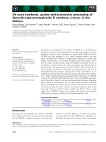
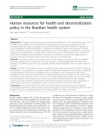
vigorous than COL in TSB containing 80 μg/ml diclofenac (Figure 1). Interestingly, SC4 also grew more vigorously in the absence of diclofenac relative to COL
(Figure 1).
The Dc RS mutants of COL and SH1000 did not
demonstrate altered MICs for the antibiotics included in
this study (Table 2). In addition, fluoroquinolone MICs
in the presence of 32- and 64- μg/ml diclofenac did not
differ between SH1000, COL and their respective DcRS
mutants. Mutation to DcRS did however alter MICs in
the presence of diclofenac for Oxa when compared to
SH1000 and COL (Table 2). For example, Oxa MICs
increased for Dc RS mutants of SH1000 at 32 μg/ml
diclofenac but not at 64 μg/ml, whereas the reverse was
true for SH1000. In addition to conferring reduced susceptibility to diclofenac, mutation to DcRS significantly
reduced susceptibility to the NSAID ibuprofen when
compared to parent strains (P < 0.05), but did not alter
susceptibility to the remaining NSAIDs, or to the salicylate analog, benzoate (Table 3).
Riordan et al. Annals of Clinical Microbiology and Antimicrobials 2011, 10:30
/>
Page 7 of 11
and diclofenac both reduce intracellular ciprofloxacin
levels, but have opposite effects on resistance to ciprofloxacin: salicylate reduces susceptibility to ciprofloxacin
[12], whereas diclofenac increases susceptibility.
Figure 1 Growth curve for S. aureus strains SH1000 (panel A)
and COL (panel B), and their respective diclofenac reducedsusceptibility (DcRS) mutant strains. Cultures of WT (circles) and
DcRS mutants (squares) were grown in TSB with (filled plots) or
without (empty plots) 80 μg/ml diclofenac. The mean optical
density is plotted as a function of time for three independent
cultures and varied by less than 5%.
Effect of diclofenac on ciprofloxacin accumulation
It has been shown previously that the reduced susceptibility of S. aureus to ciprofloxacin and ethidium bromide in the presence of salicylate correlates with
reductions in the accumulation of these antimicrobials
[10]. It was thus hypothesized that increased susceptibility of S. aureus grown with diclofenac may result from
increased ciprofloxacin accumulation. To test this, accumulation of ciprofloxacin in strain BB255 grown with
and without diclofenac was measured fluorometrically.
Surprisingly, growth with 32 μg/ml diclofenac resulted
in a 29% reduction in ciprofloxacin from 188 ± 57 to
133 ± 19 ng/mg cells (P = 0.01, N = 6). Thus, salicylate
Discussion
Diclofenac has been described as a non-antibiotic broad
spectrum antibacterial, which can act in synergy with
antibiotics to decrease bacterial cell counts. Support for
the latter claim comes from studies showing reductions
in MICs and in CFU/ml recovered from infected animals when diclofenac is administered in combination
with the protein synthesis-inhibiting aminoglycosides
streptomycin and gentamycin, and with the cell wallactive cephalosporins cefotiam and ceftriaxone
[25,26,69-71]. For S. aureus, only reductions in streptomycin MICs have been reported [17]. How diclofenac is
influencing the susceptibility of bacteria to antibiotics is
unknown.
In the present study, growth with diclofenac significantly
altered the susceptibility of lab and clinical S. aureus
strains to five of seven antibiotics not previously tested.
The study adds the fluoroquinolones ciprofloxacin, ofloxacin and norfloxacin to the list of antibiotics which significantly reduce MICs in the presence of diclofenac.
Furthermore, this is the first study to demonstrate that
growth with diclofenac can induce phenotypic resistance
to antibiotics; namely, to the cell wall-active drugs oxacillin and vancomycin. As anticipated, microarray analysis of
S. aureus strain COL grown with diclofenac revealed
alterations in genes associated with regulation of antimicrobial resistance, and drug efflux. It is thus believed that
diclofenac modifies intrinsic mechanisms of phenotypic
antimicrobial resistance in S. aureus. Similar observations
have been made for salicylate and other NSAIDs [7], suggesting that the mechanism by which these drugs influence resistance are at least partially allied. For salicylate,
this includes alterations in efflux and a PMF-independent
drug permeability barrier, as well as the involvement of
MarR-family regulators such as SarA and MgrA [8-10]. In
this study, diclofenac was not observed to significantly
alter either sarA or mgrA, but did however strongly downregulate drug efflux systems encoded by mepRAB and the
emrAB-like operon SACOL2347-2348. Both MepRAB and
EmrRAB are important for intrinsic resistance to fluoroquinolones, and emrRAB is inducible by salicylate
[38,39,45]. It was thus suspected that reduced expression
of these efflux systems, leading to intracellular accumulation of antibiotic, might explain the increased susceptibility to fluoroquinolones when grown with diclofenac
(Table 2). Instead, diclofenac was observed to reduce
intracellular ciprofloxacin levels similar to salicylate (29%
for diclofenac, vs. 19% for salicylate) [10]. Importantly,
salicylate-inducible resistance to ciprofloxacin can be
Riordan et al. Annals of Clinical Microbiology and Antimicrobials 2011, 10:30
/>
Page 8 of 11
Table 3 Susceptibility of WT and diclofenac reduced susceptibility (DcRS) mutants to NSAIDs
Drug gradient plates (mg/ml)a
Strain
Ace (0®9)
Asa (0®3.6)
Ben (0®14.4)
Dc (0®0.5)
Ibu (0®4)
Sal (0®8)
SH1000
51 ± 4.2b
24 ± 1.0
54 ± 3.2
13 ± 1.5
0
24 ± 2.1
SC-1
51 ± 3.5
25 ± 0.6
52 ± 3.2
35 ± 5.4*
28 ± 2.3*
27 ± 0.6
COL
35 ± 1.2
22 ± 0.6
39 ± 3.2
23 ± 5.8
12 ± 1.5
31 ± 1.2
SC-4
35 ± 0.6
21 ± 1.5
31 ± 1.5
35 ± 3.6*
21 ± 0*
30 ± 1.2
a
Gradient plate technique; drug gradients prepared for acetaminophen (Ace), acetyl salicylic acid (Asa), benzoate (Ben), diclofenac (Dc), ibuprophen (Ibu), and
sodium salicylate (Sal); concentration gradient provided in parentheses.
b
Average growth into NSAID gradients and standard deviation provided in mm.
* Denotes statistically significant difference between WT and DcRS by Student’s t-test (P < 0.05).
conferred independent of active efflux [10]. Thus, changes
in ciprofloxacin accumulation in the presence of diclofenac, and perhaps salicylate, may not be the direct cause of
altered susceptibility to ciprofloxacin and other fluoroquinolones. It is important to note that strain BB255, used in
ciprofloxacin accumulation assays, is a rsbU derivative,
and thus is reduced in sB activation in response to stress
[53,72]. This is perhaps significant, as an intact sigB
(encoding sB) has been shown to be involved in intrinsic
and salicylate-inducible resistance to antimicrobials [9,73],
and the expression of sigB is up-regulated by salicylate [9],
and by diclofenac (Additional File 2). Perhaps more
importantly, RsbU has been reported to control the NorA
drug efflux pump through MgrA [74]. It is therefore plausible that changes in strain BB255 which confer intrinsic
resistance to fluoroquinolones differ mechanistically from
those observed in rsbU+ strains. In support of this, ciprofloxacin MICs for BB255 were less than all other strains in
the study, and reductions in ciprofloxacin MICs in the
presence of diclofenac were more marked in rsbU+
SH1000 and in the other strains studied (Table 2).
Microarrays also revealed that growth in the presence
of diclofenac down-regulates a substantial number of
genes important for DNA stability and repair. Fluoroquinolone antibiotics interfere with DNA interactions
between gyrase (GyrAB) or topo IV (ParCE), leading to
breaks in DNA, and inducing global repair systems such
as the SOS response [75,76]. An alternative explanation
for the increased sensitivity of S. aureus grown with
diclofenac to fluoroquinolones may therefore include a
reduced ability for repair/turnover of damaged DNA
leading to cell death. Interestingly, salicylate has also
been shown to alter the expression of DNA biosynthesis/
stability genes including parE in S. aureus [8], and the
pyr genes in Bacillus subtilis [77], and to increase the frequency at which mutation to heritable antibiotic resistance occurs in S. aureus for both ciprofloxacin, and the
steroid protein synthesis inhibitor fusidic acid [11,12].
Whether or not diclofenac can select for an increased frequency of genotypic resistance to antibiotics, and the significance of these expression differences in this, are
important unanswered questions.
Diclofenac was observed to reduce susceptibility to the
cell wall active antibiotics oxacillin and vancomycin. Oxacillin is a penicillinase-resistant b-lactam, and vancomycin
is a glycopeptide antibiotic which targets D-alanyl-D-alanine
residues in the cell wall, interfering with peptidoglycan
biosynthesis. Genotypic resistance to these antibiotics is
multifactorial, and includes both lateral gene acquisition
and mutation(s) [78,79]. No mechanism of inducible phenotypic resistance to these antibiotics has been described.
Moreover, salicylates have not been shown to induce phenotypic resistance to cell-wall active antibiotics. Growth in
the presence of diclofenac led to the down-regulation of
genes encoding the cell-wall associated penicillin-binding
proteins PBP2 (pbpB) and PBP4 (pbp4), which are
required for full resistance expression to b-lactams and
vancomycin. For example, a mutation in the ORF of pbp4
which abrogates PBP4 production has been identified in
laboratory strains which express vancomycin resistance
[46], and mutations in the regulatory region of pbp4
which lead to PBP4 overproduction have been described
in methicillin resistant strains [80]. Furthermore, BoyleVavra [81] demonstrated pbpB expression was up-regulated by both oxacillin and vancomycin. It is thus possible
that pbpB and pbp4 down-regulation induced by diclofenac contributes to reduced susceptibility to these drugs,
the mechanism of which is presently unclear.
Mutation of sigB in COL, and in a vancomycin-intermediate S. aureus (VISA) strain, was shown to significantly
reduce oxacillin and vancomcyin MICs, respectively [82].
Moreover, in vitro selection of S. aureus mutants which
express reduced susceptibility to household disinfectants
has been shown to increase resistance to both oxacillin
and vancomycin in a sigB-dependent manner [73,83].
Together, these findings suggest a role for sB in intrinsic
resistance to antimicrobials which target components of
the cell envelope. As diclofenac was determined to alter
sigB expression by microarrays and qRT-PCR (Additional
File 2), the increased expression may also be important for
increased resistance to diclofenac-inducible oxacillin and
vancomycin. Concordant with this, oxacillin MICs and
growth into vancomycin gradients in the presence of
diclofenac were not altered in rsbU strain BB255, but
Riordan et al. Annals of Clinical Microbiology and Antimicrobials 2011, 10:30
/>
increased for rsbU+ strain SH1000 (Table 2 and data not
shown).
S. aureus mutants which express reduced susceptibility
to diclofenac (Dc RS) were not shown to differ in susceptibility to antibiotics compared to parent strains
SH1000 or COL. Thus, the cellular alterations which
occur at sub-MICs of diclofenac and alter antibiotic susceptibility (i.e. 32-64 μg/ml) are mechanistically-distinct
from alterations associated with mutations leading to
the DcRS phenotype selected from 1× MIC (500 μg/ml).
Diclofenac has been shown to significantly reduce S.
aureus counts from rat granulomatous tissue in the
absence of antibiotic [16]. This observation might result
from host-specific effects (i.e. immune modulation), or
bacterial-specific effects, such as inhibition of growth or
altered virulence gene expression. In support of the latter,
salicylic acid has been shown to repress sarA and SarAinducible virulence genes such as hla (a-hemolysin) and
fnbA (fibronectin-binding protein) in S. aureus, through
upregulation of sigB [15,20,84]. Although diclofenac was
also observed to up-regulate sigB, there was no attendant
change in sarA, hla or fnbA expression levels. Similarly,
up-regulation of srrAB did not lead to the down-regulation
of SrrAB-repressed virulence genes such as agr RNA III,
tsst-1 or spa. Both sigB and srrAB products contribute to
cellular functions other than pathogenesis including stress
durability and anaerobic growth.
Conclusions
In summary, growth of S. aureus with subinhibitory concentrations of diclofenac was shown to alter the expression
of hundreds of genes, including those associated with
resistance to antimicrobials and with virulence. It was
further shown that diclofenac increased the susceptibility
of S. aureus to the fluoroquinolone antibiotics ciprofloxacin, norfloxacin and ofloxacin. These observations support
previous studies which show diclofenac to increase susceptibility of S. aureus to the aminoglycoside streptomycin, and to reduce growth and survival of bacterial
pathogens in animal models. Furthermore, this is the first
study to show that diclofenac can also reduce susceptibility
(induce phenotypic resistance) to antibiotics. Significant to
S. aureus, this included the cell wall active drugs oxacillin
and vancomycin, the latter of which is critical for the treatment of severe MRSA infections. The results of this study
suggest that diclofenac modifies antimicrobial resistance
in S. aureus, in part, by altering the expression of regulatory and structural genes associated with cell wall biosynthesis/turnover and transport.
Additional material
Additional file 1: Primers used for quantitative real-time PCR (qRTPCR) in this study
Page 9 of 11
Additional file 2: Genes up-regulated following diclofenac induction
of S. aureus strain
Additional file 3: List of genes which encode hypothetical proteins
and which were significantly altered in expression in response to
diclofenac
Acknowledgements
All authors wish to acknowledge prior and ongoing support from the
National Institutes of Health: SC1GM083882-01 (J.E.G.); R25 GM07667-30
(NMSU-MARC PROGRAM); S06-GM61222-05 (NMSU-MBRS-RISE PROGRAM);
and P20RR016480 from the NM-INBRE Program of the National Center for
Research Resource.
Author details
1
Department of Cell Biology, Microbiology and Molecular Biology, University
of South Florida, Tampa, FL 33620, USA. 2Microbiology Group, Department
of Biology and Molecular Biology Program, New Mexico State University, Las
Cruces, NM 88003, USA. 3Department of Biology, Illinois State University,
Normal, IL 61790, USA. 4Department of Biological Sciences, University of
Southern Mississippi, Hattiesburg, MS 39406, USA. 5Bioinformatics and
Computational Biosciences Branch (BCBB), OCICB/OSMO/OD/NIAID/NIH,
Bethesda, MD 20892, USA.
Authors’ contributions
JR, JG and BW conceived and supervised the study, and prepared the
manuscript. JD, SCM, AKS, YS, SZ and NH performed experiments for
microarrays, antibiotic susceptibility testing, qRT-PCR and ciprofloxacin
accumulation assays. VN and ME contributed to the experimentation, design
and data analysis of DNA microarrays. All authors have read and approved
the final version.
Competing interests
The authors declare that they have no competing interests.
Received: 6 May 2011 Accepted: 21 July 2011 Published: 21 July 2011
References
1. Hiramatsu K, et al: Methicillin-resistant Staphylococcus aureus clinical
strain with reduced vancomycin susceptibility. J Antimicrob Chemother
1997, 40(1):135-6.
2. Lyon BR, Skurray R: Antimicrobial resistance of Staphylococcus aureus:
genetic basis. Microbiol Rev 1987, 51(1):88-134.
3. Neu HC: The crisis in antibiotic resistance. Science 1992,
257(5073):1064-73.
4. Goldman JD, White DG, Levy SB: Multiple antibiotic resistance (mar) locus
protects Escherichia coli from rapid cell killing by fluoroquinolones.
Antimicrob Agents Chemother 1996, 40(5):1266-9.
5. Rosner JL: Nonheritable resistance to chloramphenicol and other
antibiotics induced by salicylates and other chemotactic repellents in
Escherichia coli K-12. Proc Natl Acad Sci USA 1985, 82(24):8771-4.
6. Kumar A, HP Schweizer: Bacterial resistance to antibiotics: active efflux
and reduced uptake. Adv Drug Deliv Rev 2005, 57(10):1486-513.
7. Price CT, Lee IR, Gustafson JE: The effects of salicylate on bacteria. Int J
Biochem Cell Biol 2000, 32(10):1029-43.
8. Riordan JT, et al: Response of Staphylococcus aureus to salicylate
challenge. J Bacteriol 2007, 189(1):220-7.
9. Riordan JT, O’Leary JO, Gustafson JE: Contributions of sigB and sarA to
distinct multiple antimicrobial resistance mechanisms of Staphylococcus
aureus. Int J Antimicrob Agents 2006, 28(1):54-61.
10. Price CT, Kaatz GW, Gustafson JE: The multidrug efflux pump NorA is not
required for salicylate-induced reduction in drug accumulation by
Staphylococcus aureus. Int J Antimicrob Agents 2002, 20(3):206-13.
11. Price CT, Gustafson JE: Increases in the mutation frequency at which
fusidic acid-resistant Staphylococcus aureus arise with salicylate. J Med
Microbiol 2001, 50(1):104-6.
12. Gustafson JE, et al: Growth in the presence of salicylate increases
fluoroquinolone resistance in Staphylococcus aureus. Antimicrob Agents
Chemother 1999, 43(4):990-2.
Riordan et al. Annals of Clinical Microbiology and Antimicrobials 2011, 10:30
/>
13. Eren A, et al: Chondroprotective effect of salicylate and chloroquine in
pyogenic septic arthritis. Adv Ther 2008, 25(2):133-42.
14. Sedlacek M, et al: Aspirin treatment is associated with a significantly
decreased risk of Staphylococcus aureus bacteremia in hemodialysis
patients with tunneled catheters. Am J Kidney Dis 2007, 49(3):401-8.
15. Kupferwasser LI, et al: Acetylsalicylic acid reduces vegetation bacterial
density, hematogenous bacterial dissemination, and frequency of
embolic events in experimental Staphylococcus aureus endocarditis
through antiplatelet and antibacterial effects. Circulation 1999,
99(21):2791-7.
16. Groppo FC, et al: Effect of sodium diclofenac on serum and tissue
concentration of amoxicillin and on staphylococcal infection. Biol Pharm
Bull 2004, 27(1):52-5.
17. Dutta NK, et al: Potential management of resistant microbial infections
with a novel non-antibiotic: the anti-inflammatory drug diclofenac
sodium. Int J Antimicrob Agents 2007, 30(3):242-9.
18. Annadurai S, et al: Antibacterial activity of the antiinflammatory agent
diclofenac sodium. Indian J Exp Biol 1998, 36(1):86-90.
19. Dastidar SG, et al: The anti-bacterial action of diclofenac shown by
inhibition of DNA synthesis. Int J Antimicrob Agents 2000, 14(3):249-51.
20. Kupferwasser LI, et al: Salicylic acid attenuates virulence in endovascular
infections by targeting global regulatory pathways in Staphylococcus
aureus. J Clin Invest 2003, 112(2):222-33.
21. Muller E, et al: Mechanism of salicylate-mediated inhibition of biofilm in
Staphylococcus epidermidis. J Infect Dis 1998, 177(2):501-3.
22. Denkin S, et al: Gene expression profiling analysis of Mycobacterium
tuberculosis genes in response to salicylate. Arch Microbiol 2005,
184(3):152-7.
23. Pomposiello PJ, Bennik MH, Demple B: Genome-wide transcriptional
profiling of the Escherichia coli responses to superoxide stress and
sodium salicylate. J Bacteriol 2001, 183(13):3890-902.
24. Terada H: Uncouplers of oxidative phosphorylation. Environ Health
Perspect 1990, 87:213-8.
25. Annadurai S, et al: Experimental studies on synergism between
aminoglycosides and the antimicrobial antiinflammatory agent
diclofenac sodium. J Chemother 2002, 14(1):47-53.
26. Joly V, et al: Enhancement of the therapeutic effect of cephalosporins in
experimental endocarditis by altering their pharmacokinetics with
diclofenac. J Pharmacol Exp Ther 1988, 246(2):695-700.
27. Cohen SP, McMurry LM, Levy SB: marA locus causes decreased expression
of OmpF porin in multiple-antibiotic-resistant (Mar) mutants of
Escherichia coli. J Bacteriol 1988, 170(12):5416-22.
28. Ravel G, et al: Cytokine release does not improve the sensitivity and
specificity of the direct popliteal lymph node assay. Toxicology 2004,
200(2-3):247-54.
29. Cohen SP, et al: Salicylate induction of antibiotic resistance in Escherichia
coli: activation of the mar operon and a mar-independent pathway.
J Bacteriol 1993, 175(24):7856-62.
30. Dastidar SG, et al: Evaluation of a synergistic combination between the
non-antibiotic microbicides diclofenac and trifluoperazine. Int J
Antimicrob Agents 2003, 21(6):599-601.
31. Delgado A, et al: The fusidic acid stimulon of Staphylococcus aureus.
J Antimicrob Chemother 2008, 62(6):1207-14.
32. Tusher VG, Tibshirani R, Chu G: Significance analysis of microarrays
applied to the ionizing radiation response. Proc Natl Acad Sci USA 2001,
98(9):5116-21.
33. Nagarajan V, Elasri MO: SAMMD: Staphylococcus aureus microarray metadatabase. BMC Genomics 2007, 8:351.
34. Pfaffl MW: A new mathematical model for relative quantification in realtime RT-PCR. Nucleic Acids Res 2001, 29(9):e45.
35. O’Leary JO, et al: Effects of sarA inactivation on the intrinsic multidrug
resistance mechanism of Staphylococcus aureus. FEMS Microbiol Lett
2004, 237(2):297-302.
36. Mortimer PG, Piddock LJ: A comparison of methods used for measuring
the accumulation of quinolones by Enterobacteriaceae, Pseudomonas
aeruginosa and Staphylococcus aureus. J Antimicrob Chemother 1991,
28(5):639-53.
37. George AM, Levy SB: Amplifiable resistance to tetracycline,
chloramphenicol, and other antibiotics in Escherichia coli: involvement
of a non-plasmid-determined efflux of tetracycline. J Bacteriol 1983,
155(2):531-40.
Page 10 of 11
38. Kaatz GW, McAleese F, Seo SM: Multidrug resistance in Staphylococcus
aureus due to overexpression of a novel multidrug and toxin extrusion
(MATE) transport protein. Antimicrob Agents Chemother 2005,
49(5):1857-64.
39. Kaatz GW, DeMarco CE, Seo SM: MepR, a repressor of the Staphylococcus
aureus MATE family multidrug efflux pump MepA, is a substrateresponsive regulatory protein. Antimicrob Agents Chemother 2006,
50(4):1276-81.
40. McAleese F, et al: A novel MATE family efflux pump contributes to the
reduced susceptibility of laboratory-derived Staphylococcus aureus
mutants to tigecycline. Antimicrob Agents Chemother 2005, 49(5):1865-71.
41. Ramos JL, et al: The TetR family of transcriptional repressors. Microbiol
Mol Biol Rev 2005, 69(2):326-56.
42. Schumacher MA, et al: Structural mechanisms of QacR induction and
multidrug recognition. Science 2001, 294(5549):2158-63.
43. Brown MH, Skurray RA: Staphylococcal multidrug efflux protein QacA.
J Mol Microbiol Biotechnol 2001, 3(2):163-70.
44. Lomovskaya O, Lewis K: Emr, an Escherichia coli locus for multidrug
resistance. Proc Natl Acad Sci USA 1992, 89(19):8938-42.
45. Lomovskaya O, Lewis K, Matin A: EmrR is a negative regulator of the
Escherichia coli multidrug resistance pump EmrAB. J Bacteriol 1995,
177(9):2328-34.
46. Sieradzki K, Pinho MG, Tomasz A: Inactivated pbp4 in highly
glycopeptide-resistant laboratory mutants of Staphylococcus aureus.
J Biol Chem 1999, 274(27):18942-6.
47. Peschel A, et al: Inactivation of the dlt operon in Staphylococcus aureus
confers sensitivity to defensins, protegrins, and other antimicrobial
peptides. J Biol Chem 1999, 274(13):8405-10.
48. Wu S, de Lencastre H, Tomasz A: Sigma-B, a putative operon encoding
alternate sigma factor of Staphylococcus aureus RNA polymerase:
molecular cloning and DNA sequencing. J Bacteriol 1996, 178(20):6036-42.
49. Pane-Farre J, et al: The sigmaB regulon in Staphylococcus aureus and its
regulation. Int J Med Microbiol 2006, 296(4-5):237-58.
50. Miyazaki E, et al: The Staphylococcus aureus rsbW (orf159) gene encodes
an anti-sigma factor of SigB. J Bacteriol 1999, 181(9):2846-51.
51. Senn MM, et al: Molecular analysis and organization of the sigmaB
operon in Staphylococcus aureus. J Bacteriol 2005, 187(23):8006-19.
52. Kullik II, Giachino P: The alternative sigma factor sigmaB in
Staphylococcus aureus: regulation of the sigB operon in response to
growth phase and heat shock. Arch Microbiol 1997, 167(2/3):151-9.
53. Giachino P, Engelmann S, Bischoff M: Sigma(B) activity depends on RsbU
in Staphylococcus aureus. J Bacteriol 2001, 183(6):1843-52.
54. Pragman AA, et al: Characterization of virulence factor regulation by
SrrAB, a two-component system in Staphylococcus aureus. J Bacteriol
2004, 186(8):2430-8.
55. Throup JP, et al: The srhSR gene pair from Staphylococcus aureus:
genomic and proteomic approaches to the identification and
characterization of gene function. Biochemistry 2001, 40(34):10392-401.
56. Yarwood JM, McCormick JK, Schlievert PM: Identification of a novel twocomponent regulatory system that acts in global regulation of virulence
factors of Staphylococcus aureus. J Bacteriol 2001, 183(4):1113-23.
57. Fuchs S, et al: Anaerobic gene expression in Staphylococcus aureus.
J Bacteriol 2007, 189(11):4275-89.
58. Rogasch K, et al: Influence of the two-component system SaeRS on
global gene expression in two different Staphylococcus aureus strains.
J Bacteriol 2006, 188(22):7742-58.
59. Steinhuber A, et al: Molecular architecture of the regulatory Locus sae of
Staphylococcus aureus and its impact on expression of virulence factors.
J Bacteriol 2003, 185(21):6278-86.
60. O’Riordan K, Lee JC: Staphylococcus aureus capsular polysaccharides. Clin
Microbiol Rev 2004, 17(1):218-34.
61. Bhasin N, et al: Identification of a gene essential for O-acetylation of the
Staphylococcus aureus type 5 capsular polysaccharide. Mol Microbiol
1998, 27(1):9-21.
62. Thakker M, et al: Staphylococcus aureus serotype 5 capsular
polysaccharide is antiphagocytic and enhances bacterial virulence in a
murine bacteremia model. Infect Immun 1998, 66(11):5183-9.
63. Karakawa WW, et al: Capsular antibodies induce type-specific
phagocytosis of capsulated Staphylococcus aureus by human
polymorphonuclear leukocytes. Infect Immun 1988, 56(5):1090-5.
Riordan et al. Annals of Clinical Microbiology and Antimicrobials 2011, 10:30
/>
64. Burke KA, Lascelles J: Nitrate reductase system in Staphylococcus aureus
wild type and mutants. J Bacteriol 1975, 123(1):308-16.
65. Hoffmann T, et al: The anaerobic life of Bacillus subtilis: cloning of the
genes encoding the respiratory nitrate reductase system. FEMS Microbiol
Lett 1995, 131(2):219-25.
66. Stewart V: Nitrate respiration in relation to facultative metabolism in
enterobacteria. Microbiol Rev 1988, 52(2):190-232.
67. Bonnefoy V, et al: Autoregulation of the nar operon encoding nitrate
reductase in Escherichia coli. Mol Gen Genet 1986, 204(1):180-4.
68. Lewis K, et al: Bacterial resistance to uncouplers. J Bioenerg Biomembr
1994, 26(6):639-46.
69. Dutta NK, Mazumdar K, Park JH: In vitro synergistic effect of gentamicin
with the anti-inflammatory agent diclofenac against Listeria
monocytogenes. Lett Appl Microbiol 2009, 48(6):783-5.
70. Mazumdar K, et al: The anti-inflammatory non-antibiotic helper
compound diclofenac: an antibacterial drug target. Eur J Clin Microbiol
Infect Dis 2009, 28(8):881-91.
71. Dutta NK, et al: Activity of diclofenac used alone and in combination
with streptomycin against Mycobacterium tuberculosis in mice. Int J
Antimicrob Agents 2007, 30(4):336-40.
72. Voelker U, et al: Separate mechanisms activate sigma B of Bacillus subtilis
in response to environmental and metabolic stresses. J Bacteriol 1995,
177(13):3771-80.
73. Price CT, et al: Pine oil cleaner-resistant Staphylococcus aureus: reduced
susceptibility to vancomycin and oxacillin and involvement of SigB. Appl
Environ Microbiol 2002, 68(11):5417-21.
74. Truong-Bolduc QC, Hooper DC: Phosphorylation of MgrA and its effect on
expression of the NorA and NorB efflux pumps of Staphylococcus
aureus. J Bacteriol 192(10):2525-34.
75. Salles B, Defais M: Signal of induction of recA protein in E. coli. Mutat Res
1984, 131(2):53-9.
76. Piddock LJ, Walters RN, Diver JM: Correlation of quinolone MIC and
inhibition of DNA, RNA, and protein synthesis and induction of the SOS
response in Escherichia coli. Antimicrob Agents Chemother 1990,
34(12):2331-6.
77. Duy NV, et al: The proteome and transcriptome analysis of Bacillus
subtilis in response to salicylic acid. Proteomics 2007, 7(5):698-710.
78. Katayama Y, Ito T, Hiramatsu K: A new class of genetic element,
staphylococcus cassette chromosome mec, encodes methicillin
resistance in Staphylococcus aureus. Antimicrob Agents Chemother 2000,
44(6):1549-55.
79. Hiramatsu K: Vancomycin-resistant Staphylococcus aureus: a new model
of antibiotic resistance. Lancet Infect Dis 2001, 1(3):147-55.
80. Henze UU, Berger-Bachi B: Staphylococcus aureus penicillin-binding
protein 4 and intrinsic beta-lactam resistance. Antimicrob Agents
Chemother 1995, 39(11):2415-22.
81. Boyle-Vavra S, et al: Transcriptional induction of the penicillin-binding
protein 2 gene in Staphylococcus aureus by cell wall-active antibiotics
oxacillin and vancomycin. Antimicrob Agents Chemother 2003,
47(3):1028-36.
82. Singh VK, et al: Impact of sigB mutation on Staphylococcus aureus
oxacillin and vancomycin resistance varies with parental background
and method of assessment. Int J Antimicrob Agents 2003, 21(3):256-61.
83. Davis AO, et al: Characterization of Staphylococcus aureus mutants
expressing reduced susceptibility to common house-cleaners. J Appl
Microbiol 2005, 98(2):364-72.
84. Palma M, et al: Salicylic acid activates sigma factor B by rsbU-dependent
and -independent mechanisms. J Bacteriol 2006, 188(16):5896-903.
85. Horsburgh MJ, et al: sigmaB modulates virulence determinant expression
and stress resistance: characterization of a functional rsbU strain derived
from Staphylococcus aureus 8325-4. J Bacteriol 2002, 184(19):5457-67.
86. Kornblum J, et al: Conversion of a homogeneously methicillin-resistant
strain of Staphylococcus aureus to heterogeneous resistance by Tn551mediated insertional inactivation. Eur J Clin Microbiol 1986, 5(6):714-8.
87. Berger-Bachi B, et al: Mapping and characterization of multiple
chromosomal factors involved in methicillin resistance in
Staphylococcus aureus. Antimicrob Agents Chemother 1992, 36(7):1367-73.
doi:10.1186/1476-0711-10-30
Cite this article as: Riordan et al.: Alterations in the transcriptome and
antibiotic susceptibility of Staphylococcus aureus grown in the presence
of diclofenac. Annals of Clinical Microbiology and Antimicrobials 2011 10:30.
Page 11 of 11
Submit your next manuscript to BioMed Central
and take full advantage of:
• Convenient online submission
• Thorough peer review
• No space constraints or color figure charges
• Immediate publication on acceptance
• Inclusion in PubMed, CAS, Scopus and Google Scholar
• Research which is freely available for redistribution
Submit your manuscript at
www.biomedcentral.com/submit