Báo cáo y học: "spondyloarthritis suggests that the innate immune pathway might be of greater relevance than the Th17-mediated adaptive immune response" doc
Bạn đang xem bản rút gọn của tài liệu. Xem và tải ngay bản đầy đủ của tài liệu tại đây (1.36 MB, 9 trang )
RESEARCH ARTICLE Open Access
Analysis of IL-17
+
cells in facet joints of patients
with spondyloarthritis suggests that the innate
immune pathway might be of greater relevance
than the Th17-mediated adaptive immune
response
Heiner Appel
1*
, René Maier
1
, Peihua Wu
1,2
, Rebecca Scheer
1
, Axel Hempfing
3
, Ralph Kayser
4
, Andreas Thiel
2
,
Andreas Radbruch
2
, Christoph Loddenkemper
5
and Joachim Sieper
1,2
Abstract
Introduction: In this study, we analysed the number of IL-17
+
cells in facet joints, in the peripheral blood (PB) and
synovial fluid (SF) of spondyloarthritis (SpA) patients and compared these results with those of patients with other
rheumatic diseases and controls.
Methods: Immunohistochemical analysis of IL-17
+
cells was performed in facet joints of 33 ankylosing spondylitis
(AS) patients and compared with data from 20 osteoarthritis (OA) patients. The frequency of IL-17
+
CD4
+
T cells in
PB and SF of SpA patients (PB n = 30, SF n = 11), rheumatoid arthritis (RA) patients (PB n = 14, SF n = 7), OA
patients (PB n = 10) and healthy controls (PB n = 12) was analysed after stimulation with Staphylococcus aureus
Enterotoxin B and phorbol 12-myristate 13-acetate/ionomycin and quantified by flow cytometry.
Results: In AS facet joints, the frequency of IL-17-secreting cells was significantly higher than in samples obtained
from OA patients (P < 0.001), with a slight predominance of IL-17
+
cells among the mononuclear cells (61.5% ±
14.9%) compared to cells with polysegmental nuclei. Immunofluorescence microscopy revealed that the majority of
IL-17
+
cells were myeloperoxidase-positive (35.84 ± 13.06/high-power field (HPF) and CD15
+
neutrophils (24.25 ±
10.36/HPF), while CD3
+
T cells (0.51 ± 0.49/HPF) and AA-1
+
mast cells (2.28 ± 1.96/HPF) were less often IL-17-
positive. The frequency of IL-17
+
CD4
+
T cells in the PB and SF of SpA patients did not differ significantly compared
to RA patients, OA patients or healthy controls.
Conclusions: Our data suggest an important role for IL-17 in the inflammatory processes in AS. However, the
innate immune pathway might be of greater relevance than the Th17-mediated adaptive immune response.
Introduction
Spondyloarthritis (SpA) comprises ankylosing spondylitis
(AS), reactive arthriti s, arthritis/spondylitis with inflam-
matory bowel disease and arthritis/spondylitis with psor-
iasis. Inflammatory back pain, a similar pattern of
peripheral joint involvement with an asymmetrical
arthritis predominantly of the lower limbs and the possi-
ble occurrence of sacroiliitis, spondylitis, enthesitis and
uveitis are typical clinical features in this group of dis-
eases [1]. SpA can be split into two categories, SpA with
predominant axial involvement and SpA with predomi-
nant peripheral joint involvement, with both forms over-
lapping in about 20% to 30% of cases [1]. It has been
suggested that SpA is a T-cell-driven disease [1]. In
vitro analysis [2,3] and in situ analysis [4,5] have shown
that bot h CD4
+
T cells and CD8
+
Tcellsmightbe
involved in the pathogenesis of SpA.
* Correspondence:
1
Department of Gastroenterology, Infectiology and Rheumatology, Charité
Berlin, Campus Benjamin Franklin, Hindenburgdamm 30, D-12200 Berlin,
Germany
Full list of author information is available at the end of the article
Appel et al . Arthritis Research & Therapy 2011, 13:R95
/>© 2011 Appel et al.; licensee BioMed Central Ltd This is an open access article distributed under the terms of the Creative Commons
Attribution License ( which permits unrestricted use, distribution, and reproduction.
CD4
+
effector T cells have been classified in the past
as either Th1 cells pre dominantly secreting IFNg or Th2
cells secreting IL-4. More rec ent work could define an
IL-17-producing CD4
+
T-cell subtype termed Th17 cells
[6,7]. The first evidence of a potential role of this cyto-
kine was reported in human autoimmune diseases such
as rheumatoid arthritis (RA) [8] and multiple sclerosis
[9].
Numerous studies have been published recently inves-
tigating the frequency of Th17 cells in the peripheral
blood (PB) of SpA patients in comparison to patients
with other inflammatory joint diseases and controls.
Contradic tory results have been reported on the basis of
either flow cytometry or ELISA [10-13].
In the present study, we investigated the presence and
phenotype of IL-17
+
cells by analysing three different
compartments in patients with SpA. Our results in PB
and synovial fluid (SF) obtained from flow cytometric
analysis were compared to results obtained from
patients with RA (PB and SF) and co ntrols (PB). Addi-
tionally, we performed an extensive analysis of IL-17
+
cells in bone specimens from AS patients. To our
knowledge, this study is the first to describe in situ ana-
lysis of IL-17 expression in bone tissue samples.
Materials and methods
Patients
We obtained fresh PB from 30 patients with SpA, com-
prising 14 AS patients and 16 peripheral SpA patients,
with the latter group comprising 10 patients with reac-
tive arthrit is, 3 with Crohn’s disease and arthritis and 3
with undifferentiated peripheral SpA [14]. All AS
patients fulfilled the modified New York criteria [15],
and all peripheral SpA patients fulfilled the European
Spondylarthropathy Study Group criteria [16]. We com-
pared our data with those from 14 patients with RA
[17], 10 patients with osteoarthritis (OA) and 12 healthy
controls. We also analysed SF mononuclear cells
(MNCs) from 11 SpA patients (3 patients with AS plus
knee arthritis and 8 patients with peripheral SpA only)
and from 7 patients with RA, with all samples taken
from knee effusions. The patients’ characteristics are
shown in more detail in Table 1.
All samples from these patients and controls were
freshly analysed. All patients and controls gave their
informe d consent to participate in the study. Permission
to conduct this study was given by the local ethical
committee of the Charité University Medicine Berlin,
Campus Benjamin Franklin, Berlin, Germany.
Facet joints were obtained from 33 AS patients who
had severe kyphosis with advanced ankylosis in the lum-
bar spine and had undergone surger y for polysegmental
correcti on of rigid hyperkyphosis. Eighty-one percent of
the AS patients were male, the mean age (± SD) of
these patients was 48.6 ± 8.5 years, their mean disease
duration was 23.6 ± 10.4 years, 73% of the patients were
human leucocyte antigen B27-posit ive (HLA-B27
+
), 76%
of AS patients were taking nonsteroidal antirheumatic
drugs, four AS patients were taking non-TNF blocker
disease-modifying drugs and one AS patient had discon-
tinued adalimumab therapy four wee ks before. Pati ents
with spinal osteoporotic fractures and signs of diffuse
idiopathic skeletal hyperostosis could be excluded on
the basis of radiography [18]. Zygapophyseal joints were
also obtained from patients with OA (n =20)whohad
undergone surgery of the lumbar spine because of neu-
rological deficits in the lower limbs caused by compres-
sion of the nerve roots. Seventy percent of the OA
patients were female, and the mean age (± SD) of these
patients was 71.4 ± 9.47 years. None of the patients had
inflammatory diseases. All patients gave their informed
consent to participate in this study.
Table 1 Patient characteristics
a
Characteristics Rheumatoid arthritis Spondyloarthritides
Synovial fluid Peripheral blood Synovial fluid Peripheral blood
(n =7) (n = 14) (n = 11) (n = 30)
Age, years 34.8 ± 12.4 53.4 ± 14.5 32.4 ± 11.0 35.8 ± 13.2
Disease duration, years 4.4 ± 7.6 2.6 ± 8.2 6.6 ± 5.2 6.09 ± 5.91
ESR, mm/hour 59.3 ± 13.8 52.2 ± 26.0 36.8 ± 20.2 27.7 ± 21.5
CRP, mg/mL 6.1 ± 4.9 5.4 ± 1.6 3.6 ± 2.7 2.3 ± 3.2
RF 6/7+ 12/14+ 4/4- 11/11-
Anti-CCP 4/7+ 8/14+ 3/3- 4/4-
HLA-B27 Not done Not done 7/9+ 21/26+
DAS28 score 5.6 ± 1.4 5.4 ± 1.8
BASDAI score 2.3 ± 2.1 3.9 ± 2.6
DMARDs, n All patients 12 3 None
a
ESR = erythrocyte sedimentation rate; CRP = C-reactive protein; RF = rheumatoid factor; anti-CCP = anticyclic citrullinated peptide antibodies; HLA-B27 = human
leucocyte antigen B27; DAS28 = disease activity score in 28 joints; BASDAI = Bath Ankylosing Spondylitis Disease Activity Index (measured in AS patients only);
DMARDs = disease-modifying antirheumatic drugs. Data are expressed as means ± standard deviations unless otherwise indicated.
Appel et al . Arthritis Research & Therapy 2011, 13:R95
/>Page 2 of 9
Tissue assessment
Tissue slices from zygapophyseal joints were prepared
and examined as described before [5].
Immunohistochemistry
Immunohistochemistry of paraffin-embedded zygapo-
physeal joints was performed to detect IL-17-expressing
cells using a polyclonal anti-IL-17A antibody (R&D Sys-
tems, Wiesbaden-Nordenstadt, Germany). According to
the manufacturer’s description, this antibody has less
than 1% cross-reactivity to human IL-17B, IL-17C, IL-
17D and IL-17E and 10% cross-reactivity to IL-17F.
Control experiments were performed with isotype con-
trols a nd the anti-IL-17A antibody was also blocked by
recombinant IL-17A (R&D Systems).
We used a rabbit anti-CD3 monoclonal antibody
(clone SP-7; Thermo Scie ntific, Fremont, CA, USA) for
double-staining of CD3
+
T cells, a mouse anti-CD20
monoclonal antibody (clone L26; Dako, Hamburg, Ger-
many ) for staining of CD20
+
B cells, an anti-CD15 anti-
body (clone MMA, Acris, Herfordt, Germany) for
staining of CD15
+
neutrophils, an anti-mast cell tryptase
monoclonal antibody (clone AA1, 1:400; Abcam, Cam-
bridge, UK) for staining of mast cells, a rabbit anti-
human myeloperoxidase (MPO) polyclonal antibody
(Thermo Scientific) for staining of MPO
+
neutrophil
precursors, an an ti-CD56 monoclonal antibody (clone
1B6; Monosan, Uden, The Nethe rlands) for staining of
natural killer (NK) cells, and the anti-glucocortin mono-
clonal antibody (clone Ret 40f; Dako, Glostrup, Den-
mark) for staining of erythrocyte precursors.
Quantification was performed as described before [5].
For this analysis, tissue sections with a detectable joint
space were chosen. Areas in close proximity to these
joint spaces were analysed.
Staining for T-cell surface markers and intracellular
cytokines and analysis by flow cytometry
T cells were stained after in vitro stimulation as described
before [19]. Briefly, fresh MNCs were stimulated for six
hours in the presence of 1 μg/mL anti-CD28 antibody
alone (clone B27.2; Becton Dickinson, Heidelberg, Ger-
many), 1 μg/mL Staphylococcus aureus Enterotoxin B
(SEB) antibody (Sigma-Aldrich, Deisenhofen, Germany)
and 25 ng/mL phorbol 12-myristate 13-acetate (PMA)
(Sigma, Taufkirchen, Germany) plus 1 μg/mL ionomycin
(Sigma, Taufkirchen, Germany). After two hours of sti-
mulation, 10 μg/mL brefeldin A (Sigma Aldrich) was
added to inhibit cytokine release from cells.
The following antibodies were used: anti-human CD4
(clone SK3; Becton Dickinson, anti-IL-17 (eBio64DEC17;
eBiosciences, San Diego, CA, USA) and antihuman C-C
chemokine receptor type 6 (CCR6) (clone 11A9; Becton
Dickinson), as well as a mouse immunoglobulin G1
(IgG1) isotype contro l antibody (clone MOPC-1; Becton
Dickinson). Positive cells were subsequently quantified
by flow cytometry using a FACSCalibur flow cytometer
with CellQuest software (Becton Dickinson, San Jose,
CA, USA).
CCR6 staining of IL-17
+
T cells
For staining of the cell surface marker CCR6 on IL-17
+
T cells, CD4
+
T cells were separated by magnetic absor-
bent cell sorting (MACS) a s described be fore [3]. After
in vitro stimulation with PMA/ionomycin as described
above, CCR6
+
CD4
+
T cells were quantified by flow
cytometry. As an isotype control, we used an antimouse
IgG1 isotype antibody.
Measurement of IL-17 secretion of CD4
+
T cells by ELISA
For measurement of IL-17 secretion b y ELISA, CD4
+
T
cells were also separated by MACS. After in vitro stimu-
lation with PMA/ionomycin as described above, IL-17
was measured in the supernatant by IL-17 ELISA
(Quantikine IL-17 ELISA; R&D Systems, Abington, UK)
according to the manufacturer’s instructions.
Statistics
For data analysi s, we used the Mann -Whitne y U test or
the Wilcoxon test if appropriate and SPSS for Windows
software (SPSS, Inc., Chicago, IL, USA). Correlation ana-
lysis was performed by using the Pearson correlation
coefficient. For the correlation analysis of ELISA and
fluorescence-activated cell sorting analysis of IL-17
+
T
cells, we calculated the nonparametric Spearman’ srank
correlation coefficient.
Results
In situ analysis of IL-17A
+
cells in facet joints of AS
patients
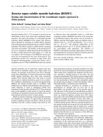
Light microscopic analysis revealed that not only MNCs
but also mature neutrophils with polysegmented nuclei
(PNCs) were IL-17A
+
(Figure 1a, top: AS facet joint; 1a
bottom: OA facet joint). The specificity of IL-17 staining
could be underlined by blocking the anti-IL- 17 antibody
and the positive staining with recombinant IL-17 in
each experiment (Figure 1a, middle). The frequenc y of
IL-17
+
MNCs (Figure 1b, top) and IL-17
+
PNCs (Figure
1b, bottom) was significantly higher in the bone marrow
of AS facet joints (mean MNCs ± SD 17.08 ± 10.41/
high-power field (HPF), PNCs 11.78 ± 9.92/HPF) com-
pared to OA facet joints (MNCs 2.9 ± 5.67/HPF, P <
0.001; PNCs 2.55 ± 5.97/HPF, P < 0.001) (Figure 1c). Of
all IL-17
+
cells, 61.5% ± 14.9% were MNCs in AS
patients and 57.0% ± 11.4% were MNCs in OA patients
(P > 0.05). There was a positive correlation between IL-
17
+
MNCs and IL-17
+
PNCs in bo th AS patients (r =
0.634, P < 0.001) and OA patients (r = 0.991, P < 0.001).
Appel et al . Arthritis Research & Therapy 2011, 13:R95
/>Page 3 of 9
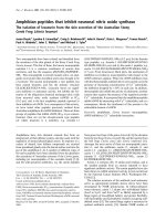
Subsequently, we identified the IL-17-producing cell
type in more detail in a subgroup of 12 AS pa tients and
10 OA patients by double-staining and immunofluores-
cence microscopy. By using this method, we could
determine that neutrophil precursors detected by MPO
staining (35.84 ± 13.04/HPF) and CD15
+
neutrophils
(24.25 ± 10.36/HPF) were by far the most frequent cell
populations expressing IL-17. Only a small proportion
of AA-1
+
mast cells (2.2 8 ± 1.16/HPF) and an even
smaller proportion of CD3
+
T cells (0.51 ± 0.49/HPF)
were IL-17
+
(Figure 2). In OA pa tients, the frequency of
all IL-17
+
cell types was significantly lower (P <0.05in
all cases), although there was a similar percentage of the
different IL-17
+
cell types: CD3
+
T c ells 0.1 ± 0.1/HPF,
AA-1
+
mast cells 1.36 ± 1.53/HPF, neutrophil precur-
sors detected by MPO staining 5.04 ± 6.15/HPF and
CD15
+
neutrophils 3.88 ± 5.75/HPF (Figure 2). Within
the population of MPO
+
cells, 63.5% and 36.5% of cells
were MNCs and PNCs in AS patients, respectively, and
65.1% and 34.9% of cells were MNCs and PNCs in OA
patients, respectively.
Double-staining with an anti-CD20 antibody directed
against B cells, with an anti-CD56 antibody directed
against natural killer cells a nd with an antibody against
erythrocyte precursors did not reveal IL-17
+
cells among
these cell types (data not shown).
In some of the AS patients and OA patients, age-
matched analysis was possible. In these patients, the
higher frequency of IL-17
+
cells in AS patients com-
pared to OA patients could be confirmed. In the three
55-, 63-and 67-year-old OA patients, 0, 0 and 0.7 IL-17
+
cells/HPF were found, respectively, compared to the
three 57-, 63-and 67-year-o ld AS patients who had 21.1,
21.0 and 1.7 cells/HPF, respectively.
OA facet joint, anti IL-17
AS facet joint, anti IL-17
AS facet joint, anti IL-17 + rIL-17
(
a
)
Mononuclear cells
Cells with
polysegmental
nuclei
(b)
0
5
10
15
20
25
30
AS OA AS OA
mononuclear cells cells with polysegmental nuclei
Number of IL-17+ cells per high power field
*
*
(c)
Figure 1 In situ analysis and quantification of IL-17
+
cells in patients with ankylosing spondylitis or osteoarthritis. (a) In situ analysis of
IL-17
+
cells in facet joints of ankylosing spondylitis (AS) (top) and osteoarthritis (OA) (bottom) patients. The specificity of IL-17 staining (top) is
shown by blocking the anti-IL-17 antibody with recombinant IL-17 (rIL-17) (middle). (b) and (c) The frequency of IL-17-secreting mononuclear
cells and cells with polysegmental nuclei in the bone marrow of AS facet joints was significantly higher in AS than in OA facet joints. *P < 0.001.
Appel et al . Arthritis Research & Therapy 2011, 13:R95
/>Page 4 of 9
Similar frequency of CD4
+
IL-17
+
cells in the peripheral
blood of SpA patients compared to RA patients, OA
patients and healthy controls
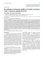
We observed similar levels of CD4
+
IL-17
+
PB T cells
(Figure 3a) after stimulation with PMA/ionomycin or
SEB in SpA patients compared to RA patients, OA
patients and healt hy controls. Except for SEB stimula-
tion (AS in comparison to con trols, P < 0.05), we did
not observe significant differen ces (Figure 3a). The low-
est frequency of CD4
+
IL-17
+
T cells was observed in
OA patients.
Similar frequency of CD4
+
IL-17A
+
cells in the synovial
fluid of SpA and RA patients
The frequency of SF CD4
+
IL-17
+
cells (Figure 3b) did
not d iffer significantly when SpA and RA patients were
compared.
Higher frequency of CD4
+
IL-17A
+
cells in the synovial
fluid in comparison to peripheral blood of SpA and RA
patients
We further addressed the question whether there was a
different frequency of IL-17
+
T cells in PB or SF. The
frequency of CD4
+
IL-17
+
T cells was higher in SF than
in PB in SpA and RA patients. However, except for SEB
stimulation of CD4
+
T cells in RA patients (P =0.041),
this finding was not statistically significant. This was
also seen in a subgroup of five AS patients with
matched SF and PB samples. The mean PB percentages
(± SD) of CD4
+
IL-17
+
cells were 0.02% ± 0.01% after
stimulation with anti-CD28, 0.19% ± 0.05% after sti mu-
lation with SEB and 0.67% ± 0.61% after stimulati on
with PMA/ionomycin (all P > 0.05). The mean SF per-
centages (± SD) of CD4
+
IL-17
+
cells were 0.02% ±
0.01% after stimulation with anti-CD28, 0.71% ± 1.24%
CD3+ T cells
(green)
CD15+ neutrophils
(green)
IL-17
(red)
IL-17
double staining
(yellow)
CD3+ T cells
(green)
CD15+ neutrophils
(green)
Ankylosing
S
pondylitis
Osteoarthritis
MPO+ cells
(green)
AA-1+ cells
(green)
MPO+ cells
(green)
AA-1+ cells
(green)
IL-17
(red)
IL-17
double staining
(yellow)
0
10
20
30
40
50
60
number of IL-17+ cells per HPF
CD3+
T cells
AA-1+
mast cells
MPO+
cells
CD15+
neutrophils
mononuclear cells
cells with polysegmental nuclei
0
10
20
30
40
50
60
number of IL-17+ cells/HPF
CD3+
T cells
AA-1+
mast cells
MPO+
cells
CD15+
neutrophils
mononuclear cells
cells with polysegmental nuclei
Figure 2 In situ immunofluorescence analysis of IL-17
+
cells. In situ analysis of IL-17
+
cells in facet joints of ankylosing spondylitis (AS)
patients and patients with osteoarthritis (OA) by using immunofluorescence microscopy. Double-staining reveals that myeloperoxidase-positive
(MPO
+
) and CD15
+
cells are the major source of IL-17 expression. The frequency of these cells was significantly higher in AS than in OA (P <
0.05 in both cases). The population of MPO
+
cells included mononuclear cells and cells with polysegmental nuclei. Th17 cells and mast cells are
also a source for IL-17
+
expression, both of which were significantly higher in AS patients than in OA patients (P < 0.05 in both cases).
Appel et al . Arthritis Research & Therapy 2011, 13:R95
/>Page 5 of 9
after stimulation with SEB and 0.92% ± 0.48% after sti-
mulation with PMA/ionomycin (all P > 0.05)
CCR6 expression in IL-17
+
T cells
Because Th17 cells express CCR6 nearly exclusively on
their surface [19], we analysed the expression of CCR6
on the cell surface of peripheral CD4
+
IL-17
+
T cells, after
MACS separation of CD4
+
Tcellsandin vitro stimula-
tion, obtained from three AS patients and three healthy
controls. The results are shown in Figure 4a. More than
90% of IL-17-secreting CD4
+
T cells were also CCR6
+
in
all cases, supporting the specific ity of our Th17 staining.
Use of an isotype control antibody revealed negative
staining, confirming the specificity of the staining. CD4
+
IL-17
+
CCR6
+
T cells were not detected without PMA/
ionomycin stimulation (Figure 4b).
IL-17 secretion by T cells measured by ELISA
The specificity of Th17 staining by flow cytometry was
further confirmed when CD4
+
T cells derived from the
PB from three AS patients and three healthy controls
were separated by MACS and IL-17 secretion was mea-
sured in the supernatant by ELISA after in vitro stimula-
tion. When IL-17 secretion after in vitro stimulation was
compared with the intracellular cytokine staining data
for CD4
+
T cells from the same patients, a good correla-
tion of r = 0.66 was found, further confirming the speci-
ficity of the IL-17 staining (Table 2).
Discussion
In this study, we analysed the frequency of IL-17
+
cells
in three different compartments of patients with spon-
dyloarthritides. The most prominent finding was a sig-
nificantly higher number of IL-17
+
cells at the primary
site of inflammation in the subchondral bone marrow of
affected facet joints [5] in AS patients compared to OA
patients. Facet joints from patients with other inflamma-
tory rheumatic diseases, such as RA patients, would
have been of interest for comparison in this analysis,
but such surgical procedures are rarely performed in RA
patients. Interestingly, IL-17
+
cells were almost similarly
distributed among the MNC and PNC populations, with
a slight predominance in the PNC population. Surpris-
ingly, immunofluorescence double-staining in situ
showed that the clear majority of the IL-17
+
cells were
found among the CD15
+
neutrophils (24.25 ± 10.36/
HPF) and among the MPO
+
cells of t he myeloid lineag e
Figure 3 Peripheral and synovial CD4
+
IL-17
+
T cell levels in
spondyloarthritis, rheumatoid arthritis and controls. Analysis of
(a) peripheral blood (PB) and (b) synovial fluid (SF) CD4
+
IL-17
+
T
cells in spondyloarthritis (SpA) patients (PB n = 30, SF n = 11),
rheumatoid arthritis (RA) patients (PB n = 14, SF n = 7), osteoarthritis
(OA) patients (PB n = 10) and healthy controls (C) (PB n = 12).
Similar levels of PB and SF CD4
+
IL-17
+
T cells after stimulation with
phorbol 12-myristate 13-acetate (PMA)/ionomycin or Staphylococcus
aureus Enterotoxin B (SEB) antibodies are seen when SpA patients
are compared to RA patients. The frequency of PB CD4
+
IL-17
+
T
cells was only significantly lower in SpA patients than in controls
when stimulated with SEB antibodies (P < 0.05).
IL1
7
CCR
6
AS patients
healthy
controls
90.8%
98.8%
93.2%
94.3%
92.3%
91.7%
(
a)
0.00%
0.01%15.1%
0.01%
0.00%
0.00%
0.00%
0.00%0.01%
0.02%
0.01%
0.23%
0.00%
0.01%
0.22%
12.8%
0.25%0.01%
w/o anti-CCR6
with anti-CCR6
with isotype control
CC
R
6
IL17
(
b)
not
stimulated
PMA/
ionomycin
Figure 4 CCR6 expression in CD4
+
IL-17
+
T cells. (a) In three
ankylosing spondylitis (AS) patients and three healthy controls, the
expression of C-C chemokine receptor type 6 (CCR6) in CD4
+
IL-17
+
T cells derived from peripheral blood was analysed after in vitro
stimulation with phorbol 12-myristate 13-acetate (PMA)/ionomycin
antibodies. Percentages indicate the relative number of CCR6
+
cells
to the total number of CD4
+
IL-17
+
T cells (top row of dot blot
analysis). (b) Using an isotype control antibody after T-cell
stimulation with PMA/ionomycin (bottom right) antibodies revealed
no positive staining, confirming the specificity of CCR6 staining
(bottom left). Without such T-cell stimulation, no CCR6
+
IL-17
+
T cells
were detected.
Appel et al . Arthritis Research & Therapy 2011, 13:R95
/>Page 6 of 9
(35.84 ± 13.04/HPF), while CD3
+
T cells (0.51 ± 0.49/
HPF) and mast cells (2.28 ± 1.16/HPF) constituted only
a small proportion of IL-17
+
cells. Staining for other cell
types (B cells, NK cells a nd erythrocyte precursors)
could exclude these cells as other sources of IL-17.
However, we cannot exclude that, in the early phase of
the disease, such a finding might be different b ecause
our current resu lts were obtained in pat ients with
advanced AS.
These data suggest that IL-17
+
-secreting cells other
than the Th17 cells are of relevan ce in local inflamma-
tion in AS. Investigators in two recent studies on syno-
vial membranes from patients with RA [20] or
peripheral SpA, including psoriatic arthritis (PsA) [21],
also showed that IL-17-producing cells other than Th17
cells are of relevance. In both RA and PsA patients,
mast cells were the major source of IL-17, while Th17
cells were rather rare among the IL-17-producing cells,
similar to the findings in our study.
There have previously been some indirect hints that
Th17 cells might play a role in the pathogenesis of SpA.
An extensive genotype analysis performed recently
revealed that AS is closely linked to polymorphisms in
the IL-23 receptor gene [22], suggesting that Th17
might be of relevance, although the functional conse-
quence of this IL-23 polymorphism has not been clari-
fied. Furthermore, in HLA-B27/human b
2
-
microglobulin-transgenic rats, a possible animal model
of SpA, HLA-B27 misfolding and the unfolded protein
response resulted in a strongly activated IL-23/IL-17
axis in the colon of B27/b
2
-microglobulin-transgenic
rats with SpA-like disease [23].
Nonetheless, our results and the studies of RA patients
[20] and peripheral SpA patients [21] indicate that T cells
might have been overestimated as the source of IL-17 in
these chronic inflammatory diseases and that an innate
immune response in the context of IL-17 might be of rele-
vance. Interestingly, a high frequency of IL-17
+
mast cells
and IL-17
+
neutrophils, as well as a low frequency of Th17
cell s, was also described in the biopsies of skin lesions of
psoriasis patients [24]. An analysis of patients with ulcera-
tive colitis revealed an elevated number of Th17 cells
located in the lamina propria of inflammatory lesions [25],
but the relative number of Th17 cells in comparison to
other IL-17
+
cells was not analysed. On the basis of the
results of our investigation, however, we cannot exclude
the possibi lity that Th17 cells are of any relevance in AS.
Although the frequency was relatively low, it was higher
than in the control group and might be sufficient to
orchestrate an immune response.
In our study, mast cells as a source of IL-17 were
much less frequent than in RA patients [20] and psoria-
sis p atients [21], and also compared to neutrophils and
their precursors. The IL-17 receptor A is highly
expressed in hematopoietic cells [26]. Whether the posi-
tive staining of neutrophils and MPO
+
precursor cells is
due to autocrine secretion or is caused by binding of IL-
17 at the IL-17 receptor could not be determined by
our staining. Interestingly, it has been w ell described
that T-cell-derived IL-17 is an important growth factor
for granulopoiesis in humans [27]. Although the involve-
ment of IL-17
+
neutrophils in inflammatory processes
has also been reported [26,28,29], though not yet in
patients with SpA, further confirmation that IL-17 is
produced by neutrophils by other methods, such as by
in situ hybrid isation, would be warranted. Very recently,
Li et al. [30] presented impressive data showing that IL-
17-producing neutrophils participated in innate immune
responses in a mouse model of kidney reperfusion
injury. Staining for MPO, which is produced during
myeloid d ifferentiation in the bone marrow by neutro-
phils and their precursors [31], is rat her specific for this
cell lineage, and these precursor cells appear in the
shape of MNCs. Nonetheless, a more detailed character-
isation of the MPO
+
precursor cells found in our pre-
sent study will be of interest in the future.
Because AS patients a re considerably younger t han
OA patients, an age-matched subanalysis was possible in
only three AS patients and three OA patients, confirm-
ing a clearly higher number of IL-17
+
cells in AS
patients than in OA patients. Nonetheless, the lack of a
larger age-matched control group, which will also not
easily be found in possible follow-up investigations, can
be seen as a limitation of our study.
Table 2 IL-17 in CD4
+
T cells: Comparison of ELISA and intracellular cytokine staining
a
Individuals ELISA for IL-17-secreting CD4
+
T cells Intracellular cytokine staining for CD4
+
IL-17
+
T cells
Without stimulation With PMA/ionomycin Without stimulation With PMA/ionomycin
AS patient 1 0 pg/nL 1,667.9 pg/nL 0.0% of CD4
+
T cells 0.98% of CD4
+
T cells
AS patient 2 0 pg/nL 967.8 pg/nL 0.0% of CD4
+
T cells 0.47% of CD4
+
T cells
AS patient 3 0 pg/nL 1,398.5 pg/nL 0.0% of CD4
+
T cells 1.72% of CD4
+
T cells
Control 1 0 pg/nL 1,784.4 pg/nL 0.0% of CD4
+
T cells 1.21% of CD4
+
T cells
Control 2 0 pg/nL 277.9 pg/nL 0.0% of CD4
+
T cells 0.57% of CD4
+
T cells
Control 3 0 pg/nL 920.5 pg/nL 0.0% of CD4
+
T cells 0.69% of CD4
+
T cells
a
PMA = phorbol 12-myristate 13-acetate; AS = ankylosing spondylitis.
Appel et al . Arthritis Research & Therapy 2011, 13:R95
/>Page 7 of 9
In PB and S F of SpA patients, w e only lo oked for the
frequency of Th17 cells and not for other IL-17-pro du-
cing cells. Here we observed no significant differences
compared to RA patients, OA patients or healthy con-
trols with regard to PB and compared to RA with regard
to SF. These results confirm our immunohistological
analysis of the bone that Th17 cells do not seem to play
an important role in AS. Pr evious investigations
reported a h igher number of Th17 cells in the PB of
SpA patients [10,11 ], while reduced levels of IL-17 were
found in the SF of SpA patients in another study. Our
data also indicate that the analysis of IL-17-producing
cells in inflamed tissue might be more informative than
in PB or SF.
It is currently unclear whether the overexpression of
IL-17 in inflammatory lesions of different autoimmune
diseases indicates an extraordinary pathogenic role of
this cytokine during inflammatory processes or whether
this is just a secondary reaction [32]. However, th e good
clinical response in trials with the anti-IL-17 antibody
secukinumab in RA patients [33], psoriasis patients and
AS patients [34] suggests that IL-1 7 might indeed play a
role in these diseases. The observed rather rapid clinical
response in these trials might be a further hint that
direct inhibition of soluble IL-17 is more important
than modulation of a T-cell response. Interestingly, a
similar discussion of the effect of TNF blockers, mainly
neutralisation of soluble TNF or modulation of T-cell
responses, has been ongoing over the past few years, but
the issue has not been resolved yet [35].
Conclusions
Our study suggest an important role for IL-17 during
inflammatory processes in SpA patients. Our data also
indicate that the innate immune pathway, mostly
mediated through neutrophils and less via mast cells,
might play a relevant role during inflammatory pro-
cesses in AS patients, while Th17 cells seem to be of
less importance.
Abbreviations
AS: ankylosing spondylitis; ELISA: enzyme-linked immunosorbent assay; IL:
interleukin; IFN: interferon; MACS: magnetic absorbent cell sorting; MNC:
mononuclear cell; MPO: myeloperoxidase; OA: osteoarthritis; PB: peripheral
blood; PNCs: cells with polysegmental nuclei; RA: rheumatoid arthritis; SF:
synovial fluid; SpA: spondyloarthritis; TNFα: tumour necrosis factor α.
Acknowledgements
This work was supported by grants from the Deutsche
Forschungsgemeinschaft (DFG): Ap82/3-1 and Si 620/11-1.
Author details
1
Department of Gastroenterology, Infectiology and Rheumatology, Charité
Berlin, Campus Benjamin Franklin, Hindenburgdamm 30, D-12200 Berlin,
Germany.
2
Deutsches Rheumaforschungszentrum Berlin, Schumannstrassse
21/22, D-10117 Berlin, Germany.
3
Center for Spine Surgery, Werner-Wicker-
Klinik, Im Kreuzfeld 4, D-34537 Bad Wildungen, Germany.
4
Department of
Trauma and Reconstructive Surgery, Charité Berlin, Campus Benjamin
Franklin, Hindenburgdamm 30, D-12200 Berlin, Germany.
5
Department of
Pathology, Charité Berlin, Campus Benjamin Franklin, Hindenburgdamm 30,
D-12200 Berlin, Germany.
Authors’ contributions
HA and JS designed the study, analysed the data and drafted the
manuscript. RH, PW and RS participated in the data collection, performed
the data analysis and helped in the drafting of the manuscript. AH and RK
participated in the data collection and helped in the drafting of the
manuscript. AT and AR analysed the data and participated in the drafting of
the manuscript. All authors were contributed to discussions and read and
approved the final manuscript.
Competing interests
The authors declare that they have no competing interests.
Received: 14 October 2010 Revised: 4 May 2011 Accepted: 6 June 2011
Published: 6 June 2011
References
1. Braun J, Sieper J: Ankylosing spondylitis. Lancet 2007, 369:1379-1390.
2. Boyle LH, Goodall JC, Opat SS, Gaston JS: The recognition of HLA-B27 by
human CD4
+
T lymphocytes. J Immunol 2001, 167:2619-2624.
3. Atagunduz P, Appel H, Kuon W, Wu P, Thiel A, Kloetzel PM, Sieper J: HLA-
B27-restricted CD8
+
T cell response to cartilage-derived self peptides in
ankylosing spondylitis. Arthritis Rheum 2005, 52:892-901.
4. Baeten D, Kruithof E, Van den Bosch F, Demetter P, Van Damme N,
Cuvelier C, De Vos M, Mielants H, Veys EM, De Keyser F:
Immunomodulatory effects of anti-tumor necrosis factor α therapy on
synovium in spondylarthropathy: histologic findings in eight patients
from an open-label pilot study. Arthritis Rheum 2001, 44:186-195.
5. Appel H, Kuhne M, Spiekermann S, Ebhardt H, Groszdanovic Z, Köhler D,
Dreimann M, Hempfing A, Rudwaleit M, Stein H, Metz-Stavenhagen P,
Sieper J, Loddenkemper C: Immunohistological analysis of zygapophyseal
joints in patients with ankylosing spondylitis. Arthritis Rheum 2006,
54:2845-2851.
6. Harrington LE, Hatton RD, Mangan PR, Turner H, Murphy TL, Murphy KM,
Weaver CT: Interleukin 17-producing CD4
+
effector T cells develop via a
lineage distinct from the T helper type 1 and 2 lineages. Nat Immunol
2005, 6:1123-1132.
7. Park H, Li Z, Yang XO, Chang SH, Nurieva R, Wang YH, Wang Y, Hood L,
Zhu Z, Tian Q, Dong C: A distinct lineage of CD4 T cells regulates tissue
inflammation by producing interleukin 17. Nat Immunol 2005,
6:1133-1141.
8. Chabaud M, Durand JM, Buchs N, Fossiez F, Page G, Frappart L, Miossec P:
Human interleukin-17: a T cell-derived proinflammatory cytokine
produced by the rheumatoid synovium. Arthritis Rheum 1999, 42:963-970.
9. Kebir H, Kreymborg K, Ifergan I, Dodelet-Devillers A, Cayrol R, Bernard M,
Giuliani F, Arbour N, Becher B, Prat A: Human T
H
17 lymphocytes promote
blood-brain barrier disruption and central nervous system inflammation.
Nat Med 2007, 13:1173-1175.
10. Jandus C, Bioley G, Rivals JP, Dudler J, Speiser D, Romero P: Increased
numbers of c irculating polyfunctional Th17 memory cells in patients
with seronegative spondylarthritides. Arthritis Rheum 2008,
58:2307-231 7.
11. Shen H, Goodall JC, Hill Gaston JS: Frequency and phenotype of
peripheral blood Th17 cells in ankylosing spondylitis and rheumatoid
arthritis. Arthritis Rheum 2009, 60:1647-1656.
12. Vandooren B, Noordenbos T, Ambarus C, Krausz S, Cantaert T,
Yeremenko N, Boumans M, Lutter R, Tak PP, Baeten D: Absence of a
classically activated macrophage cytokine signature in peripheral
spondylarthritis, including psoriatic arthritis. Arthritis Rheum 2009,
60:966-975.
13. Wendling D, Cedoz JP, Racadot E, Dumoulin G: Serum IL-17, BMP-7, and
bone turnover markers in patients with ankylosing spondylitis. Joint Bone
Spine 2007, 74:304-305.
14. Rudwaleit M, van der Heijde D, Landewé R, Akkoc N, Brandt J, Chou CT,
Dougados M, Huang F, Gu J, Kirazli Y, Van den Bosch F, Olivieri I, Roussou E,
Scarpato S, Sørensen IJ, Valle-Oñate R, Weber U, Wei J, Sieper J: The
Assessment of SpondyloArthritis International Society classification
Appel et al . Arthritis Research & Therapy 2011, 13:R95
/>Page 8 of 9
criteria for peripheral spondyloarthritis and for spondyloarthritis in
general. Ann Rheum Dis 2011, 70:25-31.
15. van der Linden S, Valkenburg HA, Cats A: Evaluation of diagnostic criteria
for ankylosing spondylitis: a proposal for modification of the New York
criteria. Arthritis Rheum 1984, 27:361-368.
16. Dougados M, van der Linden S, Juhlin R, Huitfeldt B, Amor B, Calin A,
Cats A, Dijkmans B, Olivieri I, Pasero G, Veys E, Zeidler H, the European
Spondylarthropathy Study Group: The European Spondylarthropathy
Study Group preliminary criteria for the classification of
spondylarthropathy. Arthritis Rheum 1991, 34:1218-1227.
17. Arnett FC, Edworthy SM, Bloch DA, McShane DJ, Fries JF, Cooper NS,
Healey LA, Kaplan SR, Liang MH, Luthra HS, Medsger TA Jr, Mitchell DM,
Neustadt DH, Pinals RS, Schaller JG, Sharp JG, Wilder RL, Hunder GG: The
American Rheumatism Association 1987 revised criteria for the
classification of rheumatoid arthritis. Arthritis Rheum 1988, 31:315-324.
18. Resnick D, Niwayama G: Radiographic and pathologic features of spinal
involvement in diffuse idiopathic skeletal hyperostosis (DISH). Radiology
1976, 119:559-568.
19. Thiel A, Radbruch A: Antigen-specific cytometry. Arthritis Res 1999, 1:25-29.
20. Hueber AJ, Asquith DL, Miller AM, Reilly J, Kerr S, Leipe J, Melendez AJ,
McInnes IB: Mast cells express IL-17A in rheumatoid arthritis synovium. J
Immunol 2010, 184:3336-3340.
21. Yeremenko N, Gofita I, Noordenbos T, Tak PP, Canete J, Baeten D: IL-17A
producing cells as a therapeutic target in spondyloarthritis. J Transl Med
2010, 8(Suppl 1):>P42.
22. Wellcome Trust Case Control Consortium, Australo-Anglo-American
Spondylitis Consortium (TASC), Burton PR, Clayton DG, Cardon LR,
Craddock N, Deloukas P, Duncanson A, Kwiatkowski DP, McCarthy MI,
Ouwehand WH, Samani NJ, Todd JA, Donnelly P, Barrett JC, Davison D,
Easton D, Evans DM, Leung HT, Marchini JL, Morris AP, Spencer CC,
Tobin MD, Attwood AP, Boorman JP, Cant B, Everson U, Hussey JM,
Jolley JD, Knight AS, Koch K, Meech E, et al: Association scan of 14,500
nonsynonymous SNPs in four diseases identifies autoimmunity variants.
Nat Genet 2007, 39:1329-1337.
23. DeLay ML, Turner MJ, Klenk EI, Smith JA, Sowders DP, Colbert RA: HLA-B27
misfolding and the unfolded protein response augment interleukin-23
production and are associated with Th17 activation in transgenic rats.
Arthritis Rheum 2009, 60:2633-2643.
24. Res PC, Piskin G, de Boer OJ, van der Loos CM, Teeling P, Bos JD,
Teunissen MB: Overrepresentation of IL-17A and IL-22 producing CD8 T
cells in lesional skin suggests their involvement in the pathogenesis of
psoriasis. PLoS One 2010, 5:e14108.
25. Kobayashi T, Okamoto S, Hisamatsu T, Kamada N, Chinen H, Saito R,
Kitazume MT, Nakazawa A, Sugita A, Koganei K, Isobe K, Hibi T: IL23
differentially regulates the Th1/Th17 balance in ulcerative colitis and
Crohn’s disease. Gut 2008, 57:1682-1689.
26. Korn T, Bettelli E, Oukka M, Kuchroo VK: IL-17 and Th17 cells. Annu Rev
Immunol 2009, 27:485-517.
27. Huang W, La Russa V, Alzoubi A, Schwarzenberger P: Interleukin-17A: a T-
cell-derived growth factor for murine and human mesenchymal stem
cells. Stem Cells 2006, 24:1512-1518.
28. Ferretti S, Bonneau O, Dubois GR, Jones CE, Trifilieff A: IL-17, produced by
lymphocytes and neutrophils, is necessary for lipopolysaccharide-
induced airway neutrophilia: IL-15 as a possible trigger. J Immunol 2003,
170:2106-2112.
29. Weaver CT, Hatton RD, Mangan PR, Harrington LE: IL-17 family cytokines
and the expanding diversity of effector T cell lineages. Annu Rev
Immunol 2007, 25:821-852.
30. Li L, Huang L, Vergis AL, Ye H, Bajwa A, Narayan V, Strieter RM, Rosin DL,
Okusa MD: IL-17 produced by neutrophils regulates IFN-γ-mediated
neutrophil migration in mouse kidney ischemia-reperfusion injury. J Clin
Invest 2010, 120:331-342.
31. Malech HL, Nauseef WM: Primary inherited defects in neutrophil function:
etiology and treatment. Semin Hematol 1997, 34:279-290.
32. Miossec P: Diseases that may benefit from manipulating the Th17
pathway. Eur J Immunol 2009, 39:667-669.
33. Hueber W, Patel DD, Dryja T, Wright AM, Koroleva I, Bruin G, Antoni C,
Draelos Z, Gold MH, Psoriasis Study Group, Durez P, Tak PP, Gomez-
Reino JJ, Rheumatoid Arthritis Study Group, Foster CS, Kim RY, Samson CM,
Falk NS, Chu DS, Callanan D, Nguyen QD, Uveitis Study Group, Rose K,
Haider A, Di Padova F: Effects of AIN457, a fully human antibody to
interleukin-17A, on psoriasis, rheumatoid arthritis, and uveitis. Sci Transl
Med 2010, 2:52ra72.
34. Baeten D, Sieper J, Emery P, Braun J, van der Heijde D, McInnes I, van
Laar JM, Landewé R, Wordsworth P, Wollenhaupt J, Kellner H, Paramarta J,
Bertolino AP, Wright AM, Hueber W: The anti-IL7A monoclonal antibody
secukinumab (AIN457) showed good safety and efficacy in the
treatment of active ankylosing spondylitis. Arthritis Rheum 2010,
62:2840-2841.
35. Zou J, Rudwaleit M, Brandt J, Thiel A, Braun J, Sieper J: Up regulation of
the production of tumour necrosis factor α and interferon γ by T cells in
ankylosing spondylitis during treatment with etanercept. Ann Rheum Dis
2003, 62:561-564.
doi:10.1186/ar3370
Cite this article as: Appel et al.: Analysis of IL-17
+
cells in facet joints of
patients with spondyloarthritis suggests that the innate immune
pathway might be of greater relevance than the Th17-med iated
adaptive immune response. Arthritis Research & Therapy 2011 13:R95.
Submit your next manuscript to BioMed Central
and take full advantage of:
• Convenient online submission
• Thorough peer review
• No space constraints or color figure charges
• Immediate publication on acceptance
• Inclusion in PubMed, CAS, Scopus and Google Scholar
• Research which is freely available for redistribution
Submit your manuscript at
www.biomedcentral.com/submit
Appel et al . Arthritis Research & Therapy 2011, 13:R95
/>Page 9 of 9