Báo cáo y học: "Utility of a simplified ultrasound assessment to assess interstitial pulmonary fibrosis in connective tissue disorders - preliminary results" pptx
Bạn đang xem bản rút gọn của tài liệu. Xem và tải ngay bản đầy đủ của tài liệu tại đây (863.71 KB, 7 trang )
RESEARCH ARTICLE Open Access
Utility of a simplified ultrasound assessment to
assess interstitial pulmonary fibrosis in connective
tissue disorders - preliminary results
Marwin Gutierrez
1*
, Fausto Salaffi
1
, Marina Carotti
2
, Marika Tardella
1
, Carlos Pineda
3
, Chiara Bertolazzi
1
,
Elisabetta Bichisecchi
2
, Emilio Filippucci
1
and Walter Grassi
1
Abstract
Introduction: Interstitial pulmonary fibrosis (IPF) is a frequent manifestation in patients with connective tissue
disorders (CTD). Recently the ultrasound (US) criterion validity for its assessment has been proposed; however, the
US scoring systems adopted include the study of several lung intercostal spaces (LIS), which could be time-
consuming in daily clinical practice. The aim of this study was to investigate the utility of a simplified US B-lines
scoring system compared with both the US compr ehensive assessment and the high-resolution computed
tomography (HRCT) findings of IPF in CTD patients.
Methods: Thirty-six patients with a diagnosis of CTD were enrolled. Each patient underwent chest HRCT and lung
US by an experienced radiologist and rheumatologist, respectively. Both comprehensive and simplified US B-lines
assessments were scanned. The comprehensive US assessment was performed at 50 LIS level, whereas the
simplified US assessment included bilaterally 14 LIS; for the anterior chest: the second LIS along the para-sternal
lines, the fourth LIS along the mid-clavear, anterior axillary and mid-axillary lines; for the posterior chest: the eighth
LIS along the paravertebral, sub-scapular and posterior axillary lines.
For criterion validity, HRCT was considered the gold standard. Feasibility, inter and intra-observer reliability was also
investigated.
Results: A highly significant correlation between comprehensive and simplified US assessment was found (P =
0.0001). A significant correlation was also found between the simplified US assessment and HRCT findings (P =
0.0006). Kappa values for the inter-observer simplified US assessment were in a range from 0.769 to 0.885, whereas
the concordance correlation coefficient values for the intra-observer were from 0.856 to 0.955. There was a relevant
difference in time spent on comprehensive (mean 23.3 ± SD 4.5 minutes) with respect to the simplified US
assessment (mean 8.6 ± SD 1.4) (P < 0.00001).
Conclusions: Our results provide a new working hypothesis in favor of the utility of a simplified US B-lines
assessment as an adjunct method to assess IPF in patients with CTD.
Introduction
Interstitial pulmonary fibrosis (IPF) is a frequent manifes-
tation in patients with connective tissue disorders (CTD)
[1]. The severity of lung involvement may vary consider-
ably depending on the underlying disease and frequently
it can be the cause of death of these patients [1,2].
The role of lung ultrasound (US) in the assessment of a
variety of pulmonary conditions has been reported pre-
viously [3-12]. On ly recently has it been proposed as a
criterion validity for the assessment o f IPF in patients
with CTD [13,14] compared with high-resolution com-
puted tomography (HRCT) as the concurrent “gold stan-
dard”. The US assess ment of IPF is d etermined by the
detection and quantification of B-lines, which consist of
tails generated by the reflection of the US beam from
* Correspondence:
1
Clinica Reumatologica, Via dei Colli 52, 60035, Università Politecnica delle
Marche, Jesi,Ancona, Italy
Full list of author information is available at the end of the article
Gutierrez et al. Arthritis Research & Therapy 2011, 13:R134
/>© 201 1 Gutierrez et al.; licensee BioMed Central Ltd. This is an open access article distributed under the terms of the Creative
Commons Attribution License ( , which permits unrestricted use, distribution, and
reproduction in any medium, provided the original work is properly cited.
thickened sub-pleural interlobar septa detectable in
between the lung intercostal spaces (LIS) [3,13].
To date, diverse US scoring systems to assess pulmon-
ary diseases, including systemic sclerosis, have been pro-
posed [13-15], but all of these with extensive
assessments include the study of several LIS. A compr e-
hensive US B-lines scoring system may be time consum-
ing for both the physician and patient in daily clinical
practice. Moreover, its application may impair the fol-
low-up of these patients. On the other hand, at present,
there is not enough evidence about which LIS could be
better studied for the detection of US B-lines. Therefore,
for its feasible overall assessment, the determination of
which LIS should be evaluated is necessar y. In this way,
the development of a nov el and valid simplified method
for assessing the US B-lines is a challenge absolutely
essential in both daily practice and clinical trials. Thus,
we decided to investigate the utility of a simplified US
B-lines scoring system compared with both the US com-
prehensive assessment and HRCT findings of IPF in
patients with CTD.
Materials and methods
Patients
Thirty-six consecutive patients (32 females and 4 males)
with a diagnosis of CTD (28 systemic sclerosis, 2 Sjög-
ren’s syndrome, 1 undifferentiated CTD, 2 anti-synthe-
tase syndrome, 2 dermatomyositis, 1 mixed CTD), were
included in the present study. The diagno ses were made
according to t he respective international criteria. Mean
± SD age was 57 ± 13 years (range 20 to 78 years) and
the mean ± SD disease duration was 88 ± 83.1 months
(range 4 to 252 months).
Inclusion criteria were: confirmed diagnosis of CTD
and suspicion of IPF, age > 18 years, and chest HRCT
performed no longer than 10 days prior to the begin-
ning the study. Patients with a history of pulmonary
neoplasia or other causes of int erstitial fluid, such as,
heart failure, diastolic dysfunction, asthma or pulmonary
edema were excluded from the study. All the patients
were objects of a multidisciplinary team evaluation com-
posed of car diologists, pneumologists and rheumatolo-
gists. The exclusion of other causes of interstitial fluid
was made mainly on the basis of clinical aspects; in
thos e patients with a minimal suspicion of IPF an echo-
cardiogram was made to exclude cardiac involvement.
All patients were attending the out-patient and in-
patient clinics of the Rheumatology Department of the
Università Politecnica delle Marche (Ancona-Italy).
Study design
Chest HRCT and US examinations were carried out at
the Radiology and Rheumatology Departments of the
Università Politecnica delle Marche.
All chest HRCT examinations were performed by two
experienced radiologists (EB and MC) and were succes-
sively scored by the radiologists, who were experts on
HRCT interstitial lung disease (MC) and blinded to the
clinical data.
All US examinations were performed by an experienced
rheumatologist (MG), who has eight years of experience in
musculosk eletal and four years of experience in US lung
assessment, and was blinded to both clinical and HRCT
findings. Moreover, patients were asked not to talk about
their clinical condition with the HRCT and US examiners.
A second rheumatologist sonographer (MT), with two
years of experience in musculoskeletal US and one year
of lung assessment, was blinded with respect to the first
sonograp her’s and HRCT findings. MT carried out lung
US examinations in all patients in order to assess the
inter-obser ver agreement. Prior to the study, the investi-
gators reached a consensus on the adoption of the US
scanning technique and the interpretation of US find-
ings. Additionally, a pulmonary function test, which
measured single-breath diffusing capacity for carbon
monoxide (DLco) was used.
The study was conducted according to the Declaration
of Helsinki and l ocal regulations. Ethical approval for
the study was obtained from the local Ethics Committee
and informed consent was obtained from all patients.
Chest HRCT assessment
HRCT examination was performed by standard protocol
using a CT 64 E light Speed VCT power scanner with a
rotation tube with a scanning time of 0.65 seconds. Scans
were obtained at full inspiration from the apex to the
lung base with the patients in the supine position, at 120
kV and 300 mAs, with a slice thickness of 1.25 mm and
slice spacing of 7 mm. The scans were t hen recon-
stru cted with a HR “bone” algorithm (window level, -500
to -600 HU; window width, 1,800 to 200 HU). In cases
showing increas ed opacification in the postero-basa l seg-
ments, a limited number of sections were also acquired
through the lower zones of the lung, with the patient in
the prone position, to ensure that opacification was not
due to gravity-dependent perfusion. HRCT assessment
does not include the use of contrast media agents.
Pulmonary involvement was evaluated by lung seg-
ments according the Warrickscore[16].Tocorrelate
accurately the US with HRCT findings it has been
expressed in the following semi-quantitative scoring: [0
= normal (0 points); 1 = mild (< 8 points); 2 = moderate
(from 8 to 15 points) and 3 = marked (> 15 points).
US examination
US scanning was performed using a MyLab 70 XVG
(Esaote S.p.A., Genoa, Italy) equipped with a 2 to
7 MHz broad band convex multi-frequency transducer.
Gutierrez et al. Arthritis Research & Therapy 2011, 13:R134
/>Page 2 of 7
At the first session, each patient underwent a compre-
hensive US assessment including anterior, medial and
posterior aspects of the chest wall. The anterior chest
wall was defined from clavicles to diaphragm and from
the sternum to anterior axil lary line. The medial chest
wall was delineated from armpit to diaphragm and from
the anterior to posterior axillary line, whereas the poster-
ior chest wall was defined fromalinepassingbetween
the 1
st
and 10
th
dorsal spinal apofisis, from the posterior
axillary line to the paravertebral line.
The comprehensive US assessment was performed in a
total of 50 LIS. The assessment of anterior rig ht chest
was performed from the second to fifth LIS along the
para-sternal, mid-clavear, axillary anterior and mid axil-
lary chest lines, whereas an assessment from the second
to fourth LIS along the same lines was performed for the
anterior left chest as previously proposed [13-15,17]. An
assessment of the left fifth LIS wa s not performed, since
the heart blocks correct visibility of the wall interface.
At the posterior chest l evel the US examination was
obtained from the second to eighth LIS along the para-
vertebral lines and from the seventh to eighth LIS along
both the sub-scapular and axillary posterior lines. Each
LIS was scanned in a longitudinal scan moving the probe
from the medial to lateral part along the anatomical
references lines to enable maxi mum coverage of the ana-
tomical surface area. US Greyscale imaging parameters
were set in order to obtain the maximal contrast among
all the structures under examination.
Patient positions were supine or near-supine for the
anterior chest scanning, while in a sitting position for
the posterior chest scanning.
The second time, we obtained a simplified US B-lines
model which consisted of a total of 14 LIS bilateral
scans. For the anterior chest, the authors considered the
second LIS along the para-sternal lines, the fourth LIS
along the mid-clav ear, the anterior axillary and the
mild-axillary lines. For the posterior chest, the eighth
LIS along the paravertebral, the sub-scapular and the
posterior axillary lines were selected. The simplified
scorewasobtainedbyasimplepost-hoc analysis result-
ing from US comprehensive assessment. The 14 sites
were chosen because they demonstrated both higher
prevalence of US B-lines in the comprehensive assess-
ment and easy accessibly by US.
The respective sites assessed by US for both compre-
hensive and simplified assessment are represented in
Table 1. M oreover, the time spent with each patient for
both US B-lines systems was recorded.
US interpretation
The elementa ry finding evaluated was the US B-line, an
artifact generated from the thickened interlobular septa
at lung surface level. It was defined as a hyperechoic
narrow-based reverberation type of artifact, spreading
like a laser-ray up to the edge of the screen. The US B-
lines generally are not present in healthy lungs [7,18]
(Figure 1A).
In each LIS, the number of US B-lines was recorded.
Subsequently, the US B-lines total sum of all LIS was
recorded and graded according a semi-quantitative scoring
to correlate with HRCT findings (Figure 1). For the com-
prehensive assessment the semi-quantitative score was 0 =
normal, (< 10 B-lines); 1 = mild (from 11 to 20 B-lines);
2 = moderate (from 21 to 50 B-lines) and 3 = marked
(> 50 B-lines) whereas for the simplified assessment the
semi-quantitative score was 0 = normal, (< 5 B-lines); 1 =
mild (fr om 6 to 15 B-lines); 2 = moderate (from 16 to 30
B-lines) and 3 = marked (> 30 B-lines). The semi-quantita-
tive scoring was obtained employing the distribution of
percentiles analysis.
US B-lines intra-observer reliability
The B-lines intra-observer reliability was assessed b y
recording representative dynamic clips of the full simpli-
fied baseline examination of all patients involved in the
study. The stored images of each patient were blindly
scored by the same investigator (MG) who succ essively
performed t he corresponding US lung examination two
weeks after the baseline assessment.
Table 1 Anatomical sites assessed by comprehensive and simplified US B-lines assessment
Anatomical lines Comprehensive US B-lines assessment Simplified US B-lines
assessment
Right Left Right Left
para-sternal 2
nd
,3
rd
,4
th
,5
th
LIS 2
nd
,3
rd
,4
th
LIS 2
nd
LIS 2
nd
LIS
ANTERIOR mid-clavear 2
nd
,3
rd
,4
th
,5
th
LIS 2
nd
,3
rd
,4
th
LIS 4
th
LIS 4
th
LIS
anterior axillary 2
nd
,3
rd
,4
th
,5
th
LIS 2
nd
,3
rd
,4
th
LIS 4
th
LIS 4
th
LIS
mid-axillary 2
nd
,3
rd
,4
th
,5
th
LIS 2
nd
,3
rd
,4
th
LIS 4
th
LIS 4
th
LIS
paravertebral 2
nd
,3
rd
,4
th
,5
th,
,6
th
,7
th
,8
th
LIS 2
nd
,3
rd
,4
th
,5
th,
,6
th
,7
th
,8
th
LIS 8
th
LIS 8
th
LIS
POSTERIOR sub-scapular 7
th
,8
th
LIS 7
th
,8
th
LIS 8
th
LIS 8
th
LIS
Posterior axillary 7
th
,8
th
LIS 7
th
,8
th
LIS 8
th
LIS 8
th
LIS
LIS, lung intercostal spaces; US, ultrasound.
Gutierrez et al. Arthritis Research & Therapy 2011, 13:R134
/>Page 3 of 7
Statistical analysis
Statistical analysis was performed using MedCalc, version
10.0 (Med Calc Soft ware, Mariakerke, Belgium). Standard
descriptive results were expressed as mean and standard
deviation (SD) whereas the categorical data were
expressed as proportions. Chi square analysis was used
for the comparison between the US and HRCT data,
whereas the Spearman’s rho correlation coefficient was
used for the respective correlation. P-values below 0.05
were considered statistically significant. A scatter plot
graph was used to demonstrate the correlation between
comprehensive and simplified US assessments.
To assess intra-observer and inter-observer reliability
between the two investigators, the terms of semi-quantita-
tive scoring were calculated by a weighted kappa statistic . A
kappa value of 0 to 0.20 was considered poor, 0.21 to 0.40
fair, 0.41 to 0.60 moderate, 0.61 to 0.80 g ood and 0.81 to
1.00 excellent [1 9].
The feasibility of simplified US B-lines was estimated
by comparing the time spent with respect to compre-
hensive assessment by the independent samples t-test. A
P-value less than 0.005 was considered statistically
significant.
Results
A total of 1,700 LIS was assessed for the comprehensive
US B-lines assessment, whereas 476 LIS were evaluated
for the simplified US assessment in 36 patients. Twenty-
one (58.3%) patients showed a grade 3 of IFP according
to the Warrick score. Five (13.8%) patients showed a
grade 2, and 2 (5.5 %) a grade 1. Eight patients did not
show HRCT signs of IPF. In these patients the HRCT
was performed on the basis of the results of a pulmon-
ary function test, which showed alterations in the sin-
gle-breath diffusing capacity for carbon monoxide
(DLco).
A positive correlation was found between the US B-
lines assessment and Warrick score HRCT assessment
in both comprehensive and simplified methods (P =
0.0006). Moreover a higher significant correlation
between the comprehensive (50 sites) and simplified
scoring systems (14 sites) was detected (Spearman’ s
rank test P = 0.0001).
The global kappa values for the inter-observer reliabil-
ity of comprehensive US semi-quantitative assessment at
para-sternal, mid-clavear, anterior axillary, mid-axillary,
paravertebral, sub-scapular and posterior axillary level
were: 0.943, 0.846, 0.963, 0.932, 0.958, 0.969 and 0.980
respectively.
Moreover, kappa v alues of simplified US semi-quanti-
tative assessments of inter-observer showed a good
agreement between the two investigators (Table 2). The
kappa values for the intra-observer reliability of simpli-
fied US B-lines assessment are reported in Table 2.
A significant difference between the mean time spent
on the comp rehensive US B-lines assessment (mean 23.3
± SD 4.5, range 16 to 31 minutes) and the mean time
spent on the simplified US B-lines assessment (mean 8.6
± SD 1.4, range 6 to 12 minutes, P < 0.00001) was found.
Discussion
To detect and quantify IPF in patients with CTD repre-
sents one of the primary goals in order to improve the
quality of life of these patients [1,20-24].
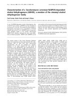
Figure 1 HRCT features of interstitial pulmonary fibrosis. A. Normal aspect of the lung. B. Mild. C. Moderate. D. Severe. A’. US examination
of healthy interlobular septa at lung surface level. Note as the pleura is a linear and regular hyperechoic band (arrow). B’-D’. US examinations
showing different scores of fibrotic pulmonary involvement: B’. Mild. C’. Moderate. D’. Severe.
Gutierrez et al. Arthritis Research & Therapy 2011, 13:R134
/>Page 4 of 7
Currently, chest HRCT is considered the “ gold-stan-
dard” for the diagnosis, disease activity and therapy
monitoring of IPF. Its value is re markable since it has
been demonstrated also to be able to detect both early
pulmonary changes and subclinical lung involvement
[21,24].
Although chest US is used to assess different lung con-
ditions, such as pulmonary interstitial edema or conges-
tion, heart and respiratory failure, atelectasis, pleural
effusions, and to guide interventional chest procedures,
such as thoracentesis or pleural lesion biopsy [2-12], its
potential role in the assessment of IPF has been recently
proposed in patients with systemic sclerosis [13,14]. The
results of these studies are encouraging since they
demonstrate a good correlation with HRCT as a concur-
rent “gold standard”. This opens up an interesting win-
dow of research focused on the US B-lines as surrogate
biomarkers of pulmonary changes in patients with CTD.
US offers particular character istics for the chest assess-
ment. From the practical viewpoint: it is a bedside proce-
dure widely available, inexpensive, readily and largely
accepted by the patient. From the technical viewpoint:
first, the surface of the lung can be easily studied by US,
so the B-lines “ artifacts” are quickly detected; second,
although small surfa ce probes with frequencies range
between 3 to 3.5 MHz were quite suitable for this specific
assessment, transducers with large surface and frequen-
cies between 5 to 7.5 MHz can be equally valuable, as
recently demonstrated by Delle Sedie et al.[25].Finally,
portable machines even without Doppler power can be
sufficient for a complete and detailed lung assessment.
In spite of these innovative data, the current US scor-
ing systems proposed to assess the B-lines are extensive,
including the study of 50 or more LIS, which is time-
consuming in daily clinical practice and difficult to
make the comparison of multi-center study results
[13-15,17]. Additionally,theyhavenottakeninto
account a semi-quantitative assessment which can facili-
tate the interpretation of the collected data. Thus, we
decided to test a simplified US semi-quantitative scoring
for assessing the B-lines in patients with CTD.
The simplified US B-li nes assessment is composed of
14 LIS, chosen on the basis of the major prevalence of
US B-lines detected during the comprehensive assess-
ment, their easy accessibility and their covering of main
pulmonary segments involved with IPF.
Our results showed a significant correlation with both
comprehensive US B-lines assessment and HRCT find-
ings. To the best of our knowledge this is the first study
providing evidence i n favor of the utility of this novel,
simplified US B-lines assessment. Results of inter and
intra-observer reliability were also highly significant.
The mean time spent in performing a simplified US B-
lines examination for each patient was much less in
respect to comprehensive assessment (8.3 minutes versus
23.3 minutes respectively). To our knowledge this remains
a contr oversial point since some aut hors previously indi-
cated that a comprehensive US B-li nes assessment could
be performed in less than 10 minutes [13,14]. Probably
this is true for patients with mild IPF, which is character-
ized by little quota of US B-lin es. In fact, most patients
included in these studies did not have a high Warrick
score . In our study , we included 19 patients (55.8%) with
severe IPF characterized by a high Warrick score.
Although the count of B-lines was more difficult in this
group, since it required both more attention and more
time, we believe that the inclusion of patients with whole
ranges of degrees of IPF may give more accurate informa-
tion about the reproducibility as well as feasib ility for
patient follow-up. Our study takes into account patients
with different CTD. In order to avoid facts that can nega-
tively influence the study, the sonographic features should
be interpreted in the light that they not provide results in
a disease driven manner but in an anatomic driven way.
The main limitation of our study is the low number of
enrolled patients, which does not permit an accurate
evaluation in terms of sensitivity and specificity which
could more strongly support these data.
Table 2 Inter-and intra-observer agreement data for simplified US B-lines assessment
Inter-observer Intra-observer
Anatomical lines weighted kappa values for the
semiquantitative system
weighted kappa values for the
semiquantitative system
2
nd
para-sternal LIS 0.885 0.864
4
th
mid-clavear LIS 0.836 0.881
4
th
anterior axillary LIS 0.863 0.868
4
th
mid-axillary LIS 0.812 0.845
8
th
paravertebral LIS 0.769 0.894
8
th
sub-scapular LIS 0.828 0.883
8
th
posterior axilary LIS 0.864 0.862
Agreement between both sonographers on both the comprehensive simplified US assessment.
Gutierrez et al. Arthritis Research & Therapy 2011, 13:R134
/>Page 5 of 7
HRCT remains the gold-standard used to assess IPF,
since it is the only imaging method that gives information
about the whole lung, and is not limited to the subpleural
interstitial lobular septa. Despite this, we believe that US
can be used as an adjunct method in the assessment of
monitoring of lung disease evolution. Additional advan-
tages of US consist of its low cost, the fact that it can also
beperformedatthebedsideandthatitisanon-ionizing
technique. This last aspect is fundamental, especially in
patients who need serial examinations for monitoring dis-
ease progression. Besides, it can pla y a relevant role for
screening purposes aimed towards the early identification
of patients that require a chest HRCT. Nevertheless, addi-
tional investigations studying a larger series of cohorts,
including sensitivity and specificity and a stratification of
Warrick score into fibrosis and alveolitis to demonstrate
which correlates better with HRCT findings, may be useful
to more strongly support these observations. In particular,
a focus aimed at deter mining sensitivity to change during
the progression of IPF could provide precious information
about the responsiveness of the simplified US assessment.
Conclusions
Theresultsofthepresentstudyprovideanewworking
hypothesis that a simplified US B-lines assessment may
be an additional, useful imaging method in the evalua-
tion of IPF in CTD patients.
Abbreviations
CTD: connective tissue disorders; DLco: diffusing capacity for carbon
monoxide; HRCT: high-resolution computed tomography; IPF: interstitial
pulmonary fibrosis; LIS: lung intercostal spaces; SD: standard deviation; US:
ultrasound
Acknowledgements
Written consent to publish was obtained from the patients.
Author details
1
Clinica Reumatologica, Via dei Colli 52, 60035, Università Politecnica delle
Marche, Jesi,Ancona, Italy.
2
S.O.D Radiologia Clinica, Dipartimento di Scienze
Radiologiche, Via Conca 1, PC 60126 Università Politecnica delle Marche,
Ancona, Italy.
3
Instituto Nacional de Rehabilitacion, Av. México-Xochimilco
289, Arenal de Guadalupe, Tlalpan 14389, Mexico City, Mexico.
Authors’ contributions
MG participated in the study development, recruitment of patients,
performed the ultrasound examinations (sonographer 1), prepared the
sonographic images, conducted data evaluation and prepared the
manuscript. FS participated in the statistical analysis and data evaluation and
manuscript preparation. MC performed the HRCT exams, prepared the HRCT
images, conducted data evaluation and prepared the manuscript. MT
performed the ultrasound examinations (sonographer 2) and gave
substantial input to data evaluation and manuscript preparation. CP gave
substantial input to the data evaluation and manuscript preparation. CB
participated actively in the recruitment of patients and manuscript
preparation. EF participated in the study development and gave substantial
input to the data evaluation and manuscript preparation. WG participated in
the study development and gave substantial input to the data evaluation
and manuscript preparation. All authors read and approved the final version
of manuscript.
Competing interests
The authors declare that they have no competing interests.
Received: 7 March 2011 Revised: 12 July 2011
Accepted: 18 August 2011 Published: 18 August 2011
References
1. Shahin AA: Pulmonary involvement in systemic sclerosis. Treat Respir Med
2006, 5:429-436.
2. Manganelli P, Salaffi F, Pesci A: Clinical and subclinical alveolitis in
connective tissue diseases as assessed by bronch- oalveolar lavage.
Semin Arthritis Rheum 1997, 26:740-754.
3. Soldati G, Copetti R, Sher S: Sonographic interstitial syndrome: the sound
of lung water. J Ultrasound Med 2009, 28:163-174.
4. Soldati G: Sonographic findings in pulmonary diseases. Radiol Med 2006,
111:507-515.
5. Lichtenstein DA, Mezière GA: Relevance of lung ultrasound in the
diagnosis of acute respiratory failure: the BLUE protocol. Chest 2008,
134:117-125.
6. Lichtenstein D, Hulot JS, Rabiller A, Tostivint I, Mezière G: Feasibility and
safety of ultrasound-aided thoracentesis in mechanically ventilated
patients. Intensive Care Med 1999, 25:955-958.
7. Lichtenstein D, Mezière G, Biderman P, Gepner A: The comet-tail artifact:
an ultrasound sign ruling out pneumothorax. Intensive Care Med 1999,
25:383-388.
8. Frassi F, Gargani L, Gligorova S, Ciampi Q, Mottola G, Picano E: Clinical and
echocardiographic determinants of ultrasound lung comets. Eur J
Echocardiogr 2007, 8:474-449.
9. Agricola E, Bove T, Oppizzi M, Marino G, Zangrillo A, Margonato A, Picano E:
“Ultrasound comet-tail images": a marker of pulmonary edema: a
comparative study with wedge pressure and extravascular lung water.
Chest 2005, 127:1690-1695.
10. Picano E, Gargani L, Gheorghiade M: Why, when, and how to assess
pulmonary congestion in heart failure: pathophysiological, clinical, and
methodological implications. Heart Fail Rev 2010, 15:63-72.
11. Sperandeo M, Varriale A, Sperandeo G, Filabozzi P, Piattelli ML, Carnevale V,
Decuzzi M, Vendemiale G: Transthoracic ultrasound in the evaluation of
pulmonary fibrosis: our experience. Ultrasound Med Biol 2009, 35:723-739.
12. Copetti R, Soldati G, Copetti P: Chest sonography: a useful tool to
differentiate acute cardiogenic pulmonary edema from acute respiratory
distress syndrome. Cardiovasc Ultrasound 2008, 6:16.
13. Gargani L, Doveri M, D’Errico L, Frassi F, Bazzichi ML, Delle Sedie A,
Scali MC, Monti S, Mondillo S, Bombardieri S, Caramella D, Picano E:
Ultrasound lung comets in systemic sclerosis: a chest sonography
hallmark of pulmonary interstitial fibrosis. Rheumatology 2009,
48:1382-1387.
14. Doveri M, Frassi F, Consensi A, Vesprini E, Gargani L, Tafuri M, Picano E,
Della Rossa A, Delle Sedie A, d’Ascanio A, Giacomelli C, Bazzichi L,
Bombardieri S: Ultrasound lung comets: new echographic sign of lung
interstitial fibrosis in systemic sclerosis. Reumatismo 2008, 60
:180-184.
15.
Jambrik Z, Monti S, Coppola V, Agricola E, Mottola G, Miniati M, Picano E:
Usefulness of ultrasound lung comets as a nonradiologic sign of
extravascular lung water. Am J Cardiol 2004, 93:1265-1270.
16. Warrick JH, Bhalla M, Schabel SI, Siver RM: High resolution computed
tomography in early scleroderma lung disease. J Rheumatol 1991,
18:1520-1528.
17. Lichtenstein D, Mézière G, Biderman P, Gepner A, Barré O: The comet-tail
artifact. An ultrasound sign of alveolar-interstitial syndrome. Am J Respir
Crit Care Med 1997, 156:1640-1646.
18. Warnecke K, Galanski M, Peters E, Hansen J: Pneumothorax: evaluation by
ultrasound. Preliminary results. J Thorac Imaging 1987, 2:76-78.
19. Landis JR, Koch GG: The measurement of observer agreement for
categorical data. Biometrics 1977, 33:159-174.
20. Strickland B, Strickland NH: The value of high definition, narrow section
computed tomography in fibrosing alveolitis. Clin Radiol 1988,
39:589-594.
21. Salaffi F, Manganelli P, Carotti M, Baldelli S: The differing patterns of
subclinical pulmonary involvement in connective tissue diseases as
shown by application of factor analysis. Clin Rheumatol 2000, 19:35-41.
Gutierrez et al. Arthritis Research & Therapy 2011, 13:R134
/>Page 6 of 7
22. Wells AU, Rubens MB, du Bois RM: Functional impairment in fibrosing
alveolitis: relationship to reversible disease on thin section computed
tomography. Eur Respir J 1997, 10:280-285.
23. Latsi PI, Wells AU: Evaluation and management of alveolitis and
interstitial lung disease in scleroderma. Curr Opin Rheumatol 2003,
15:748-755.
24. Salaffi F, Carotti M, Baldelli S, Bichi Secchi E, Manganelli P, Subiaco S,
Salvolini L: Subclinical interstitial lung involvement in rheumatic diseases.
Correlation of high resolution computerized tomography and functional
and cytologic findings. Radiol Med 1999, 97:33-41.
25. Delle Sedie A, Doveri M, Frassi F, Gargani L, D’Errico G, Pepe P, Bazzichi L,
Riente L, Caramella D, Bombardieri S: Ultrasound lung comets in systemic
sclerosis: a useful tool to detect lung interstitial fibrosis. Clin Exp
Rheumatol 2010, 28:S54.
doi:10.1186/ar3446
Cite this article as: Gutierrez et al.: Utility of a simplified ultrasound
assessment to assess interstitial pulmonary fibrosis in connective tissue
disorders - preliminary results. Arthritis Research & Therapy 2011 13:R134.
Submit your next manuscript to BioMed Central
and take full advantage of:
• Convenient online submission
• Thorough peer review
• No space constraints or color figure charges
• Immediate publication on acceptance
• Inclusion in PubMed, CAS, Scopus and Google Scholar
• Research which is freely available for redistribution
Submit your manuscript at
www.biomedcentral.com/submit
Gutierrez et al. Arthritis Research & Therapy 2011, 13:R134
/>Page 7 of 7