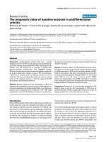
Báo cáo y học: " Phosphoinositide 3-kinase: a critical signalling event in pulmonary cells" doc
Bạn đang xem bản rút gọn của tài liệu. Xem và tải ngay bản đầy đủ của tài liệu tại đây (185.13 KB, 6 trang )
Review
Phosphoinositide 3-kinase: a critical signalling event in
pulmonary cells
Alison M Condliffe, Karen A Cadwallader, Trevor R Walker*, Robert C Rintoul*,
Andrew S Cowburn and Edwin R Chilvers
University of Cambridge School of Clinical Medicine, Addenbrooke’s and Papworth Hospitals,
Cambridge, and *University of Edinburgh Medical School, Edinburgh, UK
Abstract
Phosphoinositide 3-kinases (PI-3Ks) are enzymes that generate lipid second messenger
molecules, resulting in the activation of multiple intracellular signalling cascades. These
events regulate a broad array of cellular responses including survival, activation,
differentiation and proliferation and are now recognised to have a key role in a number of
physiological and pathophysiological processes in the lung. PI-3Ks contribute to the
pathogenesis of asthma by influencing the proliferation of airways smooth muscle and the
recruitment of eosinophils, and affect the balance between the harmful and protective
responses in pulmonary inflammation and infection by the modulation of granulocyte
recruitment, activation and apoptosis. In addition they also seem to exert a critical influence
on the malignant phenotype of small cell lung cancer. PI-3K isoforms and their downstream
targets thus provide novel therapeutic targets for intervention in a broad spectrum of
respiratory diseases.
Keywords: airways smooth muscle, lung, phosphatidylinositol 3,4,5-trisphosphate, phosphoinositide 3-kinase,
small cell lung cancer
Received: 21 April 2000
Revisions requested: 18 May 2000
Revisions received: 23 May 2000
Accepted: 23 May 2000
Published: 8 June 2000
Respir Res 2000, 1:24–29
The electronic version of this article can be found online at
/>© Current Science Ltd (Print ISSN 1465-9921; Online ISSN 1465-993X)
ARDS = acute respiratory distress syndrome; ASM = airways smooth muscle; ERK = extracellular signal-regulated protein kinase; PDGF = platelet-
derived growth factor; PDK1 = phosphoinositide-dependent kinase-1; PI-3K = phosphoinositide 3-kinase; PKB/AKT = protein kinase B;
PtdIns(3,4,5)P
3
= phosphatidylinositol 3,4,5-trisphosphate; SCLC = small cell lung cancer.
/>Introduction
Although characterised only in the late 1980s, a vast litera-
ture now exists detailing the critical roles of the ubiquitous
phosphoinositide 3-kinase (PI-3K) enzyme family in mitogen-
esis, cell survival, differentiation and activation, cytoskeletal
remodelling and vesicular trafficking. PI-3Ks are lipid
kinases — enzymes that phosphorylate membrane-associ-
ated lipids of the phosphoinositide family — and the resulting
3-phosphorylated lipids recruit and activate downstream
targets to initiate a novel set of signalling cascades, culmi-
nating in the varied cellular responses listed above (see
Figure 1). Although great progress has been made in eluci-
dating the structure and mechanism of action of the PI-3Ks
themselves, the identity and function of the downstream
targets and their interactions with other signalling cascades
within the cell are only just being unravelled.
Three classes of PI-3K are recognised on the basis of
their structure, substrate specificity and regulation. Class I
PI-3Ks are heterodimers comprising a catalytic (p110)
and a regulatory (p50, p55, p85 or p101) subunit; in the
resting unstimulated cell they are predominantly cytosolic
/>commentary
review
reports primary research
and require activation (usually by a mechanism driven by
cell-surface receptors) to display significant activity. These
enzymes preferentially phosphorylate the constitutive
plasma membrane phospholipid phosphatidylinositol 4,5-
bisphosphate to generate the critical second messenger
phosphatidylinositol 3,4,5-trisphosphate [PtdIns(3,4,5)P
3
].
PtdIns(3,4,5)P
3
is metabolised by enzymes called phos-
phatases to phosphatidylinositol 3,4-bisphosphate, which
itself can also act as a second messenger, and thence to
phosphatidylinositol 3-phosphate. Two subfamilies of
Class I PI-3K have been distinguished. Class IA com-
prises either an α, β or δ p110 catalytic subunit plus one
of a family of regulatory subunits (p85α, p85β, p55γ and
their splice variants), and are sensitive to activation by
tyrosine kinase-linked receptor transduction systems
(such as those initiated by the binding of growth factors to
their receptors). The only Class IB PI-3K so far identified
consists of a p110γ catalytic subunit and a unique p101
regulatory subunit. This enzyme is activated by βγ subunits
derived from activated G-protein-coupled receptors (eg
chemokine receptors) and, together with the Class 1A
p110δ, seems to be expressed only in haematopoietic
cells. All Class I PI-3K catalytic subunits contain a PI-
kinase domain, a protein kinase domain and a Ras-binding
domain. Recent crystallographic studies [1
•
] have shown
that p110γ has a central helical spine, with the catalytic
domain positioned to interact with phospholipid mem-
branes and the Ras-binding domain placed adjacent to
the catalytic domain, where it most probably drives the
allosteric activation of the enzyme. The monomeric Class II
PI-3Ks 3-phosphorylate phosphatidylinositol 4-phosphate
and PtdIns, but their role in mammalian systems is unclear.
Class III PI-3Ks use only PtdIns as a substrate; they do not
seem to be regulated acutely by cell-surface receptors
and have been implicated in cellular ‘housekeeping’ func-
tions, particularly protein and vesicular trafficking.
The activation of Class I PI-3Ks results in the generation of
membrane-associated PtdIns(3,4,5)P
3
, levels of which
increase substantially (up to 50-fold) in appropriately stim-
ulated cells. Proteins containing pleckstrin homology
domains bind PtdIns(3,4,5)P
3
with high affinity and thus
are recruited to the plasma membrane, thereby bringing
them into juxtaposition with their substrates and in some
cases with upstream activating enzymes. This recruitment
of pleckstrin homology domain-containing proteins in
response to PtdIns(3,4,5)P
3
generation can be imaged
directly in live cells by using fluorescently tagged target
proteins. The binding of such proteins to PtdIns(3,4,5)P
3
might result in direct allosteric activation, although defini-
tive proof for this is currently lacking. Examples of proteins
activated by PtdIns(3,4,5)P
3
include phosphoinositide-
dependent kinase-1 (PDK1), protein kinase B (PKB/AKT,
implicated in cell survival), p70
S6K
(involved in mitogenesis),
members of the protein kinase C family, phospholipase
Cγ, and several small molecular signalling intermediates
including Rac, Vav, Tiam-1 and centaurin-α. Techniques
used to identify these PtdIns(3,4,5)P
3
-binding proteins
and elucidate their functional roles include the use of
selective PI-3K inhibitors (wortmannin and LY294002),
development of constitutively active and dominant-nega-
tive forms of PI-3K and its targets, enzyme activity assays
and the use of fluorescently labelled proteins. Although
these and other methods have been applied principally in
immortalised cell lines, more recent studies have used
primary cell cultures. As exampled below, these investiga-
tions are now providing fascinating insights into the roles
of the PI-3Ks and their accompanying signalling cascades
in several tissues, including the lung.
PI3 kinase in proliferative responses
Airways smooth muscle
Although reversible airway narrowing leading to wheeze,
cough and shortness of breath is a hallmark of asthma,
patients with longstanding and severe disease can develop
fixed airways obstruction that is associated with structural
changes within the airway wall. The most prominent feature
of the remodelled airway is an increase in airways smooth
muscle (ASM). Heard and Hossain [2] demonstrated a
threefold increase in both the cross-sectional area and
number of smooth-muscle cells found within the bronchial
wall of patients with fatal asthma in comparison with those
dying from non-respiratory conditions. Subsequent mathe-
matical modelling suggests that this characteristic increase
in smooth-muscle bulk is the major cause of narrowing of
airways in such patients. Furthermore, excessive ASM DNA
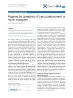
Figure 1
The PI-3K signalling network. Binding of ligand to receptors linked to
G protein or tyrosine kinase activates PI-3K-γ and PI-3K-α, PI-3K-β or
PI-3K-δ respectively. The resultant accumulation of phosphatidylinositol
3,4,5-trisphosphate [PtdIns(3,4,5)P
3
] activates downstream signalling
cascades leading to adhesion, proliferation, survival and activation
responses. Putative pathways or those demonstrated in only a limited
number of cell types are depicted by broken arrows.
Integrin
activation and
adhesion
α
PtdIns(3,4,5)
P
3
PI3K-γ
PDK
PKB
p70
S6K
GTP Rac NADPH
oxidase
Cytoskeleton
Secretion
Protection from
apoptosis
Other functions
Cell growth/division, life/death decisions, activation responses
G protein coupled receptor Tyrosine kinase-linked receptor
PI-3K- , - or -αβ δ
βγ
βγ
Respiratory Research Vol 1 No 1 Condliffe et al
synthesis has been demonstrated in at least two animal
models of airways disease, including that associated with
antigen challenge [3,4].
Although it has long been recognised that the signalling
pathways based on extracellular signal-related kinase
(ERK) and protein kinase C are involved in the mitogenic
response of ASM, the importance of PI-3K has only
recently been demonstrated. Scott et al [5] first showed
that PI-3K activity was proportional to the mitogenic
response in bovine ASM cells in culture and that the inhi-
bition of PI-3K by wortmannin substantially (more than
90%) decreased DNA synthesis. They also provided data
implicating p70
S6K
as the probable downstream mediator
of these effects. p70
S6K
is known to be activated by PI-3K
(via PDK1 and possibly PKB/AKT and/or RAC) and is now
known to be essential for the progression of cells from G
1
to S phase in the cell cycle. Further studies [6] demon-
strated a rapid activation of PI-3K and accumulation of
PtdIns(3,4,5)P
3
after stimulation of bovine ASM by throm-
bin and platelet-derived growth factor (PDGF), and
showed that the magnitude of these effects was closely
correlated with the mitogenic potential of these two
growth factors. Krymskaya et al [7
•
] confirmed the require-
ment for PI-3K activity in human ASM cell mitogenesis,
and again implicated p70
S6K
as an important mediator in
this response. Rac 1 has also now been shown to be
important as a downstream mediator of the PI-3K mito-
genic effect [8], acting via the induction of cyclin D, which
is required for cell cycle progression.
Small cell lung cancer
PI-3K activity has been shown to be critical for the inte-
grin-mediated invasive behaviour of breast and colon car-
cinoma cell lines [9], and the proto-oncogene PKB/AKT
[recruited by PtdIns(3,4,5)P
3
to the plasma membrane
and phosphorylated by the PI-3K-dependent PDK1] has
been demonstrated to be overexpressed in ovarian, breast
and pancreatic cancer. Small cell lung cancer (SCLC) is
the most aggressive and invasive form of lung cancer, with
a highly metastatic phenotype and a 5-year survival of only
3–8%. Interest in the role of PI-3K in the malignant poten-
tial of this disease was stimulated by the observation [10]
that p70
S6K
is constitutively phosphorylated and activated
in SCLC cells, and that rapamycin, which inhibits the acti-
vation of p70
S6K
, blocks SCLC proliferation. These obser-
vations were extended by Moore et al [11
••
], who
demonstrated a high constitutive activity of PI-3K,
PKB/AKT and p70
S6K
in SCLC cell lines and showed that
the proliferation of SCLC cells in liquid culture was inhib-
ited by the PI-3K inhibitor LY294002. This inhibition
resulted from a combination of decreased mitogenesis
and enhanced apoptosis (see below). PI-3K inhibition also
decreased SCLC cell colony formation in semi-solid
media. Thus the high constitutive activity of PI-3K in these
cells seems to promote growth and also anchorage-
independence, contributing to the highly aggressive
nature of this tumour. These observations have not yet
been extended to other human lung cancer cell types;
further developments are awaited.
PI3 kinase in activation responses
Neutrophils
Although not resident pulmonary cells, substantial
numbers of neutrophils are recruited to the lungs in many
respiratory disease states and have a critical role in the
pathogenesis of the acute respiratory distress syndrome
(ARDS), pulmonary fibrosis, bronchiectasis and fatal
asthma. Neutrophils cause tissue damage by their capac-
ity to release toxic oxygen radicals (generated by the
NADPH oxidase complex), the exocytosis of granules con-
taining highly histotoxic compounds such as elastase and
collagenases, and the elaboration and release of addi-
tional pro-inflammatory cytokines. PI-3Ks have been
shown to be key regulators of both neutrophil recruitment
and activation. In mice lacking the catalytic subunit of the
myeloid restricted PI-3K-γ, neutrophil migration to the
inflamed peritoneum was severely compromised [12
••
,
13
••
] and, although not examined directly, a similar defect
in granulocyte recruitment to the lungs is likely. The accu-
mulation of PtdIns(3,4,5)P
3
also seems to be correlated
precisely with respiratory burst activity, in that neutrophil
priming by agents such as tumour necrosis factor-α
markedly enhanced both the size and the duration of the
release of superoxide anions and the accumulation of
PtdIns(3,4,5)P
3
[14]. In these cells PI-3K inhibitors abolish
the production of oxygen radicals induced by physiological
agonists; neutrophils from PI-3K-γ knockout mice exhibit a
diminished respiratory burst, with residual activity most
probably attributable to the action of remaining Class IA
PI-3K. The signalling intermediates linking PtdIns(3,4,5)P
3
to activation of the oxidase are uncertain but most proba-
bly include the small GTPase Rac 2, which is both highly
expressed in neutrophils and an essential component of
the NADPH oxidase complex. The role of PI-3K in granule
exocytosis is less clearly delineated as high concentra-
tions of wortmannin only partialy inhibit this process, indi-
cating that inputs from other signalling pathways might
impinge on this response.
Eosinophils
Like neutrophils, eosinophils are non-resident pulmonary
cells that accumulate in the bronchial tree and lung
parenchyma in a number of disease states, including
asthma and eosinophilic pneumonia. Toxic eosinophil-
derived mediators such as eosinophil cationic protein,
major basic protein and oxygen radicals (again products of
the NADPH oxidase) can damage the airway epithelium
and are thought to contribute significantly to airway hyper-
responsiveness. In non-allergic subjects, eosinophils are
scarce in peripheral blood and have therefore proved
more difficult than neutrophils to study. Despite this, IL-5-
stimulated eosinophil release from bone marrow has been
shown to be inhibited by both wortmannin and LY294002
[15]; the migration of eosinophils to a number of chemoat-
tractants also seems to be sensitive to wortmannin [16].
The role of PI-3Ks in eosinophil degranulation is not
known, but these enzymes are required for activation of
the eosinophil NADPH oxidase complex [17].
Alveolar macrophages
Alveolar macrophages undertake a number of key host
defence functions within the lung. These include the
phagocytosis of inhaled particles and respiratory pathogens,
antigen presentation, and the generation of inflammatory
cytokines. Additionally, they might be important in the
resolution of acute inflammation by the ingestion of apop-
totic neutrophils. So far, although few studies have
addressed the role of PI-3Ks in the alveolar macrophage,
such data are available for monocyte-derived macro-
phages, macrophage cell lines and murine peritoneal
macrophages. If we extrapolate these results to alveolar
macrophages, it seems highly likely that PI-3Ks will again
be shown to have a critical role in the response profile of
these cells. Hence, murine PI-3K-γ-null macrophages
show decreased migration towards a variety of chemo-
tactic agents, and greatly diminished recruitment to the
inflamed peritoneum [13
••
]. The induction of cytokine gene
expression in monocytes stimulated by formylated peptide
has also been shown to be sensitive to PI-3K inhibition
[18]. Most importantly, the consequences of excessive PI-
3K activation have also been explored; mice deficient in
SHIP (SH2-containing inositol-5-phosphatase), an enzyme
that hydrolyses PtdIns(3,4,5)P
3
, suffer from lethal infiltra-
tion of the lungs by myeloid cells, principally macrophages
[19]. A remarkably similar phenotype is seen in mice with
a deletion of the tyrosine phosphatase SHP-1 (Src homol-
ogy 2 domain phosphatase-1); macrophages from these
mice display a 10–15-fold increase in the 3-phosphory-
lated products of PI-3K, with enhanced integrin-depen-
dent adhesive properties [20
•
]. Thus, as with neutrophils
and eosinophils, it seems that one or more of the PI-3Ks is
required for macrophage recruitment to the lung and for at
least a subset of activation responses.
Although comparatively little is known about the function
of the PI-3Ks in other pulmonary cells, several reports
have emerged and indicate the global importance of this
signalling pathway in other settings in the lung. For
example, Liu et al [21] have demonstrated that PI-3K is a
downstream mediator of PDGF-stimulated glycosamino-
glycan synthesis in rat foetal lung fibroblasts, suggesting a
role in the maintenance of the lung extracellular matrix.
Similarly, PI-3K has been reported to mediate lung epithe-
lial cell differentiation and surfactant protein expression
fibroblast induced by growth factor-2 [22], although sub-
sequent reports have suggested that PI-3K inhibits surfac-
tant secretion from type II alveolar cells [23]. Future work
will doubtless help to clarify the role of PI-3Ks in the differ-
entiation and function of pulmonary epithelial cells.
PI-3K in cell survival
In addition to their central role in cell proliferation and acti-
vation, Class I PI-3Ks have also been implicated as having
a key role in inhibiting apoptotic cell death. PKB/AKT, a
downstream effector of PI-3K, is believed to promote cell
survival by the phosphorylation and inactivation of both
caspase-9 (a central regulator of apoptosis) and the pro-
apoptotic factor BAD. Granulocyte apoptosis is now
thought to be important in the resolution of pulmonary
inflammation; in recent months several papers have
emerged that implicate PI-3K as a mediator of cell survival
in neutrophils [24
•
] and monocytes [25], but not
eosinophils [26]. Finally, inhibition of PI-3K activity in
SCLC cell lines results in enhanced apoptosis [11
••
], sug-
gesting that the high basal PI-3K activity observed in these
cells might contribute to the malignant phenotype by
inhibiting apoptosis.
Conclusion
Although the role played by Class I PI-3Ks in the embry-
ological development and everyday ‘housekeeping’ func-
tions of the normal lung remains unclear, it is evident that
these enzymes are of central importance in a broad spec-
trum of respiratory diseases (see Figure 2). The impor-
tance of this signalling pathway in ASM mitogenesis and
eosinophil recruitment and activation suggests that PI-3Ks
might have a key role in the pathogenesis of asthma. PI-
3Ks are also critical for the recruitment, activation and sur-
vival of neutrophils and thereby influence a wide range of
/>commentary
review
reports primary research
Figure 2
PI-3K in respiratory disease. Within the airway, activation of PI-3K is
thought to contribute to the proliferation of smooth muscle and the
accumulation of eosinophil characteristic of asthma, and to the
mitogenesis and prolonged survival of small cell lung cancer cells.
PI-3K-dependent neutrophil extravasation and activation have been
implicated in the pathogenesis of multiple respiratory diseases
including ARDS, pulmonary fibrosis, pulmonary vasculitides and
bronchiectasis.
Eosinophil Neutrophil
Airway Alveolus
Proliferating
airway smooth
muscle
Small cell lung
cancer
inflammatory and infective conditions within the lung,
including ARDS, pulmonary fibrosis and bronchiectasis.
Additionally, work on SCLC cell lines points to PI-3Ks as
being constitutively active and contributing to the malig-
nant phenotype of this tumour, perhaps via the activation
of the proto-oncogene PKB/AKT or p70
S6K
. The develop-
ment of inhibitors of specific PI-3K isoforms (particularly of
the myeloid-restricted PI-3K-δ and PI-3K-γ) and of down-
stream signalling targets might lead to novel therapeutic
strategies for a variety of respiratory diseases.
Acknowledgements
The authors’ work is supported by the Wellcome Trust, MRC, National
Asthma Campaign and British Lung Foundation. AC is a Wellcome
Advanced Fellow.
References
Articles of particular interest have been highlighted as:
•
of special interest
••
of outstanding interest
1. Walker EH, Perisic O, Stephens L, Williams RL: Structural insights
•
into phosphoinositide 3-kinase catalysis and signalling. Nature
1999, 402:313–320.
Contains the first account of the detailed three-dimensional structure of a
PI-3K catalytic subunit, correlating domain organisation and function.
2. Heard BE, Hossain S: Hyperplasia of bronchial smooth muscle in
asthma. J Pathol 1973, 110:319–332.
3. Hershenson MB, Kelleher MD, Naureckas ET, Abe MK, Rubinstein VJ,
Zimmermann A, Bendele AM, McNulty JA, Panettieri RA, Solway J:
Hyperoxia increases airway cell S-phase traversal in immature
rats in vivo. Am J Respir Cell Mol Biol 1994, 11:296–303.
4. Panettieri RA, Murray RK, Eszterhas AJ, Bilgen G, Martin JG:
Repeated allergen inhalations induce DNA synthesis in airway
smooth muscle and epithelial cell in vivo. Am J Physiol 1998, 274:
L417–L424.
5. Scott PH, Belham CM, Al-Hafidh J, Chilvers ER, Peacock AJ: A
regulatory role for cAMP in phosphatidylinositol 3-kinase/p70
ribosomal S6 kinase-mediated DNA synthesis in platelet-derived-
growth-factor-stimulated bovine airway smooth-muscle cells.
Biochem J 1996, 318:965–971.
6. Walker TR, Moore SM, Lawson M F, Panettieri RA Jr, Chilvers ER:
Platelet-derived growth factor-BB and thrombin activate phospho-
inositide 3-kinase and protein kinase B; role in mediating airway
smooth muscle proliferation. Mol Pharmacol 1998, 54:1005–1015.
7. Krymskaya VP, Penn RB, Orsini MJ, Scott PH, Plevin RJ, Walker TR,
•
Eszterhas AJ, Amrani Y, Chilvers ER, Panettieri RA Jr: Phosphatidyl-
inositol 3-kinase mediates mitogen-induced human airway
smooth muscle cell proliferation. Am J Physiol 1999, 277:L65–L78.
Using wortmannin and LY294002, plus constitutively active and dominant-
negative PI-3K constructs, these authors demonstrate that PI-3K activity is
essential for mitogenesis in human ASM and implicate p70
S6K
as a down-
stream mediator of this response.
8. Page K, Li J, Hodge JA, Liu PT, Vanden Hoek TL, Becker LB, Pestell
RG, Rosner MR, Hershenson MB: Characterisation of a rac1 sig-
nalling pathway to cyclin D
1
expression in airway smooth muscle
cells. J Biol Chem 1999, 274:22065–22071.
9. Shaw LM, Rabinovitz I, Wang HH-F, Toker A, Mercurio M: Activation
of phosphoinositide 3-OH kinase by the
αα
6
ββ
4 integrin promotes
carcinoma invasion. Cell 1997, 91:949–960.
10. Seufferlein T, Rozengurt E: Rapamycin inhibits constitutive p70
S6K
phosphorylation, cell proliferation and colony formation in small
cell lung cancer cells. Cancer Res 1996, 56:3895–3897.
11. Moore SM, Rintoul RC, Walker TR, Chilvers ER, Haslett C, Sethi T:
••
The presence of a constitutively active phosphoinositide 3-kinase
in small cell lung cancer cells mediates anchorage-independent
proliferation via a protein kinase B and p70
S6K
-dependent
pathway. Cancer Res 1998, 58:5239–5247.
This study shows that each of five SCLC cell lines tested exhibited high
basal constitutive PI-3K activity, resulting in high basal PKB/AKT and
p70
S6K
activity. Additionally, the inhibition of PI-3K activity greatly dimin-
ished SCLC cell proliferation in liquid culture (by stimulating apoptosis and
promoting cell cycle delay in G
1
) and decreased colony formation in semi-
solid media.
12. Sasaki T, Irie-Sasaki J, Jones RG, Oliveira-dos-Santos AJ, Stanford
••
WL, Bolon B, Wakeham A, Itie A, Bouchard D, Kozieradzki I, Joza N,
Mak TW, Ohashi PS, Suzuki A, Penninger JM: Function of PI3K
γγ
in
thymocyte development, T cell activation and neutrophil migra-
tion. Science 2000, 287:1040–1046.
Using mice deficient in PI-3K-γ, these authors provide evidence that in neu-
trophils, this enzyme isoform is the sole source of PtdIns(3,4,5)P
3
generated
by G protein linked agonists, and that PI-3K-γ-null neutrophils exhibit severe
defects in respiratory burst activity and migration to inflamed sites. They also
show that PI-3K-γ regulates thymocyte development and T cell activation.
13. Hirsch E, Katanaev VL, Garlanda C, Azzolino O, Pirola L, Silengo L,
••
Sozzani S, Mantovani A, Altruda F, Wymann MP: Central role for G
protein coupled phosphoinositide 3-kinase in inflammation.
Science 2000, 287:1049–1053.
This group also found that neutrophils from PI-3K-γ-knockout mice dis-
played impaired respiratory burst activity and motility, and additionally
showed that macrophages from these animals displayed severely impaired
chemotaxis and migration to the inflamed peritoneum.
14. Condliffe AM, Hawkins PT, Stephens LR, Haslett C, Chilvers ER:
Priming of human neutrophil superoxide generation by tumour
necrosis factor is signalled by enhanced phosphatidylinositol
3,4,5-trisphosphate but not inositol 1,4,5-trisphosphate accumula-
tion. FEBS Lett 1998, 439:147–151.
15. Palframen RT, Collins, PD, Severs, NJ, Rothery S, Williams TJ, Rankin
SM: Mechanisms of acute eosinophil mobilization from bone
marrow by interleukin 5: the role of specific adhesion molecules and
phosphatidylinositol 3-kinase. J Exp Med 1998, 188:1621–1632.
16. Dunzendorfer S, Meirhofer C, Wiedermann CJ: Signaling in neu-
ropeptide-induced migration of human eosinophils. J Leukoc Biol
1998, 64:828–834.
17. Hofmann C, Dichmann S, Zimpfer U, Czech W, Herouy Y, Wagner E,
Norgauer J: Metabolism and function of 3-D-phosphorylated phos-
phoinositides in C5a-stimulated eosinophils. Biochem Biophys
Res Commun 2000, 269:816–821.
18. Pan ZK, Chen LY, Cochrane CG, Zuraw BL: fMet-leu-phe stimulates
proinflammatory cytokine gene expression in human peripheral
blood monocytes: the role of phosphatidylinositol 3-kinase.
J Immunol 2000, 164:404–411.
19. Helgason CD, Damen JE, Rosten P, Grewal R, Sorensen P, Chappel
SM, Borowski A, Jirik F, Krystal G, Humphries RK: Targeted disrup-
tion of SHIP leads to hemopoietic perturbations, lung pathology,
and a shortened life span. Genes Dev 1998, 12:1610–1620.
20. Roach TI, Slater SE, White LS, Zhang X, Majerus PW, Brown EJ,
•
Thomas ML: The protein tyrosine phosphatase SHP-1 regulates
integrin-mediated adhesion of macrophages. Curr Biol 1998, 8:
1035–1038.
This report demonstrates that macrophages from mice deficient in the tyro-
sine phosphatase SHP-1 have markedly increased 3-phosphorylated lipid
levels and display greatly enhanced integrin-mediated adhesive properties
that can be abolished by PI-3K inhibition.
21. Liu J, Fitzli D, Liu M, Tseu I, Caniggia I, Rotin D, Post M: PDGF-
induced glycosaminoglycan synthesis is mediated via phos-
phatidylinositol 3-kinase. Am J Physiol 1998, 274:L702–L713.
22. Matsui R, Brody JS, Yu Q: FGF-2 induces surfactant protein gene
expression in foetal rat lung epithelial cells through a MAPK-inde-
pendent pathway. Cell Signal 1999, 11:221–228.
Respiratory Research Vol 1 No 1 Condliffe et al
/>commentary
review
reports primary research
23. White MK, Stryer DS: Surfactant protein A regulates pulmonary
surfactant secretion via activation of phosphatidylinositol 3-kinase
in type II alveolar cells. Exp Cell Res 2000, 255:67–76.
24. Klein JB, Rane MJ, Scherzer JA: Granulocyte–macrophage colony-
•
stimulating factor delays neutrophil constitutive apoptosis
through phosphoinositide 3-kinase and extracellular signal-
related kinase pathways. J Immunol 2000, 164:4286–4291.
Using PI-3K and ERK inhibitors these authors demonstrate that interleukin-8
and granulocyte–macrophage colony-stimulating factor delay neutrophil
apoptosis by stimulating these pathways, and implicate the activation of
PKB/AKT and the phosphorylation of BAD as downstream mediators of the
PI-3K effect.
25. Kelley TW, Graham MM, Doseff AI, Pomerantz RW, Lau SM,
Ostrowski MC, Franke TF, Marsh CB: Macrophage colony-stimulat-
ing factor promotes cell survival through Akt/protein kinase B.
J Biol Chem 1999, 274:26393–26398.
26. Miike S, Nakao A Hiraguri M, Kurasawa K, Saito Y, Iwamoto I: Involve-
ment of JAK2, but not PI 3-kinase/Akt and MAP kinase pathways,
in anti-apoptotic signals of GM-CSF in human eosinophils.
J Leukoc Biol 1999, 65:700–706.
Authors’ affiliations: Alison M Condliffe, Karen A Cadwallader,
Andrew S Cowburn and Edwin R Chilvers (Respiratory Medicine Unit,
Department of Medicine, University of Cambridge School of Clinical
Medicine, Addenbrooke’s and Papworth Hospitals, Cambridge, UK),
and Trevor R Walker, Robert C Rintoul (Respiratory Medicine Unit,
Department of Medicine, University of Edinburgh Medical School,
Edinburgh, UK)
Correspondence: Edwin R Chilvers, Respiratory Medicine Unit,
Department of Medicine, Box 157, Level 5, Addenbrooke’s Hospital,
Hills Road, Cambridge CB2 2QQ, UK. Tel: +44 1223 762007;
fax: +44 1223 762007; e-mail: