Báo cáo y học: " From fruitflies to mammals: mechanisms of signalling via the Sonic hedgehog pathway in lung development" potx
Bạn đang xem bản rút gọn của tài liệu. Xem và tải ngay bản đầy đủ của tài liệu tại đây (482.08 KB, 6 trang )
Review
From fruitflies to mammals: mechanisms of signalling via the
Sonic hedgehog pathway in lung development
Minke van Tuyl* and Martin Post*
†
*Hospital for Sick Children Research Institute, Toronto, and
†
University of Toronto, Toronto, Canada
Abstract
The hedgehog signalling pathway has been implicated in many different processes in fly and
vertebrate development. It is now known that the hedgehog cascade is crucial for the
patterning of the early respiratory system. Hedgehog signalling in the lung involves Gli
transcription proteins, but their potential downstream target genes have yet to be identified.
Bmp4 and Fgf10 have been shown to regulate lung branching morphogenesis but seem not
to be targets of hedgehog signalling.
Keywords: Gli proteins, lung development, patched, Sonic hedgehog
Received: 17 May 2000
Revisions requested: 1 June 2000
Revisions received: 7 June 2000
Accepted: 7 June 2000
Published: 20 June 2000
Respir Res 2000, 1:30–35
The electronic version of this article can be found online at
/>© Current Science Ltd (Print ISSN 1465-9921; Online ISSN 1465-993X)
Bmp = bone morphogenetic protein; Ci = cubitus interruptus; Dhh = Desert hedgehog; Dpp = decapentaplegic; Fgf = fibroblast growth factor;
Hh = hedgehog; Ihh = Indian hedgehog; Ptc = patched; Shh = Sonic hedgehog; Smo = smoothened.
/>Introduction
During development, the mammalian lung establishes a
large diffusible interface with the circulation to facilitate res-
piratory gas exchange at term. In the human, the appear-
ance of a midventral groove in the single foregut tube just
posterior to the pharynx at around the fourth week of
human development is the first sign of lung formation. Next,
the laryngo-tracheal groove deepens and finally constricts,
thereby dividing the foregut tube into a ventral trachea and
a dorsal esophagus. At the same time, the tracheal rudi-
ment outgrowth elongates caudally and bifurcates to form
two bronchial lung buds. In the mouse, the lung starts as a
tracheal diverticulum in the ventral half of the primitive
foregut, just anterior to the developing stomach at 9–9.5
days of gestation. The two lung buds elongate in a poste-
rior–ventral direction. At the same time, starting at the
primary branching point the single foregut tube begins to
divide into two tubes, the dorsal esophagus and the ventral
trachea. Subsequently, the primitive buds of the human and
mouse lung rudiments continue to elongate into the
splanchnic mesenchyme; outgrowth of these buds pro-
duces the left and right primary bronchi. During the next
step, the primary bronchial buds divide asymmetrically,
giving rise to two left and three right secondary bronchi in
humans and one left and four right secondary bronchi in
the mouse. Each secondary bronchus is a stem bronchus,
which will be a lobar bronchus. These stem bronchi are
destined to branch and rebranch and, with the surrounding
pulmonary mesenchyme (which will provide the elastic
tissue, smooth muscles, cartilage, vascular system and
other connective tissues), will give origin to the definitive
pulmonary lobes that characterize adult lung organization.
/>commentary
review
reports primary research
Branching of the lung buds is controlled by epithelial–
mesenchymal tissue interactions. The mesenchymal com-
ponent defines the branching pattern of the epithelium.
However, the question is: What controls the sequence of
dorsal–ventral, lateral and left–right budding and branch-
ing in the lung? Candidates for the answer to this question
are morphogens and growth factors. Morphogens are
defined as molecules that diffuse away from their source
and give positional information to surrounding cells on the
basis of their local concentrations. A candidate mor-
phogen might be the secreted signalling molecule Sonic
hedgehog (Shh), a vertebrate homolog of hedgehog (Hh)
in Drosophila. In the fly, Hh signalling patterns the
segment, leg, wing, eye and brain [1,2]. Hh is a secreted
molecule that undergoes autocatalytic cleavage to give an
active N-terminal fragment, which is modified by the addi-
tion of a cholesterol moiety at its C terminus [3]. In con-
trast with the single Hh in flies, vertebrates have at least
three Hh homologs: Sonic (Shh), Desert (Dhh) and Indian
(Ihh). Shh is the most extensively studied of the vertebrate
Hh genes and has been shown to be responsible for pat-
terning many developing organs, including the lung
[4
••
,5
••
]. Dhh and Ihh have more restricted roles and are
primarily involved in the development of the germline and
the skeletal system, respectively. Genetic and biochemical
analyses in flies and vertebrates have led to a Hh sig-
nalling pathway that has been conserved from flies
through to humans (Fig. 1). Hh binds to patched (Ptc), a
transmembrane protein, which inhibits the activity of
smoothened (Smo), another transmembrane protein,
which has some resemblance to the large family of G
protein receptors (2). On Hh binding, the repression of
Smo by Ptc is released, leading to the activation of
cubitus interruptus (Ci). In the absence of Hh, full-length
155 kDa Ci, which forms a complex with several segment
polarity proteins (fused, costal-2 and suppressor of fused)
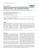
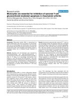
Figure 1
The Hh signalling pathway, based on what is known in flies. In the absence of Hh, Ptc inhibits signalling from Smo. Full-length Ci is cleaved after
forming a microtubule complex with several segment polarity genes (fused, costal-2 and suppressor of fused) and the 75 kDa Ci product act as a
transcriptional repressor protein. On the binding of Hh to Ptc, the inhibitory effect on Smo is released, leading to the dissociation of the
microtubule complex. Ci cleavage is prevented and Ci activates the transcription of target genes.
Respiratory Research Vol 1 No 1 van Tuyl and Post
at the microtubules, is cleaved to release a transcriptional
repressing 75 kDa N-terminal fragment. When cells bind
Hh, cleavage is prevented, the microtubule complex is dis-
sociated and the activation of target genes results [6]. At
present three vertebrate Ci genes, gli 1, 2 and 3, have
been identified but their exact role in the activation and
repression of Hh target genes remains to be elucidated.
Shh in lung development
In the developing mouse lung, shh expression is detected
in the tracheal diverticulum, the esophagus and later in the
trachea and lung endoderm [4
••
]. The gene shh is
expressed at low levels throughout the epithelium with
higher levels in the growing distal buds [7–9]. The total
amount of pulmonary shh mRNA expression declines
towards birth [8]. Shh-null mutant (shh
–/–
) mice exhibit
serious foregut defects [4
••
,5
••
]. The trachea and esopha-
gus do not separate into distinct tubes, and both
hypoplastic lung buds appear as single lobes. Both single
lobes have sac-like structures instead of an extensive
network of air sacs and when cultured for 5 days there is
no sign of epithelial branching or mesenchymal prolifera-
tion. Surprisingly, proximal–distal differentiation of lung
epithelium was normal in shh
–/–
mice [5
••
], indicating that
Shh is important for branching morphogenesis but not for
proximal–distal differentiation. In situ hybridization studies
showed that mesenchymal ptc, gli1 and gli3 expression
were downregulated in the shh
–/–
mice. Overexpression of
Shh, with the use of the SP-C enhancer/promoter,
resulted in the absence of functional alveoli and an
increase in interstitial tissue caused by an increased prolif-
eration of both epithelial and mesenchymal cells [8].
Transgenic pups die soon after birth, probably owing to
respiratory failure. Cell differentiation, as assessed by the
expression of SP-A, SP-B and SP-C and CC10 mRNA,
was normal in Shh-overexpressing mice lungs. In accord
with the decrease in the expression of pulmonary ptc in
the absence of Shh, ptc mRNA is increased in Shh-over-
expressing lungs. These results indicate a role for Shh sig-
nalling in pulmonary branching morphogenesis in mice. In
addition, there also seems to be a role for Shh in estab-
lishing asymmetry between left and right lungs [10,11]
and in the process of separating trachea and esophagus.
Ptc in lung development
Ptc is a multipass membrane protein that functions as a
Shh receptor. A clear function of Ptc is the regulation of
Shh target gene transcription. Ptc represses the transcrip-
tion of genes that are induced by Shh [12], including gli1
and ptc-1. This repression is released on Shh binding. Ptc-
1-null mutant mice die between embryonic days 9.0 and
10.5 with neural and cardiac defects [12], just at the time
that lung formation begins. In the lung, ptc is expressed at
high levels in the mesenchyme adjacent to the terminal
buds where shh is normally expressed [4
••
,8]. It is also
expressed at low levels in the distal epithelium. Ptc mRNA
levels, like shh, decline towards birth [8]. Ptc is upregu-
lated in lungs that overexpress Shh [8,13], whereas ptc is
downregulated in the lung mesenchyme of shh
–/–
mice,
indicating a positive feedback mechanism between shh
and ptc expression [4
••
,5
••
].
Gli in lung development
In mice, three zinc-finger transcription factors, Gli1, Gli2
and Gli3, have been implicated in the transduction of the
Shh signal. All three gli genes are expressed in lung meso-
derm rather than endoderm and all three are expressed at
higher levels in distal mesoderm than in proximal meso-
derm [13]. Expression levels of all three genes decline
when development proceeds. Overexpression of Shh in
the lung resulted in an upregulation of gli1 expression,
whereas gli2 and gli3 expression remained unaltered [13].
It is thought that, in developing limbs, Gli3 is a repressor
of the Shh signalling pathway [14]. Whether this also
holds for lung development is unclear: Gli3-deficient mice
are viable, but the lungs are smaller and the lobes are
changed in shape [13]. Gli3-deficient mice did not reveal
any difference in the expression of genes that are involved
in the Shh signalling pathway, such as shh, ptc, gli1 and
gli2. This suggests that Gli3 is not important for lung
development on its own, but it might have a function in
compensating for other Gli proteins. In contrast with the
mild phenotype in gli3
–/–
mutants, gli2-null mutants die at
birth with severe skeletal and neural defects. The right and
left lung in gli2
–/–
mutant mice appear together as a single
lobe reduced in size, and the primary branching in the
right lung is defective [15
••
]. Trachea and esophagus are
hypoplastic but do separate. SP-C and CC10 mRNA
expression is not different, indicating normal terminal dif-
ferentiation. Proliferation in the mesenchyme is reduced in
gli2
–/–
mice. This gli2
–/–
phenotype is very similar to that of
Shh-deficient mice, with the exception that in shh
–/–
mutants the trachea and esophagus fail to separate,
whereas separation occurs in gli2
–/–
mice. Shh gene
expression was not affected in gli2
–/–
mouse lung, although
ptc and gli1 expression were downregulated [15
••
].
In gli2
–/–
;gli3
–/+
double mutants, the trachea and esopha-
gus do not separate and the lung is hypoplastic, with no
separation into left and right lobes. Instead, the
gli2
–/–
;gli3
–/+
mutant lung consists of a single lobe.
Gli2
–/–
;gli3
–/–
double mutants die at about embryonic day
10.5 with no trachea, esophagus or lung. This indicates
that Gli3 is important for foregut formation when other Gli
proteins are missing, because one allele of the gene
rescues the formation of a fused trachea and esophagus
instead of no foregut at all. Gli1
–/–
mutants are viable and
appear normal [16
•
]. Most gli1
–/–
;gli2
–/+
double mutants
die soon after birth with multiple defects and smaller
lungs. Gli1
–/+
;gli2
–/–
mutants have smaller lungs than
gli2
–/–
mice, but they do form two lung buds. Lung lobes
of gli1
–/+
;gli2
–/+
appear normal, whereas gli1
–/–
;gli2
–/+
compound mice have only a slight reduction in the acces-
sory lobe. Gli1
–/–
;gli2
–/–
mutants have two very small
lobes, but with branching. These findings suggest that
Gli1 and Gli2 have overlapping functions during early
development of the lung. Shh
–/–
mice have four tiny lobes,
without branching [4
••
,5
••
]. Because gli1 and 3 are down-
regulated in Shh
–/–
mice, these results taken together
imply that Gli2 is important in the asymmetric pattern for-
mation of the lung.
Putative Shh target genes in lung
development
There is substantial evidence that, in Drosophila, Hh regu-
lates the expression of Decapentaplegic (dpp), the
Drosophila counterpart of bone morphogenetic proteins
(Bmp) and Wingless (wg), a member of the Wnt family of
signalling molecules. Bmp4 is a member of the transform-
ing growth factor-β superfamily, closely related to
Drosophila Dpp. The bmp4 expression pattern is very
similar to that of shh in the lung, with high expression
levels in the developing distal lung tips [7]. However,
bmp4 is also expressed in the adjacent mesenchyme,
whereas shh seems to be confined to epithelial cells.
Overexpression of Bmp4 in the distal lung epithelium
resulted in smaller lungs with less epithelial branching and
fewer, dilated airsacs [7]. In situ hybridization revealed
less SP-C expression, whereas CC10 expression was
normal. No difference was seen in the expression of shh.
Moreover, the overexpression of Shh in the distal lung
epithelium results in a different phenotype from that of
Bmp4 overexpression, and there is no major difference in
the level and pattern of bmp4 expression in shh-null
mutants [4
••
,5
••
]. These results make it less likely that
Bmp4 is a direct mesenchymal target for Shh during
mouse lung development. Bmp4-deficient mice die
between embryonic days 6.5 and 9.5 [17], which is long
before the formation of lung buds; it is therefore not possi-
ble to evaluate a role for Bmp4 in early lung development
from this knockout model. However, ectopic expression of
the Bmp antagonist Noggin throughout the distal lung
epithelium with the SP-C enhancer/promoter resulted in a
decrease in distal epithelial cell types and a concurrent
increase in proximal cell types [18
•
]. This indicates a role
for Bmp4 in controlling the proximal–distal differentiation
of endoderm in lung development. The observation that
there was no difference in the expression pattern or level
of shh in this inhibitory model of Bmp4 function also
argues against a direct interaction between Shh and
Bmp4 during lung development.
Fibroblast growth factors (Fgf) are important in many
developmental processes, and genetic analyses in fruit-
flies have demonstrated that the fly gene for Fgf (branch-
less) and its receptor (breathless) are essential for the
branching of the Drosophila tracheal respiratory system
[19]. Within the Fgf family, Fgf10 seems so far to be the
most interesting when it comes to mammalian lung devel-
opment. The importance of Fgf10 for lung development
became clear when fgf10-null mutants were generated:
these died at birth with severe lung and limb defects
[20
••
,21
••
]. In normal lung development, fgf10 is already
expressed in the mesenchyme surrounding the two small
lung buds at embryonic day 9.5 [22]. Northern blot analy-
sis revealed that the total amount of fgf10 mRNA
increases towards birth, whereas the expression stays
restricted to the distal mesenchyme, surrounding forming
lung buds. A zone of cells lacking fgf10 expression at the
distal tips develops only later, as the terminal bud under-
goes dichotomous branching. In fgf10-null mutants, the
trachea develops but the right and left primary lung buds
do not form. The ‘buds’ appear as a disorganized mes-
enchymal mass at the distal end of the trachea, with no
expression of shh, wnt2 or bmp4 [20
••
]. A similar pheno-
type developed when fgfr2, encoding the putative Fgf10
receptor, was knocked out [23]. However, Fgfr2 can also
bind Fgf7 and, in vitro, the latter has been shown to be
involved in lung branching [24]. Surprisingly the fgf7-null
/>commentary
review
reports primary research
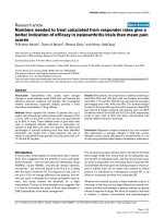
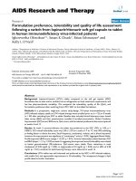
Figure 2
Schematic model of Shh/Fgf10/Bmp4 signalling during lung branching
morphogenesis. Shh is produced and secreted by epithelium in tips of
growing buds, binds Ptc in the mesenchyme and releases Smo
function, which then results in the activation of the Gli2 and Gli3
transcription factors (green). Bmp4 and Fgf10 seem not to be target
genes of Gli2 and Gli3. Fgf10 is elaborated by the mesenchyme
overlaying the tips of the epithelial buds, and binds and activates Fgfr2
in the epithelium, which then results in a chemotactic and mitogenic
response (blue). Bmp4 is expressed in the epithelium of the growing
buds but also weakly in the adjacent mesenchyme. It is secreted and
acts on Bmpr present in both mesenchyme and epithelium (red).
mutant did not show a lung phenotype [25], indicating that
Fgf7 function in lung development can be compensated
for by other Fgfs or it is simply not an essential factor for
early development. Whether Fgf10 is the mammalian
homolog of Drosophila branchless remains to be estab-
lished. In addition, it is unclear whether Fgf10 is a target
gene for Shh. fgf10 has been shown in vitro to be upregu-
lated in murine lung mesenchymal cells cultured without
epithelial cells, whereas the addition of Shh to these cells
prevented the increase in Fgf10. In contrast, when Fgf10-
containing beads were implanted in lung explants from
embryonic day 11.5, shh expression did not change [26].
Furthermore, because fgf10 expression is downregulated
in lungs that overexpress Shh [22], and fgf10 expression
increases towards birth, while shh decreases, Shh might
interact negatively with Fgf10 during the process of lung
development. However, genetic analysis suggests that
Fgf10 acts upstream of Shh in branching because no lung
buds develop in fgf10
–/–
mutants, whereas they do form in
shh
–/–
mutants. Furthermore, fgf10
–/–
mutants do develop
a separate trachea and esophagus, whereas a shh-null
mutant exhibits esophageal atresia/stenosis and tracheo-
esophageal fistula. Thus, it seems more likely that Shh and
Fgf10 function in separate but parallel pathways during
early lung development.
Concluding remarks and future
It is evident that the Shh/ptc/smo/gli pathway is important
in early pattern formation in the lung (Fig. 2). Bmp4 and
Fgf10, which are both implicated in mammalian lung
development, seem not to be regulated by Shh signalling
in the developing lung (Fig. 2). To understand Shh sig-
nalling during lung development, the identification of novel
downstream target genes is needed. Newly identified
targets are hip (hedgehog interacting protein), a mem-
brane glycoprotein, which seems to be a negative regula-
tor of Hh signalling [27], and a member of SOCS
(suppressor of cytokine signalling)-box WD proteins,
SWip1, which is one of the earliest markers to respond to
Shh in the limb [28]. It needs to be established whether
these molecules are present in the lung. Because wing-
less is a downstream target of Drosophila Hh signalling,
further investigations into the role of wnt genes in lung
development are warranted.
Acknowledgements
The work of the authors is supported by the Medical Research Council of
Canada.
References
Articles of particular interest have been highlighted as:
•
of special interest
••
of outstanding interest
1. Hammerschmidt M, Brook A, McMahon JA: The world according to
hedgehog. Trends Genet 1997, 13:14–21.
2. Ingham PW: Transducing Hedgehog: the story so far. EMBO J
1998, 17:3505–3511.
3. Porter JA, Young KE, Beachy PA: Cholesterol modification of
hedgehog signaling proteins in animal development. Science
1996, 274: 255–259.
4. Litingtung Y, Lei L, Westphal H, Chiang C: Sonic hedgehog is
••
essential to foregut development. Nat Genet 1998, 20:58–61.
In this article the authors showed for the first time that targeted deletion of
Shh in mice results in tracheal–esophageal fistula and tracheal and lung
abnormalities. The Shh
–/–
mutant lungs are hypoplastic and appear as a
single lobe.
5. Pepicelli CV, Lewis PM, McMahon AP: Sonic hedgehog regulates
••
branching morphogenesis in the mammalian lung. Curr Biol 1998,
8:1083–1086.
This article provides evidence that Shh is important for epithelial branching
morphogenesis but not proximo-distal epithelial differentiation.
6. Aza-blanc P, Ramirez-Weber FA, Laget MP, Schwartz C, Kornberg TB:
Proteolysis that is inhibited by hedgehog targets Cubitus inter-
ruptus protein to the nucleus and converts it to a repressor. Cell
1997, 89:1043–1053.
7. Bellusci S, Henderson R, Winnier G, Oikawa T, Hogan BLM: Evi-
dence from normal expression and targeted misexpression that
Bone Morphogenetic Protein-4 (Bmp-4) plays a role in mouse
embryonic lung morphogenesis. Development 1996, 122:1693–
1702.
8. Bellusci S, Furuta Y, Rush MG, Henderson R, Winnier G, Hogan BLM:
Involvement of Sonic hedgehog (Shh) in mouse embryonic lung
growth and morphogenesis. Development 1997, 124:53–63.
9. Urase K, Mukasa T, Igarashi H, Ishii Y, Yasugi S, Momoi MY, Momoi T:
Spatial expression of Sonic hedgehog in the lung epithelium
during branching morphogenesis. Biochem Biophys Res Commun
1996, 225:161–166.
10. Tsukui T, Capdevila J, Tamura K, Ruiz-Lozano P, Rodriguez-Esteban C,
Yonei-Tamura S, Magallon J, Chandraratna RA, Chien K, Blumberg B,
Evans RM, Belmonte JC: Multiple left–right asymmetry defects in
Shh
–/–
mutant mice unveil a convergence of the Shh and retinoic
acid pathways in the control of Lefty-1. Proc Natl Acad Sci USA
1999, 96:11376–11381.
11. Meyers EN, Martin GR: Differences in left–right axis pathways in
mouse and chick: functions of Fgf8 and Shh. Science 1999, 285:
403–406.
12. Goodrich LV, Milenkovic L, Higgins KM, Scott MP: Altered neural cell
fates and medulloblastoma in mouse patched mutants. Science
1997, 277:1109–1113.
13. Grindley JC, Bellusci S, Perkins D, Hogan BLM: Evidence for the
involvement of the Gli gene family in embryonic mouse lung
development. Dev Biol 1997, 188:337–348.
14. Theil T, Kaesler S, Grotewold L, Böse J, Rüther U: Gli genes and limb
development. Cell Tiss Res 1999, 296:75–83.
15. Motoyama J, Liu J, Mo R, Ding Q, Post M, Hui CC: Essential function
••
of Gli2 and Gli3 in the formation of lung, trachea and oesophagus.
Nat Genet 1998, 20:54–57.
The authors showed that the inactivation of Gli2 resulted in abnormal
growth and lobulation of the lung. No lung was formed when both Gli2 and
Gli3 were removed, whereas the presence of one allele of Gli3 in the
Gli2
–/–
background resulted in a tracheo-esophageal fistula and one lung
lobe.
16. Park HL, Bai C, Platt KA, Matise MP, Beeghly A, Hui Cc, Nakashima
•
M, Joyner AL: Mouse Gli1 mutants are viable but have defects in
SHH signaling in combination with a Gli2 mutation. Development
2000, 127:1593–1605.
Using compound mutant mice, the authors demonstrate that Gli1 and Gli2
have overlapping functions during lung development.
Respiratory Research Vol 1 No 1 van Tuyl and Post
/>commentary
review
reports primary research
17. Winnier G, Blessing M, Labosky PA, Hogan BL: Bone morpho-
genetic protein-4 is required for mesoderm formation and pat-
terning in the mouse. Genes Dev 1995, 9:2105–2116.
18. Weaver M, Yingling JM, Dunn NR, Bellusci S, Hogan BLM: Bmp sig-
•
naling regulates proximal–distal differentiation of endoderm in
mouse lung development. Development 1999, 126:4005–4015.
These authors investigated the role of Bmp4 in lung development by overex-
pressing the Bmp4 antagonist Xnoggin and, independently, a dominant-nega-
tive Bmp receptor (dnAlk6), using the Sp-C promoter. The inhibition of Bmp4
decreased Sp-C expression and increased CC10 and Hfh4 expression.
19. Metzger RJ, Krasnow MA: Genetic control of branching morpho-
genesis. Science 1999, 284:1635–1639.
20. Sekine K, Ohuchi H, Fujiwara M, Yamasaki M, Yoshizawa T, Sato T,
••
Yagishita N, Matsui D, Koga Y, Itoh N, Kato S: Fgf10 is essential for
limb and lung formation. Nat Genet 1999, 21:138–141.
Mice deficient in Fgf10 develop a normal trachea but lack the formation of
primary bronchi. Normal Shh expression was noted in the tracheal out-
growth.
21. Min H, Danilenko DM, Scully SA, Bolon B, Ring BD, Tarpley JE,
••
DeRose M, Simonet WS: Fgf-10 is required for both limb and lung
development and exhibits striking functional similarity to
Drosophila branchless. Genes Dev 1998, 12:3156–3161.
This article demonstrates that an Fgf10 knockout in mice results in trachea
formation without subsequent lung airway branching.
22. Bellusci S, Grindley J, Emoto H, Itoh N, Hogan BLM: Fibroblast
Growth Factor 10 (FGF10) and branching morphogenesis in the
embryonic mouse lung. Development 1997, 124:4867–4878.
23. Arman E, Haffner-Krausz R, Gorivodsky M, Lonai P: Fgfr2 is required
for limb outgrowth and lung-branching morphogenesis. Proc Natl
Acad Sci USA 1999, 96:11895–11899.
24. Post M, Souza P, Liu J, Tseu I, Wang J, Kuliszewski M, Tanswell AK:
Keratinocyte growth factor and its receptor are involved in regu-
lating early lung branching. Development 1996, 122:3107–3115.
25. Guo L, Degenstein L, Fuchs E: Keratinocyte growth factor is
required for hair development but not for wound healing. Genes
Dev 1996, 10:165–175.
26. Lebeche D, Malpel S, Cardoso WV: Fibroblast growth factor interac-
tions in the developing lung. Mech Dev 1999, 86:125–136.
27. Chuang PT, McMahon AP: Vertebrate Hedgehog signalling modu-
lated by induction of a Hedgehog-binding protein. Nature 1999,
397:617–621.
28. Vasiliauskas D, Hancock S, Stern CD: SWiP-1: novel SOCS box con-
taining WD-protein regulated by signalling centres and by Shh
during development. Mech Dev 1999, 82:79–94.
Authors’ affiliations: Minke van Tuyl (Lung Biology Research Program,
Hospital for Sick Children Research Institute, Toronto, Canada) and
Martin Post (Lung Biology Research Program, Hospital for Sick
Children Research Institute, Department of Pediatrics, Physiology and
Laboratory Medicine and Pathology, University of Toronto, Toronto,
Canada)
Correspondence: Martin Post, Hospital for Sick Children,
555 University Avenue, Toronto, Ontario, Canada M5G 1X8.
Tel: +1 416 813 6772; fax: +1 416 813 5002;
e-mail: