Báo cáo y học: "Marked alveolar apoptosis/proliferation imbalance in end-stage emphysema" pdf
Bạn đang xem bản rút gọn của tài liệu. Xem và tải ngay bản đầy đủ của tài liệu tại đây (817.31 KB, 13 trang )
BioMed Central
Page 1 of 13
(page number not for citation purposes)
Respiratory Research
Open Access
Research
Marked alveolar apoptosis/proliferation imbalance in end-stage
emphysema
Fiorella Calabrese*
1
, Cinzia Giacometti
†1
, Bianca Beghe
†2
, Federico Rea
†3
,
Monica Loy
†3
, Renzo Zuin
†2
, Giuseppe Marulli
†3
, Simonetta Baraldo
†2
,
Marina Saetta
†2
and Marialuisa Valente
†1
Address:
1
Institute of Pathology, University of Padua, Italy,
2
Department of Clinical and Experimental Medicine, Section of Respiratory Diseases,
University of Padua, Italy and
3
Department of Gastroenterological Sciences, Section of Thoracic Surgery, University of Padua, Italy
Email: Fiorella Calabrese* - ; Cinzia Giacometti - ;
Bianca Beghe - ; Federico Rea - ; Monica Loy - ;
Renzo Zuin - ; Giuseppe Marulli - ; Simonetta Baraldo - ;
Marina Saetta - ; Marialuisa Valente -
* Corresponding author †Equal contributors
apoptosisproliferationend-stage emphysema
Abstract
Background: Apoptosis has recently been proposed to contribute to the pathogenesis of emphysema.
Methods: In order to establish if cell fate plays a role even in end-stage disease we studied 16 lungs (9
smoking-associated and 7 α1antitrypsin (AAT)-deficiency emphysema) from patients who had undergone
lung transplantations. Six unused donor lungs served as controls. Apoptosis was evaluated by TUNEL
analysis, single-stranded DNA laddering, electron microscopy and cell proliferation by an
immunohistochemical method (MIB1). The role of the transforming growth factor (TGF)-β1 pathway was
also investigated and correlated with epithelial cell turnover and with the severity of inflammatory cell
infiltrate.
Results: The apoptotic index (AI) was significantly higher in emphysematous lungs compared to the
control group (p ≤ 0.01), particularly if only lungs with AAT-deficiency emphysema were considered (p ≤
0.01 vs p = 0.09). The proliferation index was similar in patients and controls (1.9 ± 2.2 vs 1.7 ± 1.1). An
increased number of T lymphocytes was observed in AAT-deficiency lungs than smoking-related cases (p
≤ 0.05). TGF-β1 expression in the alveolar wall was higher in patients with smoking-associated emphysema
than in cases with AAT-deficiency emphysema (p ≤ 0.05). A positive correlation between TGF-βRII and AI
was observed only in the control group (p ≤ 0.005, r
2
= 0.8). A negative correlation was found between
the TGF-β pathway (particularly TGF-βRII) and T lymphocytes infiltrate in smoking-related cases (p ≤ 0.05,
r
2
= 0.99)
Conclusion: Our findings suggest that apoptosis of alveolar epithelial cells plays an important role even
in end-stage emphysema particularly in AAT-deficiency disease. The TGFβ-1 pathway does not seem to
directly influence epithelial turnover in end-stage disease. Inflammatory cytokine different from TGF-β1
may differently orchestrate cell fate in AAT and smoking-related emphysema types.
Published: 10 February 2005
Respiratory Research 2005, 6:14 doi:10.1186/1465-9921-6-14
Received: 29 July 2004
Accepted: 10 February 2005
This article is available from: />© 2005 Calabrese et al; licensee BioMed Central Ltd.
This is an Open Access article distributed under the terms of the Creative Commons Attribution License ( />),
which permits unrestricted use, distribution, and reproduction in any medium, provided the original work is properly cited.
Respiratory Research 2005, 6:14 />Page 2 of 13
(page number not for citation purposes)
Background
Pulmonary emphysema, a significant global health prob-
lem, is a pathological condition characterized by enlarge-
ment of the airspaces distal to the terminal bronchiole,
destruction of the alveolar walls, without and/or with
mild fibrosis [1]. To date the pathogenesis remains enig-
matic. The most prevailing hypothesis since the 1960s has
been the elastase/antielastase imbalance theory of inflam-
mation [2]. Briefly, the concept is that activated inflam-
matory cells release large quantities of elastases,
overwhelming local antiprotease activity with consequent
damage to the alveolar wall matrix [3]. However the
emphasis on alveolar matrix destruction by a combina-
tion of inflammation and excessive proteolysis has failed
to fully explain the loss of lung tissue, particularly when
compared to alterations seen in other inflammatory lung
diseases.
Recently more attention has been paid to alveolar epithe-
lial injury in addition to alveolar matrix destruction. The
presence of apoptosis has recently been described in ani-
mal models of emphysema [4,5] and in a few studies of
human disease [6-9].
The majority of investigations have focused the attention
on smoking-related emphysema keeping in mind that cig-
arette smoking was the main cause of apoptotic cell death.
Cigarette smoke may induce alveolar cell apoptosis either
directly by a cytotoxic effect on pneumocytes or indirectly
by decreasing the production of vascular endothelial
growth factor (VEGF) via altered epithelial cells [7]. To
date smoking-associated centrilobular emphysema is the
only studied form of emphysema in which apoptosis, and
more recently also proliferation, have been investigated
[9]. Alterations of lung epithelial cell turnover in end-
stage emphysema, either smoking-associated emphysema
or α1-antitrypsin (AAT)-deficiency emphysema, are up to
now not well distinguished.
Moreover apoptotic phenomenon has been previously
investigated in moderate/severe smoking-related forms of
emphysematous lungs obtained almost exclusively from
lung volume reduction surgery [6,7,9]. If cell fate is a sta-
ble, progressive and/or a decreasing process in end-stage
disease is to date unknown.
Among the growth factors, transforming growth factor
(TGF)-β1 could play a crucial role in the remodeling proc-
ess occurring in emphysematous parenchyma. TGF-β1,
other than its known profibrogenetic [10] and anti-
inflammatory effects [11,12], has an important influence
on epithelial cell growth [14]. It has been demonstrated
that it has an inhibitory effect on the growth of lung epi-
thelial cells, particularly for airway epithelium [14,15].
The cytokine has been shown to be over-expressed in
patients with a history of smoking and chronic obstructive
pulmonary disease (COPD) [16,17]. Paracrine (mainly
produced by macrophages) and autocrine (released by
epithelial cells) activity of this growth factor could play an
important role in the loss of the alveolar walls by inducing
apoptotic cell death.
In the present work the degree of apoptotic cell death and
epithelial proliferation in the lungs of patients with differ-
ent types of end-stage emphysema was studied. The sever-
ity of inflammatory cell infiltrate (ICI) was also quantified
and correlated with epithelial cell turnover. Further, the
TGF-β1 pathway was detected and correlated with the
apoptotic index (AI), the proliferative index (PI) and the
ICI.
Methods
Lung tissue preparation
Lung tissue used in the present study comprised material
from 16 patients undergoing lung transplantation for
end-stage emphysema at the Thoracic Surgery Unit of the
University of Padua Medical School. Cold ischemia pres-
ervation was 60 minutes and 120 minutes, respectively,
for single and double lung transplantations. Small-sized
pieces from all lobes were cut and immediately fixed in
Karnovsky's solution for electron microscopy. The lungs
were then gently fixed in 10% phosphate-buffered forma-
lin by airway perfusion and processed for sectioning (3
µm). Samples were selected from specimens that showed
features of excellent tissue preservation and adequate lung
inflation. In particular, large thin blocks approximately 30
× 25 mm were cut from the subpleural areas of the apical
anterior and lingular segments of the upper lobes, as well
as the apical and basal segments of the lower lobes. A
more centrally placed block was taken to sample the seg-
mented airways and blood vessels. The right lung was
sampled in the same way with the middle lobe being
treated in the same way as the lingula [18]. Adult control
lungs were obtained from unused donor lungs for trans-
plantation (6 cases). The Local Research Ethics Commit-
tee approved the study.
TUNEL analysis
The terminal deoxynucleotidyl transferase-mediated
dUTP-biotin nick end-labeling method (TUNEL) was
used to investigate the presence of apoptosis. Sections
were processed in accordance with Gavrieli et al's method
[19]. Briefly, after deparaffinization and rehydration, sec-
tions were digested with proteinase K (Boehringer Man-
nheim, Mannheim, Germany) at a concentration of 20
µg/ml for 15 minutes. The slides were then incubated with
TdT/biotinylated dUTP diluted in buffer (Boehringer
Mannheim, Mannheim, Germany). The slides were devel-
oped by using diaminobenzidine and 30 ml hydrogen
Respiratory Research 2005, 6:14 />Page 3 of 13
(page number not for citation purposes)
peroxide. For negative controls, some slides were incu-
bated in buffer without TdT or biotinylated UTP. For pos-
itive controls, some slides were incubated with 1 µg/ml
DNAse (Sigma-Aldrich, Milan, Italy).
Electron microscopy
Lung specimens fixed in Karnovsky's solution (2% para-
formaldehyde, 2.5% glutaraldehyde in Millonig, pH 7.3)
for 24 hours were post-fixed with 1% osmium tetroxide
(Millonig, pH 6.8) for 1 hour, and then progressively
dehydrated in alcohol and embedded in epon. Semi-thin
sections were stained with 0.1% toluidine blue for light
microscopic examination. Ultra-thin sections were
stained with uranyl acetate and lead citrate for transmis-
sion electron microscopy performed using a Hitachi H-
7000 (Hitachi Ltd., Tokyo, Japan).
Oligonucleosomal-length DNA laddering
The presence of oligonucleosomal-length DNA cleavage
was investigated with APO-DNA1 (Maxim Biotech Inc,
San Francisco, CA, USA) in 12 cases (4 AAT-emphysema
patients, 4 smoking-related emphysema patients and 4
controls) in which frozen tissue was available. Briefly,
DNA was obtained from lung tissue samples using protei-
nase K-phenol extraction. Dephosphorylated adaptors
were ligated to 5' phosphorylated blunt ends with T
4
DNA
ligase to 500 ng of lung sample DNA (for 16 h at 16°C).
These then served as primers in LM-PCR under the follow-
ing conditions: hot start (72°C for 8 min), 30 cycles
(94°C for 1 min, and 72°C for 3 min) and extension
(72°C for 15 min). Every reaction set included thymus
DNA as a positive control and normalization of the
amount of reaction products. Amplified DNA was sub-
jected to electrophoresis on 1.2% agarose gel containing
ethidium bromide. Images were scanned and the DNA
fragmentation levels were based on the density of the
bands ranging between 1000 base pairs (bp) and 300 bp.
The percentage of DNA fragmentation was quantified by
scanning densitometry.
Immunohistochemistry for TGF-
β
1, TGF-
β
RII and MIB1
All lung sections were subjected to antigen retrieval by
heating in a microwave oven on high power for 8 minutes
in 0.01 mol/l citrate buffer (ph 6.0) and then incubated
with a mouse monoclonal anti-TGF-β
1
-β
2
and-β
3
primary
antibody to active TGF-β1 (150 µg/ml; dilution 1:20,
Genzyme Diagnostics, Cambridge, MA), with polyclonal
antibody against TGF-β receptor type II (200 µg/ml, dilu-
tion 1:200, Santa Cruz Biotechnology Inc., Santa Cruz)
and monoclonal MIB-1 antibody (1:50 Dako, Santa Bar-
bara, CA, U.S.A.), which recognizes the Ki-67 antigen in
paraffin-embedded tissue sections. Immunohistochemi-
cal investigations were done on the sections from the
same paraffinembedded specimens processed for TUNEL
analysis.
The detection system was the Vectastain ABC kit (Vector
Peterborough, UK) with 3-amino-9-ethylcarbazole (for
TGF-β1, TGF-βRII) and with a mixture of 3,3'-diamino-
benzidine tetra7 hydrochloride (Dako) and hydrogen per-
oxide as the chromogenic substrates. Sections were coun-
terstained with Mayer's hematoxylin.
Immunohistochemistry for inflammatory cell infiltrate
(ICI)
In all samples, immunohistochemistry for the characteri-
zation of ICI was carried out by using the following anti-
body panel: CD20 (1.100), CD45RO (1.100), CD4
(1:20), CD8 (1:50), CD3 (1:100), CD68 (1:50) (Dako,
Santa Barbara, CA, U.S.A.). The detection system was the
Vectastain ABC kit, as described above.
For all immunohistochemistry experiments, negative con-
trols were performed by incubation of the sections with
the omission of primary antibody and using the antibody
diluents alone or the appropriate non-immune IgG in
each case.
Double immune-labeling
For simultaneous detection of DNA fragmentation and
cell proliferation a double labeling was also performed.
The TUNEL technique was first performed and the stain-
ing achieved was diaminobenzidine as chromogen. For
MIB1 immunolocalization in the second staining
sequence the sections were stained with 5-bromo-4-
chloro-3-indoxyl phosphate/nitro blue tetrazolium
(BCIP/NBT alkaline phosphatase Kit II, Vector Laborato-
ries (Vector Peterborough, UK).
Image analysis
Immunoassay for TGF-β1 and TGF-βRII was detected by
using digital quantitative analysis (Image Pro Plus soft-
ware version 4.1, Media Cybernetics, Silver Spring MD) as
previously described [13]. Quantification of TUNEL,
MIB1 positive cells and ICI was restricted to the alveolar
wall. Images for each lung section from the upper and
lower lobes were acquired with a 40X lens.
In each case at least 50 microscopic randomly chosen
fields were analyzed. A total of 5,000 epithelial cells were
counted for AI and PI and the values were expressed as
percentages.
Statistical analysis
To avoid observer bias the cases were coded and measure-
ments were made without knowledge of clinical data. Dif-
ferences between groups were detected using the analysis
of variance for clinical data and the Kruskall-Wallis test for
histological data. The Mann-Whitney U test was per-
formed after the Kruskall-Wallis test when appropriate.
The statistical tests used were two-sided.
Respiratory Research 2005, 6:14 />Page 4 of 13
(page number not for citation purposes)
Correlation coefficients were calculated using Spearman's
rank method. Probability values of 0.05 or less were
accepted as significant. Group data were expressed as
means and SD or as medians and range when appropriate.
Results
Clinical data and histological findings
Major clinical data for patients with emphysema are
shown in Table 1.
Average patient age was 54.4 ± 7.5 years. FEV1 mean was
19 ± 8.9 (predicted for sex, age, and body weight). Bilat-
eral single lung transplantation was performed in 12 out
of 16 patients. All patients had been heavy smokers: 7
were only smoking-associated emphysema cases (51 ± 28
packs-year) and 9 were both AAT-deficiency emphysema
and smoking cases (55 ± 27 packs-year). For the sake of
brevity, the abbreviation AAT-deficiency emphysema for
smoking patients with AAT-deficiency will be used
throughout the manuscript.
All patients had quit smoking at least 1 year before under-
going surgery.
The average control patient age was 34 ± 16.8 years and
cerebral trauma was the cause of death. All the donors
stayed less than two days in intensive care without evi-
dence of lung infection or other complications. During
artificial ventilation, airway pressure (P
aw
) was 20,9 ± 1.5
mmHg and inspiratory oxygen fraction (FI, O
2
) was 0.4 ±
0.1.
All the samples showed various degrees of emphysema-
tous changes. In particular, all the patients with AAT-defi-
ciency showed diffuse destruction of alveolar tissue,
consistent with panlobular emphysema. In contrast, rela-
tively preserved lower portions of the lungs were observed
in patients with smoking-associated emphysema, consist-
ent with centrolobular emphysema.
Immunophenotype analysis
Emphysema patients had an increased number of ICI
(CD20, CD3, CD8, CD68, CD45RO, CD4 and PMN) as
compared with controls (p ≤ 0.01). An increased number
of CD3 (p ≤ 0.05), CD8 (p ≤ 0.05) and CD45RO (p ≤
0.001) was seen in AAT-deficiency emphysema compared
to smoking-related emphysema (Table 2).
Analysis of epithelial apoptosis and proliferation
Labeling of the DNA breaks by TUNEL demonstrated pos-
itive cells that were localized to peribronchiolar, intra-
alveolar and septal sites in both normal and emphysema-
tous lungs. Quantification was limited to the alveolar
wall. Apoptotic bodies that were very close to each other
were counted as one dying cell. Intra-alveolar apoptotic
cells were not included in the cell count.
In emphysematous lungs AI ranged from 0.68 to 11.92
(mean 6.3 ± 3.5). The TUNEL-positive cells were more fre-
quently detected within more enlarged alveolar walls.
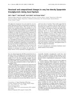
Apoptotic cells and/or bodies were frequently seen in
intra-alveolar lumen that presumably represented apop-
totic cells detached from the alveolar wall (Fig. 1). AI was
significantly higher in patients than in controls (6.5 ± 3.5
Table 1: Subject Characteristics
Case Sex Age Emphysema type Packs/year FEV
1*
FEV
1
/FVC* Transplantation
1 M§ 49 AAT deficiency 27 27 27 BSLT *
2 M 59 Smoking 36,5 31 38 RtSLT †
3 F|| 62 Smoking 7 17 55 BSLT
4 F 62 Smoking 36,5 13 30 LtSLT ‡
5 M 62 Smoking 108 15 45 BSLT
6F49Smoking731233BSLT
7M47Smoking542260BSLT
8 M 59 AAT deficiency 36,5 20 56 BSLT
9 M 64 Smoking 54 6 42 LtSLT
10 M 63 Smoking 54 24 37 BSLT
11 M 51 AAT deficiency 54 11 25 BSLT
12 M 53 AAT deficiency 108 8 14 BSLT
13 M 45 AAT deficiency 73 17 24 BSLT
14 M 56 Smoking 36,5 15 32 RtSLT
15 F 41 AAT deficiency 54 35 38 BSLT
16 M 51 AAT deficiency 36,5 34 36 BSLT
*BSLT: bilateral single lung transplantation, † RtSLT: right transplant single lung transplantation, ‡ LtSLT: left transplant single lung transplantation,
§M: male, || F: female. FEV
1
and FVC are given as percentages of predicted values.
Respiratory Research 2005, 6:14 />Page 5 of 13
(page number not for citation purposes)
vs 2.7 ± 2.6, p ≤ 0.01) (Fig 2). If separately compared with
the control group only the AAT-deficiency emphysema
showed a statistically significant difference (p ≤ 0.01 vs p
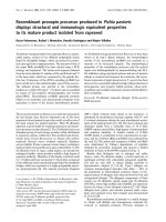
= 0.09). Increased levels of oligonucleosomal-length DNA
fragments were also detected in emphysema patients, par-
ticularly in AAT-deficiency emphysema, than control
lungs (Fig. 3a,b). The PI of patients ranged from 0.19% to
4.81% (mean 1.9 ± 2.2). Similar numbers of MIB1-posi-
tive alveolar septal cells were observed in both types of
emphysema and control lungs (1.7 ± 1.1).
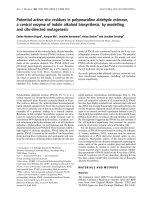
TUNEL-positive/MIB1-negative nuclei detected by double
staining were seen in all cases, whereas MIB1 was never
expressed in any of the TUNEL-positive nuclei (Fig. 4a,b).
In each group, no statistically significant correlations were
found between AI and PI as well as between AI/PI and ICI.
Table 2: Inflammatory Cells (Total Cells/MM Alveolar Wall)
AAT Smokers Controls * P†
CD20 4.77 2.1 0.0 ns
CD3 25.39 17.84 2.5 <0,05
CD8 12.32 5.07 0.99 <0,05
CD68 4.35 6.88 0.75 ns
CD45RO 25.42 8.34 2.31 0,001
CD4 12.1 12.67 1.89 ns
PMN 18.07 20.78 0.0 ns
Definition of abbreviation. PMN: Polymorphonuclear cells
* The values of control group were all statistically significant compared to both emphysema groups.
† Significant differences between AAT-deficiency and smoking patients.
AAT-deficiency emphysema case 1Figure 1
AAT-deficiency emphysema case 1: Micrograph show-
ing strong specific staining for DNA strand breaks in the alve-
olar epithelial cells and in two cells detaching from the wall.
TUNEL (original magnification 160×).
AI in controls vs emphysema patientsFigure 2
AI in controls vs emphysema patients: Significantly
higher AI was found in emphysema patients (6.5 ± 3.5 vs 2.7
± 2.6, p ≤ 0.01) = AAT-deficiency emphysema; ❍ = smok-
ing-related emphysema.
Respiratory Research 2005, 6:14 />Page 6 of 13
(page number not for citation purposes)
Gel-electrophoresisFigure 3
Gel-electrophoresis: a) Oligonucleosomal-length DNA laddering in emphysematous and control lungs. Lane 1: DNA
marker; Lanes 2–5: control donor lungs (4 cases); Lanes 6–9: AAT-deficiency emphysema patients (4 cases); Lanes 10–13:
smoking-related emphysema patients (4 cases); Lane 14: positive control. b) Quantification of DNA laddering based on scan-
ning densitometry of bands approximately between 1000 bp and 300 bp (arrowhead) followed by normalization with the den-
sity obtained with the equivalent band of the thymus DNA positive control (lung sample/control = densitometric ratio) which
was included in every oligonucleosomal DNA laddering assay.
Respiratory Research 2005, 6:14 />Page 7 of 13
(page number not for citation purposes)
At electron microscopy typical features of early apoptosis
with margination of chromatin at the nuclear membrane
and late apoptosis with completely dense nuclear chroma-
tin, including apoptotic bodies in various stages of degra-
dation, were seen in pneumocytes, endothelial cells and
fibroblasts. Typical features of reduplication of vessel
basal membranes were frequently seen in cases with more
evident apoptosis (5 a-f). Ultrastructural analysis showed
more frequent mitotic features in type II pneumocytes.
TGF-
β
1 and TGF-
β
RII receptor analysis
In emphysema patients and controls TGF-β1 and TGF-
βRII were localized in bronchiolar and alveolar epithelial
cells and macrophages. Quantitative analysis of TGF-β1
measured in the alveolar wall showed no statistically sig-
nificant difference between emphysema patients and con-
trols. A higher cytokine expression was noted in patients
with smoking-associated emphysema compared with
AAT-deficiency disease (mean 8.8 ± 1.7 vs 5.2 ± 3.9, p ≤
0.05) (Fig. 6). A positive significant correlation between
TGF-βRII and AI (p = 0.005; r
2
= 0.8) was seen in control
lungs (Fig. 7). A significant negative correlation was found
between TGF-β pathway (particularly TGF-βRII) and T
lymphocytes infiltrate (CD3+) (p ≤ 0.05, r
2
= 0.99) in
smoking-related cases. No correlation was noted between
the TGF-β1 pathway (TGF-β1 and its RII) and the AI/ PI of
emphysematous lungs.
Discussion
In the present study we have analyzed for the first time
apoptosis and proliferation in different types of end-stage
emphysema. The detection of a high AI in emphysema-
tous lungs even in end-stage disease emphasizes the
importance of the phenomenon in the development, and
overall, in the progression of emphysema.
In general there are two main forms of cell death: oncosis
and apoptosis. The latter process results in characteristic
biochemical features and cellular morphology such as cell
shrinkage condensation and fragmentation of the nucleus
into well contained fragments called apoptotic bodies.
Perturbation of normal rates of apoptosis has been impli-
cated in many pathologic conditions such as neuro-vege-
tative, cardiovascular and liver disorders and cancer [20-
22]. As stated in Tuder's recent review on apoptosis and its
role in emphysema, cell damage, apoptosis, apoptotic cell
removal, and cellular replacement are ongoing and pre-
sumably highly regulated in order to maintain homeosta-
sis of the entire alveolar unit. The concept of the
irreversible destruction of alveolar walls due to the loss of
homeostasis of the alveolar unit is a critical point. Lung
inflammation, protease/antiprotease imbalance, oxida-
tive stress and apoptosis could work together in the
irreversible changes seen in emphysema [23]. Over-induc-
Smoking-related emphysema case 3Figure 4
Smoking-related emphysema case 3: a) double labeling TUNEL/MIB1 (marker of cell proliferation) showing two apop-
totic cells (dark) and one MIB1-positive cell (blue), on the surface of the same alveolar wall (original magnification 160×). b)
Note alveolar cell in proliferation close (blue) to apoptotic pneumocyte (dark) (Original magnification 160×).
Respiratory Research 2005, 6:14 />Page 8 of 13
(page number not for citation purposes)
AAT-deficiency emphysema (case 1)Figure 5
AAT-deficiency emphysema (case 1): Electron micrograph showing (a) early apoptosis with perinuclear chromatin
condensation (arrow) and (b) late apoptosis with nuclear dense chromatin of pneumocytes (arrow). (c) An endothelial cell
with condensed chromatin is well visible (arrow). (d) Note reduplication of the vessel basal membrane (arrow). In (e) and (f)
apoptotic bodies in various degrees of degradation close to a macrophage and an intraluminal apoptotic body are well visible
(arrows).
Respiratory Research 2005, 6:14 />Page 9 of 13
(page number not for citation purposes)
tion of apoptosis and inefficient cellular replenishment,
modifying alveolar homeostasis, would both be expected
to disrupt the alveolar wall thus inducing the develop-
ment of emphysema.
Recently the causal role of apoptosis has been increasingly
recognized in the destruction of alveolar walls and air-
space enlargement [6-9]. Among constitutive cell popula-
tions of the alveolar wall, epithelial cells are more
frequently susceptible to programmed cell death [6,9]. In
our study the AI of epithelial cells was significantly higher
in end-stage emphysema cases compared to the control
group (p ≤ 0.01) and this was particularly more evident
for those with AAT-deficiency.
To avoid a bias of under or over-estimated alveolar cell
apoptosis and proliferation due to regional disease activ-
ity we analyzed large specimens taken from different lung
regions (upper and lower lobes). The lower AI detected in
our control lungs underlines an important concept: in
non-emphysematous lungs apoptosis is an irrelevant
process adequately balanced by proliferation. The
increased number of apoptotic cells in patients with
emphysema (not adequately replaced by new epithelial
cells) suggests a new mechanism, namely "epithelial
apoptosis/proliferation imbalance" in the pathogenesis of
disease. In our study, different from a recent clinical study
by Yokohori et al [9], a PI similar to that of the control
group was detected in the alveolar epithelial cells of
emphysema patients. The discrepancies between the two
studies can be attributed to several factors: 1) a different
monoclonal antibody used for detection of cell prolifera-
tion (MIB1 vs PCNA); 2) different case series including
patients affected by emphysema in end-stage status and
overall of different types (smoking-associated and AAT-
deficiency emphysema), and 3) more analysis of extensive
areas (upper and lower lobes) of emphysematous lung
parenchyma. Regarding the monoclonal antibody used
for proliferation detection, Ki-67 is now well recognized
as the most reliable immunohistochemical marker for the
analysis of cell proliferation in formalin-fixed, paraffin-
embedded tissue [24]. Immunoassaying for proliferating
nuclear cell antigen (PCNA) can also be used in paraffin-
embedded tissue, but it may overestimate the prolifera-
tion rate given the long half-life of this antigen [25]. More-
over, the simultaneous positive staining of TUNEL and
PCNA in the same cells has been reported [26]. In fact, it
has also been demonstrated that PCNA expression can
increase without a corresponding increase in S-phase
DNA synthesis [27].
DNA nicks may be seen in cells with DNA synthesis/repair
thus sometimes producing false TUNEL positive cells.
False positive TUNEL staining can also be generated
through non-apoptotic mechanisms: RNA synthesis and
splicing, necrosis as well as artifacts due to preservation
methods. Consequently, some authors have stressed the
importance of associating other techniques, such as Taq
polymerase-based DNA in situ ligation, DNA gel electro-
phoresis or electron microscopy, in order to avoid false
positive labeling and to assess the reliability of apoptosis
[28].
Our TUNEL findings have been corroborated by employ-
ing an additional quantitative apoptosis assay. Moreover,
the presence of different stages of apoptosis was con-
firmed and the cells involved in programed cell death
were well characterized by using electron microscopy
investigation, which is considered the gold-standard tech-
nique for apoptotic cell detection. In our work double-
immune labeling showed that all TUNEL positive cells
were consistently negative for MIB1 thus suggesting the
true epithelial DNA fragmentation. Although the high AI
detected in our patients could be mainly explained by the
high rate of apoptotic cell death, an impaired clearance
mechanism of apoptotic cells/bodies should also be con-
sidered. A frequent finding of apoptotic bodies in alveolar
walls and within lumen may support the latter theory as
TGF-β1 expression in smoking-related vs AAT-deficiency emphysemaFigure 6
TGF-β1 expression in smoking-related vs AAT-defi-
ciency emphysema: the graphic shows the different
cytokine expression in both types of emphysema. A signifi-
cantly higher TGF-β1 expression was found in smoking-
related emphysema versus AAT-deficiency emphysema
(mean value 8.8 ± 1.7 vs 5.2 ± 3.9, p ≤ 0.05).
Respiratory Research 2005, 6:14 />Page 10 of 13
(page number not for citation purposes)
an important contributing factor for a high percentage of
AI.
The principal trigger of epithelial injury leading to apop-
totic cell death is up to now still unclear. The cytotoxic
effects of cigarette smoke, one of the most clearly proven
etiologic factors in the development of emphysema and in
general of COPD, have been suggested to suppress
epithelial proliferation and to induce cell death. In partic-
ular oxidants and aldehydes, major constituents in the
volatile phase of cigarette smoke, have been reported to
induce apoptosis of lung cells [29].
Different DNA and RNA viruses have been identified as
viral pathogens associated with the disease. Double-
strand DNA viruses such as adenovirus have the ability to
persist in airway epithelial cells long after the acute infec-
tion has cleared. The expression of adenoviral trans-acti-
vating proteins has been demonstrated in the airway
epithelium of both human and animal lungs and is asso-
ciated with an amplification of cigarette smoke-induced
inflammatory response [30].
Different adenovirus early proteins, in particular E4orf4,
have been reported in the shutoff of host protein synthesis
and in the promotion of a p53-independent cell death
program [31]. It is likely that many and various noxious
agents all come together to play an important role in the
progression of cell death in end-stage disease, justifying
the high AI in the alveolar wall, as detected in our study.
Correlation between TGF-βRII and AIFigure 7
Correlation between TGF-βRII and AI: the graphic shows the correlation between TGF-βRII expression and AI in con-
trols and emphysema patients. The degree of TGF-βRII is linearly related to the extension of apoptosis in the control group (p
≤ 0.005, r
2
= 0.8).
Respiratory Research 2005, 6:14 />Page 11 of 13
(page number not for citation purposes)
The specific molecular pathogenetic pathways that regu-
late both cell fate and proliferation are also under investi-
gation. Previous studies demonstrated an inhibitory effect
of the TGF-β1 pathway on the growth of lung epithelial
cells [14,15].
As the TGF-β1 pathway is well-known for its anti-inflam-
matory activity, a higher epithelial expression of TGF-β1
in patients with smoking-related emphysema compared
with AAT-deficiency may partially justify the different pat-
terns of inflammation in the two types of emphysema, as
found in our study. A significantly higher increase of
inflammation, particularly due to T lymphocytes, was
found in AAT-deficiency emphysema (panlobular type)
than in smoking-related disease (centrilobular type), with
the latter displaying an increased expression of the TGF-β
pathway (as demonstrated by the negative correlation
with T lymphocyte infiltrate). Similar findings have been
previously reported in both in vitro and in vivo studies
[32,33]. An increased pro-apoptotic milieu of inflamma-
tory related cytokines may contribute to the higher cell
death rate detected in AATdeficiency emphysema. Moreo-
ver, additional cigarette smoke-mediated damage should
also be considered in AAT-deficiency emphysema
patients, in that in our study they were all heavy smokers.
In our work a direct correlation between TGF-βRII and AI
was found in the control group thus showing that this
cytokine could play a role in alveolar homeostasis in
physiologic conditions. Instead no correlation was found
between the AI and TGF-β1 pathway in either type of
emphysema, suggesting that the TGF-β1 regulated mecha-
nism is lost in the disease. Other cytokines besides TGF-β1
could be involved in uncontrolled programed cell death
inducing the progressive disappearance of the alveolar
unit.
Decreased expression of VEGF and VEGF R2 has been
demonstrated to be significantly correlated with apoptosis
of both epithelial and endothelial cells in cigarette smok-
ing-induced emphysema [7]. It has been shown that VEGF
receptor signaling is extremely important for the mainte-
nance of alveolar structures. Hence an impairment of its
trophic endothelial activity may be one of several factors
facilitating alveolar septal cell apoptosis [4,7]. Signifi-
cantly reduced levels of VEGF have also been detected in
induced sputum of emphysema patients compared to that
of normal individuals and patients with asthma [34].
More recently in an experimental model some authors
have shown that over-expression of placenta growth factor
(PIGF) causes a phenotype and pulmonary dysfunction
similar to human lung emphysema by inducing apoptotic
events in the alveolar septa [35]. Although epithelial cells
have been demonstrated to be more susceptible to apop-
tosis [7], endothelial cells are also an important target for
programed cell death. Our ultrastructural analysis showed
evidence of endothelial cell apoptosis mainly in those
cases with more increased alveolar programed cell death.
The presence of a multi-layered vessel basement mem-
brane, as found in many of our emphysematous lungs,
may also reflect additional data supporting the increased
apoptotic rate of endothelial cells.
In summary, our work has demonstrated for the first time
that apoptotic phenomenon is extensive also in end-stage
emphysema patients. This unique case series, and overall
the large variety of lung tissue samples examined, (not
only subpleural emphysematous regions as those from
lung volume reduction surgery in which apoptosis could
already be switched off) may account for the differences in
our AI findings compared to other studies [9]. The higher
rate of apoptotic cell death in patients with AAT-defi-
ciency emphysema, partially influenced by the higher
degree of inflammation, may allow us to consider this
peculiar emphysema subtype as an additional modifier of
apoptosis.
Whether the "apoptosis/proliferation imbalance" occurs
before, after or at the same time as the "elastase/antie-
lastase imbalance" is still unknown and should be the
subject of future studies.
Limitations of the study
Our study had a few limitations. Firstly, the patients with
AAT-deficiency can not be considered pure AAT-deficiency
emphysema cases because these patients were also smok-
ers. Panlobular emphysema occurs at a younger age in
alpha-1-antitrypsin patients, especially if the patients
smoke cigarettes, as in our case series (49.8 ± 5.7 yrs AAT-
deficiency vs 58.2 ± 6.3 yrs smokers, p ≤ 0.01). Patients
with AAT-deficiency who are smokers develop lung
impairment function earlier and in a more severe form
than their non-smoking counterparts. Thus, it is extremely
rare to have patients who are non-smokers with AAT-defi-
ciency as candidates for lung transplantation. Secondly,
the clinical characterization of the donor was poor and
according to the guidelines for the selection of donor
lungs, smokers were not excluded [36]. Smokers could
have been included in the control group, and it is well
known that smoking itself may induce apoptosis.
However, if this was the case, the AI difference between
emphysema and control patients would have been even
higher because of the lower AI in healthy patients. A third
potential bias is that all the donors were mechanically
ventilated before lung transplantation and it is known
that mechanical ventilation may induce lung apoptosis
[37]. Again, the difference observed in our study would be
even higher than non-ventilated controls, thus confirming
Respiratory Research 2005, 6:14 />Page 12 of 13
(page number not for citation purposes)
the findings that enhanced apoptosis may act as a leading
mechanism in the pathogenesis of emphysema.
Conclusions
Our study analyzed apoptosis and proliferation in end-
stage emphysema. In particular the work described for the
first time a high AI in patients with AAT-deficiency
emphysema. Ultrastructural investigation, TUNEL analy-
sis and oligonucleosomal-length DNA laddering, per-
formed in different lung regions were all used for
detection of apoptotic phenomenon. The increase of
apoptotic cells in patients with emphysema not ade-
quately replaced by new epithelial cells suggests a new
mechanism, namely "epithelial apoptosis/proliferation
imbalance" in the pathogenesis of disease. More inflam-
mation, particularly due to T lymphocytes, was observed
in AAT-deficiency emphysematous lungs. An increased
pro-apoptotic milieu of inflammatory related cytokines
may contribute to the higher cell death rate detected in
AAT-deficiency emphysema. While a direct correlation
between TGF-βRII and AI was found in the control group,
no relation was found between the AI and TGF-β1 path-
way in end-stage emphysema, suggesting that the
influence of the TGF-β pathway on epithelial turnover is
lost in the disease. Knowledge of the mechanism respon-
sible for activation and progression of the apoptotic cas-
cade could offer new information in the near future, on
more appropriate stratification and treatment of the
disease.
Authors' contributions
FC: conceived of the study and participated in its design
and coordination
CG: substantial contribution in study design and data
interpretation
BB: acquisition of clinical data and critical revision for
important intellectual content
FR: thoracic surgeon providing lung specimen and critical
revision for important technical aspects
ML: thoracic surgeon providing lung specimen and critical
revision for important technical aspects
RZ: critical revision for important intellectual content
GP: acquisition of clinical data
SB: acquisition of clinical data and performed the statisti-
cal analysis
MS: participated in the design of the study and gave criti-
cal revision for important intellectual content
MV: substantial contribution in study design and data
interpretation
All the authors read and approved the final manuscript.
Acknowledgements
We would like to thank Alessandra Dubrovich and Giovanna Mattiazzo for
their excellent technical assistance and Dr. Judith Wilson for revision of the
English manuscript. Sources of support: Grant Project "Chronic obstructive
pulmonary disease (COPD) and lung transplantation: morphologic and
molecular study of pathogenetic substrates of disease progression" and
"Structure-function relationships in chronic obstructive pulmonary dis-
ease", Ministery of Education, University and Research, Rome, Italy
References
1. Pauwels RA, Buist AS, Calverley PM, Jenkins CR, Hurd SS: GOLD
Scientific Committee: Global strategy for the diagnosis,
management, and prevention of chronic obstructive pulmo-
nary disease. NHLBI/WHO Global Initiative for Chronic
Obstructive Lung Disease (GOLD) Workshop summary. Am
J Respir Crit Care Med 2001, 163:1256-1276.
2. Snider GL: Chronic obstructive pulmonary disease: a defini-
tion and implications of structural determinants of airflow
obstruction for epidemiology. Am Rev Respir Dis 1989, 140:S3-S8.
3. Hautamaki RD, Kobayashi , Senior RM, Shapiro SD: Requirement
for macrophage elastase for cigarette smoke-induced
emphysema in mice. Science 1997, 277:2002-2004.
4. Kasahara Y, Tuder RM, Taraseviciene-Stewart L, Le Cras TD, Abman
S, Hirth P, Waltenberger J, Voelkel NF: Inhibition of VEGF recep-
tors causes lung cell apoptosis and emphysema. J Clin Invest
2000, 106:1311-1319.
5. Aoshiba K, Yokohori N, Nagai A: Alveolar wall apoptosis causes
lung destruction and emphysematous changes. Am J Respir Cell
Mol Biol 2003, 28:555-562.
6. Segura-Valdez L, Pardo A, Gaxiola M, Uhal BD, Becerril C, Selman M:
Upregulation of gelatinases A and B, collagenases 1 and 2,
and increased parenchymal cell death in COPD. Chest 2000,
117:684-694.
7. Kasahara Y, Tuder RM, Cool CD, Lynch CD, Flores SC, Voelkel NF:
Endothelial cell death and decreased expression of vascular
endothelial growth factor and vascular endothelial growth
factor receptor 2 in emphysema. Am J Respir Crit Care Med 2001,
163:737-744.
8. Majo J, Ghezzo H, Cosio MG: Lymphocyte population and apop-
tosis in the lungs of smokers and their relation to
emphysema. Eur Respir J 2001, 17:946-953.
9. Yokohori N, Aoshiba K, Nagai A: Respiratory Failure Research
Group in Japan: Increased levels of cell death and prolifera-
tion in alveolar wall cells in patients with pulmonary
emphysema. Chest 2004, 125:626-632.
10. Massague J: The transforming growth factor-beta family. Annu
Rev Cell Biol 1990, 6:597-641.
11. Haneda K, Sano K, Tamura G, Shirota H, Ohkawara Y, Sato T, Habu
S, Shirato S: Transforming growth factor-beta secreted from
CD4(+) T cells ameliorates antigeninduced eosinophilic
inflammation: a novel high-dose tolerance in the trachea. Am
J Respir Cell Mol Biol 1999, 21:268-274.
12. Barbato A, Turato G, Baraldo S, Bazzan E, Calabrese F, Tura M, Zuin
R, Beghè B, Maestrelli P, Fabbri LM, Saetta M: Airway inflammation
in childhood asthma. Am J Respir Crit Care Med 2003, 168:798-803.
13. Lutz M, Knaus P: Integration of the TGF-β pathway into the
cellular signalling network. Cell Signal 2002, 14:977-988.
14. Hodge S, Hodge G, Flower R, Reynolds PN, Scicchitano R, Holmes M:
Up-regulation of production of TGF-beta and IL-4 and down-
regulation of IL-6 by apoptotic human bronchial epithelial
cells. Immunol Cell Biol 2002, 80:537-543.
15. Fjellbirkeland L, Cambier S, Broaddus VC, Hill A, Brunetta P, Dolga-
nov G, Jablons D, Nishimura SL: Integrin alpha beta8-mediated
activation of transforming growth factorbeta inhibits human
airway epithelial proliferation in intact bronchial tissue. Am J
Pathol 2003, 163:533-542.
Publish with BioMed Central and every
scientist can read your work free of charge
"BioMed Central will be the most significant development for
disseminating the results of biomedical research in our lifetime."
Sir Paul Nurse, Cancer Research UK
Your research papers will be:
available free of charge to the entire biomedical community
peer reviewed and published immediately upon acceptance
cited in PubMed and archived on PubMed Central
yours — you keep the copyright
Submit your manuscript here:
/>BioMedcentral
Respiratory Research 2005, 6:14 />Page 13 of 13
(page number not for citation purposes)
16. Vignola AM, Chanez P, Chiappara G, Merendino A, Pace E, Rizzo A,
la Rocca AM, Bellia V, Monsignore G, Bousquet J: Transforming
growth factor-beta expression in mucosal biopsies in asthma
and chronic bronchitis. Am J Respir Crit Care Med 1997,
156:591-599.
17. Takizawa H, Tanaka M, Takami K, Ohtoshi T, Ito K, Satoh M, Okada
Y, Yamasawa F, Nakahara K, Umeda A: Increased expression of
transforming growth factor-beta1 in small airway epithelium
from tobacco smokers and patients with chronic obstructive
pulmonary disease (COPD). Am J Respir Crit Care Med 2001,
163:1476-1483.
18. Sheppard MN: Practical pulmonary pathology. Edward Arnold Ed.
London, UK 1995.
19. Gavrieli Y, Sherman Y, Ben-Sasson SA: Identification of pro-
grammed cell death in situ via specific labelling of nuclear
DNA fragmentation. J Cell Biol 1992, 119:493-501.
20. Thompson CB: Apoptosis in the pathogenesis and treatment
of disease. Science 1995, 267:1456-1462.
21. Valente M, Calabrese F, Tiene G, Angelini A, Basso C, Nava A, Rossi
L: In Vivo evidence of apoptosis in arrhythmogenic right ven-
tricular cardiomyopathy. Am J Pathol 1998, 152:479-484.
22. Calabrese F, Pontisso P, Pettenazzo E, Benvegnù L, Alessandro V,
Chemello L, Alberti A, Valente M: Liver cell apoptosis in chronic
hepatitis C correlates with histological but not biochemical
activity or serum HCV-RNA levels. Hepatology 2000,
31:1153-1159.
23. Tuder RM, Petrache I, Elias JA, Voelkel NF, Henson PM: Apoptosis
and emphysema. The missing link. Am J Respir Cell Mol Biol 2003,
28:551-554.
24. Cattoretti G, Becker MH, Key G, Duchrow M, Schluter C, Galle J,
Gerdes J: Monoclonal antibodies against recombinant parts of
the Ki-67 antigen (MIB 1 and MIB 3) detect proliferating cells
in microwave-processed formalin-fixed paraffin sections. J
Pathol 1992, 168:357-363.
25. Linden MD, Torres FX, Kubus J, Zarbo RJ: Clinical application of
morphologic and immunocytochemical assessments of cell
proliferation. Am J Clin Pathol 1992, 97:S4-S13.
26. Kanoh M, Takemura G, Misao J, Hayakawa Y, Aoyama T, Nishigaki K,
Noda T, Fujiwara T, Fukuda K, Minatoguchi S, Fujiwara H: Signifi-
cance of myocytes with positive DNA in situ nick end-labe-
ling (TUNEL) in hearts with dilated cardiomyopathy: not
apoptosis but DNA repair. Circulation 1999, 99:2757-2764.
27. Hall PA, McKee PH, Menage HD, Dover R, Lane DP: High levels of
p53 protein in UVirradiated normal human skin. Oncogene
1993, 8:203-207.
28. Kockx MM, Muhring J, Knaapen MW, de Meyer R: RNA synthesis
and splicing interferes with DNA in situ end labeling tech-
niques used to detect apoptosis. Am J Pathol 1998, 152:885-888.
29. Pryor WA, Stone K: Oxidants in cigarette smoke. Radicals,
hydrogen peroxide, peroxynitrate and peroxynitrite. ANN N
Y Acad Sci 1993, 686:12-27.
30. Ogawa E, Elliott WM, Hughes F, Eichholtz TJ, Hogg JC, Hayashi S:
Latent adenoviral infection induces production of growth
factors relevant to airway remodeling in COPD. Am J Physiol
Lung Cell Mol Physiol 2004, 286:L189-197.
31. Shtrichman R, Sharf R, Barr H, Dobner T, Kleinberger T: Induction
of apoptosis by adenovirus E4orf4 protein is specific to trans-
formed cells and requires an interaction with protein phos-
phatase 2A. Proc Natl Acad Sci USA 1999, 96:10080-10085.
32. Arora PK, Miller HC, Aronson LD: Alpha-1 antitrypsin is an
effector of immulogical stasis. Nature 1978, 274:589-590.
33. Takubo Y, Guerassimov A, Ghezzo H, Triantafillopoulos A, Bates JH,
Hoidal JR, Cosio MG: Alpha1-antitrypsin determines the pat-
tern of emphysema and function in tobacco smoke-exposed
mice: parallels with human disease. Am J Respir Crit Care Med
2002, 166:1596-1603.
34. Kanazawa H., Asai K, Hirata K, Yoshikawa J: Possible effects of vas-
cular endothelial growth factor in the pathogenesis of
chronic obstructive pulmonary disease. Am J Med 2003,
114:354-358.
35. Tsao PN, Su YN, Li H, Huang PH, Chien CT, Lai YL, Lee CN, Chen
CA, Cheng WF, Wie SC, Yu CJ, Hsieh FJ, Hsu SM: Overexpression
of placenta growth factor contributes to the pathogenesis of
pulmonary emphysema. Am J Respir Crit Care Med 2004,
169:505-511.
36. Orens JB, Boehler A, de Perrot M, Estenne M, Glanville AR, Keshavjee
S, Kotloff R, Morton J, Studer SM, Van Raemdonck D, Waddel T, Snell
GI: Pulmonary Council, International Society for Heart and
Lung Transplantation: A review of lung transplant donor
acceptability criteria. J Heart Lung Transplant 2003, 22:1183-1200.
37. May M, Ströbel P, Preisshofen T, Seidenspinner S, Marx A, Speer CP:
Apoptosis and proliferation in lungs ventilated and oxygen-
treated preterm infants. Eur Respir J 2004, 23:113-121.