Báo cáo y học: "Expression profiles of hydrophobic surfactant proteins in children with diffuse chronic lung disease" pptx
Bạn đang xem bản rút gọn của tài liệu. Xem và tải ngay bản đầy đủ của tài liệu tại đây (530.25 KB, 11 trang )
BioMed Central
Page 1 of 11
(page number not for citation purposes)
Respiratory Research
Open Access
Research
Expression profiles of hydrophobic surfactant proteins in children
with diffuse chronic lung disease
Matthias Griese*
1
, Silja Schumacher
1
, Mohammed Tredano
2
,
Manuela Steinecker
1
, Annika Braun
1
, Susan Guttentag
3
, Michael F Beers
4
and
Michel Bahuau
2
Address:
1
Kinderklinik and Poliklinik, Dr. von Haunersches Kinderspital, Ludwig-Maximilians University, Munich, Germany,
2
Service de
Biochimie et Biologie Moléculaire, Hôpital d'Enfants Armand-Trousseau (AP-HP), Paris, France,
3
Division of Neonatology, Childrens' Hospital of
Philadelphia, University of Pennsylvania School of Medicine, Philadelphia, Pennsylvania 19104-4318, USA and
4
Pulmonary and Critical Care
Division, University of Pennsylvania School of Medicine Philadelphia, Pennsylvania 19104-6160, USA
Email: Matthias Griese* - ; Silja Schumacher - ;
Mohammed Tredano - ; Manuela Steinecker - ; Annika Braun - ;
Susan Guttentag - ; Michael F Beers - ; Michel Bahuau -
* Corresponding author
SFTPBSFTPCSP-B deficiencySP-Cpro-SP-Cprocessingpulmonary alveolar proteinosis (PAP)unexplained respiratory distressinterstitial lung diseasechildreninfantneonate
Abstract
Background: Abnormalities of the intracellular metabolism of the hydrophobic surfactant
proteins SP-B and SP-C and their precursors may be causally linked to chronic childhood diffuse
lung diseases. The profile of these proteins in the alveolar space is unknown in such subjects.
Methods: We analyzed bronchoalveolar lavage fluid by Western blotting for SP-B, SP-C and their
proforms in children with pulmonary alveolar proteinosis (PAP, n = 15), children with no SP-B (n
= 6), children with chronic respiratory distress of unknown cause (cRD, n = 7), in comparison to
children without lung disease (n = 15) or chronic obstructive bronchitis (n = 19).
Results: Pro-SP-B of 25–26 kD was commonly abundant in all groups of subjects, suggesting that
their presence is not of diagnostic value for processing defects. In contrast, pro-SP-B peptides
cleaved off during intracellular processing of SP-B and smaller than 19–21 kD, were exclusively
found in PAP and cRD. In 4 of 6 children with no SP-B, mutations of SFTPB or SPTPC genes were
found. Pro-SP-C forms were identified at very low frequency. Their presence was clearly, but not
exclusively associated with mutations of the SFTPB and SPTPC genes, impeding their usage as
candidates for diagnostic screening.
Conclusion: Immuno-analysis of the hydrophobic surfactant proteins and their precursor forms
in bronchoalveolar lavage is minimally invasive and can give valuable clues for the involvement of
processing abnormalities in pediatric pulmonary disorders.
Published: 22 July 2005
Respiratory Research 2005, 6:80 doi:10.1186/1465-9921-6-80
Received: 01 March 2005
Accepted: 22 July 2005
This article is available from: />© 2005 Griese et al; licensee BioMed Central Ltd.
This is an Open Access article distributed under the terms of the Creative Commons Attribution License ( />),
which permits unrestricted use, distribution, and reproduction in any medium, provided the original work is properly cited.
Respiratory Research 2005, 6:80 />Page 2 of 11
(page number not for citation purposes)
Introduction
Pulmonary surfactant is a highly surface active complex of
lipids and specific proteins, including surfactant proteins
(SP-) A, B, C and D [1]. The maintenance of the patency
of the airspaces at end-expiration is heavily dependent on
the phospholipid components and their interaction with
SP-B and SP-C [2]. SP-B is encoded by a single gene
(SFTPB) [3] and translated in the alveolar type II cells into
a preproprotein (~40 kDa). Post-translational processing
of pro-SP-B to yield mature SP-B is a multistep entirely
intracellular process involving multiple sites and enzymes
[4-7]. SP-C is encoded by the SFTPC gene on chromosome
8 [8] and the SP-C proprotein processing [9-11] is inte-
grally linked to the metabolism of SP-B in that infants and
mice with genetic SP-B deficiency exhibit incompletely
processed pro-SP-C peptides of 6–14 kDa in intra- and
extracellular surfactant [12,13]. In lung homogenates of
most infants with SFTPB mutations, aberrant pro-SP-C
forms (Mr 6–12 kD) are observed [14]. Similarly, pro-SP-
B forms of variable sizes have been detected in lung
homogenates from some children with chronic lung dis-
ease but were predominantly absent in patients with
SFTPB mutations [14].
Bronchoalveolar lavage (BAL) is a commonly used first
line diagnostic tool to sample the alveolar space content
and this technique is much less invasive than open lung
biopsy. Thus the profiles of SP-B, SP-C and their propep-
tide precursors present in the extracellular, intraalveolar
space represent a potential diagnostic tool for assessment
of neonatal and childhood lung disease.
Neonates with respiratory distress of unknown cause are
likely candidates for abnormalities of SP-B and SP-C
metabolism. Similarly, but much less appreciated, SP-B
and SP-C abnormalities might play a role in infants or
older children with chronic respiratory distress develop-
ing beyond the neonatal period. Pediatric pulmonary
alveolar proteinosis (PAP) is a rare abnormality of the sur-
factant metabolism, characterized by the accumulation of
large amounts of surfactant in the alveolar space, leading
to gas exchange abnormalities [15,16]. In contrast to the
adult form of acquired PAP where GM-CSF autoantibod-
ies appear to play a pathogenic role, the causes of pediatric
PAP are as yet unresolved. In particular the characteristics
of SP-B and SP-C peptides and their precursors in the alve-
olar space of pediatric patients with lung disease have not
been described.
Using defined pediatric patient populations, Western
blotting of BAL identified several distinct banding profiles
for the hydrophobic surfactant proteins and their precur-
sors. These data support the feasibility of using immuno-
analyses of BAL fluid to evaluate chronic pediatric
pulmonary disorders in more detail.
Patients, Materials and methods
Patients
The lavage effluents from 15 children without lung dis-
ease and 19 children with chronic obstructive bronchitis
were used as controls or disease controls, for comparison
with the lavage effluents that were available from our pre-
viously described cohort of neonatal, pediatric or juvenile
patients with respiratory distress of unknown cause. These
children were seen in western European medical hospital
centers (mainly from France and Germany) and were ana-
lyzed for a genetic defect leading to deficiency in SP-B and
SP-C [17,18].
The lavage effluents from the children without lung dis-
ease were aliquots obtained previously in a study that
assessed inflammation in children with chronic tracheos-
toma in comparison to these controls [19]. The lavage
effluents were obtained during anesthesia for elective sur-
gery for minor conditions. The usage of this material and
that of the children with chronic bronchitis for this study
was approved by the ethics committee at the University of
Munich. Written informed consent was obtained from the
patients where appropriate from age and from the
caregivers.
Children with chronic obstructive bronchitis in whom
anomalies of the airways, cystic fibrosis, primary ciliary
dyskinesia, gastro-esophageal reflux, immuno deficien-
cies, allergic asthma and passive smoke exposure were
excluded as causes and in whom a lavage was performed
during the diagnostic work up, were used as a disease con-
trol group. The obstruction was determined by chest aus-
cultation during the course of the disease. Details of these
patients are given in table 1.
From the cases with SP-B deficiency we initially described,
sufficient BAL material for analysis was available from 6
neonates (URD 6-II.1, 2-II.1, 7-II.1, 4-II.1, 3-II.1, 9-II.4),
now labeled no-SP-B 1–6. All these babies had respiratory
distress, and alveolar infiltrates with various degrees of
interstitial involvement. A congenital heart disease or a
lung disease due to mycoplasma, chlamydia, and viruses
had also been ruled out. Details on the subjects are given
in table 1. All but 2 subjects had mutations of SP-B as the
cause for the SP-B deficiency.
From the cases with pulmonary alveolar proteinosis, suf-
ficient BAL material for analysis was available from 15
children (URD 10-II.1, 11-II.3, 17-II.2, 25-II.3, 19-II.1,
20-II.2, 21-II.1, 16-II.2, 27-II.3, 22-II.1, 26-II.1, 23-II.3,
13-II.1, 13-II.2, 18-II.2), now labeled PAP 1–15. Most of
these cases were less severely affected, had dyspnea and
progressive cough, sometimes accompanied by cyanosis,
finger clubbing, failure to thrive in the younger ones, and
asthenia or weight loss in the others. Chest x-ray showed
Respiratory Research 2005, 6:80 />Page 3 of 11
(page number not for citation purposes)
typical alveolar as well as interstitial infiltrates (table 1).
In all these patients mutations of SP-B were excluded, 3
patients (PAP 04, PAP 10 and PAP 12) had heterozygous
mutations in SFTPC. None of these children was investi-
gated for ABCA3 mutations. All known secondary causes
of PAP were excluded.
In addition, 7 subjects with chronic respiratory distress of
unknown cause, in the absence of SP-B deficiency or alve-
olar proteinosis were investigated. BAL was available from
6 (URD 31-II.3, 40-II.1, 36-II.2, 30-II.1, 39-II.1, 37-II.2) of
the initial 15 patients and from another infant born at 36
wks of gestation, with acute respiratory distress and devel-
opment of chronic respiratory distress of unknown cause,
after exclusion of SP-B, SP-C deficiency, and pulmonary
alveolar proteinosis. None of these children was investi-
gated for ABCA3 mutations. The children were labeled
cRD 1–7 and their outcomes are given in table 1.
Bronchoalveolar lavage
Routinely, the fluid recovered from BAL (4 × 1 ml 0.9%
NaCl/kg body weight, b.w.) was pooled and the cells sep-
arated before analysis. Alternatively, in very sick neonates,
repetitive tracheal aspirates after the instillation of 1 ml
0.9%NaCl/kg b.w. were collected over time periods of sev-
eral hours up to a week, pooled and used for biochemical
analyses.
Antisera
All antisera used were polyclonal and raised in rabbits.
The antibodies against SP-B (c329) and SP-C (22/96)
were gifts from Dr W. Steinhilber, Altana AG, Konstanz,
FRG and were used at a dilution of 1:10,000 [20]. The
antisera against pro-SP-B were raised against peptides of
pro-SP-B, which were also used to determine the specifi-
city of the signals on the immunoblots in all cases. The
abbreviations and location of these peptides in the pro-
SP-B sequence is indicated in figure 1. NFPROX was raised
against SRQPEPEQEPGMSDPL, NFLANK against QAR-
PGPHTQDLSEQQ, both were used at 1:2000 dilution.
CFLANK was raised against GPRSPTGEWLPRDSECHL-
CMS, used at 1:1000 dilution and CTERMB was raised
against LDREKCKQFVEQHTPQLLTL, used at 1:5000 dilu-
tion. Pro-SP-C was detected by anti-serum used at 1:5.000
dilution and raised previously against ESPPDYSTGPRSQ,
i.e. Glu10–Gln23 of the amino acid sequence in pro-SP-C.
The characteristics of all these antibodies has been
described previously in detail [21-23].
Surfactant protein characterization
Total protein content of the samples was determined with
the Biorad Protein Assay Kit (Biorad, Richmond, CA),
which is based on the method by Bradford [24]. Ten to
twenty-five µg of total protein were separated under
reducing conditions on NuPage10% Bis-Tris gels using a
NOVEX X-cell II Mini-Cell system (Novex, San Diego,
CA). At least two sets of gels were prepared in parallel for
each patient. Following electrophoresis the gels were
either silver stained [25], or subjected to Western transfer.
For immunodetection, the proteins in the gels were trans-
fered onto a PVDF membrane (ImmobilonP, Millipore,
Bedford, MA) with a NuPage Blot module (Novex, San
Table 1: Patient characteristics, lavage protein content and apparent molecular weight of SP-B and SP-C
Children N (males) Age (y) Time of follow up (years),
outcome
Protein (µg/ml) SP-B M
r
of band
(kDa)
SP-C M
r
of band
(kDa)
without lung
disease
15 (8) 5.4 (0.5–12) not applicable 62 (21–275) 7.1 (5.9–11.6) 4.8 (4.3–5.8)
with chronic
obstructive
bronchitis
19 (13) 5.3 (1–15) 4 (0.3–10) years, 14/19
better, 3/19 same, 1/19
worse, 1/19 unknown
76 (17–207) 11.0 (8–13.5) 5.2 (3.9–5.6)
with no SP-B 6 (3) neonates 5 pts [2–6] died at 0.3 (0.1–
0.4) years, pt [1] alive with
corticosteroids
318* (131–2048) no SP-B bands
in any pt
5.6 (3.6–6.5) pt [4]
no SP-C
with pulmonary
alveolar
proteinosis
15 (9) 1.4 (0.6–4) Pts [6,10,14,15] died at ages
1.3 and 1.7 years and at 4
and 5 months of age. 11/15
alive with repetitive whole
lung lavages and oxygen-
dependence
495** (87–2099) 10.5 (8.8–12.5) 4.8 (3.6–5.4)
with chronic
respiratory
distress of
unknown cause
7 (7) neonates, one
subject 4 months
4 died at age 8 days to 4
months, 3 [3,6,7] lost on
follow up
449* (184–474) 9.7 (6.3–11.2) 5.6 (4.3–7)
All data are medians and range, n.d. = not determined. Significantly higher compared to children without lung disease or children with obstructive
bronchitis, which did not differ *p < 0.01, **p < 0.001 by Kruskal-Wallis-Analysis followed by Dunn's multiple comparisons test
Respiratory Research 2005, 6:80 />Page 4 of 11
(page number not for citation purposes)
Diego, CA) according to the manufacturers
recommendations.
Surfactant proteins and their pro-forms were detected on
the PVDF membrane by immunoblot using the polyclo-
nal rabbit antisera described in detail above, and the
enhanced chemiluminescence assay (Amersham Bio-
sciences, Buckinghamshire, UK) with horseradish peroxi-
dase conjugated goat anti-rabbit polyclonal anti-IgG
(1:10,000; Dianova, Hamburg, FRG).
To verify the specificity of the antibodies used to probe the
pro-forms of SP-B and SP-C, a duplicate blot was prepared
in each case and probed with an antibody solution con-
taining 1 µM of the peptide, against which the antibody
was raised. Antigen specific bands on the blot disappeared
under these conditions. The blots were developed by
exposure of X-ray film (Hyperfilm ECL, Amersham Bio-
sciences, Buckinghamshire, UK) to the blots.
In the group of controls blots were first incubated with
antibody against CTERMB and after that with the SP-B
antibody respectively first with antibody against SP-C and
after that with the pro-SP-C antibody, with and without
competing peptide. In the other groups there were sepa-
rate blots for each incubation with antibodies against SP-
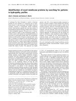
Schematic diagram of pro-SP-B and its processing to SP-BFigure 1
Schematic diagram of pro-SP-B and its processing to SP-B. Upper panel: Indicated are the antibodies used, the symbols for their
identification, the amino acid stretches against which the antibodies were developed, and a diagram of the structure of pro-SP-
B. Lower panel: The molecular weight and the reactivity of the antibodies (in the absence, but not in the presence of the com-
peting peptides) during Western blotting is indicated. The sizing of the letters used for indication of the molecular weights is
proportional to the frequency at which the bands were observed (biggest: common >75% of subjects, 2
nd
biggest: frequent, in
<75 but >50% of the subjects, 3
rd
biggest: sporadic, in <50 but >25% of the subjects, smallest: rare, in <25% of the subjects).
The sequence of SP-B within the pro-SP-B sequence is indicated in pink. All bands were analyzed under reducing conditions.
Respiratory Research 2005, 6:80 />Page 5 of 11
(page number not for citation purposes)
B and SP-C and their proforms, with and without compet-
ing peptide. Under these conditions the assay could detect
about 2.5 ng of SP-B or SP-C per lane. In several experi-
ments, aliquots of a patient with pro-SP-C forms were run
in parallel as a positive control for pro-SP-C forms.
Immunoblots and silver stained gels were scanned with
the Fluor-S MultiImager (Biorad, Richmond, CA) gel doc-
umentation system, and the resulting images were ana-
lyzed with the Software MultiAnalyst (Biorad, Richmond,
CA).
Deglycosylation
To determine if the proteins that reacted with the
CTERMB antibody on the immunoblots were glyco-
sylated, the samples were deglycosylated before applying
them on the gel (4). In brief, 1 unit of recombinant N-gly-
cosidase F (Roche Molecular Biochemicals, Mannheim)
was added to 500 µl incubation buffer (100 mM Na-phos-
phate, 25 mM EDTA, 1% β-mercaptoethanol, 0.5% Triton
X-100, 0,1% SDS, pH 7.2). The vacuum dried sample was
resuspended in 20 µl of this solution and incubated for 15
h at 37°C. The sample was then vacuum dried and ana-
lyzed by Western immunoblot.
Genetic analysis
For SFTPB mutation screening, first the 121ins2 frame-
shift mutation was searched using the restriction enzyme
cleavage SfuI endonuclease by PCR. In 121ins2-negative
patients, SFTPB exons 1–11 and the promoter region were
PCR-amplified and the purified PCR products served as
templates in the sequencing reaction using Ready Reac-
tion Dye Terminator Cycle Sequencing Kit With Ampli-
Taq
®
DNA Polymerase, FS (PEBiosystems, Foster City, CA)
with forward and reverse PCR oligonucleotides used as
extension primers. Extension products were analyzed
using the ABIPRISM™ 310 Genetic analysis System (PEBi-
osystems), as previously reported in detail [18]. Similarly,
SFTPC exons 1–6 were analysed [17].
Statistical analysis
Statistical calculations were performed with the Software
GraphPad Prism 4.0 (GraphPad Software, San Diego,
CA). Differences in nonparametric values were calculated
with the Kruskal-Wallis test. For pair wise comparisons of
groups we used Dunn's test (2). Differences in frequencies
were calculated with the Fisher exact test. Correlation
coefficients were determined according to Pearson.
Results with a p ≤ 0.05 were considered significant.
Children with chronic bronchitisFigure 2
Children with chronic bronchitis. Representative Western
blotting pattern of BAL from child with chronic bronchitis
(patient control 03). After SDS-PAGE and transfer, the mem-
branes were probed with different antibodies directed
against SP-B, certain sequences of the pro-SP-B, in the
absences (-) and presence (+) of excess of the peptides, used
to raise the antibodies, SP-C and against pro-SP-C, in the
absence (-) and presence (+) of excess of the N-terminal
peptide, used to raise these antibodies. The numbers next to
the lanes indicate the molecular weight in kDa. The arrow
heads indicate bands of interest, as described in the text. All
bands were analyzed under reducing conditions.
SP-B deficiencyFigure 3
SP-B deficiency. Western blotting of a lavage from patient SP-
B 06 homozygous for the 121ins2 SFTPB mutation. After
SDS-PAGE and transfer, the membranes were probed with
the antibodies indicated. The pro-forms were probed in the
absence (-) and presence (+) of an excess of the peptide used
to raise this antibody. Note that bands that are not displaced
by the competing peptide were not considered as specific
bands (marked by an asterisk). The numbers next to the
lanes indicate the molecular weight in kDa. The closed
arrowheads indicate the absence of SP-B and of proforms of
SP-B. Arrows show the presence of SP-C (open arrow) and
of abberant pro-SP-C (closed arrows). Some aberrant pro-
SP-C can also be seen on the SP-C blot, above the SP-C
band, which is indicated by an open arrowhead. All bands
were analyzed under reducing conditions.
Respiratory Research 2005, 6:80 />Page 6 of 11
(page number not for citation purposes)
Results
Children without lung disease and children with chronic
bronchitis
The children with chronic obstructive bronchitis had a
slight increase in neutrophils (3% (2; 15)(data are median
and (25.; 75. percentile)) compared to children without
lung disease (1% (1; 2); p = 0.035) and a somewhat lower
viability (80% (70; 90) and 90% (80; 97) in children
without lung disease; p = 0,035). The other variables did
not differ and were within the normal range, i.e. children
with chronic obstructive bronchitis: total cell count 150/
µl (82; 275), macrophages 80% (69; 90) of total cells,
lymphocytes 10% (4/14), eosinophils 0% (0; 2) and
recovery was 54% (39; 70) and the children without lung
disease: total cell count 115/µl (82; 180), macrophages
87% (82; 92) of total cells, lymphocytes 11.5% (7; 14.5),
eosinophils 0% (0; 0.5) and recovery was 48% (42; 62).
Mature SP-B was regularly detected in all lavages from
normal children and from those with chronic bronchitis
at a median molecular weight of 7 kDa (Tab. 1, Fig. 2).
Similarly, pro-SP-B forms with a molecular weight of 25–
26 kDa were commonly observed using an antibody
against the C-terminal flanking propeptide of pro-SP-B
(Tab. 2, Fig. 2). Those bands never reacted with NFPROX,
but showed reactivity with NFLANK, demonstrating that
this was a processing intermediate generated by removal
of the proximal N-terminal amino acids. A similar, but
somewhat more truncated, 19–21 kDa pro-SP-B fragment
was detected sporadically in these children (Tab. 2, Fig. 2).
The pro-SP-B forms at 25–26 and 19–21 kDa were glyco-
sylated as treatment with N-glycosidase F resulted in a sig-
nificant drop in size for both peptides (not shown). A 40–
42 kDa form and a 34–36 kDa form of pro-SP-B were
rarely detected. Except for a single case when a 9 kDa C-
terminal cleavage fragment was observed, in these chil-
dren no other cleavage products of pro-SP-B processing
Table 2: Pro-SP-B and pro-SP-C in the comparison groups, i.e. children without lung disease and in children with chronic bronchitis.
pro-SP-B pro-SP-C
Detecting antibody CTERMB CFLANK NFLANK NFPROX
M
r
of band 40–42 34–36 25–26 19–21 9 25–26
Children without
lung disease (n = 15)
7%
[8]
0% 80%
[1,3,4,6–8,10–15]
7%
[3]
nd nd nd no bands
Chronic obstructive
bronchitis (n = 19)
5%
[14]
26%
[12–14,18]
100%
[1–19]
37%
[4–6,10,13,15,18]
5%
[15]
21%
[4–6,9]
no bands no bands
Percent of subjects with bands and identification numbers of those subjects in whom bands reacting with the anti-pro-SP-B-antibodies CTERMB,
NFLANK, CFLANK and NFPROX displaced by the CTERMB, CFLANK, NFLANK or NFPROX peptides, or the anti-pro-SP-C-antibody NPRO-SP-
C-C2 and displaced by the respective peptide, were identified. The identification numbers of the patients are given in square brackets []. Numbers
in bold refers to bands not identified by the CTERMB antibodies. Due to shortage of lavage material in the normal controls (no lung disease), not all
4 antibodies were tested in this group (nd = not done).
Table 3: Pro-SP-B and pro-SP-C in children with no SP-B
pro-SP-B pro-SP-C
Detecting antibody CTERMB NFLANK NPROSP-C-C2
M
r
of band (kDa) 34–36 25–26 19–21 25–26
Subject Genetic analysis of SFTPB
no SP-B 01 no SFTPB mutation; marker exclusion - ++ - - -
no SP-B 02 496delG homozygote + + + - -
no SP-B 03 121ins2 homozygote - + - - 6 and 7.9 kDa
no SP-B 04 no SFTPB mutation - ++ - - -
no SP-B 05 457delC/121ins2 compound heterozygotes - - - - -
no SP-B 06 121ins2 homozygote - - - + 6.6. and 9 kDa
Bands reacting with the anti-pro-SP-B-antibodies CTERMB, NFLANK, CFLANK and NFPROX displaced by the CTERMB, NFLANK, CFLANK or
NFPROX peptides are indicated by "+", or the anti-pro-SP-C-antibody NPRO-SP-C-C2 and displaced by the respective peptide are indicated by the
molecular weight directly.
Respiratory Research 2005, 6:80 />Page 7 of 11
(page number not for citation purposes)
were identified. Mature SP-C with M
r
of 5.0 kDa was
present in both controls and children with chronic bron-
chitis, whereas pro-SP-C forms were never detected in BAL
(Tabs. 1 and 2, Fig. 2).
Children with no SP-B
6 of all children investigated did not have SP-B in their
lavages. Of these, 4 had lethal mutations of the SFTPB
gene, i.e. SP-B deficiency (Tab. 3). Pro-SP-B processing
products were not found in patient 5, having a 457delC/
121ins2 compound heterozygote mutation (Fig. 3, Tab.
3). Unexpectedly, patients 3 and 6, homozygous for
121ins2, and patient 2 homozygous for 496delG had
small but specific (competitive) pro-SP-B bands at about
19–21, 25–26 or 34–36 kDa (Tab. 3). Aberrant pro-SP-C
bands previously thought to be diagnostic of SP-B muta-
tions were only detected in 121ins2-mutations but not
with 457delG [17,26] or with 496delG mutations.
In the other two infants with no SP-B in the lavages,
SFTPB and SFTPC mutations were excluded [17,18].
These patients had significant amounts of pro-SP-B at 25–
26 kDa, similar to that observed in the comparison
groups. They also did not have pro-SP-C forms in their
lavages, providing additional indirect evidence against SP-
B processing defects. However, one of these two patients,
i.e. patient 4 (Tab. 1), also lacked mature SP-C. This infant
died at the age of 1 month from respiratory failure. This
case suggests the presence of SP-B and SP-C processing
defects arising by means other than from mutations of
these genes, i.e. alterations in the protein processing
machinery or in the lipid transporters, like ABCA3, as
recently shown [27]. The other child (patient 1, Tabs. 1
and 3) is still alive with corticosteroids.
Children with PAP
In all subjects with PAP, except patient 5, antibodies
against GM-CSF in their sera or lavages were excluded in
the pathogenesis of their disease. Although SP-B was
abundantly present and mutations of SFTPB were
excluded [18], alterations of SP-B processing from other
causes have not been excluded. In general, the same pro-
SP-B processing products were observed as in the control
and the chronic bronchitis group, however, the 25–26
kDa band was stained by NFLANK at increased frequency
(Tab. 4, Fig. 4, lanes 4 and 5). In addition, 15 kDa and 13
kDa bands were present that were only stained by
NFPROX. These peptides represent the N-terminal cleaved
processing fragments, which were detected only in these
patients and not in the respective control group (Tab. 4,
Fig. 4, lanes 6 and 7). Three of the PAP patients (PAP 14,
PAP 05 and PAP 10) had bands reacting merely with
CFLANK or NFLANK. These bands were at 8, 9, 11 and 12
kDa. These may represent imprecisely processed SP-B, still
having not completely removed small N- or C-terminal
peptide stretches (Figs. 1, Tab. 4).
Among the PAP patients, only 2 had consistent pro-SP-C
bands (Tab. 4). Subject PAP 08, a patient with a hetero-
zygous SFTPC mutation and previously described in
detail, had 3 bands, and subject PAP 04, in whom no SP-
C mutation was detected, had one band at 6 kD [17].
Those 2 patients with the SFTPC mutation g.2125G>A
[17] had no pro-SP-C bands with this antibody.
Table 4: Pro-SP-B and pro-SP-C in 15 children with pulmonary alveolar proteinosis
pro-SP-B pro-SP-C
Detecting antibody CTERMB CFLANK NFLANK NFPROX NPROSP-C-C2
M
r
of bands(kDa)
40–42 7% [5]
34–36 7% [5]
25–26 93% [1–5,7–15] 20% [3,5,15] 87%
+
[1–5,7–12,14,15] - -
19–21 87%* [1–5,7–13,15] 7% [5] 20% [4,5,9] 7% [4] 7% [8]
15 7% [8] - 7% [8] 33%
+
[2,8,9,11,12]7% [8]
13 20% [3,8,9]-
12 - - 7% [14]- -
11 - 7% [14]- - 7% [8]
9-7% [10]14% [5,10]- -
6 7% [4]
Percent of subjects with bands and identification numbers of those subjects in whom bands reacting with the anti-pro-SP-B-antibodies CTERMB,
NFLANK, CFLANK, NFPROX and displaced by the CTERMB, NFLANK, CFLANK, NFPROX peptides, or the anti-pro-SP-C-antibody NPRO-SP-
C-C2 and displaced by the respective peptide, were identified. Differences in the frequency of bands of all the disease groups were evaluated by the
Fisher exact test and those with a P ≤ 0.05 were indicated by an * for comparison with the healthy control group or by a
+
for comparison with the
disease control group, bronchitis (see table 2). The identification numbers of the patients are given in square brackets []. Numbers in bold indicate
bands not identified by the CTERMB antibodies.
Respiratory Research 2005, 6:80 />Page 8 of 11
(page number not for citation purposes)
Infants with chronic respiratory distress of unknown cause
The infants with chronic respiratory distress of unknown
cause had no mutations of SFTPB or SFTPC, and normal
SP-B and SP-C in their lavages (Tab. 1). Nevertheless,
aberrant pro-SP-C was detected in one of these infants at
9 kDa (Tab. 5). Concerning the processing of pro-SP-B sig-
nificant deviations from the pattern observed in the con-
trol groups were observed in some of these children with
cRD. Indeed a pro-SP-B precursor at 40–42 kDa was
observed more frequently in these patients (Fig. 5, Tab. 5).
Similarly, as in PAP, bands reacting with NFPROX, repre-
senting fragments of the cleaved N-terminus, were
detected (Tab. 5, Figs. 1 and 5).
Discussion
In this study we defined the presence and characteristics of
SP-B, SP-C and their processing forms in bronchoalveolar
lavages from children with severe chronic respiratory
distress and in comparison groups of normal children and
children with chronic obstructive bronchitis (Fig. 1). The
major findings are the presence of mature SP-B and SP-C
in all children, except those with SP-B deficiency,
supporting the view that analysis of BAL for these sur-
factant proteins may aid in the diagnostic work up of chil-
dren with severe respiratory distress. Overall pro-SP-C
forms were rarely detected, and their presence was spe-
cific, but not pathognomonic for a SP-B deficiency due to
SFTPB mutations. In addition, using epitope specific
antisera, we identified unique pro-SP-B forms containing
residues 145–160 of proSP-B (i.e. the "NFPROX" epitope)
exclusively in BAL from patients with alveolar proteinosis
and chronic respiratory distress. Taken together, the data
suggest that immunobiochemical analysis of BAL can
detect abnormalities in surfactant biosynthesis and
metabolism associated with a variety of parenchymal lung
diseases.
Of the 6 patients with SP-B deficiency defined as a lack of
mature SP-B on Western blotting, 4 had mutations in
SFTPB (Tab. 3). Based on our results, the biochemical
analysis of BAL fluid for mature SP-B, previously thought
to be diagnostic for SP-B deficiency, is not 100% specific,
as there are additional cause(s) leading to a lack of SP-B.
Possible mechanisms include mutations or secondary
changes in regulatory elements or other defects in the
synthesis and secretion of surfactant, as recently shown
for the ABCA3 transporter [27].
An important finding of this study is the regular detection
of certain pro-SP-B peptides in BAL from children without
bronchoalveolar disease. Most prominent was a 25–26
kDa band, detected in almost all patients. This protein
corresponds to removal of N'-terminal peptides from pro-
SP-B, liberating 13–15 kDa fragments. SP-B is synthesized
as a proprotein by alveolar type II epithelial cells and non-
Children with pulmonary alveolar proteinosisFigure 4
Children with pulmonary alveolar proteinosis. Western blot-
ting of a lavage from patient PAP 12 (only NFPROX bands)
and PAP 04 (all other bands) to demonstrate the most fre-
quent abnormalities. After SDS-PAGE and transfer, the mem-
branes were probed with the antibodies indicated. The pro-
forms were probed in the absence (-) and presence (+) of an
excess of the peptide used to raise this antibody. Note that
bands that are not displaced by the competing peptide were
not considered as specific bands and they are marked by an
asterisk. The numbers next to the lanes indicate the molecu-
lar weights in kDa. The arrowheads indicate the abundance
of SP-B, the bands at 19–21 and 25–26 kDa using CTERMB
which also react with NFLANK, and some of the break-down
fragments reacting with NFPROX which are more frequently
seen in this condition and in cRD as compared to the other
lung diseases (see figure 5). All bands were analyzed under
reducing conditions.
Children with chronic respiratory distress of unknown cause (cRD)Figure 5
Children with chronic respiratory distress of unknown cause
(cRD). Western blotting of a lavage from patient cRD 06
(NFLANK) and from patient cRD 07 (all other blots), per-
formed as described in detail in the legend to figure 4. An
asterisk marks non-specific bands, i.e. bands not displaced by
the competing peptide. The arrowheads indicate the bands
reacting with CTERMB at 40–42 kDa which are more fre-
quently observed in these conditions than in the others. Sim-
ilarly, with CFLANK, bands are seen at 40–42, 25–26, and
19–21 kDa. Cut off fragments likely generated during protein
processing react with NFLANK or NFPROX. All bands were
analyzed under reducing conditions.
Respiratory Research 2005, 6:80 />Page 9 of 11
(page number not for citation purposes)
ciliated bronchiolar (Clara) cells; however, complete
processing of the precursor to the biologically active,
mature peptide occurs only in type II cells. Clara cells
merely generate the 25 and 42 kDa precursors [28]. Thus,
this intermediate represents a normal pro-SP-B processing
intermediate of SP-B biosynthesis and could result from
either constitutive secretion of this form by type II cells or
from the physiologic release of 25 kD pro-SP-B into the
airways by Clara cells. The 25–26 kD bands of pro-SP-B
have previously been described in amniotic fluid from a
24-week-old human fetus, in lung tissue from an infant
with severe bronchopulmonary dysplasia at the time of
lung transplantation, as well as in normal adult lung tis-
sue and lavages and plasma [21,29]. Here we show that
these peptides are released into the bronchoalveolar space
in normal patients. Since lamellar bodies do not contain
pro-SP-B, this likely occurs via constitutive, non-regulated
secretory pathways.
In children with pulmonary alveolar proteinosis we dis-
covered increased amounts of a 19–21 kD intermediate
which reacted against C-terminal pro-SP-B antisera and
with the NFLANK SP-B antibody. This finding of a com-
plex pro-SP-B intermediate containing both the C-termi-
nal propeptide and a vestigial N-terminal propeptide
(approximate residues 186–201) extends the work of Bra-
sch and colleagues who also noted the presence of pro-SP-
B forms containing C-terminal propeptide epitopes [30].
Consistent with our data, this group also found that, in
contrast to patients with congenital respiratory distress
due to SP-B deficiency, the appearance of pro-SP-C forms
in these PAP patients was a rare occurrence. Thus, despite
similar chest x-rays and histopathological findings, the
BAL profile for SP-B, SP-C and their proforms appears use-
ful in distinguishing PAP from SP-B deficiency of any
etiology.
Children with chronic respiratory distress of unknown
cause (cRD) exhibited the 40–42 kD proprotein with
increased frequency. The N'-terminal peptides liberated
from pro-SP-B pre-protein during intracellular processing,
i.e. 13–15 kDa peptides or smaller fragments and reacting
with NFPROX, were found exclusively in both cRD and
PAP (Fig. 1, Tab. 4, Tab. 5). As such they may give diagnos-
tic hints for the involvement of processing defects in,
especially in pediatric PAP.
Other peptides reacted with the antibodies directed to the
flanking aminoacids next to the SP-B core (NFLANK and
CFLANK). The presence of these relatively rarely observed
bands at 11 to 15 kDa was not related to specific clinical
features of the subjects, i.e. more pronounced lung injury,
high protein to phospholipids ratio or high abundance of
SP-B. Both, a 9 kDa intermediate, reactive to NFLANK [21]
and a 9 kDa band reacting with antibodies directed to the
C'-terminal flanking of pro-SP-B, have previously been
observed in human isolated type II cells and fetal lung.
Such bands were indeed detected in the lavages we inves-
tigated, although very rarely.
Pro-SP-C peptides were never detected in the control
groups. This is in agreement with an earlier observation
on a limited number of samples [31]. However, we found
pro-SP-C forms that were clearly, but not exclusively, asso-
ciated with SP-B deficiency or SFTPC mutation. On the
other hand, not all infants with SFTPB (496delC) or
SFTPC (R167Q) mutations had pro-SP-C in their lavages.
Thus the presence of pro-SP-C in lavages may give strong,
Table 5: Pro-SP-B and pro-SP-C in 7 children with chronic respiratory distress of unknown cause (cRD)
pro-SP-B pro-SP-C
Detecting antibody CTERMB CFLANK NFLANK NFPROX NPROSP-C-C2
M
r
of bands (kDa)
40–42 57%* [2,5–7] 14% [7]
25–26 71% [2,3,5–7] 38%
+
[4,6,7] 57% [2,4,5,6] - -
19–21 - - 14% [6]14%§ [6]-
15 29%
§
[2,7]-
914% [6]14% [7] 14% [6]
3.6 14%
§
[3]-
Percent of subjects with bands and identification numbers of those subjects in whom bands reacting with the anti-pro-SP-B-antibodies CTERMB,
NFLANK, CFLANK, NFPROX and displaced by the CTERMB, NFLANK, CFLANK, NFPROX peptides, or the anti-pro-SP-C-antibody NPRO-SP-
C-C2 and displaced by the respective peptide, were identified. Differences in the frequency of bands of all the disease groups were evaluated by the
Fisher exact test and those with a P < 0.05 were indicated by an * for comparison with the healthy control group or by a
+
for comparison with the
disease control group, bronchitis (see table 2). §indicates a significant difference to the disease control group, bronchitis, when all NFPROX reactive
bands were combined (P < 0.01). The identification numbers of the patients are given in square brackets []. Numbers in bold indicate bands not
identified by the CTERMB antibodies.
Respiratory Research 2005, 6:80 />Page 10 of 11
(page number not for citation purposes)
but surely not definitive, diagnostic evidence for SP-B and
SP-C processing defects.
The aberrant pro-SP-C species observed in patients with
SP-B deficiency carrying the 121ins2 mutation consists of
a N-terminal extension of SP-C by the N-flanking 12 ami-
noacids of pro-SP-C [13]. The pro-SP-C forms observed in
patients not bearing a SFTPB mutation clearly differed in
molecular weights from those detected in SP-B deficiency,
suggesting that several processing defects may result in
aberrant pro-SP-C in the alveolar space.
Conclusion
Here we defined the presence and characteristics of SP-B,
SP-C and their processing forms in bronchoalveolar
lavage fluids from children with severe chronic respiratory
distress and in comparison groups of normal children and
children with chronic obstructive bronchitis. Pro-SP-B of
25–26 kD was commonly detected in all groups, suggest-
ing that this form currently does not appear to be of great
diagnostic value for processing defects. In contrast, pro-
SP-B of 19–21 kD was increased in children with alveolar
proteinosis while the cleaved flanking propeptides liber-
ated during intracellular processing of pro-SP-B were
exclusively found in these children and in chronic respira-
tor distress of unknown cause. Furthermore, although
identified at low frequency, pro-SP-C forms when present
in the BAL suggest the presence of one of the parenchymal
diseases studied in this report. Though often associated
with mutations in SFTPB and SFTPC genes, this was not
an exclusive finding limiting the usage of pro-SP-C as a
surrogate for SFTP/SFTPC diagnostic screening proce-
dures. Taken together, our results demonstrate that signif-
icant perturbations in the metabolism of these
hydrophobic surfactant proteins occur in a variety of
chronic lung diseases.
Competing interests
The author(s) declare that they have no competing
interests.
Authors' contributions
MG designed the study, categorized and organized the
subjects, wrote initial drafts of the manuscript, SS
performed the blots, MT and MB determined the genotype
of the patients, MG, SS, MS, AB, MT and MB collected the
case histories, reviewed the subjects data and clinical
courses, SG and MFB participated in the design for the
methods to blot for the surfactant proteins, helped to
organize the data and the results, and to prepare the man-
uscript. All authors read and approved the final
manuscript.
Acknowledgements
The authors are grateful to Andrea Schams and Yvonne Wüst from Ludwig-
Maximilians Universität, Munich, for expert technical assistance. We thank
Dr. Wolfram Steinhilber, ALTANA Pharma AG, Konstanz, Germany for
donating antibodies to the surfactant proteins B and C. Supported by: DFG
Gr 970/7-1 (MG), HL 076064 (MFB), and P50-HL56401 (MFB).
References
1. Griese M: Pulmonary surfactant in health and human lung dis-
eases: state of the art. Eur Respir J 1999, 13:1455-1476.
2. Weaver TE, Conkright JJ: Functions of surfactant proteins B and
C. Annu Rev Physiol 2001, 63:555-578.
3. Weaver TE: Synthesis, processing and secretion of surfactant
proteins B and C. Biochimica et Biophysica Acta-Molecular Basis of
Disease 1998, 1408:173-179.
4. Brasch F, Ochs M, Kahne T, Guttentag S, Schauer-Vukasinovic V, Der-
rick M, et al.: Involvement of napsin A in the C- and N-terminal
processing of surfactant protein B in type-II pneumocytes of
the human lung. J Biol Chem 2003, 278:49006-49014.
5. Ueno T, Linder S, Na CL, Rice WR, Johansson J, Weaver TE:
Processing of Pulmonary Surfactant Protein B by Napsin and
Cathepsin H. J Biol Chem 2004, 279:16178-16184.
6. Brasch F, Johnen G, Winn-Brasch A, Guttentag SH, Schmiedl A, Kapp
N, et al.: Surfactant Protein B in Type II Pneumocytes and
Intra-Alveolar Surfactant Forms of Human Lungs. Am J Respir
Cell Mol Biol 2004, 30:449-458.
7. Guttentag S, Robinson L, Zhang P, Brasch F, Buhling F, Beers M:
Cysteine protease activity is required for surfactant protein
B processing and lamellar body genesis. Am J Respir Cell Mol Biol
2003, 28:69-79.
8. Wood S, Yaremko ML, Schertzer M, Kelemen PR, Minna J, West-
brook CA: Mapping of the Pulmonary Surfactant SP5
(SFTP2) Locus to 8p21 and Characterization of a Microsat-
ellite Repeat Marker That Shows Frequent Loss of Hetero-
zygosity in Human Carcinomas. Genomics 1994, 24:597-600.
9. Glasser SW, Korfhagen TR, Perme CM, Pilot-Matias TJ, Kister S,
Whitsett JA: Two SP-C genes encoding human pulmonary sur-
factant proteolipid. J Biol Chem 1988, 263:10326-10331.
10. Qanbar R, Cheng S, Possmayer F, Schurch S: Role of the palmi-
toylation of surfactant-associated protein C in surfactant
film formation and stability. Am J Physiol (Lung Cell Mol Physiol)
1996, 271:L572-L580.
11. Stults JT, Griffin PR, Lesikar DD, Naidu A, Moffat B, Benson BJ: Lung
surfactant protein SP-C from human, bovine, and canine
sources contains palmityl cysteine thioester linkages. Am J
Physiol (Lung Cell Mol Physiol) 1991, 261:L118-L125.
12. Nogee LM, de Mello DE, Dehner LP, Colten HR: Brief-report: defi-
ciency of pulmonary surfactant protein B in congenital alve-
olar proteinosis. N Engl J Med 1993, 328:406-410.
13. Li J, Ikegami M, Na CL, Hamvas A, Espinassous Q, Chaby R, et al.: N-
terminally extended surfactant protein (SP) C isolated from
SP-B-deficient children has reduced surface activity and
inhibited lipopolysaccharide binding. Biochemistry 2004,
43:3891-3898.
14. Nogee LM, Wert SE, Proffit SA, Hull WM, Whitsett JA: Allelic het-
erogeneity in hereditary surfactant protein B (SP-B)
deficiency. Am J Respir Crit Care Med 2000, 161:973-981.
15. Mahut B, Delcourt C, Scheinmann P, de Blic J, Mani T, Fournet J, et al.:
Pulmonary alveolar proteinosis: Experience with eight pedi-
atric cases and a review. Pediatrics 1996, 97:117-122.
16. Seymour JF, Presneill JJ: Pulmonary alveolar proteinosis:
progress in the first 44 years. Am J Resp Crit Care Med 2002,
166:215-235.
17. Tredano M, Griese M, Brasch F, Schumacher S, de Blic J, Marque S, et
al.: Mutation of SFTPC in infantile pulmonary alveolar protei-
nosis with or without fibrosing lung disease. Am J Med Genet
2004, 126A:18-26.
18. Tredano M, Griese M, de Blic J, Lorant T, Houdayer C, Schumacher
S, et al.: Analysis of 40 sporadic or familial neonatal and pedi-
atric cases with severe unexplained respiratory distress:
Relationship to SFTPB. Am J Med Genet 2003, 119A:324-339.
19. Griese M, Felber J, Reiter K, Strong P, Reid K, Belohradsky BH, et al.:
Airway inflammation in children with tracheostomy. Pediatr
Pulmonol 2004, 37:356-361.
20. Schmidt R, Steinhilber W, Ruppert C, Grimminger F, Seeger W,
Günther A: An ELISA technique for quantification of sur-
factant apoprotein (SP)-C in bronchoalveolar lavage fluid.
Am J Respir Crit Care Med 2002, 165:470-474.
Publish with BioMed Central and every
scientist can read your work free of charge
"BioMed Central will be the most significant development for
disseminating the results of biomedical research in our lifetime."
Sir Paul Nurse, Cancer Research UK
Your research papers will be:
available free of charge to the entire biomedical community
peer reviewed and published immediately upon acceptance
cited in PubMed and archived on PubMed Central
yours — you keep the copyright
Submit your manuscript here:
/>BioMedcentral
Respiratory Research 2005, 6:80 />Page 11 of 11
(page number not for citation purposes)
21. Guttentag S, Beers MF, Bieler BM, Ballard PL: Surfactant protein B
processing in human fetal lung. Am Phys Soc 1998:L559-L566.
22. Beers MF, Kim CY, Dodia C, Fisher AB: Localization, synthesis,
and processing of surfactant protein SP-C in rat lung ana-
lyzed by epitope-specific antipeptide antibodies. J Biol Chem
1994, 269:20318-20328.
23. Johnson AL, Braidotti P, Pietra GG, Russo SJ, Kabore A, Wang WJ, et
al.: Posttranslational processing of surfactant protein C pro-
protein. Targeting motifs in the NH2-terminal flanking
domain are cleaved in late compartments. Am J Respir Cell Mol
Biol 2001, 24:253-263.
24. Bradford MM: A rapid and sensitive method for the quantita-
tion of microgram quantities of protein utilizing the princi-
ple of protein-dye binding. Anal Biochem 1976, 72:248-254.
25. Heukeshoven J, Dernick R: Improved silver staining procedure
for fast staining in PhastSystem Development Unit. I. Stain-
ing of sodium dodecyl sulfate gels. Electrophoresis 1988, 9:28-32.
26. Tredano M, van Elburg RM, Kaspers AG, Zimmermann LJ, Houdayer
C, Aymard P, et al.: Compound SFTPB 1549C-GAA (121ins2)
and 457delC heterozygosity in severe congenital lung dis-
ease and surfactant protein B (SP-B) deficiency. Hum Mutat
1999, 14:502-509.
27. Shulenin S, Nogee LM, Annilo T, Wert SE, Whitsett JA, Dean M:
ABCA3 Gene Mutations in Newborns with Fatal Surfactant
Deficiency. N Engl J Med 2004, 350:1296-1303.
28. Lin S, Na CL, Akinbi HT, Apsley KS, Whitsett JA, Weaver TE: Sur-
factant protein B (SP-B) -/- mice are rescued by restoration
of SP-B expression in alveolar type II cells but not Clara cells.
J Biol Chem 1999, 274:19168-19174.
29. Doyle IR, Bersten AD, Nicholas TE: Surfactant proteins-A and -B
are elevated in plasma of patients with acute respiratory
failure. Am J Respir Crit Care Med 1997, 156:1217-1229.
30. Brasch F, Birzele J, Ochs M, Guttentag S, Schoch OD, Boehler A, et
al.: Surfactant proteins in pulmonary alveolar proteinosis in
adults. Eur Respir J 2004, 24:426-435.
31. Vorbroker DK, Profitt SA, Nogee LM, Whitsett JA: Aberrant
processing of surfactant protein C in hereditary SP-B
deficiency. Am J Physiol 1995, 268:L647-L656.