Báo cáo y học: " CD8+ T lymphocytes in lung tissue from patients with idiopathic pulmonary fibrosis" pot
Bạn đang xem bản rút gọn của tài liệu. Xem và tải ngay bản đầy đủ của tài liệu tại đây (805.55 KB, 8 trang )
BioMed Central
Page 1 of 8
(page number not for citation purposes)
Respiratory Research
Open Access
Research
CD
8+
T lymphocytes in lung tissue from patients with idiopathic
pulmonary fibrosis
Zoe Daniil
1
, Panagiota Kitsanta
2
, George Kapotsis
1
, Maria Mathioudaki
2
,
Androniki Kollintza
1
, Marilena Karatza
3
, Joseph Milic-Emili
4
,
Charis Roussos
1
and Spyros A Papiris*
1
Address:
1
Department of Critical Care and Pulmonary Services, National and Capodistrian University of Athens, "Evangelismos" Hospital, Athens,
Greece,
2
Pathology Department, "Evangelismos" Hospital, Athens, Greece,
3
Hematology Department, "Evangelismos" Hospital, Athens, Greece
and
4
Meakins-Cristie Laboratories, McGill University, Montreal, Quebec, Canada
Email: Zoe Daniil - ; Panagiota Kitsanta - ; George Kapotsis - ;
Maria Mathioudaki - ; Androniki Kollintza - ; Marilena Karatza - ;
Joseph Milic-Emili - ; Charis Roussos - ; Spyros A Papiris* -
* Corresponding author
Abstract
Background: Several studies have implicated a role of inflammation in the pathogenesis of lung damage in idiopathic
pulmonary fibrosis (IPF). Parenchymal lung damage leads to defects in mechanics and gas exchange and clinically manifests
with exertional dyspnea. Investigations of inflammatory cells in IPF have shown that eosinophils, neutrophils and CD
8+
TLs may be associated with worse prognosis. We wished to investigate by quantitative immunohistochemistry infiltrating
macrophages, neutrophils and T lymphocytes (TLs) subpopulations (CD
3+
, CD
4+
and CD
8+
) in lung tissue of patients with
IPF and their correlation with lung function indices and grade of dyspnoea.
Methods: Surgical biopsies of 12 patients with IPF were immunohistochemically stained with mouse monoclonal
antibodies (anti-CD
68
for macrophages, anti-elastase for neutrophils, and anti-CD
3
, anti-CD
4
, anti-CD
8
for CD
3+
TLs,
CD
4+
TLs, and CD
8+
TLs respectively). The number of positively stained cells was determined by observer-interactive
computerized image analysis (SAMBA microscopic image processor). Cell numbers were expressed in percentage of
immunopositive nuclear surface in relation to the total nuclear surface of infiltrative cells within the tissue (labeling Index).
Correlations were performed between cell numbers and physiological indices [FEV
1
, FVC, TLC, DLCO, PaO
2
, PaCO
2
and P(A-a)O
2
)] as well as dyspnoea scores assessed by the Medical Research Council (MRC) scale.
Results: Elastase positive cells accounted for the 7.04% ± 1.1 of total cells, CD
68+
cells for the 16.6% ± 2, CD
3+
TLs for
the 28.8% ± 7, CD
4+
TLs for the 14.5 ± 4 and CD
8+
TLs for the 13.8 ± 4. CD
8+
TLs correlated inversely with FVC %
predicted (r
s
= -0.67, p = 0.01), TLC % predicted (r
s
= -0.68, p = 0.01), DLCO % predicted (r
s
= -0.61, p = 0.04), and
PaO
2
(r
s
= -0.60, p = 0.04). Positive correlations were found between CD
8+
TLs and P(A-a)O
2
(r
s
= 0.65, p = 0.02) and
CD
8+
TLs and MRC score (r
s
= 0.63, p = 0.02). Additionally, CD
68+
cells presented negative correlations with both FVC
% predicted (r
s
= -0.80, p = 0.002) and FEV
1
% predicted (r
s
= -0.68, p = 0.01).
Conclusion: In UIP/IPF tissue infiltrating mononuclear cells and especially CD
8+
TLs are associated with the grade of
dyspnoea and functional parameters of disease severity implicating that they might play a role in its pathogenesis.
Published: 24 July 2005
Respiratory Research 2005, 6:81 doi:10.1186/1465-9921-6-81
Received: 26 May 2005
Accepted: 24 July 2005
This article is available from: />© 2005 Daniil et al; licensee BioMed Central Ltd.
This is an Open Access article distributed under the terms of the Creative Commons Attribution License ( />),
which permits unrestricted use, distribution, and reproduction in any medium, provided the original work is properly cited.
Respiratory Research 2005, 6:81 />Page 2 of 8
(page number not for citation purposes)
Background
In usual interstitial pneumonia (UIP)/idiopathic pulmo-
nary fibrosis (IPF) the role of inflammation in the patho-
genesis of fibrosis is debatable [1-3]. Traditionally, UIP/
IPF was regarded to develop in response to chronic
inflammation of the lung parenchyma [4]. This view was
advanced from previous studies implicating a role of the
inflammatory cells including neutrophils, macrophages,
eosinophils and T lymphocytes (TLs), based on the obser-
vation of their accumulation in sites of disease activity [5-
8] or on their presence in high numbers in bronchoalveo-
lar lavage [9-12].
Actually, the current pathogenetic theory that holds in
UIP/IPF, implicates that fibrosis per se might progress
despite a paucity of interstitial inflammation [13]. How-
ever, even in this case, recent data still indicate the conten-
tion that the type of the inflammatory response may
modulate tissue injury, fibrosis or both [3,4]. Animal
studies imply that TLs might play a role in the initiation
and the evolution of pulmonary fibrosis. They also sug-
gest that different TLs subpopulations, including both
CD
4+
and CD
8+
subsets, might contribute through their
ability to secrete fibrogenic cytokines [14,15]. Ultimately,
in UIP/IPF, the inflammatory response is considered to
resemble closely the type-2 T lymphocytic pattern [16-18]
and drives the process in a profibrogenic direction.
The present study was designed to investigate by quantita-
tive immunohistochemistry the inflammatory cell pattern
in lung tissue of patients with UIP/IPF (macrophages,
neutrophils, and CD
3+
, CD
4+
, CD
8+
TLs) and to correlate
their population numbers with the lung function indices
and grade of dyspnoea.
Methods
1. Subjects
The study population consisted of 12 untreated patients
with IPF and included 7 ex-smokers, and 5 never smokers
(Table 1). They were recruited from the respiratory outpa-
tient clinic of the "Evangelismos" General Hospital, Ath-
ens, Greece over a period of 3 years. The diagnosis of UIP/
IPF was based on standard criteria [19], which included
clinical findings (exertional dyspnoea, non-productive
cough, fine bibasilar inspiratory crackles), pulmonary
function tests (restrictive pattern and impaired gas
exchange), and high resolution computerized tomogra-
phy findings (bibasilar reticular abnormalities with mini-
mal ground-glass opacities consistent with the diagnosis
of IPF). The diagnosis of UIP/IPF was confirmed by video-
assisted thoracoscopic lung biopsy in all patients. Pathol-
ogy examination of these specimens clearly documented
UIP according to Katzenstein's and American Thoracic
Society – European Respiratory Society criteria (histologic
variation with alternating zones of interstitial fibrosis,
inflammation, honeycomb change, and normal lung
[1,19]. A right thoracic approach was done and two or
three samples were taken from the right lower or middle
lobe in the region of the greater fissure. Biopsy of the lin-
gular tip was avoided, as changes in this area may be par-
ticularly advanced and unrepresentative. All patients
experienced a normal and uncomplicated postoperative
course. Secondary causes of lung fibrosis were excluded:
none of the patients included in this study had a history
of environmental or occupational exposure, drug toxicity
or connective tissue disease, as documented by patient's
history and thorough clinical and immunological work
out. The study was approved by the institutional ethics
committee and informed consent was obtained from each
patient.
2. Pulmonary function tests
The pulmonary function tests included FEV
1
, FVC, FEV
1
/
FVC ratio × 100, total lung capacity (TLC), residual vol-
ume (RV) and carbon monoxide transfer factor (DLCO).
TLC and RV were measured by the helium dilution
method with a Master Screen apparatus (Erich Jaeger
GmbH, Wuerzburg, Germany), and DLCO by the single
breathholding helium dilution method [20,21]. Lung
function measurements (Table 1) were expressed as per-
centages of predicted values [20,21]. In all patients, the
arterial PaO
2
and PaCO
2
were also measured at rest, and
P(A-a)O
2
calculated.
3. Dyspnea
Dyspnea was assessed with the modified (6-point) MRC
dyspnoea self-administered questionnaire [22] that con-
sists of six questions about perceived breathlessness: cate-
gory 0, no dyspnoea; category 1, slight degree of dyspnoea
(troubled by shortness of breath when hurrying on the
Table 1: Demographic, clinical and lung function data of all
patients at presentation
Variables Values
Age (yr) 60 ± 2
Sex (M/F) 5/7
MRC dyspnoea score 1.8 ± 0.3
FEV
1
(% pr) 85 ± 4
FVC (% pr) 78 ± 4
FEV
1
/FVC (ratio × 100) 85 ± 4
TLC (% pr) 64 ± 3
RV (% pr) 54 ± 4
DLCO (% pr) 50 ± 5
PaO
2
(mmHg) 75 ± 2
P(A-a)O
2
30 ± 3
PaCO
2
(mmHg) 36 ± 3
Data are presented as means ± SEM; M = Male; F = Female; pr:
predicted
Respiratory Research 2005, 6:81 />Page 3 of 8
(page number not for citation purposes)
level or walking up a slight hill); category 2, moderate
degree of dyspnoea (walks slower than people of the same
age on the level because of breathlessness); category 3,
moderately severe degree of dyspnoea (has to stop
because of breathlessness when walking at own pace on
the level); category 4, severe degree of dyspnoea (stops for
breath after walking about 100 yards or after a few min-
utes on the level); category 5, very severe degree of dysp-
noea (too breathless to leave the house or breathless when
dressing or undressing).
4. Histology
Open lung biopsies from the 12 patients were used. They
were taken for diagnostic and staging purposes and were
analyzed according to the Katzenstein's criteria [1] by two
pathologists. Specimens were fixed in 4% formalin and
after dehydration embedded in paraffin. Tissue sections
were orientated and serial sections of 4 µm thickness were
cut and immunohistochemistry was performed according
to the Streptavidin-Biotin method.
5. Immunohistochemistry
To evaluate macrophages, neutrophils and the lym-
phocyte-subpopulations in lung tissues, 4 µm paraffin
sections were immunohistochemically stained with the
following mouse monoclonal antibodies (all from
DAKO, Glostrup, Denmark): macrophages-histiocytes
with anti CD
68
, (clone: M814, dilution 1:4000), neu-
trophils with anti-elastase (clone:M752 dilution 1:4000),
T-cells with anti-CD
3
(dilution 1:200), anti-CD
4
(dilution
1:100), and anti-CD
8
(dilution 1:4), according to the
labeled Streptavidin-Biotin Complex method. The sec-
tions were deparaffinized and rehydrated with Tris-Buff-
ered Saline (TBS: 0.005 M Tris, 0.15 M NaCl), pH = 7.6 for
10 minutes. Endogenous peroxidase was blocked with 3%
hydrogen peroxide for 5 minutes. Then they were washed
in TBS and incubated with primary antibodies at the
appropriate dilutions for one hour. Biotinylated anti-
mouse IgG was used as a secondary antibody (DAKO),
followed by peroxidase-conjugated streptavidin (DAKO).
The peroxidase reaction was developed using 3,3'-diamin-
benzidine tetrachloride (0.25 mg dissolved in 1 ml of
0.02% hydrogen peroxide) for 3 min.
6. Lung Parenchyma Computer Image Analysis
The number of positively stained cells was determined by
observer-interactive computerized image analysis
(SAMBA microscopic image processor), whose hardware
and software have been described by Brugal and associates
[23]. This system is fitted with a standard Zeiss axioplan
microscope, a color video camera (Sony Corporation
Tokyo, Japan), an image analysis processor (matrox) and
an IBM compatible Pentium 2, 166 MHZ computer. Esti-
mation of the standard error of the mean within 95% con-
fidence limits required a maximum of at least randomly
selected 15 High Power fields (X400-Zeiss microscope)
(Analysis per area of approximately 110000 µm
2
). The
immunostaining was analyzed as dark brown color with
counterstained cells as false blue. Formal scoring (labeling
Index) for each antibody was then performed in one sec-
tion for each paraffin block. Interobserver variability was
very low (<0.03%). The results were expressed in percent-
age of immunopositive nuclear surface in relation to the
total nuclear surface of infiltrative cells within the tissue
(labeling Index), as previously described [24,25]. Blood
vessels, connective tissue and cartilage structures were
excluded.
7. Statistical Analysis
Data were expressed as means and standard error (SEM).
Correlation coefficients were calculated using Spearman's
rank method. A p-value of less than 0.05 was considered
statistically significant. Analysis was performed using the
SAS System software.
Results
Demographic characteristics, MRC dyspnoea score and
lung function data of all patients are listed in Table 1. All
patients claimed some degree of dyspnoea (MRC score >
0) and most patients had a restrictive lung function pat-
tern characterized by a decrease in TLC (mean value was
64% of predicted) and an increased in FEV
1
/FVC ratio
×100 (mean value was 85% of predicted). The DLCO was
decreased in all patients (mean value was 50% of
predicted).
Among the inflammatory cells studied T lymphocytes
(CD
3+
) appeared as the most numerous cells infiltrating
the lung parenchyma. They were found in aggregates,
within lymphoid follicles or diffusely within the fibrotic
lung parenchyma and mainly in the areas with moderate
or severe thickening of the alveolar wall. They were also
observed within the wall of the alveoli. The CD
4+
subpop-
ulation was found in aggregates inside or around lym-
phoid follicles. A little portion of them infiltrated the lung
parenchyma diffusely, especially the alveolar wall. On the
other hand, the CD
8+
cells infiltrated the parenchyma
mainly diffusely (Fig 1); they were also found within the
alveolar wall, around the fibrotic foci and in the areas with
alveolar thickening. Less commonly they were distributed
within aggregates or lymphoid follicles. Macrophages
(CD
68+
cells) were preferentially located in the lamina
propria of the airways compared with the surface epithe-
lium and the submucosa. They were also distributed in
large aggregates in the dilated alveolar spaces. Neutrophils
(Elastase+) were mainly observed in the surface epithe-
lium and within the alveolar wall.
Elastase positive cells accounted for the 7.04% ± 1.1 of
total cells, CD
68+
cells for the 16.6% ± 2, CD
3+
TLs for the
Respiratory Research 2005, 6:81 />Page 4 of 8
(page number not for citation purposes)
28.8% ± 7, CD
4+
TLs for the 14.5 ± 4 and CD
8+
TLs for the
13.8 ± 4. Among the infiltrating inflammatory cells,
CD
8+
TLs were inversely correlated with FVC % predicted
(r
s
= -0.67, p = 0.01) (Fig 2), TLC % predicted (r
s
= -0.68,
p = 0.01) (Fig 3), DLCO % predicted (r
s
= -0.61, p = 0.04),
PaO
2
(r
s
= -0.60, p = 0.04). Positive and statistically signif-
icant correlations were found between CD
8+
TLs and P(A-
a)O
2
(r
s
= 0.65, p = 0.02) and CD
8+
TLs and MRC score (r
s
= 0.63, p = 0.02) (Fig 4). Additionally, the CD
68+
cells pre-
sented significant negative correlations with the FVC %
predicted (r
s
= -0.80, p = 0.002) and the FEV
1
% predicted
(r
s
= -0.68, p = 0.01). Elastase positive cells, CD
3+
TLs, and
the CD
4+
TLs did not correlate with any of the functional
indices as well as the MRC score.
Discussion
Several studies have implicated a role of inflammation in
the pathogenesis of lung damage in IPF. Parenchymal
lung damage leads to defects in mechanics and gas
exchange and consequently to exertional dyspnoea, the
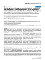
The CD
8+
TLs infiltrate diffusely the lung parenchyma (scale bar = 25 µm)Figure 1
The CD
8+
TLs infiltrate diffusely the lung parenchyma (scale bar = 25 µm).
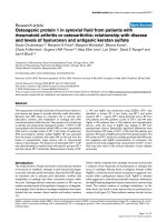
Relationship between CD
8+
TLs (% of total cells) and TLC (% predicted), (r
s
= -0.68, p = 0.01)Figure 3
Relationship between CD
8+
TLs (% of total cells) and TLC (%
predicted), (r
s
= -0.68, p = 0.01).
Respiratory Research 2005, 6:81 />Page 5 of 8
(page number not for citation purposes)
most prominent and disabling symptom in these patients.
This study shows that the type of the inflammatory infil-
trate in lung tissue of patients with UIP/IPF was
predominantly mononuclear, and that among the differ-
ent inflammatory cells, CD
8+
TLs correlated significantly
with both functional [FVC, TLC, DLCO, PaO
2
, P(A-a)O
2
]
and clinical indices (the MRC chronic dyspnoea score) of
the disease severity and extent studied and macrophages
(CD
68+
cells) with some of the functional indices studied
(FVC and FEV
1
).
Previous studies have shown that a patchy chronic inter-
stitial inflammatory process coexists with an abnormal
extracellular matrix deposition, foci of fibroblasts, and
alveolar collapse in lung tissue of subjects with IPF
[1,2,8,26]. This inflammatory component in tissue speci-
mens appeared to consist primarily of mononuclear cells
(macrophages, lymphocytes and plasma cells) while the
presence of the other inflammatory cells such as neu-
trophils and eosinophils appeared less numerous
[5,8,26,27]. Previous studies of immunohistochemical
analysis including the TLs subpopulations of lung tissue
in IPF [5,8,28,29] and other interstitial pneumonias [30]
have shown that the inflammatory process is mainly
mononuclear, and that both CD
4+
and CD
8+
TLs were well
represented and diffusely distributed in the interstitium,
with an additional component of the CD
4+
TLs observed
inside lymphoid follicles. Our findings do not contrast
these observations.
Few studies have attempted to evaluate the structure-func-
tion relationship in IPF, and their findings are not always
in agreement [31-35]. The selection of patients and the
different methodologies might, at least in part, explain
discrepancies. Our findings come into agreement with the
studies by Fulmer and coworkers [31] and Chinet and
coworkers [35] who found significant correlations
between the degree of inflammation and lung volumes
and some index of gas exchange. Gaensler and Carrington
[34] also reported correlations between physiological
indices and an estimate of functional impairment from
histology (designated by them as pathological severity) in
a large but mixed population (502 patients) with "inter-
stitial lung disorders" including 64 patients with UIP.
However, comparisons with previous studies are not
always possible for the following reasons. First, we used a
different methodology than the above-cited works. This is
the first study attempted to correlate cell counts including
TLs subpopulations in tissue biopsies with clinical and
physiological parameters. This should be in relation to the
fact that reliable cell counts were not feasible with the past
technologies. Second, older studies might have included a
mixed population of patients with UIP/IPF, patients with
non-specific interstitial pneumonia and patients with
pulmonary fibrosis associated with collagen vascular dis-
orders since there were not defined strict criteria for these
entities, by that time [36]. Finally, the patients' selection
and the effect of previous treatment might have influ-
enced the results.
Inflammatory cells including subpopulations of TLs have
been also studied in IPF by bronchoalveolar lavage (BAL)
[4] and many of the conclusions regarding the role of
inflammation in interstitial lung disorders have been
drawn from these studies. The possible role of lym-
phocytes in the pathogenesis of IPF traditionally received
little investigation since increase in their number is an
uncommon finding in BAL samples. Hence, early BAL
studies have driven attention into neutrophils as well as
macrophages. However, evidence from animal models
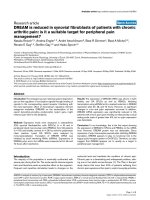
Relationship between CD
8+
TLs (% of total cells) and FVC (% predicted), (r
s
= -0.67, p = 0.01)Figure 2
Relationship between CD
8+
TLs (% of total cells) and FVC (%
predicted), (r
s
= -0.67, p = 0.01).
Relationship between CD
8+
TLs (% of total cells) and MRC dyspnea score, (r
s
= 0.63, p = 0.02)Figure 4
Relationship between CD
8+
TLs (% of total cells) and MRC
dyspnea score, (r
s
= 0.63, p = 0.02).
Respiratory Research 2005, 6:81 />Page 6 of 8
(page number not for citation purposes)
appears to suggest that lymphocytes do play a role in
fibrosis [4]. Furthermore, studies on BAL lymphocytes
have shown that CD
8+
TLs are prominent in BAL in IPF
[36] and may also be associated with a worse prognosis
[37]. T lymphocytes and their phenotypic and functional
characteristics have been more extensively studied in scle-
roderma fibrosis [38-43]. IPF and scleroderma fibrosis are
two fibroses with different prognoses [41]. This might be
related to the fact that most patients with scleroderma
develop a less aggressive form of fibrosing interstitial
pneumonia, the non-specific interstitial pneumonia
(NSIP) [42]. Indeed, studies that compared the prognosis
of patients with "idiopathic" NSIP to that of patients with
usual interstitial pneumonia type/IPF have clearly shown
that the former present a far better prognosis than the lat-
ter [43]. Recent studies with BAL in scleroderma patients
have shown that a subset of them, who present more than
15% lymphocytes in BAL [38], or have activated, long-
lived CD
8+
T cells [40], or produce type 2 cytokines (IL-4
and IL-5) by the CD
8+
TLs [39], present a more aggressive
form of interstitial pneumonia. Hence, TLs and in partic-
ular the CD
8+
subset may be associated with progressive
fibrosis in scleroderma resembling more patients with
IPF.
Notwithstanding correlations do not imply direct cause-
effect relationships, we think that the significant negative
correlations observed in this group of patients with IPF
between CD
8+
TLs and functional indices and the positive
correlation observed between the same cells and clinical
indices estimating the disease severity as well as the corre-
lation between CD
68+
cells with FVC and FEV
1
might sug-
gest a potential pathophysiologic relevance for
mononuclear cells and especially CD
8+
TLs in the patho-
genesis of pulmonary fibrosis. However, the mechanisms
related to these correlations and the relationship of
inflammatory and immune parameters to structural
changes in the lung parenchyma still remain unknown
and further studies are needed for their clarification.
The increase in CD
8+
TLs observed in lung surgical biop-
sies in patients with IPF appears intriguing. Classically,
the major role of CD
8+
TLs in the inflammatory response
has been considered the rapid resolution of viral infec-
tions [44]. It has also become evident that CD
8+
TLs may
contribute to lung injury [45,46]. Viruses have been impli-
cated in the pathogenesis of IPF, and a higher incidence of
viral infections (Ebstein Barr Virus, influenza, cytomega-
lovirus, and possibly Hepatitis C virus) has been reported
in these patients [19]. Recently, it has been hypothesized
that in patients with IPF an excessive recruitment of CD
8+
TLs may occur in response to recurrent or persistent viral
infections, and this excessive response may play a role for
the development of lung damage [47]. The above hypoth-
esis has received some experimental confirmation by the
studies of Enelow and coworkers [47] and Small and cow-
orkers [48] who have shown that antigen-specific CD
8+
T
cell recognition of an alveolar epithelial "autoantigen" is
sufficient to trigger an inflammatory cascade that results
in the histological and physiological manifestations of
interstitial pneumonia. CD
8+
TLs can differentiate into
cells that make IFN-γ but no IL-4 (Tc1 cells) promoting
attenuation of fibrosis and cells that make IL-4 but not
IFN-γ (Tc2 cells) leading to exuberant fibrosis [49].
Though further studies are necessary to address the spe-
cific role of the Tc2 cells in pulmonary fibrosis, some data
such as the upregulation of genes encoding immunoglob-
ulins and extracellular matrix proteins in IPF lung tissue
[50] appear to suggest that the predominance of type-2
immune response in IPF [51] is what drives the process in
profibrogenic direction.
Conclusion
We found that the type of the inflammatory cell infiltrate
in surgical biopsies of patients with UIP/IPF was predom-
inantly mononuclear. Among the different inflammatory
cells revealed by immunohistochemistry, the CD
8+
TLs
correlated significantly with both functional [FVC, TLC,
DLCO, PaO
2
, P(A-a)O
2
] and clinical indices (the MRC
chronic dyspnoea score) of disease severity and extent
studied and the CD
68+
cells with FVC and FEV
1
. These data
might suggest a potential role for mononuclear cells and
especially CD
8+
TLs in the pathogenesis of pulmonary
fibrosis. However, because of the relatively small size of
the population studied, further studies are needed to sup-
port our findings.
Competing interests
The author(s) declare that they have no competing
interests.
Authors' contributions
ZD participated in the design of the study and collection
of the clinical data, performed the statistical analysis and
drafted the manuscript. PK carried out the histology and
the immunohistochemical analysis and revised the article.
GK participated in the collection of the data and helped to
draft the manuscript. MD helped in biopsy evaluation,
diagnosis and analysis. AK participated in tissue collection
and data analysis. MK participated in data analysis. JM-E
helped in the interpretatiuon of the data and revised the
article. CR participated in the interpretation of the data
and revised the article. SP conceived of the study, partici-
pated in its design and coordination and helped to draft
the manuscript. All authors read and approved the final
manuscript.
Acknowledgements
Supported by the "Thorax" Foundation, Athens, Greece.
Respiratory Research 2005, 6:81 />Page 7 of 8
(page number not for citation purposes)
References
1. Katzenstein A, Zisman AD, Litzky AL, Nguyen TB, Kotloff MR: Usual
interstitial pneumonia. Histologic study of biopsy and
explant specimens. Am J Surg Pathol 2002, 26:1567-1577.
2. Flaherty KR, Travis WD, Colby TV, Toews BG, Kazerooni AE, Gross
HB, Jain A, Strawderman LR III, Flint A, Lynch PJ III, Martinez JF: His-
topathologic variability in usual and nonspecific interstitial
pneumonias. Am J Respir Crit Care Med 2001, 164:1722-1727.
3. Gross TJ, Hunninghake GW: Idiopathic pulmonary fibrosis. N
Engl J Med 2001, 345:517-525.
4. Riches WHD, Worthen GS, Augustin A, Lapadat R, Chan DE:
Inflammation in the pathogenesis of interstitial lung disease.
In Interstitial Lung Disease Edited by: Schwartz JM, King ET Jr. Hamilton
London BC Decker; 2003:187-220.
5. Kradin RL, Divertie MB, Colvin RB, Ramirez J, Ryu J, Carpenter HA,
Bhan AK: Usual interstitial pneumonitis is a T-cell alveolitis.
Clin Immunol Immunopathol 1986, 40:224-235.
6. Crystal RG, Fulmer JD, Roberts WC, Moss ML, Line BR, Reynolds
HY: Idiopathic pulmonary fibrosis. Clinical, histologic, radio-
graphic, physiologic, scintigraphic, cytologic and biochemical
aspects. Ann Intern Med 1976, 85:769-788.
7. Hunninghake GW, Kawanami O, Ferrans VJ, Young RC, Roberts WC,
Crystal RG: Characterization of the inflammatory and
immune effector cells in the lung parenchyma of patients
interstitial lung disease. Am Rev Respir Dis 1981, 123:407-412.
8. Campell DA, Poulter LW, Janossy G, du Bois RM: Immunohistolog-
ical analysis of lung tissue from patients with cryptogenic
fibrosing alveolitis suggesting local expression of immune
hypersensitivity. Thorax 1985, 40:405-411.
9. Haslam PL, Turton CW, Lukoszek A, Salsbury AJ, Dewar A, Collins
JV, Turner-Warwick M: Bronchoalveolar lavage fluid cell counts
in cryptogenic fibrosing alveolitis and their relation to
therapy. Thorax 1980, 35:328-339.
10. Reynolds HY, Fulmer JD, Kazmierowski JA, Roberst WC, Frank MM,
Crystal RG: Analysis of cellular and protein content of bron-
cho-alveolar lavage fluid from patients with idiopathic pul-
monary fibrosis and chronic hypersensitivity pneumonitis. J
Clin Invest 1977, 59:165-175.
11. Peterson MW, Monick M, Hunninghake GW: Prognostic role of
eosinophils in pulmonary fibrosis. Chest 1987, 92:51-56.
12. Lynch JP 3rd, Standiford TJ, Rolfe MW, Kunkel SL, Strieter RM: Neu-
trophilic alveolitis in idiopathic pulmonary fibrosis. The role
of interleukin-8. Am Rev Respir Dis 1992, 145:1433-1439.
13. Selman M, King TE Jr, Pardo A: Idiopathic pulmonary fibrosis:
prevailing and evolving hypotheses about its pathogenesis
and implications therapy. Ann Intern Med 2001, 134:136-151.
14. Piguet PF, Collart MA, Grau GE, Kapanci Y, Vassali P: Tumor necro-
sis factor/cachectin plays a key role in bleomycin induced
pneumopathy and fibrosis. J Exp Med 1989, 170:655-663.
15. Hu H, Stein-Streilein J: Hapten-immune pulmonary interstitial
fibrosis (HIPIF) in mices requires both CD
4+
and CD
8+
T-lym-
phocytes. J Leukocyte Biol 1993, 54:414-422.
16. Furuie H, Yamasaki H, Suga M, Ando M: Altered accessory cell
function of alveolar macrophages: a possible mechanism for
induction of Th 2 secretory profile in idiopathic pulmonary
fibrosis. Eur Respir J 1997, 10:787-794.
17. Hancock A, Armstrong L, Gama R, Millar A: Production of inter-
leukin 13 by alveolar macrophages from normal and fibrotic
lung. Am J Respir Cell Mol Biol 1998, 18:60-65.
18. Romagnani S: Th1/Th2 cells. Inflamm Bowel Dis 1999, 5:285-294.
19. American Thoracic Society: Idiopathic pulmonary fibrosis: diag-
nosis and treatment. International consensus statement. Am
J Respir Crit Care Med 2000, 161:646-664.
20. Quanjer PhH, Tammeling GJ, Cotes JE, Pedersen OF, Peslin R, Yer-
nault J-C: Lung volumes and forced ventilatory flows. Report
working party, Standardization of lung function tests, Euro-
pean Community for steel and coal. Official Statement of
the European respiratory Society. Eur Respir J 1993:5-40.
21. Cotes JE, Chinn DJ, Quanjer PhH, Roca J, Yernault J-C: Standardi-
zation of the measurement of transfer factor (Diffusing
Capacity). Report working party, Standardization of lung
function tests, European Community for steel and coal. Offi-
cial Statement of the European respiratory Society. Eur
Respir J 1993:41-52.
22. Eltayara L, Becklake MR, Volta CA, Milic-Emili J: Relationship
between chronic dyspnea and expiratory flow limitation in
patients with chronic obstructive pulmonary disease. Am J
Respir Crit Care Med 1996, 154:1726-1734.
23. Brugal T, Arnorssen R, Bengtsson A, Wilander E: A double scan-
ning micromorphometer for image analysis: hardware, soft-
ware and biomedical applications. J Histochem Cytochem 1979,
27:144-152.
24. Charpin C, Martin P, Jacquemin J, Jacquemier J, Lavaut MN, Pourreau-
Schneider N, Toga M: Estrogen receptor immunocytochemical
assay (ER-ICA): Computerized image analysis system,
immunoelectron microscopy and comparisons with estra-
diol binding assays in 115 breast assays in 115 breast
carcinomas. Cancer Res 1986:4271-4277.
25. Costes V, Marty-Ane C, Picot MC, Serre I, Pujol J-L, Mary H, Baldet
P: Typical and Atypical Bronchopulmonary Carcinoid
Tumors: A Clinicopathologic and Ki-67-Labeling Study. Hum
Pathol 1995, 26:740-745.
26. White SR, Lazar HM, Thannichal JV: Pathogenetic mechanisms in
usual interstitial pneumonia/idiopathic pulmonary fibrosis. J
Pathol 2003, 201:343-354.
27. Haslam LP, Turton WGC, Heard B, Lukoszek A, Collins VJ, Slasbury
A, Turner-Warwick : Bronchoalveolar lavage in pulmonary
fibrosis: comparison of cells obtained with lung biopsy and
clinical features. Thorax 1980, 35:9-18.
28. Haslam LP: Evaluation of alveolitis by studies of lung biopsies.
Lung 1990:984-992.
29. Paradis LI, Dauber HJ, Rabin SB: Lymphocyte phenotypes in
bronchoalveolar lavage and lung biopsies in sarcoidosis and
idiopathic pulmonary fibrosis. Am Rev Respir Dis 1986,
133:855-860.
30. Yamadori I, Fujiata J, Kajitani H, Tokuda M, Yang Y, Ohtsuki Y, Yoshi-
nouchi T, Kamei T, Ishida T: Lymphocytic subsets in lung tisuue
of non-specific interstitial pneumonia and pulmonary fibrosis
associated with collagen vascular disorders: correlation with
CD4/CD8 ratio in bronchoalveolar lavage. Lung 2000,
178:361-370.
31. Fulmer JD, Roberts WC, Von Gal R, Crystal RG: Morphologic-
physiologic correlates of the severity of fibrosis and degree
of cellular infiltration in idiopathic pulmonary fibrosis. J Clin
Invest 1979, 63:665-676.
32. Green GM, Graham GB, Hanson JS, Gump DW, Phillips CA, Brody
AR, Sylvester DW, Landis JN, Davis GS, Craighead JE: Correlate
studies of interstitial pulmonary disease. Chest 1976:263.
33. Gaensler EA, Carrington CB, Coutu RE, FitzGerald MX: Radiologic-
physiologic-pathologic correlations in interstitial
pneumonias. Prog Respir Res 1975, 8:223-241.
34. Gaensler EA, Carrington CB: Open lung biopsy for chronic dif-
fuse infiltrative lung disease: clinical, roentgenographic and
physiologic correlations in 502 patients. Ann Thorac Surg 1980,
30:411-426.
35. Chinet T, Jaubert F, Dusser D, Danel C, Chretien J, Huchon JG:
Effects of inflammation and fibrosis on pulmonary function in
diffuse lung fibrosis. Thorax 1990, 45:675-768.
36. Groen H, Hamstra M, Aalbert R, Van Der Mark WT, Koëter HG,
Postma SD: Clinical evaluation of lymphocyte sub-populations
and oxygen radical production in sarcoidosis and idiopathic
pulmonary fibrosis. Respir Med 1994, 88:55-64.
37. Fireman E, Vardinon N, Burke M, Soizer S, Levin S, Endler A, Stav D,
Topilsky M, Mann A, Schwarz S, Greif J: Predictive value of
response to treatment of T-lymhocyte subpopulations in idi-
opathic pulmonary fibrosis. Eur Respir J 1998, 11:706-711.
38. Behr J, Vogelmeier C, Beinert T, Meurer M, Krombach F, Konig G,
Fruhmann G: Bronchoalveolar lavage for evaluation and man-
agement of scleroderma disease of the lung. Am J Respir Crit
Care Med 1996, 154:400-6.
39. Atamas SP, Yurovsky VV, Wise R, Wigley FM, Goter Robinson CJ,
Henry P, Alms WJ, White B: Production of type 2 cytokines by
CD8+ lung cells is associated with greater decline in pulmo-
nary function in patients with systemic sclerosis. Arthritis
Rheum 1999, 42:1168-78.
40. Luzina IG, Atamas SP, Wise R, Wigley FM, Choi J, Xiao HQ, White B:
Occurrence of an activated, profibrotic pattern of gene
expression in lung CD8+ T cells from scleroderma patients.
Arthritis Rheum 2003, 48:2262-74.
41. Papiris SA, Vlachoyiannopoulos PG, Maniati MA, Karakostas KX,
Constantopoulos SH, Moutsopoulos HH: Idiopathic pulmonary
fibrosis and pulmonary fibrosis in diffuse systemic sclerosis:
Publish with BioMed Central and every
scientist can read your work free of charge
"BioMed Central will be the most significant development for
disseminating the results of biomedical research in our lifetime."
Sir Paul Nurse, Cancer Research UK
Your research papers will be:
available free of charge to the entire biomedical community
peer reviewed and published immediately upon acceptance
cited in PubMed and archived on PubMed Central
yours — you keep the copyright
Submit your manuscript here:
/>BioMedcentral
Respiratory Research 2005, 6:81 />Page 8 of 8
(page number not for citation purposes)
two fibroses with different prognoses. Respiration 1997,
64:81-5.
42. Flaherty KR, Colby TV, Travis WD, Toews GB, Mumford J, Murray S,
Thannickal VJ, Kazerooni EA, Gross BH, Lynch JP 3rd, Martinez FJ:
Fibroblastic foci in usual interstitial pneumonia: idiopathic
versus collagen vascular disease. Am J Respir Crit Care Med 2003,
167:1410-5.
43. Daniil ZD, Gilchrist FC, Nicholson AG, Hansell DM, Harris J, Colby
TV, du Bois RM: A histologic pattern of nonspecific interstitial
pneumonia is associated with a better prognosis than usual
interstitial pneumonia in patients with cryptogenic fibrosing
alveolitis. Am J Respir Crit Care Med 1999, 160:899-905.
44. Ramsay A, Ruby J, Ramshaw I: A case for cytokine as effector
molecules in the resolution of virus infection. Immunol Today
1993, 14:155-157.
45. Alwan WH, Kozlowska WJ, Opensaw PJ: Distinct types of lung
disease caused by functional subsets of antiviral T cells. J Exp
Med 1994, 179:81-89.
46. Guidotti LG, Chisari FV: To kill or to cure: options in host
defense against viral infection. Curr Opin Immunol 1996,
8:478-483.
47. Enelow RI, Mohammed AZ, Stoler MH, Ning Liu A, Young JS, Lou Y-
H: Structural and functional consequences of alveolar cell
recognition by CD8
+
T Lymphocytes in experimental lung
disease. J Clin Invest 1998, 102:1653-1661.
48. Small BA, Dressel SA, Lawrence CW, Drake DR III, Stoler MH,
Enelow RI, Braciale TJ: CD8
+
T Cell-mediated Injury In Vivo
Progresses in the Absence of Effector T Cells. J Exp Med 2001,
194:1835-1846.
49. Croft M, Carter L, Swain SL, Dutton RWL: Generation of polar-
ized antigen-specific CD8 effector populations: reciprocal
action of interleukin (IL)-4 and IL-12 in promoting type 2
versus type 1 cytokine profiles. J Exp Med 1994, 180:1715-1728.
50. Zuo F, Kaminski N, Eugui E, Allard J, Yakhini Z, Ben-Dor A, Lollini L,
Morris D, Kim Y, DeLustro B, Sheppard D, Pardo A, Selman M, Heller
RA: Gene expression analysis reveals matrilysin as a key reg-
ulator of pulmonary fibrosis in mice and humans. Proc Natl
Acad Sci USA 2002, 99:6292-6297.
51. Strieter RM, Keane MP: Innate immunity dictates cytokine
polarization relevant to the development of pulmonary
fibrosis. J Clin Invest 2004, 114:165-168.