Báo cáo khoa học: "In vivo and in vitro evaluation of an Acetobacter xylinum synthesized microbial cellulose membrane intended for guided tissue repair" ppt
Bạn đang xem bản rút gọn của tài liệu. Xem và tải ngay bản đầy đủ của tài liệu tại đây (2.28 MB, 8 trang )
BioMed Central
Page 1 of 8
(page number not for citation purposes)
Acta Veterinaria Scandinavica
Open Access
Research
In vivo and in vitro evaluation of an Acetobacter xylinum synthesized
microbial cellulose membrane intended for guided tissue repair
Péricles Nóbrega Mendes
1
, Sheila Canevese Rahal
1
, Oduvaldo Câmara
Marques Pereira-Junior*
1
, Viciany Erique Fabris
2
, Sara Lais Rahal Lenharo
3
,
João Ferreira de Lima-Neto
1
and Fernanda da Cruz Landim-Alvarenga
1
Address:
1
Department of Veterinary Surgery and Anesthesiology, School of Veterinary Medicine and Animal Science, São Paulo State University
(Unesp), Botucatu, SP, Brazil,
2
Department of Pathology, Botucatu Medical School, Unesp, Botucatu, SP, Brazil and
3
Department of Federal Police,
Brasília, Distrito Federal, Brazil
Email: Péricles Nóbrega Mendes - ; Sheila Canevese Rahal - ; Oduvaldo Câmara
Marques Pereira-Junior* - ; Viciany Erique Fabris - ; Sara Lais
Rahal Lenharo - ; João Ferreira de Lima-Neto - ; Fernanda da Cruz Landim-
Alvarenga -
* Corresponding author
Abstract
Background: Barrier materials as cellulose membranes are used for guided tissue repair.
However, it is essential that the surrounding tissues accept the device. The present study
histologically evaluated tissue reaction to a microbial cellulose membrane after subcutaneous
implantation in mice. Furthermore, the interaction between mesenchymal stem cells and the
biomaterial was studied in vitro to evaluate its ability to act as cellular scaffold for tissue engineering.
Methods: Twenty-five Swiss Albino mice were used. A 10 × 10 mm cellulose membrane obtained
through biosynthesis using Acetobacter xylinum bacteria was implanted into the lumbar
subcutaneous tissue of each mouse. The mice were euthanatized at seven, 15, 30, 60, and 90 days,
and the membrane and surrounding tissues were collected and examined by histology.
Results: A mild inflammatory response without foreign body reaction was observed until 30 days
post-surgery around the implanted membrane. Polarized microscopy revealed that the membrane
remained intact at all evaluation points. Scanning electron microscopy of the cellulose membrane
surface showed absence of pores. The in vitro evaluation of the interaction between cells and
biomaterial was performed through viability staining analysis of the cells over the biomaterial, which
showed that 95% of the mesenchymal stem cells aggregating to the cellulose membrane were alive
and that 5% were necrotic. Scanning electron microscopy showed mesenchymal stem cells with
normal morphology and attached to the cellulose membrane surface.
Conclusion: The microbial cellulose membrane evaluated was found to be nonresorbable,
induced a mild inflammatory response and may prove useful as a scaffold for mesenchymal stem
cells.
Published: 24 March 2009
Acta Veterinaria Scandinavica 2009, 51:12 doi:10.1186/1751-0147-51-12
Received: 14 January 2009
Accepted: 24 March 2009
This article is available from: />© 2009 Mendes et al; licensee BioMed Central Ltd.
This is an Open Access article distributed under the terms of the Creative Commons Attribution License ( />),
which permits unrestricted use, distribution, and reproduction in any medium, provided the original work is properly cited.
Acta Veterinaria Scandinavica 2009, 51:12 />Page 2 of 8
(page number not for citation purposes)
Background
The composition and structure of a membrane are impor-
tant factors in determining its medical applications [1,2].
Membranes constructed of synthetic or semisynthetic
materials (polytetrafluoroethylene, expanded poly-
tetrafluoroethylene, polylactic acid, copolymer of polylac-
tic acid and polyglycolic acid, cellulose acetate, and
others) or of natural origin (type I bovine collagen, por-
cine type I collagen, bovine type I atecollagen, and others)
have been developed and tested, with some of them
showing promising results as barrier material [3-7].
Barrier materials have been used to promote guided bone
regeneration or guided tissue regeneration in maxillofa-
cial bone defects, cranial defects, and periodontal bone
defects [5-8]. The same biological concept has been used
in the treatment of segmental defects in long bones
[3,9,10]. Both resorbable and nonresorbable membranes
have been developed, each of which with advantages and
disadvantages. Within dentistry, the problems associated
with nonresorbable barrier materials include requirement
of a second surgical procedure for removal of the mem-
brane besides gingival recession and membrane exposure
[11]. On the other hand, the resorbable membranes
should preferably be resorbed in a time period that is pre-
dictable and compatible with the bone regeneration and
the degradation should not interfere with bone regenera-
tion [12].
Membranes composed of bacterial cellulose produced by
Acetobacter species have been tested clinically and experi-
mentally for different applications, such as wound dress-
ing material, duraplasty, nerve anastomosis, artificial
blood vessels or barrier to bone defects [2,13-19]. Differ-
ences in the manufacturing according to initial concentra-
tions of carbon sources, surface/volume ratios, strain of
Acetobacter and extended times of fermentation interfere
in the final product obtained [2,19]. In addition, the
choice of a particular cellulose structure will depend on
the clinical application [2].
The multipotent mesenchymal stem cells are becoming a
subject of increasing interest because of their potential in
tissue engineering applications [20]. They can be obtained
from adult bone marrow and are capable to differentiate
into other phenotypes including the cells of the bone, car-
tilage, tendons, ligaments, fat, and other connective tis-
sues [21]. According to tissue engineering concepts, it is
possible to regenerate various tissues using living cells and
an appropriate scaffold [22].
Thus, the aim of the present study was to histologically
evaluate tissue reaction to a cellulose membrane obtained
through biosynthesis using Acetobacter xylinum and its in
vitro ability as scaffold to mesenchymal stem cells.
Materials and methods
Biomaterial
The membrane evaluated in this study was provided by
the manufacturer (Bionext; São Paulo, Brazil). It is
approved for medical use by the Brazilian Health Agency
– ANVISA (register number 80255120001). The mem-
brane is semi-transparent, flexible, hydrophilic, selective
permeable, pH 6.0–7.0, 0.05 mm thick, and gamma radi-
ation-sterilized.
Evaluation of membrane surface
For the evaluation of the biomaterial's surface, membrane
pieces of 1 cm
2
were submitted to scanning electron
microscopy in a Quanta 200 3D Scanning Electron Micro-
scope (Fei Company, Hillsboro, USA). No previous prep-
aration of the samples was needed.
Tissue reaction evaluation
Twenty-five male Swiss Albino mice, approximately 1
month old and weighing 25 g were used. The animals
were randomly divided into five groups according to post-
operative observation points (G1 = 7 days, G2 = 15 days,
G3 = 30 days, G4 = 60 days, G5 = 90 days). Each group of
five mice was housed in a polyethylene cage (30 × 20 × 13
cm) with a stainless steel top. Commercial rat chow diet
and water were provided ad libitum. Guidelines for the
care and use of laboratory animals were followed and the
study was approved by the Ethics Committee at the
School of Veterinary Medicine and Animal Science, São
Paulo State University.
Before surgery, anesthesia was induced by intramuscular
injection of a combination of xylazine 2%, 10 mg/kg
(Bayer S.A., São Paulo, SP, Brazil) and ketamine 5%, 150
mg/kg (Vetbrands Brasil Ltda., Paulínia, SP, Brazil). Each
mouse was positioned in ventral recumbency and the
lumbar area was prepared aseptically for surgery. A skin
incision (1 cm) was made dorsolaterally in the right flank
area. A piece of membrane (10 × 10 mm) was placed
below the dorsal midline after blunt dissection through
the subcutaneous tissues. Skin incision was closed using
simple interrupted sutures of monofilament nylon 4-0.
Buprenorphine (Schering Plough, Rio de Janeiro, RJ, Bra-
zil) was administered intramuscularly immediately after
surgical procedure (25 mg/kg).
Groups of five mice were euthanized at seven, 15, 30, 60,
and 90 days post-surgery by intraperitoneal administra-
tion of an overdose of sodium pentobarbital. The mem-
brane and surrounding tissues were collected and stored
in 10% phosphate buffered formalin. After fixation, spec-
imens were washed in tap water for 5 h, dehydrated in eth-
anol, cleared with xylene and embedded in paraffin.
Histological sections of 5 μm thickness were stained with
hematoxylin and eosin. The specimens were evaluated
Acta Veterinaria Scandinavica 2009, 51:12 />Page 3 of 8
(page number not for citation purposes)
under polarized and light microscopy. Descriptive analy-
sis of inflammatory infiltrate, vascular density and fibrosis
was done. Semi-quantitative tissue analysis using previ-
ously established scores was performed as follows: absent
(0), mild (1), intense (2), and severe (3).
The data were submitted to statistical analyses. Analysis of
variance followed by the Tukey-Kramer Multiple Compar-
isons Test was used to evaluate the five different time
points using the GraphPad InStat software. Differences
were considered statistically significant at P < 0.05.
Biomaterial in cell culture
Canine bone marrow (5 ml obtained from the humerus)
was collected for cell culture and centrifuged at 300 g for
10 min to remove serum and fat. The cell rich sediment
was then diluted at the proportion of 1/1 with Dulbecco's
Modified Eagle Medium (DMEM) high glucose with L-
glutamin (GIBCO BRL; Grand Island, USA). Four ml were
transferred to a tube containing 4 ml of Ficoll-Paque
(1.077 g/ml) for density gradient centrifugation at 300 g
for 40 min. After this, the mononuclear cell ring was col-
lected and washed with DMEM twice. The cells were then
diluted in 1 ml DMEM with 20% fetal calf serum (FCS)
and transferred to culture bottles of 25 cm
2
with 5 ml of
DMEM (with L-glutamine), FCS, penicillin and strepto-
mycin. Once the cells achieved 80% of sub-confluence at
15 days of culture, they were re-suspended to a concentra-
tion of 2 × 10
7
cells/ml.
To confirm the mesenchymal stem cell lineage, CD34 and
CD44 specific surface antibodies (AbD Serotec, Oxford,
UK) were used to mark mesenchymal cells. The cell pop-
ulations isolated on primary culture were prepared
according to the antibodies manufacturer protocols. The
CD34 antibody FITC (Fluorescein Isothiocyanate) conju-
gated was negative at direct immunofluorescence staining
for flow cytometry. The CD44 antibody associated to RPE
secondary antibody (R. Phycoerythrin) was positive at
indirect immunofluorescence staining for flow cytometry.
The tests were performed by a flow cytometer (FACS Cal-
ibur – BD).
The microbial cellulose membrane was cut to fit into a 6-
well plate. The membrane was damped with FCS and each
well was filled with 5 ml of a medium containing DMEM,
20% FCS, penicillin, streptomycin and amphotericin B
before mesenchymal stem cells were added. The stem cells
were placed over the cellulose membrane at a concentra-
tion of 1–2 × 10
6
cells/ml. The cells were incubated at
37.5°C in a 5% CO
2
atmosphere. The cell growth was fol-
lowed over 10 days and the cells were subsequently sub-
mitted to cell viability staining with Hoescht 33342
(Sigma Chemical Co, St. Louis, USA) and Propidium
Iodide (Sigma Chemical Co). Scanning electron micros-
copy was used to evaluate the cell attachment and growth.
The membrane samples associated to the mesenchymal
stem cells were removed from the 6-well plate and directly
assessed in a Quanta 200 3D Scanning Electron Micro-
scope (Fei Company, Hillsboro, USA). No previous prep-
aration of the samples was needed.
Results
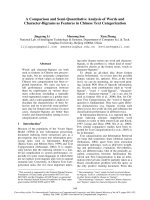
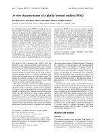
Scanning electron microscopy of the microbial cellulose
membrane showed absence of pores throughout its sur-
face. One side of the membrane was completely smooth
(Fig. 1a), while the other side was distinctly rough (Fig.
1b).
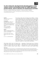
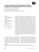
The viability staining analysis of the cells over the bioma-
terial showed that 95% of the mesenchymal stem cells
aggregating to the cellulose membrane were alive while
5% were necrotic. Scanning electron microscopy of the
mesenchymal stem cells showed normal morphology and
attachment to the cellulose membrane surface (Fig. 2a
and 2b).
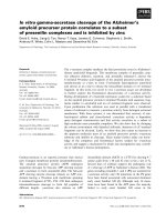
Descriptive analysis of the histological sections by light
microscopy on day seven post surgery demonstrated an
intact membrane surrounded by a mild inflammatory
infiltrate of mainly polymorphonuclear cells and lym-
phocytes (Fig. 3a). Immature granulation tissue was
evinced by intense presence of newly formed vessels and
capillaries close to the membrane and mononuclear cells.
Examination of mice 15 days post-surgery showed a
reduced inflammatory infiltrate, especially due to lower
numbers of lymphocytes (Fig. 3b). Granulation tissue
appeared similar to that observed at day 7 post surgery.
The membrane showed no signs of resorption. No poly-
morphonuclear cells and only a few lymphocytes were
observed at 30 days post-surgery. There was a reduced
number of newly formed vessels and collagen fibers began
to be oriented parallel to the implant's surface that was
apparently intact (Fig. 3c). At 60 and 90 days post-surgery,
no inflammatory infiltrate was observed. Angiogenesis
was markedly reduced and the connective tissue sur-
rounding the membrane was mature. The membrane was
still present with no signs of resorption (Figs 3d and 3e).
Foreign body reaction or connective tissue cells penetrat-
ing the membrane were not observed at any time point
throughout the study period. Polarized microscopy
revealed that the membrane remained intact at all evalua-
tion points (Fig. 4). Table 1 summarizes the mean score
values (semi-quantitative analysis) regarding the intensity
of inflammatory infiltrate, polymorphonuclear cells,
multinucleated giant cells, lymphocytes, angiogenesis,
and fibrosis. No significant differences were observed
among the time points regarding presence of polymor-
phonuclear cells, multinucleated giant cells, and lym-
Acta Veterinaria Scandinavica 2009, 51:12 />Page 4 of 8
(page number not for citation purposes)
Scanning electron microscopy of a microbial cellulose membrane after 10 days in cell cultureFigure 2
Scanning electron microscopy of a microbial cellulose membrane after 10 days in cell culture. Observe the mes-
enchymal stem cells with normal morphology and attached to the membrane surface. (×1200).
Scanning electron microscopy of both sides of a microbial cellulose membraneFigure 1
Scanning electron microscopy of both sides of a microbial cellulose membrane. One side of the membrane is com-
pletely smooth (a), and the other side is distinctly rough (b).
Acta Veterinaria Scandinavica 2009, 51:12 />Page 5 of 8
(page number not for citation purposes)
Histomorphology of a microbial cellulose membrane implanted subcutaneously in mice and surrounding tissue reaction 7(a), 15(b), 30(c), 60(d) and 90(e) days postoperativelyFigure 3
Histomorphology of a microbial cellulose membrane implanted subcutaneously in mice and surrounding tis-
sue reaction 7(a), 15(b), 30(c), 60(d) and 90(e) days postoperatively. Observe the presence of the intact membrane
(*) surrounded by immature granulation tissue and newly formed vessels and capillaries (a). At 15 days post-surgery, a reduc-
tion in inflammatory infiltrate, especially of lymphocytes, is observed (b). At 30 days postoperatively observe the collagen fibers
commencing orientation parallel to the implant's surface (c). No inflammatory infiltrate is observed and the connective tissue
surrounding the membrane is mature at 60 (d) and 90 (e) days post-surgery. (HE, Obj. ×10).
Acta Veterinaria Scandinavica 2009, 51:12 />Page 6 of 8
(page number not for citation purposes)
phocytes (P > 0.05). Angiogenesis and fibrosis decreased
throughout the evaluation periods (P < 0.05).
Discussion
The chemical composition of a membrane intended for
guided tissue repair determines the type, duration and
degree of inflammatory and immune response, means of
disintegration and its longevity in the host tissue [1]. The
inflammatory response may delay the healing process
[23]. In the present study a low inflammatory response to
the implanted membrane was seen at seven, 15, and 30
days post-surgery with absence of foreign body reaction at
any time, suggesting that the microbial cellulose mem-
brane was well tolerated by the organism. Low cellular
reaction was also found in a duraplasty study in dogs [14],
and no gross or histological signs of inflammation includ-
ing giant cell reaction were observed when pieces of bac-
terial cellulose membranes were implanted
subcutaneously in rats for one to 12 weeks [24]. In addi-
tion, the infection rate was decreased in humans that
received cellulose membrane as wound and burn dress-
ings [13]. The absence of multinucleated giant cells sug-
gests absence of foreign body reaction [1,25]. Absence of
foreign body reaction is important as such reactions may
demand additional surgery for removal of the device [1].
In general, chemically nonreactive smooth-surfaced
implants are surrounded by fibroblasts and collagen ori-
ented parallel to the implant's surface within 2 weeks of
implantation [1,25]. Connective tissue surrounding but
not penetrating the membrane was observed as early as 7
days postoperatively in the present study. Later on, espe-
cially 30 days after surgery, improved collagen deposition
was seen. Similar enveloping of a cellulose membrane by
connective tissue has been observed in association with
duraplasty [14]. Other authors have noticed that fibrob-
Polarized microscopy showing the structural organization of the cellulose membraneFigure 4
Polarized microscopy showing the structural organization of the cellulose membrane. There is no evidence of
structural organization alteration at 7 (a), 15 (b), 30 (c), 60 (d) and 90 (d) days postoperatively. (Obj. ×10).
Table 1: Scores
1
attributed to the level of infiltration with polymorphonuclear cells (PMNs), multinucleated giant cells (MGCs), and
lymphocytes, and development of angiogenesis and fibrosis at seven, 15, 30, 60 and 90 days post-operatively.
Time points of evaluation (days)
Cells/event 7 15 30 60 90
PMNs 0.5
2
(0/1)
3,a
0.5 (0/2)
a
0 (0/0)
a
0 (0/0)
a
0 (0/0)
a
MGCs 0 (0/0)
a
0 (0/0)
a
0 (0/0)
a
0 (0/0)
a
0 (0/0)
a
Lymphocytes 1 (0/2)
a
0 (0/0)
a
0.2 (0/1)
a
0 (0/0)
a
0 (0/0)
a
Angiogenesis 2 (1/3)
a
2 (1/3)
ab
1 (1/2)
ab
1 (1/1)
ab
1 (0/1)
b
Fibrosis 1.5 (1/3)
a
1.5 (1/3)
a
1 (1/1)
ab
1 (0/1)
ab
0 (0/1)
b
1
Scores: absent (0), mild (1), intense (2), and severe (3);
2
Mean score of analysis;
3
The number inside parenthesis represents the minimal and the maximal score observed in the event;
Values followed by different letters (a or b) on horizontal were significantly different (P < 0.05)
Acta Veterinaria Scandinavica 2009, 51:12 />Page 7 of 8
(page number not for citation purposes)
lasts are able to penetrate the more porous bottom side of
a cellulose membrane implanted into rats [24].
In the present study no signs of membrane structural
changes or membrane absorption were detected by light
and polarized light microscopy. In a clinical study com-
paring cellulose membrane and expanded polytetrafluor-
oethylene as barrier membrane in the treatment of class II
furcation in human patients, both materials were
removed 4 weeks after placement [15]. On the other
hand, in a duraplasty study in dogs the membrane was
invaded by connective tissue and membrane filaments
had loosened and separated from each other, resulting in
its partial disappearance when evaluated 270 days postop-
eratively [14]. Differences in production techniques prob-
ably influence the results [2,19]. However, since the
membrane used in the present experiment seems nonre-
sorbable, problems associated with membrane durability
may emerge [11].
Guided bone regeneration presents some requirements
such as prevention of bacterial infection, maintenance of
space beneath the barrier membrane, and separation of
osteogenic cells from the competing nonosteogenic cells
[1,26]. Since the tested cellulose membrane was flexible,
especially when wet, it is probably unable to prevent soft
tissue collapse into a bony defect, which would necessi-
tate the placement of a bone graft or biomaterial together
with the membrane as a space-holder [1,3]. This scenario
was probably one of the factors that influenced the
incomplete bone regeneration in circular defects per-
formed on rabbit tibia [18].
Some authors describe the microbial cellulose membrane
as highly porous material with pore sizes from several
nanometers to micrometers [2]. However, the absence of
pores renders the membrane used in the present study
cell-occlusive, suggesting that it will prevent cellular
ingrowth from the adjacent connective tissue. In a study
utilizing three different expanded polytetrafluoroethylene
membrane qualities with different porosities placed on
denuded rat calvaria, it was observed that there was a
porosity range within which osteogenesis beneath the
membrane is optimal, and the material with the smallest
internodal distance did not integrate well with the sur-
rounding soft tissue [27]. Thus, the production of a micro-
bial cellulose membrane with different pore sizes will be
important according to its application.
Microbial cellulose membrane has been used as a scaffold
to substances in order to augment its therapeutic proper-
ties [2]. For example, Wan et al. [28] developed a micro-
bial cellulose membrane coated with hydroxyapatite, an
important compound for bone formation due to its oste-
oconductivity and bioactivity properties. In the present
study, the mesenchymal stem cells aggregated to the cellu-
lose membrane despite the difference of surface texture
observed by electron microscopy thus demonstrating its
ability to scaffold cells. The maintenance of normal mes-
enchymal stem cell morphology also suggested biocom-
patibility of the product. In a study using a cellulose
membrane produced by Acetobacter aceti the growth of
eight types of cells on the membrane was comparable to
that obtained in plastics Petri dishes [29]. However, both
modification of the ionic charge and adsorption of colla-
gen to membrane were used to promote cellular adhesion.
Native and chemically modified bacterial cellulose mate-
rials from A xylinum was also evaluated as scaffold to
chondrocytes suggesting a good potential [30]. On the
other hand, tissue culture and full-thickness transcortical
bone defects induced in rat's mandible showed better
results using expanded polytetrafluoroethylene than
alkali-cellulose membrane that showed predominantly
endochondral regeneration [16]. According to the authors
the alkali-cellulose membrane induced severe inflamma-
tory reaction and appeared to disintegrate.
Conclusion
The tested microbial cellulose membrane was nonresorb-
able, induced low inflammatory response, and may be
used as a scaffold for stem cells. Further investigations are
necessary to confirm its use as barrier material.
Competing interests
The authors declare that they have no competing interests.
Authors' contributions
PNM participated in the study design and experimental
work. SCR participated in the study design, participated in
its coordination and drafted the manuscript. OCMPJ par-
ticipated in the study design and experimental work,
interpreted the results and was responsible for the data
analysis and helped to draft the manuscript. VEF per-
formed the histopathological study. SLRL performed the
scanning electron microscopy of the biomaterial. JFLM
participated in the study design and experimental work.
FCLA helped to draft the manuscript. All authors read and
approved the final manuscript.
References
1. Linde A, Alberius P, Dahlin C, Bjurstam K, Sundin Y: Osteopromo-
tion: a soft-tissue exclusion principle using a membrane for
bone healing and bone neogenesis. J Periodontol 1993,
64:1116-1128.
2. Czaja WK, Young DJ, Kawecki M, Brown RM Jr: The future pros-
pects of microbial cellulose in biomedical applications.
Biomacromolecules 2007, 8:1-12.
3. Zellin G, Linde A: Treatment of segmental defects in long
bones using osteopromotive membranes and recombinant
human bone morphogenetic protein-2. Scand J Plast Reconstr
Surg Hand Surg 1997, 31:97-104.
4. Simonpietri-C JJ, Novaes AB Jr, Batista EL Jr, Filho EJF: Guided tissue
regeneration associated with bovine-derived anorganic bone
Publish with BioMed Central and every
scientist can read your work free of charge
"BioMed Central will be the most significant development for
disseminating the results of biomedical research in our lifetime."
Sir Paul Nurse, Cancer Research UK
Your research papers will be:
available free of charge to the entire biomedical community
peer reviewed and published immediately upon acceptance
cited in PubMed and archived on PubMed Central
yours — you keep the copyright
Submit your manuscript here:
/>BioMedcentral
Acta Veterinaria Scandinavica 2009, 51:12 />Page 8 of 8
(page number not for citation purposes)
in mandibular class II furcation defects. 6-month results at
re-entry. J Periodontol 2000, 71:904-911.
5. Lekovic V, Camargo PM, Weinlaender M, Kenney EB, Vasilic N:
Combination use of bovine porous bone mineral, enamel
matrix proteins, and a bioabsorbable membrane in intrab-
ony periodontal defects in humans. J Periodontol 2001,
72:583-589.
6. Stal S, Tjelmeland K, Hicks J, Bhatia N, Eppley B, Hollier L: Compart-
mentalized bone regeneration of cranial defects with biode-
gradable barriers: an animal model. J Craniofac Surg 2001,
12:41-47.
7. Takata T, Wang HL, Miyauchi M: Migration of osteoblastic cells
on various guided bone regeneration membranes. Clin Oral
Impl Res 2001, 12:332-338.
8. Kellomäki M, Niiranen H, Puumanen K, Ashammakhi N, Waris T,
Törmälä P: Bioabsorbable scaffolds for guided bone regenera-
tion and generation. Biomaterials 2000, 21:2495-2505.
9. Meinig RP, Rahn B, Perren SM, Gogolewski S: Bone regeneration
with resorbable polymeric membranes: treatment of dia-
physeal bone defects in the rabbit radius with poly(L-Lac-
tide) membrane. A pilot study. J Orthop Trauma 1996,
10:178-190.
10. Teixeira JOC, Urist MR: Bone morphogenetic protein induced
repair of compartmentalized segmental diaphyseal defects.
Arch Orthop Trauma Surg 1998, 117:27-34.
11. Buser D, Bragger U, Lang NP, Nyman S: Regeneration and
enlargement of jaw bone using guided tissue regeneration.
Clin Oral Implants Res 1990, 1:22-32.
12. Aaboe M, Pinholt EM, Hjorting-Hansen E, Solheim E, Praetorius F:
Guided tissue regeneration using degradable and non-degra-
dable membranes in rabbit tibia. Clin Oral Impl Res 1993,
4:172-176.
13. Peixoto R, Santos DLN: Biofill: use and clinical evaluation of a
cellulose graft in cutaneous lesions. Rev Bras Cir 1988,
78:141-145.
14. Mello LR, Feltrin LT, Fontes Neto PT, Ferraz FA: Duraplasty with
biosynthetic cellulose: an experimental study. J Neurosurg
1997, 86:143-150.
15. dos Anjos B, Novaes AB Jr, Meffert R, Barboza EP: Clinical compar-
ison of cellulose and expanded polytetrafluoroethylene
membranes in the treatment of class II furcations in mandib-
ular molars with 6-month re-entry. J Periodontol 1998,
69:454-459.
16. Salata LA, Hatton PV, Devlin AJ, Craig GT, Brook IM: In vitro and
in vivo evaluation of e-PTFE and alkali-cellulose membranes
for guided bone regeneration. Clin Oral Impl Res 2001, 12:62-68.
17. Torres MFP, Graça DL, Farias ELP: Microsurgical repair of
peripheral nerve by means of suture, fibrin glue or BioFill
sheat in Wistar rats. Arq Bras Med Vet Zoo 2003, 55:557-561.
18. Macedo NL, Matuda FS, Macedo LGS, Monteiro ASF, Valera MC, Car-
valho YR: Evaluation of two membranes in guided bone tissue
regeneration: histological study in rabbits. Braz J Oral Sci 2004,
3:395-400.
19. Czaja WK, Krystynowicz A, Bielecki S, Brown RM: Microbial cellu-
lose–the natural power to heal wounds. Biomaterials 2006,
27:145-151.
20. Awad HA, Butler DL, Boivin GP, Smith FNL, Malaviya P, Huibregtse
B, Caplan AI: Autologous mesenchymal stem cell-mediated
repair of tendon. Tis Eng 1999, 5:267-277.
21. Caplan AI: Mesenchymal stem cells: cell-based reconstructive
therapy in orthopedics. Tis Eng 2005, 11:1198-1211.
22. Boo JS, Yamada Y, Okazaki Y, Hibino Y, Okada K, Hata K, Yoshikawa
T, Sugiura Y, Ueda M: Tissue-engineered bone using mesenchy-
mal stem cells and a biodegradable scaffold. J Craniofacial Surg
2002, 13:231-239.
23. Mendenhall HV: Surgical Procedures. In Handbook of Biomaterials
Evaluations Edited by: von Recum AF. Philadelphia: Taylor & Francis;
1999:481-491.
24. Helenius G, Bäckdahl H, Bodin A, Nannmark U, Gatenholm P, Risberg
B: In vivo biocompatibility of bacterial cellulose. J Biomed
Mater Res A 2006, 76:431-438.
25. Dee KC, Puleo DA, Bizios R: An Introduction to Tissue-Biomaterial Inter-
actions New Jersey: Wiley-Liss; 2002.
26. Nielsen FF, Karring T, Gogolewski S: Biodegradable guide for
bone regeneration. Polyurethane membranes tested in rab-
bit radius defects.
Acta Orthop Scand 1992, 63:66-69.
27. Zellin G, Linde A: Effects of different osteopromotive mem-
brane porosities on experimental bone neogenesis in rats.
Biomaterials 1996, 17:695-702.
28. Wan YZ, Huang Y, Yuan CD, Raman S, Zhu Y, Jiang HJ, He F, Gao C:
Biomimetic synthesis of hydroxyapatite/bacterial cellulose
nanocomposites for biomedical applications. Mater Sci Eng C
2007, 27:855-864.
29. Watanabe K, Eto Y, Takano S, Nakamori S, Shibai H, Yamanaka S: A
new bacterial cellulose substrate for mammalian cell cul-
ture. Cytotechnology 1993, 13:107-114.
30. Svensson A, Nicklasson E, Harrah T, Panilaitis B, Kaplan DL, Brittberg
M, Gatenholm P: Bacterial cellulose as a potential scaffold for
tissue engineering of cartilage. Biomaterials 2005, 26:419-431.