Báo cáo khoa học: "Effects of osteoprotegerin from transfection of pcDNA3.1(+)/chOPG on bioactivity of chicken osteoclasts" potx
Bạn đang xem bản rút gọn của tài liệu. Xem và tải ngay bản đầy đủ của tài liệu tại đây (343.13 KB, 7 trang )
RESEARCH Open Access
Effects of osteoprotegerin from transfection of
pcDNA3.1(+)/chOPG on bioactivity of chicken
osteoclasts
Lele Hou, Jiafa Hou
*
, Jing Yao and Zhenlei Zhou
Abstract
Background: Osteoprotegerin (OPG) has been reported to prevent bone resorption by inhibiting the formation,
function, and survival of osteoclasts in a variety of animal models. However, the effects of OPG on bone
metabolism in avian species have not been described. The objective of this study was to investigate the effects of
chicken OPG (chOPG) expressed in chicken embryo fibroblasts (CEFs) on chicken osteoclast function in vitro.
Methods: The chOPG sequence containing the open reading frame (ORF) was amplified from chicken embryo
frontal bone and inserted into the pcDNA3.1 (+) vector. PcDNA3.1 (+)/chOPG was transiently transfected into CEFs
by lipofectamine 2000. Transcription of OPG mRNA and expression of chOPG recombinant protein were detected
by reverse transcriptio n polymerase chain reaction (RT-PCR) and indirect immunofluorescence. The level of chOPG
recombinant protein was detected by enzyme-linked immunosorbent assay. The suspension of osteoclasts was
separated from chicken embryos and divided into three groups (control group, pcDNA3.1 (+) group and pcDNA3.1
(+)/chOPG group). The percentage of osteoclast apoptosis was detected by flow cytometry. The tartrate-resistant
acid phosphatase (TRAP) secreted by osteoclasts was measured by the diazol method. The resorbing activity of
osteoclasts was evaluated by the area of lacunae on bone flaps and the concentration of calcium in the
supernatant liquid of osteoclasts.
Results: 48 h after transfection, the exogenous OPG gene transcription was detected by RT-PCR. After 72 h, the
CEFs transfected from pcDNA3.1 (+)/chOPG displayed green fluorescence and the concentration of chOPG protein
was 15.78 ± 0.22 ng/mL. After chicken osteoclasts were cultured for 5 d in a me dium containing supernatant from
transfected CEFs, the percentage of osteoclast apoptosis was increased significantly, the concentration of TRAP, the
area of lacunae on bone flaps and calcium concentration were decreased significantly in the pcDNA3.1(+)/OPG
group compared to the control group and the pcDNA3.1 (+) group.
Conclusion: Constructed pcDNA3.1 (+)/chOPG transfe cted into CEFs expressed bioactive OPG protein that was
able to inhibit osteoclast function.
Background
Osteoporosis in laying hens is a condition that involves
a progressive loss of bone resulting in bone fragility and
increased risk of fracture. Surveys of laying flocks in
Europe have indicated that about 30% of the birds
experience one or more bone fractures due to osteo-
porosis during their lifetime. The high fracture rates
show that osteoporosis not only leads to production
losses, but also to severe welfare problems in hens [1].
In laying hens, the main types of bone providing
structural integrity are cortical and trabecular bone. In
addition to these, medullary bone, an extremely labile
source for calcium that develops in specific bones of
female birds at the onset of sexual maturity, provides a
labile source of calcium for shell formation. Bones
undergo a co nstant process of remodelling, which at the
cell ular level involves a coordinated regulation of osteo-
blasts and osteoclasts. As hens mature sexually, bone
formation of osteoblasts switches from structural bone
* Correspondence:
College of Veterinary Medicine, Nanjing Agricultural University, Nanjing,
Jiangsu, 210095, China
Hou et al. Acta Veterinaria Scandinavica 2011, 53:21
/>© 2011 Hou et al; licensee BioMed Central Ltd. This is an Open Access article distributed under the terms of the Creative Commons
Attribution License (h ttp://cre ativecommons.org/lice nses/by/2.0), which permits unrestricted use, distribution, and rep roduction in
any medium, provided the original work is properly cited.
to medullary bone [ 2]. In the absence of structural bone
formation, continued osteoclastic resorption of struc-
tural bone will result in a depletion of structural bone,
ultimately leading to osteoporosis.
The differentiation and function of osteoclasts are
regulated by soluble cyto kines from osteoblasts, such as
osteoprotegerin (OPG) and the receptor activator of
nuclear factor ligand (RANKL; also called OPG ligand)
[3]. OPG is a soluble decoy receptor that inhibits osteo-
clast formation, function, and survival by preventing the
binding of RANKL to the receptor activator of nuclear
factor B (RANK), a membrane-bound protein that is
found on chondrocytes, dendritic cells, osteoclast pre-
cursors, and mature osteoclasts [4]. Many cytokines and
effectors are known to influence the osteoclastic bone
resorption via the OPG/RANK/RANKL trio of proteins
[5,6]. Changes of expression levels of OPG/RANK/
RANKL would be expected to cause bone disorders
such as postmenopausal osteoporosis, glucocorticoid-
induced osteoporosis, and sporadic Paget’ s disease in
man [7].
Although the importance of OPG in the osteoclasto-
genesis has been established in mammalian models, it is
not yet clear how O PG regulates the function of osteo-
clasts in avian species. To elucidate the function of OPG
in laying hens in vivo, we amplified the open reading
frame (ORF) of the chicken OPG (chOPG) sequence,
constructed the pcDNA3.1 (+)/chOPG plasmid and
transiently transfected it into chicken embryo fibroblasts
(CEFs). We tested whether pcDNA3.1 (+)/chOPG
expressed OPG protein at a level able to inhibit the bio-
logical activity of osteoclasts in vitro.
Methods
Cloning of the ORF of chOPG
Total RNA was extracted from chicken embryonic fron-
talbone(AnimalHusbandryIndustryCo.,Nanjing,
China) with TRIzol
®
Reagent (Invitrogen, Inc. Carlsbad,
CA, USA) according to the manufacturer’sinstruction.
RNA purity was determined by 260 nm and 280 nm
absorbance ratios and integrity was checked by 1% agar-
ose/formaldehyde gel electrophoresis. A Biometra DNA
Thermal Cycler was used for reverse transcription poly-
merase chain reaction (RT-PCR). RT-PCR was per-
formedinthepresenceofDTT, oligo(d T)18, dNTP,
RNase inhibitor, first-strand buffer and Moloney murine
leukaemia virus reverse transcriptase (TakaRa Bio Inc.
Japan). The final mixture was reacted at 42°C for
60 min and at 70°C for 15 min to denature the enzyme.
On the basis of the published nucleotide sequence of
chOPG (DQ098013), one pair of PCR primers (Invitro-
gen) were designed. Primers P1 and P2 were used to
amplify the ORF of chOPG sequence. Nhe|andXho|
(TakaRa Bio Inc.) restriction sites were inserted into pri-
mers P1 and P2, respectively:
P1: 5’-CAT
GCTAGCATGAACAAGTTCCTGTGC-3’
(sense strand, positions 10-27 of cDNA sequence);
P2: 5’-CCGG
CTCGAGTTAGAC ACATCTTACTTT-3’
(antisense strand, positions 1,201-1,218 of cDNA
sequence).
PCRmix (TakaRa Bio Inc.), primers P1 and P2, and
cDNA were mixed and ampli fied for 30 cycles under the
following conditions: denaturation for 30 s at 94°C,
annealing for 45 s at 47°C, and extension for 50 s at 72°C.
The products were subsequently sequenced (Invitrogen)
after 1% agarose electrophoresis, recovery and purification.
pcDNA3.1 (+)/chOPG construction
The eucaryote expression vector pcDNA3.1 (+) (Invitro-
gen) and OPG products were digested with Nh e|and
Xho|. After purification, two fragments were ligated with
T4DNA ligase (TakaRa Bio Inc.) at 16°C (overnight).
The ligation product was subsequently transformed into
DH5a competent cells (Nanji ng Agricultural University,
Nanjing, China)). The transformed cells were plated on
Luria-Bertani agar (Invitrogen-Gibco, Grand Island, NY,
USA) contai ning ampicillin (Invi trogen-Gibco). The
positive clones were identified by PCR (PCRmix,
pcDNA3.1 (+) consensus primer P3 (5’-CTGGCTAAC-
TAGAGAACCCAC-3’), P4 (5’-TAGAAGGCACAGTC-
GAGG-3’ )). DNA of positive clones were mixed and
amplified for 30 c ycles under t he following conditions:
denaturation for 30 s at 94°C, annealing for 45 s at 49°C,
and extension for 50 s at 72°C and double restriction
digestion, followed by agarose gel analysis. Then
pcDNA3.1 (+)/chOPG and pcDNA3.1 (+) were prepared
with non-endotoxi n plamid extraction kit (Sigma Chemi-
cals. St. Louis, MO, USA).
Cell culture and DNA transfection
CEFs were prepared from two 10-days old chicken
embryos (Animal Husbandry Industry Co) and were
grown according to standard procedures, cultured
in Dulbecco’ s modified Eagle’ smedium(DMEM)
(Invitrogen-Gibco) supplemented with 5% fetal bovine
serum (Invitrogen-Gibco). The number of cells was
adjusted to 2 × 10
5
cell s/ml and incubated in 24-well tis-
sue culture plates (Bo Quan Sci&Tech. Co. Ltd. Nanjing,
China) containing cover glass at 37°C in a humid atmo-
sphere of 5% CO
2
for 24 h. Pr ior to each t est, CE Fs were
washed three times with phosphate buffered solution
(PBS), transfected with 1 μg/well pcDNA3.1 (+)/OPG
plasmid and pcDNA3. 1 (+) vector using 3 μl/well lipofec-
tamine 2000 (Invit rogen), respectively, followed by incu-
bation at 37°C in 5% CO
2
for 48 h and 72 h. The culture
medium was renewed every 2nd day.
Hou et al. Acta Veterinaria Scandinavica 2011, 53:21
/>Page 2 of 7
RT-PCR analysis of chOPG mRNA
The cells (both floating and adherent cells) were har-
vested 48 h post transfection. The total RNA was
extracted with TRIzol
®
Reagent according to the manu-
facturer’ s instruction. RNA samples were then treated
with DNase I (1 U/μg) (TakaRa Bio Inc.) before the RT
step to avoid the interference with contaminating geno-
mic DNA. P5 (5’-ATGAACAAGTTCCTGTGC-3’)and
P6 (5’-TTAGACACATCTTACTTT-3’ ) were subjected
to PCR using upstream and downstream primers.
Immunocytochemical analysis of chOPG product in CEFs
CEFs (2 × 10
5
)culturedfor72honglasscoverslips
(6 mm × 6 mm) were replated into 24-well plates. Glass
coverslips were washed with 0.01 M PBS and fixed in
4% formaldehyde for 45 min. Detergent extraction with
3% Triton X-100 was performed for 10 min. Coverslips
were saturated with PBS containing 5% bovine serum
albumin (Wuhan Boster Biotechnology Company,
China) for 1 h at room temperature with gentle rocking,
processed with rabbit anti-chOPG polyclonal antibody
(Nanjing Agricultural University) for 1 h at 37°C and
followed by FITC-goat-anti-rabbit IgG (Wuhan Boster
Biotechnology Company) for 1 h at 37°C and then
stained by DAPI staining solution (Wuhan Boster Bio-
technology Company). Coverslips were washed by PBS
for 30 min prior to each treatment. Finally, coverslips
were mounted on slides and fluorescence signals were
analyzed by a Fluoview microscopy (Olympus, Japan).
ELISA analysis of chOPG product in supernatant
The concentration of the chOPG product in the super-
natant was determined using an enzyme-linked immuno-
sorbent assay (ELISA) kit (R&D Systems, Minneapolis,
MN, USA) according to the manufacturer’sinstructions.
The concen tration was determined for three wells of
each sample by measuring the optical density (OD) at
450 nm wavelength by an ELISA reader (Immuno Mini
NJ-2300, InterMed, Japan).
Effect of chOPG on osteoclast bioactivity
Tibias and humeri were isolated from 15 18-days old
chicken embryos. Osteoclast cultures were prepared as
previously described [8]. Briefly, a cell suspension was
seeded at a concentration of 2 × 10
5
cells per well in 24-
well dishes containing either glass coverslips or bovine
bone slices (4 mm × 4 mm × 50 μm) (Nanjing Agricultural
University). Non-adherent cells were washed off after 2 h.
The adherent cells were grown for another 2 d and then
cultured in DMEM containing OPG supernatant. The
medium was changed every 48 h. Glass coverslips, bovine
bone slices and supernatant were harvested after 5 d. The
percentage of osteoclast apoptosis was detected by flow
cytometry. TRAP secreted to the supernatant by
osteoclasts was measured at a OD of 530 nm by the diazol
method using TRAP test kit (Bo Quan Sci&Tech. Co. Ltd.
Nanjing, China). The resorption lacunae on the bone slice
was visualized by toluidine blue staining after removal of
osteoclasts using 50 mM NH
4
OH and brief sonication [9].
The co ncentration of calcium in the supernatant was
determined by atomic absorption spectrometry (wave-
length 422.7 nm, electric current 3.0 mA, spectrum width
0.4 nm) after 5 times dilution.
Statistical analysis
Allvalueswereexpressedasmeans±thestandard
deviation (SD). Differences between mean val ues of nor-
mally distributed data were assessed by the one-way
ANOVA test and Student’s t-test. Statistical difference
was accepted at P < 0.05.
Results
Cloning of the ORF of chOPG and construction pcDNA3.1
(+)/chOPG
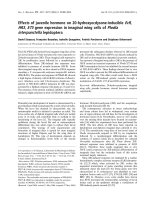
The size of the specific gene fragment amplified was, as
expected, about 1.2 kbp (Figure 1A). The positive clones
were identified by PCR amplification and the double
restriction digestion with Nhe|andXho| (Figure 1B and
Figur e 1C). Analysis of the PCR products by agarose gel
electrophoresis showed that both constructs contained a
DNA insert of the correct size and in the correct orien-
tation. The result of sequencing showed that it had
100% homology with that report ed in GenBank
(DQ098013) indicating that the OPG gene has an exten-
sive hereditary conservation and that no mutations were
present in this region of the vector.
Expression of chOPG in CEFs transfected with pcDNA3.1
(+)/chOPG
RT-PCR analysis indicated that CEFs in the group with
pcDNA3.1(+)/chOPG t ransfection expressed OPG
mRNA, but there was no expression of OPG mRNA in
the control group and pcDNA3.1(+) group (Figure 1D).
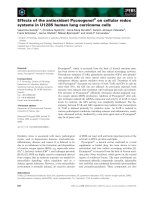
Immunofluorescence studies showed that chOPG pro-
tein was distributed in the cytoplasma and CE Fs show-
ing green fluorescence were observed in the pcDNA3.1
(+)/chOPG group, but were not present in the other
groups (Figure 2A).
In the culture supernatant of the pcDNA3.1
(+)/chOPG group transfected from CEFs, the concentra-
tion of chOPG was 15.78 ± 0.22 ng/ml, whereas chOPG
was not be demonstrated in media from the control
group or the pcDNA3.1 (+) group.
Effect of product from transfected CEFs on chicken
osteoclast bioactivity in vitro
The morphology of osteoclasts after culturing for 5 d is
shown in Figure 2B. Osteoclasts grew well in the control
Hou et al. Acta Veterinaria Scandinavica 2011, 53:21
/>Page 3 of 7
and pcDNA3.1 (+) transfected CEF groups, whereas
major nuclei disappeared, many vacuoles and lipid dro-
plet appeared in the cytoplasm and many non-adherent
and dead osteoclasts were observed in the culture solu-
tion of the pcDNA3.1 (+)/chOPG transfected CEF
group. The percentage of osteoclast apoptosis in the
control, pcDNA3.1 (+ ) and pcDNA3.1 (+)/chOPG
groups was 10.32%±1.50%, 12.61%±0.95%, 20.59%
±2.83%, respectively (Figure 2C). TRAP enzyme activity
in the pcDNA3.1 (+)/chOPG group was significantly
decreased compared to the control group (P < 0.01)
(Figur e 3A). An individual resorption event was seen as
a dark border of toluidine blue stain surrounding an
excavation. The data were recorded for each resorption
event separately (Figure 2D). The quantity and area of
lacunae reflected bone resorption by osteoclasts (Table 1).
DMEM culture solution did not contain Ca
2+
until after
culturing thus suggesting osteoclast activity (Figure 3B).
Discussion
Bone is an exceedingly complex tissue with multisyste-
mic regulation. Skeletal metabolism depends on the
dynamic balance of bone formation by osteoblasts and
bone resorption by osteoclasts. The discovery of the
OPG/RANKL/RANK system in the mid 1990s has led
to major advances in our understanding of how bone
modeling and remodeling are regulated [10]. Current
research has focused on OPG in humans and mice,
while reports on avian OPG are lacking. In our labora-
tory, chOPG mRNA was extracted from chicken embryo
frontal bone. The OPG coding region was successfully
amplified and sequence analysis indicated that OPG is
Figure 1 Gel electrophoresis of chOPG. 1A: Gel electrophoresis of reverse transcription polymerase chain reaction (RT-PCR) product. Total RNA
extracted from chicken embryo frontal bone was analyzed using RT-PCR with specific primers. About 1.2 kbp gene of chicken osteoprotegerin
(chOPG) was amplified (lane 1); DL2000 marker (lane 2); 1B: Gel electrophoresis of pcDNA3.1 (+)/chOPG PCR product. chOPG fragment was
inserted into the eucaryon expression vector pcDNA3.1 (+) between Nhe| and Xho|. Negative plasmid (lane 1) and positive plasmid (lane 2) were
chosen using PCR; marker (lane 3); 1C: Gel electrophoresis of pcDNA3.1 (+)/chOPG double restriction enzyme assay. Positive plasmid (lane 3) was
identified by Nhe|and Xho|double restriction digestion and showed pcDNA3.1 (+) and OPG (lane 2); marker (lane 1); 1D: Gel electrophoresis of
RT-PCR analysis showing the expression of chOPG gene at 48 h. Amplification of chOPG using cDNA from lane 1 (control group) and lane 2
(pcDNA3.1 (+) transfected CEFs group) showing negative result. Amplification of chOPG using cDNA from lane 4 (pcDNA3.1 (+)/chOPG
transfected chicken embryo fibroblasts group) showing about 1200 bp gene of chOPG. Lane 3: marker.
Hou et al. Acta Veterinaria Scandinavica 2011, 53:21
/>Page 4 of 7
highly conserved evolutionary. The sequence reported
here had a 68.76%, 68.60% and 68.29% homonology to
human, rat and mouse OPG, respectively. The sequence
similarity suggests a similar function across species.
Bone is particularly intriguing in laying hens because
of the huge demands for calcium for eggshell formation
and the occurrence of medullary bone. On the surface
of the medullary bone, osteoclasts undergo cyclical func-
tional modifications during the egg-laying cycle [11].
In this study, chOPG induced osteoclast apoptosis
after in vitro incubation for 5 d. This result was similar
to that reported by Lacey et al. [12 ], who demonstrated
that OPG inhibited bone resorption and induced osteo-
clast apoptosis though inhib ition of F-actin ring
Figure 2 The expression of chO PG protein and effect on osteoclast morphology, apoptosis and r esorption. 2A : Immunofluorescence
assay for a possible chicken osteoprotegerin (chOPG) protein. Chicken embryo fibroblasts (CEFs) were grown on coverslips, fixed, and examined
by indirect immunofluorescence. Cells were incubated with rabbit anti-chOPG serum. The secondary antibody was fluorescein-conjugated goat
anti-rabbit immunoglobulin G (green). The nuclei of the corresponding cells were visualized by DAPI staining (blue). Fluorescence signals were
analyzed by Fluoview microscopy (×200). Negative results are shown on card l (control group) and card 2 (pcDNA3.1 (+) transfected CEFs
group), positive green fluorescence for CEFs are shown on card 3 (pcDNA3.1 (+)/chOPG transfected CEFs group). 2B: The morphology of
osteoclasts was observed by inverted phase contrast microscope (×200). The adherent osteoclasts were cultured in Dulbecco’s modified Eagle’s
medium (DMEM) containing supernatant of control group (l), pcDNA3.1 (+) transfected CEF group (2) and pcDNA3.1 (+)/chOPG transfected CEF
group (3) for 5 d. 2C: Effect of the supernatant of three groups on the apoptosis of osteoclasts by flow cytometry. 2D: Toluidine blue staining of
bone slices showing resorption lacunae (×200). The adherent osteoclasts were cultured in DMEM containing supernatant for 5 d in three groups.
Hou et al. Acta Veterinaria Scandinavica 2011, 53:21
/>Page 5 of 7
formation of mature osteoclasts or altered interaction
between stroma cell and osteoclasts.
The results suggest that the secretion of TRAP by
osteoclasts was significantly decreased; further demon-
strating that recombinant chOPG could inhibit the
activity of osteoclasts in vitro.Chamberet al. [13] and
Boyde et al. [14] provided evidence for a direct associa-
tion between the quantity, area and depth of absorption
and the capability of osteoclasts to resorption bone. The
present study showed that chOPG inhibited osteoclast
Figure 3 The change of TRAP enzyme activity and concentration of Ca2+ in three groups. 3A: Effect of culture supernatant from chicken
embryo fibroblasts transfected on osteoclastic TRAP enzyme activity (Mean ± SD; n = 8). ** indicates P < 0.01 compared with the control group.
3B: The concentration of Ca
2+
in the supernatant containing bovine bone slices (Mean ± SD; n = 8). ** indicates P < 0.01 compared with the
control group.
Table 1 Effect of culture supernatant of chicken embryo fibroblasts on the quantity and area of osteoclast resorption
lacunae in three groups
control group pcDNA3.1 (+) group pcDNA3.1 (+)/chOPG group
Number of lacunae 10.7 ± 1.2 9.0 ± 1.0 5.4 ± 0.5
Areas of lacunae (μm2) 5755.2003 ± 234.7778 4987.7468 ± 124.5471 739.4407 ± 150.1978**
Note: compared with the control group, ** P < 0.01.
Hou et al. Acta Veterinaria Scandinavica 2011, 53:21
/>Page 6 of 7
bone resorption and consequently the concentration of
Ca
2+
in the supernatant was significantly reduced. How-
ever, the mechanisms by which OPG exerts its biological
activity as well as the nature of its molecular interac-
tions with osteoclasts are not well defined. Hakeda et al.
[15] reported the first evidence of a direct biological
activity of OPG on isolated osteoclasts via a 140 kDa
OPG-binding protein. The e xact nature of osteoclastic
OPG receptors was not further characterized. Direct
biological activities of OPG on osteoclasts were recently
showed by Wit trant et al. [16] demonstrating OPG
enhanced proMMP-9 activity along with several other
parameters (TRAP, TIMP, cathepsin K) in purified
osteoclasts. Theoleyre et al. [17] showed that OPG sti-
mulates proMMP-9 activity of osteoclasts by the ras/
MAPK pathway in volving p38 and ERK1/2 phosphoryla-
tions. Moreover, OPG-induced MAPK pathway depends
on RANKL. In general, OPG is not only a soluble decoy
receptor for RANKL as described in the literature but
may be also considered as a direct effector of osteoclast
functions.
Conclusions
ChOPG is capable of inhibiting bone resorption as well
as promoting osteoclast apoptosis. The study also indi-
cates that pcDNA3.1 (+)/chOPG may be a target for
regulating bone metabolism in chicken bone metabolic
diseases such as osteoporosis.
Abbreviations
CEFs: chicken embryo fibroblasts; chOPG: chicken OPG; DMEM: Dulbecco’s
modified Eagle’s medium; OD: optical density; OPG: osteoprotegerin; ORF:
open reading frame; PBS: phosphate buffered solution; RANKL: receptor
activator of nuclear factor κB ligand; RANK: receptor activator of nuclear
factor κB; RT-PCR: reverse transcription polymerase chain reaction; TRAP:
tartrate-resistant acid phosphatase.
Acknowledgements
This work was supported by the National Natural Science Foundation of
China (30972234, 30671546) and the key program of Education Ministry of
China (200803070021).
Authors’ contributions
LH and JH conceived of the study, and participated in its design and
coordination and helped to draft the manuscript. ZZ participated in the data
collection. JY cultured the chicken embryo osteoclasts. LH performed the
other experiments. All authors have been involved in drafting the
manuscript and have read and approved the final manuscript.
Competing interests
The authors declare that they have no competing interests.
Received: 7 November 2010 Accepted: 24 March 2011
Published: 24 March 2011
References
1. Beck MM, Hansen KK: Role of estrogen in avian osteoporosis. Poult Sci
2004, 83:200-206.
2. Whitehead CC: Overview of bone biology in the egg-laying hen. Poult Sci
2004, 83:193-199.
3. Lacey DL, Timms E, Tan HL, Kelley MJ, Dunstan CR, Burgess T, Elliott R,
Colombero A, Elliott G, Scully S, Hsu H, Sullivan J, Hawkins N, Davy E,
Capparelli C, Eli A, Qian YX, Kaufman S, Sarosi I, Shalhoub V, Senaldi G,
Guo J, Delaney J, Boyle WJ: Osteoprotegerin ligand is a cytokine that
regulates osteoclast differentiation and activation. Cell 1998, 93:165-176.
4. Simonet WS, Lacey DL, Dunstan CR, Kelley M, Chang MS, Luthy R,
Nguyen HQ, Wooden S, Bennett L, Boone T, Shimamoto G, DeRose M,
Elliott R, Colombero A, Tan HL, Trail G, Sullivan J, Davy E, Bucay N,
Renshaw-Gegg L, Hughes TM, Hill D, Pattison W, Campbell P, Sander S,
Van G, Tarpley J, Derby P, Lee R, Boyle WJ: Osteoprotegerin: a novel
secreted protein involved in the regulation of bone density. Cell 1997,
89:309-319.
5. Kwan Tat S, Padrines M, Théoleyre S, Heymann D, Fortun Y: IL-6, RANKL,
TNF-alpha/IL-1: interrelations in bone resorption pathophysiology.
Cytokine Growth Factor Rev 2004, 15:49-60.
6. Anderson DM, Maraskovsky E, Billingsley WL, Dougall WC, Tometsko ME,
Roux ER, Teepe MC, DuBose RF, Cosman D, Galibert L: A homologue of the
TNF receptor and its ligand enhance T-cell growth and dendritic-cell
function. Nature 1997, 390:175-179.
7. Hofbauer LC, Schoppet M: Clinical implications of the Osteoprotegerin/
RANKL/RANK system for bone and vascular diseases. JAMA 2004,
292:490-495.
8. Yao J, Zhang J, Hou JF: Effects of ipriflavone on caged layer bone
metabolism in vitro and in vivo. Poult Sci 2007, 86:503-507.
9. Wang Y, Hou JF, Zhou ZL: Chicken receptor activator of nuclear factor-κB
ligand induces formation of chicken osteoclasts from bone marrow cells
and also directly activates mature osteoclasts. Poult Sci 2008,
87:2344-2349.
10. Boyc BF, Xing LP: Functions of RANKL/RANK/OPG in bone modeling and
remodeling. Arch Biochem Biophys 2008, 473:139-146.
11. Miller SC: Osteoclast cell-surface specializations and nuclear kinetics
during egg-laying in Japanese quail. Am J Anat 1981, 162:35-43.
12. Lacey DL, Tan HL, Lu J: Osteoprotegerin ligand modulates murine
osteoclastsurvival in vitro and vivo. Am J Pathol 2000, 157:435-448.
13. Chamber TJ, Thomson BM, Fuller K: Resorption of bone by isolated rabbit
osteoclasts. J. Cell Sci 1984, 66:383-399.
14. Boyde A, Ali NN, Jones SJ: Resorption of dentine by isolated osteoclasts
in vitro. Br Dent J 1984, 156:216-230.
15. Hakeda Y, Kobayashi Y, Yamaguchi K, Yasuda H, Tsuda E, Higashio K,
Miyata T, Kumegawa M: Osteoclastogenesis inhibitory factor (OCIF)
directly inhibits bone-resorbing activity of isolated mature osteoclasts.
Biochem Biophys Res Commun 1998, 251:796-801.
16. Wittrant Y, Couillaud S, Theoleyre S, Dunstan C, Heymann D, Redini F:
Osteoprotegerin differentially regulates protease expression in
osteoclast cultures. Biochem Biophys Res Commun 2002, 293:38-44.
17. Theoleyre S, Wittrant Y, Couillaud S, Vusio P, Berreur M, Dunstan C,
Blanchard F, Redini F, Heymann D: Cellular activity and signaling induced
by osteoprotegerin in osteoclasts: involvement of receptor activator of
nuclear factor κB ligand and MAPK. Biochimica Biophysica Acta 2004,
1644:1-7.
doi:10.1186/1751-0147-53-21
Cite this article as: Hou et al.: Effects of osteoprotegerin from
transfection of pcDNA3.1(+)/chOPG on bioactivity of chicken
osteoclasts. Acta Veterinaria Scandinavica 2011 53:21.
Submit your next manuscript to BioMed Central
and take full advantage of:
• Convenient online submission
• Thorough peer review
• No space constraints or color figure charges
• Immediate publication on acceptance
• Inclusion in PubMed, CAS, Scopus and Google Scholar
• Research which is freely available for redistribution
Submit your manuscript at
www.biomedcentral.com/submit
Hou et al. Acta Veterinaria Scandinavica 2011, 53:21
/>Page 7 of 7