Báo cáo y học: "Science review: Extracellular acidosis and the immune response: clinical and physiologic implications" potx
Bạn đang xem bản rút gọn của tài liệu. Xem và tải ngay bản đầy đủ của tài liệu tại đây (71.96 KB, 6 trang )
331
bHS = 6% hetastarch in a balanced electrolyte solution; IL = interleukin; iNOS = inducible nitric oxide synthase; LPS = lipopolysaccharide; LR =
lactated Ringer’s; MAP = mean arterial pressure; NF-κB = nuclear factor-κB; NO = nitric oxide; NS = normal (0.9%) saline; pH
i
= intracellular pH;
pH
o
= extracellular pH; SBE = standard base excess; TNF = tumor necrosis factor.
Available online />Introduction
Critical illness is exemplified by a state of profound disruption
in normal homeostatic mechanisms. Patients who remain
critically ill may progress to a poorly understood condition
known as multiple organ failure, which is characterized by
widespread alterations in both individual organ function and
integrative function across organs. Although our under-
standing of this condition is extremely limited, numerous
observations suggest that alterations in the immune response
are not only caused by but may also be the cause of ongoing
organ injury, and these alterations may adversely affect
patients’ ability to recover. Both increased inflammation and
immune suppression have been implicated in the
pathogenesis of multiple organ failure. Little is known about
the influences that therapies have on the immune response.
Emerging evidence suggests that ventilator-associated lung
injury results in increased systemic inflammation [1] and that
systemic inflammation resulting from local tissue injury
appears to have effects on remote organs [2]. Drugs that
appear to modify the course of organ injury such as activated
protein C and corticosteroids appear to have a broad range
of effects on the immune system [3,4]. Abnormalities in
systemic acid–base balance may also induce significant
alterations in the immune response. The clinical significance
of these alterations is not yet known, but their magnitude
suggests that they may play an important role in the
development or maintenance of immune dysfunction. If this is
the case, then they represent attractive targets (or even tools)
for therapy. Extracellular pH (pH
o
) for circulating leukocytes
(i.e. blood pH) is easily altered and thus, for good or bad,
changes in pH may rapidly alter the immune response in
these cells.
Review
Science review: Extracellular acidosis and the immune response:
clinical and physiologic implications
John A Kellum
1
, Mingchen Song
2
and Jinyou Li
3
1
Associate Professor, Critical Care Medicine and Medicine, Co-Director, The MANTRA (Mechanisms And Novel Therapies for Resuscitation and Acute
illness) Laboratory, Department of Critical Care Medicine, University of Pittsburgh School of Medicine, Pittsburgh, Pennsylvania, USA
2
Research Fellow, Department of Critical Care Medicine, The MANTRA Laboratory, Department of Critical Care Medicine, University of Pittsburgh
School of Medicine, Pittsburgh, Pennsylvania, USA
3
Visiting Researcher, Department of Critical Care Medicine, The MANTRA Laboratory, Department of Critical Care Medicine, University of Pittsburgh
School of Medicine, Pittsburgh, Pennsylvania, USA
Corresponding author: John A Kellum,
Published online: 16 June 2004 Critical Care 2004, 8:331-336 (DOI 10.1186/cc2900)
This article is online at />© 2004 BioMed Central Ltd
Abstract
Metabolic acidosis is among the most common abnormalities seen in patients suffering from critical
illness. Its etiologies are multiple and treatment of the underlying condition is the mainstay of therapy.
However, growing evidence suggests that acidosis itself has profound effects on the host, particularly
in the area of immune function. Given the central importance of immune function to the outcome of
critical illness, there is renewed interest in elucidating the effects of this all too common condition on
the immune response. In this review we concentrate on the effects of extracellular acids on production
and release of inflammatory mediators, and we demonstrate that different acids produce different
effects despite similar extracellular pH. Finally, we discuss potential clinical implications.
Keywords acidosis, cytokines, immune response, pH, sepsis
332
Critical Care October 2004 Vol 8 No 5 Kellum et al.
Effects of extracellular acidosis on
inflammatory mediator release
There are now several studies documenting the effects of
decreased pH
o
on the synthesis and release of inflammatory
mediators, especially tumor necrosis factor (TNF) and nitric
oxide (NO). Most of these studies were conducted in resident
macrophages or macrophage-like cell lines and yielded
conflicting results (Table 1). However, studies using HCl have
consistently shown proinflammatory effects at the level of
nuclear factor-κB (NF-κB) DNA binding or TNF synthesis
provided pH
o
was not less than 6.0 [5–7], although TNF
secretion was reduced even at pH
o
as high as 7.0 [5,7,8].
Studies of nonstimulated resident peritoneal macrophages
[6] and lipopolysaccharide (LPS)-stimulated RAW 264.7
cells [9] have shown increased NO formation at moderately
reduced pH
o
(7.0–7.2). However, more severely acidic pH
o
reduces NO formation [6,9], and there is an apparent
dissociation between the pH
o
effects on inducible nitric oxide
synthase (iNOS) mRNA, protein, and final NO release [9].
Thus, HCl appears to affect inflammatory mediators
differently at different stages in their synthesis and release.
Little is known about the effects of HCl on other cytokines or
on the kinetics of pH
o
mediated effects.
Lactic acid has been studied in an even more limited way
than HCl. Lactic acid (pH
o
6.75) was shown in one study
[10] to result in increased TNF release in LPS-stimulated
peritoneal macrophages. This finding is surprising in light of
the growing evidence of a protective effect of lactic acid in
neuronal injury [11–13]. Several studies have sought to
explore the effect of dialysis solutions on the immune
response [14,15]. These acidic, lactate-based solutions have
been shown to decrease various aspects of the immune
response, including TNF synthesis and release [14,15].
Douvdevani and coworkers [15] also demonstrated a
decrease in LPS-induced NF-κB DNA binding in human
blood-derived macrophages when incubated with dialysis
solution. Although these solutions are also hyperosmolar and
have excessive glucose concentrations – variables that are
known to influence immune function [14,16] – they provide
additional evidence of a potential anti-inflammatory role of
lactate and highlight potential differences between various
acids and their effects on the immune response.
We conducted a series of experiments in LPS-stimulated
RAW 264.7 murine macrophage-like cells in which we
decreased the pH
o
of the medium using different acids.
Remarkably, dramatically different patterns of inflammatory
mediator expression occurred with different acids, despite
normalization to the same pH
o
. In our first set of experiments
[17] we acidified the cell culture medium using HCl and
stimulated the cells with 10 ng/ml LPS (Escherichia coli
0111:B4) for 24 hours. Acidic medium itself barely affected
the release of inflammatory mediators, including NO, IL-6, and
IL-10. However, compared with pH
o
7.4, acidosis (pH
o
7.0)
was associated with significantly increased NO release in
response to LPS stimulation. Interestingly, under more
extreme acidic conditions (pH
o
6.5), NO release decreased in
response to LPS and was again similar to pH
o
7.4 (Table 2).
At pH
o
6.5, release of both IL-6 and IL-10 was significantly
less than at pH
o
7.0 or 7.4. However, IL-10 release was
reduced to a far greater extent than was IL-6, and thus the
ratio of IL-6 to IL-10 increased significantly from 5:1 at pH
o
7.4 to 55:1 at pH
o
6.5.
These findings suggest a proinflammatory effect of HCl,
which is consistent with the existing literature on the effects
of HCl on TNF synthesis [5–7]. Furthermore, the paradox in
which mild and severe acidosis induced by HCl results in
opposite effects on NO has now been explained. Pedoto and
colleagues [18] first suggested that the optimal intracellular
pH (pH
i
) for iNOS was near 7.0 and that the addition of acid
Table 1
Effects of acids on inflammatory mediators in macrophages
Acid pH
o
Cells LPS Effect Reference
HCl 6.5 Alveolar macrophages (+) ↑TNF mRNA 5
HCl 5.5 Alveolar macrophages (+) ↑TNF mRNA/↓TNF secretion 5
HCl 5.5 RAW (+) No ∆TNF mRNA/↓TNF secretion 7
HCl 7.0 Alveolar macrophages (+) ↓TNF secretion 8
HCl 7.0 Peritoneal macrophages (–) ↑NO, ↑TNF*, ↑NF-κB6
HCl 7.2 RAW (+) ↑NO 9
LA 6.7 Peritoneal macrophages (+) ↑TNF mRNA/↑TNF secretion 10
DS 6.0 Peritoneal macrophages (+) ↓TNF mRNA/↓TNF secretion 14
DS 6.5 Human blood-borne macrophages (+) ↓TNF mRNA, ↓NF-κB15
*Tumor necrosis factor (TNF) was not measured directly. DS, lactate-based dialysis solution; LA, lactic acid; LPS, lipopolysaccharide; NF-κB,
nuclear factor-κB; NO, nitric oxide; NR, not recorded; pH
o
, extracellular pH.
333
would lower the pH
i
toward the optimal value, thus increasing
iNOS activity and NO production. Further addition of acid
would cause pH
i
to fall below the optimal value, leading to
decreased NO production [18]. This hypothesis was recently
tested by Huang and coworkers [9], who demonstrated that
the optimal pH
o
for NO formation by iNOS was 7.2 in RAW
264.7 cells. However, they also noted that alkaline pH
o
favored
expression of iNOS protein but that post-transcriptional
mechanisms predominated, resulting in increased NO release
at slightly acidotic pH
o
.
To clarify the mechanism by which HCl influenced the release
of cytokines from LPS-stimulated cells, we measured NF-κB
DNA binding using electrophoretic mobility shift assay after
exposure to different concentrations of HCl [17]. Again,
acidosis (pH
o
7.0) significantly increased LPS-induced NF-κB
activation, as compared with pH
o
7.4, whereas more extreme
acidosis (pH
o
6.5) actually attenuated NF-κB activation. Thus,
different degrees of hyperchloremic acidosis have differing
effects on inflammatory mediator release as well as on NF-κB
activation. Overall, the effects of HCl appear to be
proinflammatory. These findings are in accordance with those
of a study conducted in resident peritoneal macrophages by
Bellocq and colleagues [6]. Those investigators found that
these cells produced more NO when incubated in medium at
pH
o
7.0 than at pH 7.4, and that this effect was associated
with upregulation of iNOS mRNA as well as with activation of
NF-κB.
By contrast, our data using lactic acid demonstrates that this
acid is anti-inflammatory to RAW 264.7 cells, as indicated by
decreased cytokine expression and NF-κB activation [17]. In
these experiments, increasing concentrations of lactic acid
(0–30 mmol/l) caused increasing acidification of the media,
and trypan blue exclusion and lactate dehydrogenase release
demonstrated that lactic acid did not reduce cell viability.
However, lactic acid inhibited LPS-induced NF-κB DNA
binding (Table 2). Lactic acid also significantly decreased
LPS-induced expression of NO, IL-6, and IL-10, both RNA
and protein, in a dose-dependent manner.
The mechanisms by which these acids exert their effects on
innate immunity are presently unknown. The effects are not
limited to LPS-stimulated cells, however, because the results
have been (preliminarily) reproduced in interferon-γ stimulated
RAW 264.7 cells [19], suggesting that the effects are not
mediated through pH-induced changes in the LPS molecule
or LPS-binding protein, or at the receptor. The effects may be
partly mediated through NF-κB because DNA binding of this
transcription factor is generally consistent with effects on NO
and IL-6 (Table 2). However, extracellular acids also have
effects on IL-10, which is outside the NF-κB pathway. What
is apparent is that the effects of extracellular acids are not
limited to the effects on pH
o
because different acids produce
different effects despite similar pH
o
. Whether different effects
can be explained by differences in pH
i
are as yet unknown,
although the patterns of response (Table 2) suggest that this
is likely.
Effects of extracellular acidosis on other
aspects of immune cell function
While this review focuses on the effects of extracellular acids
on inflammatory mediator release, there is evidence that
acidosis influences other aspects of the immune response.
As detailed in the excellent review by Lardner [20],
extracellular acidosis has far reaching effects on the immune
response. For example, leukocyte chemotaxis is impaired at
extreme acidic pH
o
, generally beginning between pH 6.0 and
5.5 [21–23] with an additive effect of hypoxia [22,24].
Activation of oxygen burst in neutrophils [25], production of
reactive oxygen species [26–28], neutrophil phagocytosis
[25,29], and intracellular killing [30] all appear to be
influenced by pH
o
, as does neutrophil apoptosis [31,32].
Finally, there is evidence that complement activation by C-
reactive protein may be the result of a pH
o
-dependent
conformational change in the protein [33].
Available online />Table 2
Summary of effects of lactic acid versus HCl on lipopolysaccharide-stimulated RAW 264.7 cells
Lactic acid (pH 7.0) Lactic acid (pH 6.5) HCl (pH 7.0) HCl (pH 6.5)
NO ↓↓↓↑ –
iNOS mRNA ↓↓↓↑↑↑
IL-6 ↓↓↓– ↓
IL-6 mRNA ↓↓↓– ↓
IL-10 ↓ ↓↓ ↓ ↓↓↓
IL-10 mRNA ↓↓ ↓↓ ––
IL-6 :IL-10 ratio – – – ↑↑
NF-κB ↓↓↓↑ ↓
IL, interleukin; iNOS, inducible nitric oxide synthase; NO, nitric oxide. Adapted from Kellum and coworkers [19].
334
Thus, pH
o
, or the effects of the separate ions involved,
appears to influence multiple aspects of the inflammatory
response. In addition, extracellular acidification may exert its
effects by altering pH
i
. Indeed, several studies have identified
a relationship between pH
i
and pH
o
, regardless of which
milieu is altered experimentally [34,35]. For example, when
pH
o
was increased a subsequent increase in pH
i
, mediated
by the N
+
/H
+
exchanger (NHE-1), was observed, along with
augmented leukotriene release by neutrophils [34]. These
events were followed by extracellular acidification. Of note,
studies conducted in bicarbonate-buffered medium [32] have
shown effects on neutrophil function that are at odds with
other literature. Those investigators hypothesized that acid
titration of bicarbonate with generation of CO
2
leads to a
rapid decrease in pH
i
. Alternatively, the CO
2
effect may be
independent from the effect on pH
i
.
In vivo
effects of hyperchloremic acidosis
Experiments using cells in culture exposed HCl or lactic acid
provide a highly reproducible but less clinically relevant model
for study. By contrast, saline resuscitation is an extremely
common cause of hyperchloremic acidosis. By using a
mathematical model based on a physicochemical acid–base
analysis, we accurately predicted the serum Cl
–
concentra-
tion and resulting arterial blood pH changes in healthy dogs
given large volumes of intravenous 0.9% saline [36]. By
applying this model to dogs given an intravenous bolus of
LPS (1 mg/kg) and subsequent large volume saline resuscita-
tion (100 ml/kg over 3 hours), we quantified the effects on
acid–base balance [36]. The total acid load was calculated
from the change in standard base excess (SBE) attributable
to each source. In LPS-treated animals mean arterial pH
decreased from 7.32 to 7.11 (P < 0.01); partial CO
2
tension
and lactate were unchanged. Saline accounted for 38% of
the total acid load. Although serum Na
+
did not change,
serum Cl
–
increased (128 to 137 mmol/l; P = 0.016). From
these experiments we concluded that saline resuscitation alone
accounts for more than a third of the acidosis seen in this
canine model of acute endotoxemia, whereas lactate accounts
for less than 10%. Furthermore, a large amount of the
unexplained acid load in this model appears to be attributable
to differential Na
+
and Cl
–
shifts, presumably from extravascular
to vascular or intracellular to extracellular spaces.
In a recent study [37], we found that normal (0.9%) saline
(NS) resuscitation resulted in a decreased survival time and
reduced the SBE by 5–10 mEq/l as compared with a
balanced colloid solution. In this experiment, we studied 60
rats for 12 hours after intravenous infusion of LPS (20 mg/kg).
We resuscitated to maintain a mean arterial pressure (MAP)
above 60 mmHg using NS, 6% hetastarch in a balanced
electrolyte solution (bHS), or lactated Ringer’s (LR). We
showed that mean survival time among animals treated with
NS or LR was 45% less than in bHS-treated animals
(P < 0.0001) and that overall survival (at 12 hours) was 0%
with NS or LR versus 20% with bHS (P = 0.05). After
resuscitation with NS, arterial SBE and plasma apparent
strong ion difference were both significantly lower and
plasma Cl
–
was significantly higher than with bHS.
Resuscitation with LR resulted in a SBE and plasma Cl
–
between those with NS and bHS. Importantly, we observed
an inverse relationship between the change in serum Cl
–
and
survival time in these animals (R
2
= 0.37; P < 0.001). From
these data we concluded that, as compared with bHS,
volume resuscitation with NS was associated with more
metabolic acidosis and shorter survival in this experimental
animal model of septic shock. Furthermore, we hypothesized
that hyperchloremia may play a role in reducing short-term
survival, but that other factors must also be involved because
LR-treated rats fared no better than did those treated with
NS, even if they had less hyperchloremia.
Metabolic acidosis might reduce survival from sepsis through
a variety of mechanisms. First, acidosis has been associated
with hemodynamic instability [38], although the association is
not always consistent [39] and the underlying mechanisms
are uncertain. Pedoto and colleagues [18] recently showed
that metabolic acidosis may increase iNOS expression in
animals and that this could exacerbate vasodilation and
shock. Second, acidosis, even in the absence of sepsis or
endotoxemia, is associated with gut barrier dysfunction
[40,41]. Finally, acidosis can lead to oxidative stress by
promoting delocalization of protein-bound iron stores in cells
leading to Fenton-type biochemistry and redox stress [42],
and by causing protonation of the peroxynitrite anion
(ONOO
–
) and thereby increasing the tendency of this moiety
to behave like the potent free radical hydroxyl (OH
•
) [43,44].
Pedoto and colleagues demonstrated that hyperchloremic
acidosis increases lung [18] and intestinal injury [45] in
healthy rats.
In order to control for other effects of large-volume
resuscitation (e.g. cell swelling), we next increased serum Cl
–
concentration by infusing a dilute HCl solution into rats with
sepsis induced by cecal ligation and puncture [46]. Eighteen
hours after cecal ligation and puncture, we randomly assigned
24 rats to three groups. In groups 2 and 3 we began an 8-
hour intravenous infusion of 0.1 N HCl to reduce the SBE by
5–10 and 10–15 mEq/l, respectively. We measured MAP,
arterial blood gases, electrolytes, and plasma nitrate/nitrite
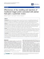
levels at 0, 3, 6 and 8 hours. MAP remained stable in group 1
but decreased in groups 2 and 3 (P < 0.001), such that at
8 hours MAP was much higher in group 1 than in either group
2 or group 3 (Fig. 1). This change in MAP correlated with the
increase in plasma Cl
–
(R
2
= 0.50; P < 0.0001) and less well
with the decrease in pH (R
2
= 0.24; P < 0.001). After 6 hours
of acidosis plasma nitrite levels were significantly higher in
group 2 animals than in group 1 or group 3 animals
(P < 0.05). We concluded that moderate acidosis, induced by
HCl infusion, worsened blood pressure and increased plasma
nitrate/nitrite levels in septic rats. Some other mechanism is
needed to account for the further reduction in MAP in group 3
Critical Care October 2004 Vol 8 No 5 Kellum et al.
335
animals, however, because NO release was not increased in
that group. Our results are in general agreement with reports
by Pedoto and coworkers [18,45] that demonstrated that
metabolic acidosis increased iNOS, leading to vasodilation
and shock in healthy rats. Our study extends these findings by
examining the effects of acidosis in nonshocked, septic
animals. These data are also consistent with our data from
RAW 264.7 cells (presented above), in which a decreased
pH
o
(7.0) resulted in increased NO release but more severe
acidosis (pH
o
= 6.5) did not [17].
Clinical implications
Understanding the effects of acid–base balance on the
inflammatory response is highly relevant to clinical medicine
for a variety of reasons. First, current deficiencies in our
understanding of the effects of acidosis on a wide range of
cellular processes have led to controversy in the way in which
patients are managed in a variety of clinical settings. Most
clinicians tend to ignore the effects of exogenous Cl
–
on pH
o
,
but many will treat even mild forms of acidemia. In addition, all
forms of metabolic acidosis appear to be associated with
prolonged hospital and intensive care unit length of stay [47].
Because metabolic acidosis is both commonly caused and
treated by clinicians, an understanding of the physiologic
consequences of altered pH
o
is imperative.
Second, our ability to alter acid–base balance as a tool with
which to manipulate cellular processes will be dependent on
an improved understanding of the relationship between pH
o
and the synthesis and release of inflammatory molecules.
Investigators continue to seek means to modulate the
inflammatory response as primary therapy for sepsis and
related conditions. These efforts have focused not only on
reducing proinflammatory mediators in an effort to reduce
tissue injury, but also on the converse – augmenting the
inflammatory response to infection. This interest also extends
into other fields, including autoimmune disease and cancer
therapy. For example, decreased lymphocyte function has
been documented with decreased pH
o
in human lymphokine-
activated killer cells [48], human IL-2 stimulated lymphocytes
[49], as well as murine natural killer cells [50]. The
mechanisms responsible for these effects are unknown but
probably do not include energy substrate depletion [50].
Third, even when it is not practical or desirable to manipulate
pH
o
as a primary means of altering the inflammatory response,
an understanding of how pH
o
affects this response is necessary
to interpret data from studies of immunomodulation; to avoid
unintended immunomodulation in clinical and laboratory
settings; and to explore the capacity of pH
o
to improve the
effectiveness of existing treatments. Finally, an understanding of
how pH
o
is involved in the regulation of inflammation by
intracellular signaling pathways or other mechanism might
ultimately lead to other strategies for immunomodulation.
Conclusion
Little is currently known about the effects of acid–base
abnormalities on innate immunity. Acidosis produces
significant effects on immune effector cell function in vitro.
The regulation of NO release and synthesis has been found
to be significantly effected by pH
o
both in vitro and in vivo,
and may be partially responsible for acidosis-associated
hemodynamic instability. Production of inflammatory cyto-
kines, as well as DNA-binding of transcription factors in their
control pathways, appears to be sensitive to pH
o
as well.
However, emerging evidence suggests that different forms of
acidosis (respiratory versus metabolic) and even different
types of metabolic acidosis (lactic versus hyperchloremic)
produce different effects. Overall, lactic acid appears to be
anti-inflammatory whereas HCl is proinflammatory. The extent
to which these effects apply to the clinical situation has yet to
be determined, but given that acidosis is an extremely
common problem in the intensive care unit, and immune
function is of critical importance, efforts to elucidate these
relationships are quite justified.
Competing interests
JAK has received research grants and consulting fees from
Abbott Laboratories.
References
1. Chu EK, Whitehead T, Slutsky AS: Effects of cyclic opening and
closing at low- and high-volume ventilation on bronchoalveo-
lar lavage cytokines. Crit Care Med 2004, 32:168-174.
2. Burne-Taney MJ, Kofler J, Yokota N, Weisfeldt M, Traystman RJ,
Rabb H: Acute renal failure after whole body ischemia is char-
acterized by inflammation and T cell-mediated injury. Am J
Physiol Renal Physiol 2003, 285:F87-F94.
3. Joyce DE, Grinnell BW: Recombinant human activated protein
C attenuates the inflammatory response in endothelium and
monocytes by modulating nuclear factor-kappaB. Crit Care
Med 2002, Suppl:S288-S293.
Available online />Figure 1
Mean arterial pressure for septic animals (induced by cecal ligation
and puncture) after infusion of 0.1 N HCl acid to reduce the base
deficit (BD) by 5–10 mEq/l (white bars) or 10–15 mEq/l (black bars).
A control group was given a similar volume of lactated Ringer’s (gray
bars). Shown are group means (n = 8) ± SEM. *P < 0.05. Adapted
from Kellum and coworkers [46].
0
20
40
60
80
100
120
140
0368
Time (hours)
mmHg
BD 0
BD 5–10
BD 10–15
*
*
336
4. Annane D, Cavaillon JM: Corticosteroids in sepsis: from bench
to bedside? Shock 2003, 20:197-207.
5. Heming TA, Dave SK, Tuazon DM, Chopra AK, Peterson JW,
Bidani A: Effects of extracellular pH on tumour necrosis
factor-alpha production by resident alveolar macrophages.
Clin Sci 2001, 101:267-274.
6. Bellocq A, Suberville S, Philippe C, Bertrand F, Perez J, Fou-
queray B, Cherqui G, Baud L: Low environmental pH is respon-
sible for the induction of nitric-oxide synthase in
macrophages. Evidence for involvement of nuclear factor-
kappaB activation. J Biol Chem 1998, 273:5086-5092.
7. Heming TA, Tuazon DM, Dave SK, Chopra AK, Peterson JW,
Bidani A: Post-transcriptional effects of extracellular pH on
tumour necrosis factor-alpha production in RAW 246.7 and
J774 A.1 cells. Clin Sci 2001, 100:259-266.
8. Bidani A, Wang CZ, Saggi SJ, Heming TA: Evidence for pH sen-
sitivity of tumor necrosis factor-alpha release by alveolar
macrophages. Lung 1998, 176:111-121.
9. Huang CJ, Haque IC, Slovin PN, Nielsen RB, Fang X, Skimming
JW: Environmental pH regulates LPS-induced nitric oxide for-
mation in murine macrophages. Nitric Oxide 2002, 6:73-78.
10. Jensen JC, Buresh C, Norton JA: Lactic acidosis increases
tumor necrosis factor secretion and transcription in vitro. J
Surg Res 1990, 49:350-353.
11. Himmelseher S, Pfenninger E, Georgieff M: Basic fibroblast
growth factor reduces lactic acid-induced neuronal injury in
rat hippocampal neurons. Crit Care Med 1998, 26:2029-2036.
12. Schurr A, Payne RS, Miller JJ, Rigor BM: Brain lactate is an
obligatory aerobic energy substrate for functional recovery
after hypoxia: further in vitro validation. J Neurochem 1997, 69:
423-426.
13. Schurr A, Payne RS, Miller JJ, Rigor BM: Brain lactate, not
glucose, fuels the recovery of synaptic function from hypoxia
upon reoxygenation: an in vitro study. Brain Res 1997, 744:
105-111.
14. Jorres A, Gahl GM, Frei U: In vitro studies on the effect of dialy-
sis solutions on peritoneal leukocytes. Perit Dial Int 1995, 15:
S41-S45.
15. Douvdevani A, Abramson O, Tamir A, Konforty A, Isakov N,
Chaimovitz C: Commercial dialysate inhibits TNF alpha mRNA
expression and NF-kappa B DNA-binding activity in LPS-stim-
ulated macrophages. Kidney Int 1995, 47:1537-1545.
16. AlKharfy KM, Kellum JA, Matzke G: Unintended immunomodula-
tion: part I. Effect of common clinical conditions on cytokine
biosynthesis. Shock 2000, 13:333-345.
17. Kellum JA, Song M, Li J: Lactic, and hydrochloric acids induce
different patterns of inflammatory response in LPS-stimulated
RAW 264.7 cells. Am J Physiol Regul Integr Comp Physiol 2004,
286:R686-R692.
18. Pedoto A, Caruso JE, Nandi J: Acidosis stimulates nitric oxide
production and lung damage in rats. Am J Respir Crit Care
Med 1999, 159:397-402.
19. Lu Y, Song M, Zuo Y, Kellum JA: The inhibitory effect of lactic
acid on NO release by stimulated RAW cells is not specific to
LPS [abstract]. Crit Care Med 2003, Suppl:A45.
20. Lardner A: The effects of extracellular pH on immune function.
J Leukoc Biol 2001, 69:522-530.
21. Nahas GG, Tannieres ML, Lennon JF: Direct measurement of
leukocyte motility: effects of pH and temperature. Proc Soc
Exp Biol Med 1971, 138:350-352.
22. Rotstein OD, Fiegel VD, Simmons RL, Knighton DR: The delete-
rious effect of reduced pH and hypoxia on neutrophil migra-
tion in vitro. J Surg Res 1988, 45:298-303.
23. Rabinovich M, DeStefano MJ, Dziezanowski MA: Neutrophil
migration under agarose: stimulation by lowered medium pH
and osmolality. J Reticuloendothel Society 1980; 27:189-200.
24. Simchowitz L, Cragoe EJ Jr: Regulation of human neutrophil
chemotaxis by intracellular pH. J Biol Chem 1986, 261:6492-
6500.
25. Leblebicioglu B, Lim JS, Cario AC, Beck FM, Walters JD: pH
changes observed in the inflamed gingival crevice modulate
human polymorphonuclear leukocyte activation in vitro. J Peri-
odontol 1996, 67:472-477.
26. Araki A, Inoue T, Cragoe EJ Jr, Sendo F: Na+/H+ exchange
modulates rat neutrophil mediated tumor cytotoxicity. Cancer
Res 1991, 51:3212-3216.
27. Simchowitz L: Intracellular pH modulates the generation of
superoxide radicals by human neutrophils. J Clin Invest 1985,
76:1079-1089.
28. Gabig TG, Bearman SI, Babior BM: Effects of oxygen tension
and pH on the respiratory burst of human neutrophils. Blood
1979, 53:1133-1139.
29. Beachy JC, Weisman LE: Acute asphyxia affects neutrophil
number and function in the rat. Crit Care Med 1993, 21:1929-
1934.
30. Craven N, Williams MR, Field TR, Bunch KJ, Mayer SJ, Bourne FJ:
The influence of extracellular and phagolysosomal pH
changes on the bactericidal activity of bovine neutrophils
against Staphylococcus aureus. Vet Immunol Immunopathol
1986, 13:97-110.
31. Nakagawara A, Nathan CF, Cohn ZA: Hydrogen peroxide
metabolism in human monocytes during differentiation in
vitro. J Clin Invest 1981, 68:1243-1252.
32. Trevani AS, Andonegui G, Giordano M, Lopez DH, Gamberale R,
Minucci F, Geffner JR: Extracellular acidification induces
human neutrophil activation. J Immunol 1999, 162:4849-4857.
33. Miyazawa K, Inoue K: Complement activation induced by
human C-reactive protein in mildly acidic conditions. J
Immunol 1990, 145:650-654.
34. Osaki M, Sumimoto H, Takeshige K, Cragoe EJ, Jr., Hori Y,
Minakami S: Na+/H+ exchange modulates the production of
leukotriene B4 by human neutrophils. Biochem J 1989, 257:
751-758.
35. Grinstein S, Furuya W: Cytoplasmic pH regulation in phorbol
ester-activated human neutrophils. Am J Physiol 1986, 251:
C55-C65.
36. Kellum JA, Bellomo R, Kramer DJ, Pinsky MR: Etiology of meta-
bolic acidosis during saline resuscitation in endotoxemia.
Shock 1998, 9:364-368.
37. Kellum JA: Fluid resuscitation and hyperchloremic acidosis in
experimental sepsis: improved survival and acid-base
balance with a synthetic colloid in a balanced electrolyte solu-
tion compared to saline. Crit Care Med 2002, 30:300-305.
38. Opie L: Effect of extracellular pH on function and metabolism
of isolated perfused rat heart. J Appl Physiol 1965, 209:1975-
1980.
39. Cooper D, Herbertson M, Werner H, Walley K: Bicarbonate
does not increase left ventricular contractility during L-lactic
acidemia in pigs. Am Rev Resp Dis 1993, 148:317-322.
40. Salzman AL, Wang H, Wollert PS, Vandermeer TJ, Compton CC,
Denenberg AG, Fink MP: Endotoxin-induced ileal mucosal
hyperpermeability in pigs: role of tissue acidosis. Am J Physiol
1994, 266:G633-G646.
41. Menconi MJ, Salzman AL, Unno N, Ezzell RM, Casey DM, Brown
DA, Tsuji Y, Fink MP: Acidosis induces hyperpermeability in
Caco-2BBe cultured intestinal epithelial monolayers. Am J
Physiol 1997, 272:G1007-G1021.
42. Gonzalez PK, Doctrow SR, Malfroy B, Fink MP: Role of oxidant
stress and iron delocalization in acidosis-induced intestinal
epithelial hyperpermeability. Shock 1997, 8:108-114.
43. Unno N, Menconi MJ, Smith M, Aguirre DE, Fink MP: Hyperperme-
ability of intestinal epithelial monolayers is induced by NO: effect
of low extracellular pH. Am J Physiol 1997, 272:G923-G934.
44. Unno N, Hodin RA, Fink MP: Acidic conditions exacerbate inter-
feron-gamma-induced intestinal epithelial hyperpermeability:
role of peroxynitrous acid. Crit Care Med 1999, 27:1429-1436.
45. Pedoto A, Nandi J, Oler A, Camporesi EM, Hakim TS, Levine RA:
Role of nitric oxide in acidosis-induced intestinal injury in
anesthetized rats. J Lab Clin Med 2001, 138:270-276.
46. Kellum JA, Song M, Venkataraman R: Effects of hyperchloremic
acidosis on arterial pressure and circulating inflammatory
molecules in experimental sepsis. Chest 2004, 125:243-248.
47. Gunnerson KJ, Saul M, Kellum JA: Lactic versus nonlactic meta-
bolic acidosis: outcomes in critically ill patients [abstract]. Crit
Care 2003, Suppl 2:17.
48. Severin T, Muller B, Giese G, Uhl B, Wolf B, Hauschildt S, Kreutz
W: pH-dependent LAK cell cytotoxicity. Tumour Biol 1994, 15:
304-310.
49. Loeffler DA, Juneau PL, Masserant S: Influence of tumour
physico-chemical conditions on interleukin-2-stimulated lym-
phocyte proliferation. Br J Cancer 1992, 66:619-622.
50. Loeffler DA, Juneau PL, Heppner GH: Natural killer-cell activity
under conditions reflective of tumor micro-environment. Int J
Cancer 1991, 48:895-899.
Critical Care October 2004 Vol 8 No 5 Kellum et al.