Báo cáo y học: " Unintended spread of a biosafety level 2 recombinant retrovirus" ppsx
Bạn đang xem bản rút gọn của tài liệu. Xem và tải ngay bản đầy đủ của tài liệu tại đây (637.25 KB, 6 trang )
BioMed Central
Open Access
Page 1 of 6
(page number not for citation purposes)
Retrovirology
Short report
Unintended spread of a biosafety level 2 recombinant
retrovirus
Alexander Stang
1
, Elisabeth Petrasch-Parwez
2
, Sabine Brandt
1
,
Rolf Dermietzel
2
, Helmut E Meyer
3
, Kai Stühler
3
, Sven-T Liffers
3
,
Klaus Überla*
1
and Thomas Grunwald
1
Address:
1
Department of Molecular and Medical Virology, Ruhr-University Bochum, D-44780 Bochum, Germany,
2
Department of Neuroanatomy
and Molecular Brain Research, Ruhr-University Bochum, D-44780 Bochum, Germany and
3
Medical Proteome Center, Ruhr-University Bochum,
D-44780 Bochum, Germany
Email: Alexander Stang - ; Elisabeth Petrasch-Parwez - ;
Sabine Brandt - ; Rolf Dermietzel - ; Helmut E Meyer - ;
Kai Stühler - ; Sven-T Liffers - ; Klaus Überla* - ;
Thomas Grunwald -
* Corresponding author
Abstract
Background: Contamination of vertebrate cell lines with animal retroviruses has been documented
repeatedly before. Although such viral contaminants can be easily identified with high sensitivity by PCR,
it is impossible to screen for all potential contaminants. Therefore, we explored two novel methods to
identify viral contaminations in cell lines without prior knowledge of the kind of contaminant.
Results: The first hint for the presence of contaminating retroviruses in one of our cell lines was obtained
by electron microscopy of exosome-like vesicles released from the supernatants of transfected 293T cells.
Random amplification of particle associated RNAs (PAN-PCR) from supernatant of contaminated 293T
cells and sequencing of the amplicons revealed several nucleotide sequences showing highest similarity to
either murine leukemia virus (MuLV) or squirrel monkey retrovirus (SMRV). Subsequent mass
spectrometry analysis confirmed our findings, since we could identify several peptide sequences originating
from monkey and murine retroviral proteins. Quantitative PCRs were established for both viruses to test
currently cultured cell lines as well as liquid nitrogen frozen cell stocks. Gene fragments for both viruses
could be detected in a broad range of permissive cell lines from multiple species. Furthermore,
experimental infections of cells negative for these viruses showed that both viruses replicate rapidly to
high loads. We decided to further analyze the genomic sequence of the MuLV-like contaminant virus.
Surprisingly it was neither identical to MuLV nor to the novel xenotropic MuLV related retrovirus (XMRV)
but showed 99% identity to a synthetic retrovirus which was engineered in the 1980s.
Conclusion: The high degree of nucleotide identity suggests unintended spread of a biosafety level 2
recombinant virus, which could also affect the risk assessment of gene-modified organisms released from
contaminated cell cultures. The study further indicates that both mass spectrometry and PAN-PCR are
powerful methods to identify viral contaminations in cell lines without prior knowledge of the kind of
contaminant. Both methods might be useful tools for testing cell lines before using them for critical
purposes.
Published: 22 September 2009
Retrovirology 2009, 6:86 doi:10.1186/1742-4690-6-86
Received: 23 April 2009
Accepted: 22 September 2009
This article is available from: />© 2009 Stang et al; licensee BioMed Central Ltd.
This is an Open Access article distributed under the terms of the Creative Commons Attribution License ( />),
which permits unrestricted use, distribution, and reproduction in any medium, provided the original work is properly cited.
Retrovirology 2009, 6:86 />Page 2 of 6
(page number not for citation purposes)
Findings
The first evidence for a retroviral contamination was
obtained by electron microscopy, originally performed to
characterize the production of exosome-like vesicles
released from transfected 293T cells. Vesicles were puri-
fied from supernatants by ultracentrifugation through a
20% sucrose cushion. Resulting pellets were fixed with
2,5% glutaraldehyde and 1% paraformaldehyde in 0.1 M
sodium phosphate (pH: 7.4), postfixed with 2% osmium
tetroxide, dehydrated and embedded in araldite (Serva).
Ultrathin sections (100 nm) were contrasted with uranyl
acetate and lead citrate, viewed in a Philips EM 420 elec-
tron microscope and documented by the digital system
DITABIS (Digital Biomedical Imaging System). Surpris-
ingly, in addition to the expected exosome-like vesicles
the supernatants displayed two main types of enveloped
viruses each of which with a diameter of around 100 nm
(Fig. 1). One type exhibits a centrally located spherical
electron-dense core closely resembling the type-C mor-
phology of retroviruses that are shown by both murine
leukemia viruses (MLV) and squirrel monkey retrovirus
[1]. The other type which was much less frequent also
shows a spherical outer shape, but displays an excentri-
cally located core (Fig. 1). Of note, the latter type does not
resemble the characteristic morphology of the two retrovi-
ruses identified below. Both types of particles were
present in supernatants from both transfected and
untransfected cells indicating that they were produced
independently of proteins expressed by the transfected
plasmids.
For identification of the unknown viruses a method based
on the random amplification of particle associated RNAs
(PAN-PCR) [2] was used. Isolation of nucleic acids from
viral particles was done as described before [2] using 30
ml of supernatant from 293 T cells. Pellets were resus-
pended in 0.5 ml PBS and used for purification of viral
RNA by means of the DNA Blood Mini Kit (Qiagen). For
RNA preparation residual DNA was degraded by an
RNase-free DNase (Ambion) and purified RNA was subse-
quently reverse transcribed to double stranded cDNA
using the cDNA Synthesis Kit (Roche). Thereafter DNA
and RNA (double stranded cDNA) were further processed
equally.
Our previously published protocol of PAN-PCR [2] was
slightly modified by including a random amplification
protocol described elsewhere [3,4]. In detail, double
stranded cDNA was digested with MseI and ethanol-pre-
cipitated in the presence of 1 μg glycogen. Adapter ligation
was carried out in a total volume of 10 μl containing the
MseI digested DNA, 400 U T4 DNA-Ligase and T4 DNA-
Ligase buffer (New England Biolabs) and 20 pmol adapter
composed by the hybridized oligonucleotides NBam24
(AGGCAACTGTGCTATCCGAGGGAG) and NCsp11
(TACTCCCTCGG) for 1 h at 4°C followed by 6 h at 16°C.
2 μl of this reaction were used for PCR amplification in a
total volume of 25 μl containing 0.2 mM each dNTP, 10
mM Tris-HCl (pH 9.0), 1.5 mM MgCl
2
, 50 mM KCl, 1.25
U of Taq DNA Polymerase (Amersham Biosciences) and
the primer NBam24 (1 mM). Two-step thermocycling was
done by 30 cycles 95°C for 30 s and 72°C for 2 min fol-
lowed by 10 min at 72°C. Products were used for cloning
into the pCRII Vector as described by the manufacturer
(Invitrogen). At least 40 colonies were picked and tested
directly by PCR as described [2]. 25 PCR products of dif-
ferent size were sequenced and analyzed for homologies
to viral sequences by a nucleotide-nucleotide (BLASTn)
and translated BLAST search (BLASTx) at the NCBI web-
site [5].
Results from PAN-PCR using supernatants of 293T cell
cultures confirmed our assumption of retroviral contami-
nations (Fig 2). While there were no viral sequences
detected in the DNA preparation, we found 12 sequences
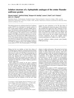
Electron microscopical analysis of viral contaminantsFigure 1
Electron microscopical analysis of viral contami-
nants. Viral particles from the supernatant of 293T cells
were pelleted through a 20% sucrose cushion. Ultrathin sec-
tions of the fixed pellet show two types of particles resem-
bling retroviruses. The more abundant one (*) exhibits a
central electron-dense core, while the core of the other type
of particles (→) is located excentrically. Scale bar: 100 nm.
Retrovirology 2009, 6:86 />Page 3 of 6
(page number not for citation purposes)
in the RNA preparation that showed high similarity scores
(97-100%) to five different regions within either the gag
and pol gene or the LTR region of squirrel monkey retro-
virus (SMRV, Fig. 2A) by analyses on nucleotide level.
Additionally we isolated seven clones that bore six differ-
ent sequences with high similarity scores of 97-100% to
murine leukemia virus strains (MuLV) in a nucleotide
BLAST search. These sequences were located within the
LTR and the gag, pol and env genes. (Fig. 2B).
To get detailed sequence information for classification of
the contaminants and to clarify, if the murine contami-
nant virus is related or identical to the recently discovered
xenotropic MuLV related retrovirus (XMRV) [6] we
sequenced the regions of both viruses between the most
outwards located subgenomic fragments derived from
PAN-PCR. Therefore these regions were amplified by PCR
using genomic DNA from infected cells and the primers
SMRV-1s (GTTGGGAACCCAGGCTAAGCTG) and SMRV-
8057a (GTAGGAGGGGAACCGGCTAC) for SMRV and
T3R03s (AGGGGATTTATTGGATACACG), T3R32a
(CATCGTGACCTGGGAAGC) for the murine retrovirus.
PCR products were sequenced directly by primer walking
and resulting proviral genomic sequences were analyzed
by a BLASTn search.
The resulting sequence of the simian retrovirus (7.968
kbp) confirmed our preliminary identification as squirrel
monkey retrovirus [GenBank: M23385.1
] with an overall
sequence identity of 98,5%. In contrast the 7.4 kbp
sequence of the murine retrovirus was neither identical to
one of the murine leukemia viruses nor to XMLV but
showed an overall similarity score of 99% to pAMS [Gen-
Bank: AF010170
], a plasmid carrying the proviral
sequence of a recombinant hybrid virus. This construct
was engineered in the 1980s and is composed of
sequences from Moloney murine leukemia virus
(MoMLV) and amphotropic mouse leukemia virus clone
4070A [7,8]. In the current GenBank entry it is described
as " reference retrovirus for FDA validation of retrovirus vec-
tors used for human gene therapy ". Neither the hybrid
virus itself nor the plasmid pAMS were ever used in our
laboratory. Due to the high identity of nucleotide
sequence and the fact that the structure of MoMLV and
amphotropic leukemia virus related segments is 100%
identical to that of pAMS (data not shown) we can
exclude that the contaminant virus is a natural recom-
binant of MoMLV and amphotropic leukemia virus.
In order to confirm our data from PAN-PCR we applied
mass spectrometry to supernatants of the 293T cells, sus-
pected to be contaminated. This method was chosen as an
additional option - besides PAN-PCR - which allows for
identification of contaminants without prior knowledge
or assumptions about the agent in question. Therefore,
pelleted material from supernatants of transfected and
untransfected 293T cells was separated by SDS-PAGE and
digested with trypsin. For protein identification peptides
were analyzed by nanoLC-ESI-MSMS and uninterpreted
ESI-MS/MS-spectra were correlated with the NCBI-protein
sequence database applying the SEQUEST™ algorithm [5].
Besides peptides similar to human gene products, we
found peptides derived from squirrel monkey retrovirus
(SMRV) and murine leukemia virus (MuLV). Additionally
there was one hit with the peptide sequence KAADTES-
GPSSGRT to 'Pol [synthetic construct]' [GenBank:
gi|2281588] corresponding to the integrase of the hybrid
amphotropic/Moloney murine leukemia virus (Tables 1
and 2).
For quantification of viral genomes from supernatants of
cell culture and proviral genomes within cellular DNA we
established SybrGreen based Real-Time PCRs (and RT-
PCRs). Cellular DNA (from >10
5
cells) and viral RNA
from supernatants (200 μl) were both purified using the
DNA Blood Mini Kit (Qiagen) as recommended by the
manufacturer. RNA was treated with RNase-free DNase
(Ambion) prior to RT-PCR. Primers were chosen to match
one clone derived from PAN-PCR for each virus respec-
tively. For SMRV we used the primers T3R34s (CTGCCCT-
GTATCATCTGAACC) and T3R34a (CTCCCCTGAC ATT
CAACGC) amplifying the gag gene of the viral genome
whereas for the murine hybrid retrovirus primers T3R27s2
(CAGGGAGAACATGGTAATAGGA) and T3R27a2
(ACGACCTCTCCAAAGTATCCA) were used to amplify a
region within the env gene. As standards for PCR we used
1 μl of the corresponding plasmid quantified by photom-
Mapping of mass spectrometry hits and PAN-PCR products to retroviral genomesFigure 2
Mapping of mass spectrometry hits and PAN-PCR
products to retroviral genomes. Asterisks mark the
position of peptides identified by mass spectrometry to be
derived from pAMS-MLV (A) or SMRV (B), while grey bars
indicate genomic regions highly homologous to PAN-PCR
products. The hatched box (A) represents proviral
sequences of pAMS with high sequence similarity to ampho-
tropic MLV.
pol envgagLTR LTR
pol envgagLTR LTR
pAMS
SMRV
A)
B)
***
****** * **
Retrovirology 2009, 6:86 />Page 4 of 6
(page number not for citation purposes)
etry and diluted to suitable concentrations in water con-
taining herring sperm DNA as carrier/noise DNA (100 ng/
μl). Reaction was done using the QuantiTect SybrGreen
PCR Kit (Qiagen) in a total volume of 20 μl containing 5
μl purified DNA and 5 pmol of each primer. RNA stand-
ards for RT-PCRs were in vitro transcripts from the corre-
sponding plasmids. RT-PCR was performed using the
QuantiTect SybrGreen RT-PCR Kit (Qiagen) in a total vol-
ume of 20 μl with 5 μl purified RNA and 5 pmol of each
primer. All quantitative PCRs were performed on a Rotor-
Gene 3000 (Corbett Research). After an initial tempera-
ture step of 95°C for 15 min 40 cycles of 95°C for 10 s,
55°C for 30s, 72°C for 30 s and acquiring SybrGreen flu-
orescence at 81°C were performed followed by a standard
melting curve analysis of products. In case of RT-PCR a
reverse transcription step of 20 min at 50°C was added
before cycling.
Quantitative RT-PCRs could confirm high loads in the
supernatant of infected 293T cells of up to 10
12
RNA cop-
ies/ml for the recombinant MLV virus and up to 10
11
RNA
copies/ml for SMRV. Although these values are rather
high, they are not unexpected. Viral RNA copies do not
reflect the infectious titer due to a large excess of non-
infectious particles. In lentiviral RNA packaging studies
we also observed that the RNA copy numbers detected in
the supernatants are approximately 1000-fold higher than
the infectious titer [9]. Consistently, Münch et al., could
increase the titer of HIV by up to 5 orders of magnitude by
adding the SEVI peptide, indicating that a vast excess of
particles are not detected by conventional titration of
infectious virus without SEVI [10]. In addition, viral RNAs
released from dying cells independent of budding parti-
cles could also contribute to the RNA copies detected in
the supernatant of infected cells.
To get further insight into the degree of contamination we
quantified proviral genomes within purified cellular DNA
from various currently cultured cell lines as well as nitro-
gen-frozen cell stocks in our laboratory (Table 3). Both
viruses showed a broad range of permissiveness for cell
lines from multiple species and a first contamination
event dated back to the years 2002 (SMRV) and 2003
(hybrid amphotropic/Moloney murine leukemia virus).
For both viruses highest infection scores were found for
293T cells, where the mean copy number of proviral
genomes per cell reached values of up to 208 for SMRV
and 2874 for the pAMS related virus (Table 3). Proviral
genomic copy numbers per cell were calculated by the
ratio or proviral genomic copy numbers and the DNA
content measured by spectrophotometry. To verify the
accuracy of determination of cell numbers, standardiza-
tion by single copy gene PCR for myostatine and quantifi-
cation of cellular genomic DNA by QBit Kit (Invitrogen)
were also performed. Each of the methods used gave com-
parable results with a maximum deviation of 2.2-fold.
It has to be mentioned that each of both viruses was found
in at least one aliquot of all cell lines tested. Thus, we can-
not report a single cell line which is not permissive for one
Table 2: Results of mass spectrometry (nano-LC-ESI-MSMS) -Concentrated and purified supernatant from untransfected 293T cells.
Accession number Descriptive Name
gi|773422 gag protein [SMRV]
gi|40796131 pp12 [Murine leukemia virus] gag
gi|18338742 gPr80 glycosylated gag polyprotein [Moloney murine leukemia virus]
gi|42543698 Chain A, The Crystal Structure Of The Human Hsp70 Atpase Domain
gi|1065227 Heat Shock Cognate 70kd Protein (44kd Atpase N-Terminal Fragment)
gi|74589 gag polyprotein [SMRV-H]
gi|9626961 Pr180 [Murine leukemia virus] gag/pol
gi|2281588 pol [synthetic product]
gi|30583573 programmed cell death 6 interacting protein [Homo sapiens]
Table 1: Results of mass spectrometry (nano-LC-ESI-MSMS) - Concentrated and purified supernatant from transfected 293T cells.
Accession number Descriptive Name
gi|773422 gag protein [SMRV]
gi|40796131 pp12 [Murine leukemia virus] gag
gi|9626959 Pr65 [Murine leukemia virus] gag
gi|4691418 heat shock protein 72 [Homo sapiens]
gi|74589 gag polyprotein [SMRV-H]
gi|9626961 Pr180 [Murine leukemia virus] gag/pol
gi|1045516 gPr80 envelope protein [Murine leukemia virus]
gi|2281588 pol [synthetic product]
gi|30583573 programmed cell death 6 interacting protein [Homo sapiens]
Retrovirology 2009, 6:86 />Page 5 of 6
(page number not for citation purposes)
of both viruses. Furthermore, experimental infections of
retrovirus-negative aliquots of selected cell lines showed
that both viruses are highly infectious and propagate to
high viral loads as determined by viral RNA copy numbers
in the supernatants and proviral genome copies of
extracted cellular DNA (data not shown). In the early
stage of infection even some cytopathic effects (CPE)
could be observed. In contrast, no CPE was seen in persist-
ently infected cultures possibly due to adaptation of cells
to the retroviral infection (data not shown). To evaluate a
potential contamination of cell lines from tissue culture
respositories, we also directly performed PCR analyses of
a frozen 293T cell stock obtained from ECACC. Neither
proviral DNA of SMRV nor chimeric MLV could be
detected.
In summary, there have been numerous publications
about retroviral contaminations like recent reports of eco-
tropic murine leukemia virus in various cell lines [11,12].
The most frequent retrovirus found in this context is
squirrel monkey retrovirus (SMRV) [13-16]. One study
even reported the detection of SMRV related sequences in
commercial interferon preparations in 1998 [17].
Although the sequences were found only as DNA and
therefore rather derived from cellular DNA carrying provi-
ral genomes than viral particles, it clearly demonstrated
the contamination of the interferon producing cell line
with SMRV. Germany's Central Commission of Biosafety
(ZKBS) recently reported that SMRV was detectable in 128
samples of 4279 cell cultures from different laboratories
throughout the country [18].
The present report extents these studies by identifying for
the first time a presumably synthetic chimeric retrovirus as
a contaminant. This gene-modified organism seems to
have replicated and spread intensely in a broad set of cell
lines for several years without being noticed. This hybrid
amphotropic/Moloney murine leukemia virus was engi-
neered in the 1980s [7,8] and neither the virus itself nor
the plasmid (pAMS) containing its proviral genome were
ever used in our laboratory. Although the precise source
for the contamination could not be traced back, sharing
cell lines with other laboratories seems the most likely
explanation. A frozen aliquot of 293T cells (HEK
293tsA201), which we obtained from ECACC, was not
contaminated. While SMRV contaminations were
detected in different laboratories, testing of three other
laboratories' cell lines did not reveal contaminations with
the hybrid amphotropic/Moloney murine leukemia virus
(data not shown).
This study also shows that both PAN-PCR and mass spec-
trometry are powerful tools to identify viral contamina-
tions without prior knowledge or assumptions about the
viruses in question. Therefore, both methods might be
useful in routine controls for viral contaminations of cell
cultures.
Competing interests
The authors declare that they have no competing interests.
Authors' contributions
AS, EPP, KU and TG wrote the manuscript. EPP performed
electron microscopy. EPP and RD were responsible for
interpretation of electron micrographs. SB was involved in
vesicle production and purification, SL performed mass
spectrometry. KS and HEM provided advice for the mass
spectrometry analyses. All other experiments and study
design were done by AS, KU and TG.
Acknowledgements
We would like to acknowledge Rosemarie Bohr, Regina Buetermann, Bet-
tina Tippler, Hans-Werner Habbes and Marlen-Loebbecke-Schumacher for
their excellent technical assistance.
Table 3: Proviral load in contaminated cell lines.
Proviral DNA copies/cell
Cell line Hybrid amphotropic/Moloney murine leukemia virus Squirrel monkey retrovirus
CHO 0.0007 0.00002
A549 0.0004 26
Cos 7 0.0005 0.26
HEp2 0.13 0.001
L929 1.44 0.002
Vero E6 3.84 12
3T6 4.58 0.00001
HeLa 5.74 115
293A 1622 20
T-Rex 293 2596 8.11
293T 2874 208
Viral DNA copy numbers were determined by quantitative PCR and are given as the maximum proviral DNA copies/cell detected.
Publish with BioMed Central and every
scientist can read your work free of charge
"BioMed Central will be the most significant development for
disseminating the results of biomedical research in our lifetime."
Sir Paul Nurse, Cancer Research UK
Your research papers will be:
available free of charge to the entire biomedical community
peer reviewed and published immediately upon acceptance
cited in PubMed and archived on PubMed Central
yours — you keep the copyright
Submit your manuscript here:
/>BioMedcentral
Retrovirology 2009, 6:86 />Page 6 of 6
(page number not for citation purposes)
References
1. Gonda MA, Fine DL, Gregg M: Squirrel-Monkey Retrovirus -
Electron-Microscopy of A Virus from New World Monkeys
and Comparison with Mason-Pfizer Monkey Virus. Archives of
Virology 1978, 56:297-307.
2. Stang A, Korn K, Wildner O, Uberla K: Characterization of virus
isolates by particle-associated nucleic acid PCR. J Clin Microbiol
2005, 43:716-720.
3. Allander T, Emerson SU, Engle RE, Purcell RH, Bukh J: A virus dis-
covery method incorporating DNase treatment and its
application to the identification of two bovine parvovirus
species. Proceedings of the National Academy of Sciences of the United
States of America 2001, 98:11609-11614.
4. Klempa B, Kruger DH, Auste B, Stanko M, Krawczyk A, Nickel KF, et
al.: A novel cardiotropic murine adenovirus representing a
distinct species of mastadenoviruses. J Virol 2009,
83:5749-5759.
5. National Center for Biotechnology Information [http://
www.ncbi.nlm.nih.gov]
6. Urisman A, Molinaro RJ, Fischer N, Plummer SJ, Casey G, Klein EA,
Malathi K, Magi-Galluzzi C, Tubbs RR, Ganem D, Silverman RH,
DeRisi JL: Identification of a Novel Gammaretrovirus in Pros-
tate Tumors of Patients Homozygous for R462Q RNASEL
Variant. PLoS Pathog 2006, 2:e25.
7. Miller AD, Law MF, Verma IM: Generation of Helper-Free
Amphotropic Retroviruses That Transduce A Dominant-
Acting, Methotrexate-Resistant Dihydrofolate-Reductase
Gene. Molecular and Cellular Biology 1985, 5:431-437.
8. Miller AD, Buttimore C: Redesign of Retrovirus Packaging Cell-
Lines to Avoid Recombination Leading to Helper Virus Pro-
duction. Molecular and Cellular Biology 1986, 6:2895-2902.
9. Brandt S, Blissenbach M, Grewe B, Konietzny R, Grunwald T, Uberla
K: Rev proteins of human and simian immunodeficiency virus
enhance RNA encapsidation. PLoS Pathog 2007, 3:e54.
10. Münch J, Rücker E, Ständker L, Adermann K, Goffinet C, Schindler M,
Wildum S, Chinnadurai R, Rajan D, Specht A, Giménez-Gallego G,
Sánchez PC, Fowler DM, Koulov A, Kelly JW, Mothes W, Grivel JC,
Margolis L, Keppler OT, Forssmann WG, Kirchhoff F: Semen-
derived amyloid fibrils drastically enhance HIV infection. Cell
2007, 131:1059-1071.
11. Takeuchi Y, McClure MO, Pizzato M: Identification of {gamma}-
retroviruses constitutively released from cell lines used for
HIV research. J Virol 2008, 82(24):12585-8.
12. Hartley JW, Evans LH, Green KY, Naghashfar Z, Macias AR, Zerfas
PM, Ward JM: Expression of infectious murine leukemia
viruses by RAW264.7 cells, a potential complication for stud-
ies with a widely used mouse macrophage cell line. Retrovirol-
ogy 2008, 5:1.
13. Gelderblom H, Bauer H, Ogura H, Wigand R, Fischer AB: Detection
of Oncornavirus-Like Particles in Hela-Cells. 1. Fine-Struc-
ture and Comparative Morphological Classification. Interna-
tional Journal of Cancer 1974, 13:246-253.
14. Popovic M, Kalyanaraman VS, Reitz MS, Sarngadharan MG: Identifi-
cation of the Rpmi-8226 Retrovirus and Its Dissemination As
A Significant Contaminant of Some Widely Used Human
and Marmoset Cell-Lines. International Journal of Cancer 1982,
30:93-99.
15. Middleton PG, Miller S, Ross JA, Steel CM, Guy K: Insertion of
Smrv-H Viral-Dna at the C-Myc Gene Locus of A Bl Cell-Line
and Presence in Established Cell-Lines. International Journal of
Cancer 1992, 52:451-454.
16. Sun R, Grogan E, Shedd D, Bykovsky AF, Kushnaryov VM, Grossberg
SE, Miller G: Transmissible Retrovirus in Epstein-Barr Virus-
Producer B95-8 Cells. Virology 1995, 209:374-383.
17. Pienkowska M, Seth A: Detection of squirrel monkey retroviral
sequences in interferon samples. Journal of Hepatology 1998,
28:396-403.
18. Bundesamt für Verbraucherschutz und Lebensmittelsicher-
heit [ />06__Gentechnik/00__doks__downloads/SMRV-Info.html__nnn =
true]