Báo cáo y học: " Response of a simian immunodeficiency virus (SIVmac251) to raltegravir: a basis for a new treatment for simian AIDS and an animal model for studying lentiviral persistence during antiretroviral therapy" pptx
Bạn đang xem bản rút gọn của tài liệu. Xem và tải ngay bản đầy đủ của tài liệu tại đây (1.66 MB, 19 trang )
RESEA R C H Open Access
Response of a simian immunodeficiency virus
(SIVmac251) to raltegravir: a basis for a new
treatment for simian AIDS and an animal model
for studying lentiviral persistence during
antiretroviral therapy
Mark G Lewis
1†
, Sandro Norelli
2†
, Matt Collins
1
, Maria Letizia Barreca
3
, Nunzio Iraci
3
, Barbara Chirullo
2
,
Jake Yalley-Ogunro
1
, Jack Greenhouse
1
, Fausto Titti
4
, Enrico Garaci
5
, Andrea Savarino
2*
Abstract
Background: In this study we successfully created a new approach to ART in SIVmac251 infected nonhuman
primates. This drug regimen is entirely based on drugs affecting the pre-integration stages of replication and
consists of only two nucleotidic/nucleosidic reverse transcriptase inhibitors (Nt/NRTIs) and raltegravir, a promising
new drug belonging to the integrase strand transfer inhibitor (INSTI) class.
Results: In acutely infected human lymphoid CD4
+
T-cell lines MT-4 and CEMx174, SIVmac251 replication was
efficiently inhibited by raltegravir, which showed an EC
90
in the low nanomolar range. This result was confirmed in
primary macaque PBMCs and enriched CD4
+
T cell fractions. In vivo monotherapy with raltegravir for only ten days
resulted in reproducible decreases in viral load in two different groups of animals. When emtricitabine (FTC) and
tenofovir (PMPA) were added to treatment, undetectable viral load was reached in two weeks, and a parallel
increase in CD4 counts was observed. In contrast, the levels of proviral DNA did not change significantly during
the treatment period, thus showing persistence of this lentiviral reservoir during therapy.
Conclusions: In line wi th the high conservation of the three main amino acids Y143, Q148 and N155 (responsible
for raltegravir binding) and molecular docking simulations showing similar binding modes of raltegravir at the
SIVmac251 and HIV-1 IN active sites, raltegravir is capable of inhibiting SIVmac251 replication both in tissue culture
and in vivo. This finding may help to develop effective ART regimens for the simian AIDS model entirely based on
drugs adopted for treatment in humans. This ART-treated AIDS nonhuman primate model could be employed to
find possible strategies for virus eradication from the body.
Background
Integration of proviral DNA into the host’sgenomeisa
fundamental step in lentiviral infections, initiating the
latency period, and allowing the virus to exploit the cel-
lular transcriptional and translational machinery [1,2].
The recent approval of the integrase strand transfer
inhibitor (INSTI) raltegravir for first -line HIV-1 therapy
thus provides a further option for treatment of drug-
naïve HIV-1 infected patients [3]. INSTIs selectively
inhibit the strand transfer re action, catalyzed by HIV-1
integrase (IN) after 3’ processing, which generates a
reactive 3’-hydroxylgroup in proviral DNA. Raltegravir
represents a major success in the history of antiretro-
viral therapy (ART) and is the result of a drug develop-
ment process which encountered exceptional difficulties
[1,4,5].
Despite this and other major successes in antiretro-
viral drug discovery and the availabi lity of several drug
* Correspondence:
† Contributed equally
2
Department of Infectious, Parasitic and Immune-mediated Diseases, Istituto
Superiore di Sanità, Viale Regina Elena, 299, 00161, Rome, Italy
Lewis et al. Retrovirology 2010, 7:21
/>© 2010 Lewis et al; licensee BioMed Central Ltd. This is an Open Access article distributed under the terms of the Creative Commons
Attribu tion License ( which permits unrestricted use, distribution, and reproduction in
any medium, provided the original work is properly cited.
options for obtaining sustained suppression of viral load
in HIV-1 infected individuals, ART cannot eradicate the
virus from the body [6], at least in a reasonable time [7].
The grounds for HIV-1 persistence during therapy lie in
the presence o f long-lived viral reservoirs (mainly the
memory T CD4
+
cell subset), which harbour silent
copies of proviral DNA that cannot be targeted by drugs
or the immune system [6,8,9]. Alternative/co mplemen-
tary strategies are therefore being actively researched, in
order to facilitate the purging of HIV-1 from reservoirs.
To this end, the so-called “ shock and kill” strategies
have been proposed [8,10]. These strategies should
induce, throu gh drugs, HIV-1 activation from quies-
cence (i.e.the“shock” phase), in the presence of ART
(to block viral spread), followed by the elimination of
infected cells (i.e.the“kill” phase), through either nat-
ural means (e.g. immune response, viral cytopathogeni-
city) or artificial means (e.g. drugs).
One major obstacle which has been encountered by
the studies on such “ HIV-1 purging” strategies is the
availability of reliable animal models. Such models
should mimic the long-term effects of ART in humans.
Interesting low-cost models include the new SCID mice
technology [11] and feline immunodeficiency virus
(FIV)-i nfected cats [12,13]; however, the macaque AIDS
model has encountered the largest consensus in the
AIDS researchers’ community. This model is based on
lentiviruses derived from African sooty mangabeys intro-
duced into the non-natural host, Asian macaque species
(Macaca sp.), which results in the development of illness
similar to that described in AIDS patients [14]. Recently,
also chimpanzees were found to develop disease when
naturally infected with SIVcpz, the ancestor of HIV-1
group M [15]. However, the close phylogenetic relation-
ships w ith humans restrict the use of these apes in the
laboratory.
The simian AIDS model presents its own profile of
response to HIV-1 drugs, rendering it difficult to treat
with the ART protocols adopted for treatment of HIV-
1/AIDS. For example, SIVmac251, one of the most com-
monly adopted viral s trains for laboratory infection of
macaques, is fully sensitive to nucleotidic and nucleosi-
dic reverse transcriptase inhibitors (NtRTIs/NRTIs),
retains limited sensitivity to some, but not all of the
protease inhibitors (PIs) designed for HIV-1, and shows
approximately 200-fold less sensitive to non-nucleosidic
reverse transcriptase inhibitors [16]. Treatment with
NtRTI tenofovir (also referred to as PMPA) and NRTI
emtricitabine (FTC) represents a valuable option for
studying the gene expression profiles activated during
suppression of viral load and immune restor ation [17].
However, this type of treatment can hardly be used to
model long-term lentivi ral persistence during ARTs
designed for humans, which comprise three or more
active drugs and at least two drug targets. The poor
response of the laboratorysimianlentivirusesto
NNRTIs prompted some to replace the reverse tran-
scriptase (RT) gene of the simian lentivirus with a gene
encoding HIV-1 RT [18]. This substitution is extremely
useful for studying the occurrence of drug resistance
mutations in vivo [19], and for preclinical testing of
novel NRTIs. However, an impact of the RT substitution
on the natural history of the disease cannot be excluded
so far. Indeed, apart from altering immunogenicity,
replacement of a simian lentivirus’s RT with its counter-
part from HIV-1 might alter susceptibility of some cell
populations to the virus. For example, RT-bound, elon-
gating proviral DNA is the substrate of APOBEC3G, a
species-specific cellular restrict ion factor to infection by
primate lentiviruses [14]. Upon clarification of these
issues, this simian/human immunodeficiency virus
(SHIV) chimera could become an extremely useful tool
to model ART consisting of two Nt/NRTIs and an
NNRTI [20], a commonly adopted regimen for first-line
treatment of HIV-1/AIDS.
New strategies for treatment of the macaque AIDS
model may exploit the novel INSTI drug class. Hazuda
et al. [21] evaluated, in SHIV 89.6P-infected rhesus non-
human primates, the effects of naphthyridine carboxa-
mide, L-870,812 , an INSTI belonging to a chemical class
distinct from that of raltegravir. This study provided the
first proof of concept for an antiretroviral effect of IN
inhibition in vivo. Moreover, L-870,812 monotherapy of
macaques allowed the isolation of drug-resistant viruses
presenting the N155H mutation, which later proved an
important drug resistance mutation in HIV-1-infected
individuals failing raltegravir-based regimes [22]. On this
basis, an ART-treated nonhuman primate model was
recently developed by Dinoso et al. using L-870,812 in
comb ination with PMPAand two PIs, i.e.saquinavirand
atazanavir in macaque s infected simultaneously with
SIV/17E-Fr and SIV/Delta B670 [23]. Sustained suppres-
sion of viral load was obtained until the end of follow-
up. However, one limitation of this model is that this
type of drug regimen is not adopted in humans. More-
over, the authors used two PIs at a relative dosage much
higher than that adopted for humans.
Thesusceptibilityofnon-humanprimatelentiviruses
to naphthyridine carboxamides is probably due to the
high level of conservation of IN CCDs [12]. A three-
dimensional (3D) structure [PDB: 1C6V] is available for
the catalytic core domain (CCD) and C-terminal domain
of the IN of SIVmac251 [24]. SIVmac251 IN catalyses
reactions similar to those of HIV-1 IN, and t he crystal
structure shows that the IN of SIVmac251 shares the
highly conserved three-dimensional (3D) architecture of
retroviral INs [see Additional file 1] [24]. Accordingly,
SIVmac251 has been reported to be susceptible also to
Lewis et al. Retrovirology 2010, 7:21
/>Page 2 of 19
the investigational HIV-1 INSTI, CHI/1043, belonging
to the 1H-benzylindole drug class [25]. Despite this bulk
of evidence, response o f SIVmac251 to raltegravir has
not yet been studied in detail. An extension of the data
from other INSTI classes to raltegravir may not be
obvious, because diff erent classes of INSTIs may have
different binding modes, as sho wn by the partially over-
lapping yet different dr ug resistance mutation profiles
and molecular docking calculations [26,27].
The assessment of the response of a simian lentivirus
laboratory strain t o raltegravir may have important
repercussions on the development of antiretrov iral
therapies for the simian AIDS model using a drug com-
bination adopted in humans. Moreover, the response of
a non-human lentivirus to this drug may furnish impor-
tant insights into the requirements for susceptibility to
this new and important drug class.
Results
Raltegravir inhibits SIVmac251 replication in tissue
culture
To test susceptibility of SIVmac251 to raltegravir, MT-4
cells were in fected with SIVmac251, washe d and incu-
bated with decreasing raltegravir concentrations.
Response to raltegravir was assessed by the widely vali-
dated MTT assay, when the majority of cells in the
untreated controls were dead, i.e., approx. fifteen days
post-infection. Results showed that raltegravir inhibited
SIVmac251 replication in the low nanomolar range (Fig.
1A). The EC
50
was approximately one ord er of magni-
tude lower than that obtained using HIV-1 IIIB, which
was calculated on the basis of data collected at five days
post-infection due to the more rapid kinetics of viral
cytopathogenicity (Fig. 1A). Data from HIV-2(strain:
CDC 77618 [28])-infected MT-4 cell cultures showed
A
0.1 1 10 100
EC50
EC90
EC95
SIVmac251
HIV-1
HIV-2
raltegravir conc. [nM]
B
0.1 1 10 100
EC50
EC90
EC95
SIVmac251
HIV-1
raltegravir conc. [nM]
C
0.1 1 10 100
EC50
EC90
EC95
SIVmac251
HIV-1
HIV-2
raltegravir conc. [nM]
D
1 10 100
EC50
EC90
EC95
CD4+ T-cells
PBMCs
raltegravir conc. [nM]
EC
95
EC
90
EC
50
EC
95
EC
90
EC
50
EC
95
EC
90
EC
50
EC
95
EC
90
EC
50
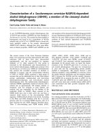
Figure 1 SIVmac251 s usceptibility to ralteg ravir in tissue culture. The effective concentrations at 50%, 9 0% and 95% (respectively, EC
50
,
EC
90
, and EC
95
) are presented (means ± SEM from at least two independent experiments) for inhibition of: lentiviral cythopathogenicity in MT-4
cells (Panel A), viral core antigen release in supernatants of acutely infected MT-4 cells (Panel B), syncytium formation in acutely infected
CEMx174 cells (Panel C), viral core antigen release in supernatants of acutely SIVmac251-infected rhesus peripheral blood mononuclear cells
(PBMCs) and enriched CD4
+
T-cell fractions (Panel D). In panel A, the inhibitory concentrations were determined by the methyl tetrazolium (MTT)
method when the majority of control infected cells (in the absence of drug treatment) were dead at light microscopy examination. In panel B,
values were derived by quantifying, using antigen-capture ELISA assays, SIVmac251 p27 and HIV-1 p24 in supernatants from five-day old cultures.
In panel C, values were calculated on the basis of the numbers of syncytia per well at five days post-infection, Syncytia were counted in
triplicate on three different occasions by light microscopy. In panel D, values are representative for supernatants of primary cells from three
different donors at Day 5 post-infection.
Lewis et al. Retrovirology 2010, 7:21
/>Page 3 of 19
intermediate characteristics between those obtained
from SIVmac251- and HIV-1-infected cultures. Apart
from being phylogenetically closer to SIVmac251 than
to HIV-1, the H IV-2 strain that we used killed the
majority of the infected cells in eight days following
infection, thus showing viral cytopathogenicity kinetics
slower than HIV-1 and more rapid than SIVmac251.
To assess whether the difference in the EC
50
values
for SIVmac251 and HIV-1 IIIB cytopathic effects were
attributable to the different kinetics of viral cytopatho-
genicity, we measured, by antigen-capture ELISA assays,
the viral core antigen in supernatants collected at five
days post-infection from both the SIVmac251 and HIV-
1 infected cell cultures. In this ca se, the ranges of the
EC
50
values for SIVmac251 and HIV-1 obtained in the
different experimental set-ups were overlapping (Fig.
1B). We concluded that raltegravir inhibits SIVmac251
replication in human T-cell lines with similar potency as
shown against HIV-1.
Asdifferenttypesofkitshadtobeusedtocompare
inhibition of SIVmac251 p27 and HIV-1 p24 production,
we decided to confirm the results using another method
allowing simultaneous and homogeneous measurements
of antiviral efficacy against SIVmac251, HIV-1, and H IV-
2. We used syncytia counts in CEMx174 cells as a mea-
sure of lentiviral repl ication. SIVmac251 replication
induces syncytia at an earlier time point as compared to
the cytopathic effect induced in MT-4 cells, in which len-
tiviral replication mostly induces apoptotic and necrotic
cell death [29]. The effectiveness of syncytia counts as a
parameter for detection of the antiretroviral effects was
confirmed by correlation analyses of syncytium formation
and viral core antigen production in the presence of anti-
retroviral drugs (an example using raltegravir is given in
the additional mater ial [see Additional file 2]). CEMx174
cells were infected with SIVmac251, HIV-1, and HIV-2
viral stocks at the same multiplicity of infection (MOI),
and syncytia were counted by optical microscopy at 4-5
days post-infection. Results confirmed that raltegravir
exerted potent and reproduci ble anti-SIVmac2 51 activity
(Fig 1C).
To assess the anti-SIVmac251 effects of raltegravir
under c onditions more closely resembling those occur-
ring in vivo, 3 day-old PHA-stimulated peripheral blood
mononuclear cells (PBMCs) from uninfected rhesus
macaques (Macaca mulatta)wereinfectedwithSIV-
mac251, and viral replicatio n was quantified in superna-
tants by ELISA at five days post-infection, in order to
allow comparison with the results reported in the pre-
vious paragraph. Also in this case, raltegravir displayed
an EC
50
in the low nanomolar range (Fig 1D).
To assess the effect of raltegravir in the rhesus CD4
+
T cell population, i.e., the main target of SIVmac251
in vivo, we separated the CD4
+
T cells from fresh
unstimulated PBMCs using magnetic beads. Flow cyto-
metric analysis of the enriched CD4
+
T cell fraction
showed that 94 to 100% of cells expressed the CD4 anti-
gen (data not shown). Cells were PHA-stimulated for
three days, infected with SIVmac251, and, a gain, viral
replication was quantitated in supernatants by EL ISA at
five days post-infection. Again, results confirmed the
potent inhibitory effect of raltegravir (Fig 1D).
We concluded that raltegravir inhibits SIVmac251 in
different tissue culture assays at least with similar
potenc y as observed in human primary cell-base d assays
[30,31]. The EC
95
values are within the mean trough
concentration (142 nM) measured in pharmacokinetic
studies in humans [32].
Raltegravir decreases viral load in SIVmac251-rhesus
macaques and stably maintains suppressed viral loads
when associated with RT inhibitors PMPA and FTC
To confirm s usceptibility of SIVmac251 to raltegravir
in vivo, we tested the effects of the drug in six rhesus
macaques with stabilized infection by SIVmac251 (hen-
ceforth referred to as Group 1). The macaques had been
challenged with SIVmac251 by either the rectal or vagi-
nal route and were between 5 months and two years
post infections prior to the start of raltegravir treatment.
The macaqu es were randomized to receive 50 or 100 mg
of raltegravir twice daily with food (bid). Monotherapy
was continued for ten days. At day ten, raltegravir
treatment resulted in a significant decrease in viral
load (P = 0.031, Wilcoxon signed rank test) (Fig. 2A).
The 100 mg treatment subgroup apparently had higher
decreases in viral load than the 50 mg treatment sub-
group, although the numbers of animals did not allow
statistical evaluation of differences between subgroups.
Of note, one animal treated with the 100 mg bid
dosage showed an undetectable viral load (detection
threshold: 40 copies of viral RNA ml
-1
). Virological
response to raltegravir was associated with a significant
increase in CD4 counts (P = 0.017, Wilcoxon signed
rank test), detectable in all animals (Fig. 2B). We con-
cluded that raltegravir-treated animals showed viro-
immunological improvement.
This group of nonhuman primates had been released
by another study showing that viral loads had been
stable before initiating raltegravir treatment (data not
shown). In the prior study, unfortunately, viral load had
been measured by another technique (NASBA), thus
rendering incorrect a possible statistical comparison
between the historical values and the pre-and post ralte-
gravir treatment values from the present study.
Comparison of the CD4 values after raltegravir mono-
therapy with historical data derived from flow-cytometric
determination of CD4 numbers was instead possible. The
data available from the time of SIVmac251 inoculations
Lewis et al. Retrovirology 2010, 7:21
/>Page 4 of 19
showed that the CD4 counts prior to raltegravir treat-
ment had been gradually decreasing, or maintained at
levels lower than pre-inoculation values, as a sign of the
ongoi ng lentiviral infection [33]. Our results showed that
raltegravir abruptly changed the trends in the CD4
counts(Fig.2B).ForfiveoftheGroup1animals,itwas
possible to make a multiple comparison between values
at ten days prior to treatment start, at Day 0, and Day 10
of ralteg ravir monotherapy. Repea ted-measures ANOVA
reported an extremely significant difference (P = 0.0014).
The CD4 counts post-monotherapy significantly deviated
from values at Day 0 and ten days prior t o raltegravir
admini stration (P < 0.05 in both cases; Bonferroni’spost-
test for multiple comparisons), whereas no significant dif-
ference was found between values prior to treatment start
and Day 0 (P > 0.05). We concluded that there was a sig-
nificant association between CD4 rise and raltegravir
treatment.
Figure 2 Effect of raltegravir (RAL), alone and in combination with PMPA and FTC, on viral load (panel A) and CD4 counts (panel B) in
SIVmac251-infected macaques (Group 1). SIVmac251-infected rhesus macaques (Macaca mulatta) were randomized to receive 50 (marked by
the blue symbols) or 100 (red symbols) mg of raltegravir twice daily with food (bid). Monotherapy was continued for ten days. At day 11,
nonhuman primates treated with 50 mg of raltegravir bid were switched to the 100 mg regimen, and two RT inhibitors, i.e. the NtRTI, tenofovir
(PMPA) and the NRTI emtricitabine (FTC), were added to treatment (henceforth referred to as ART) in all animals. Viral load values positioning on
the dotted line parallel to the x axis should read as undetectable.
Lewis et al. Retrovirology 2010, 7:21
/>Page 5 of 19
At day 11, nonhuman primates treated with 50 mg o f
raltegravir bid were switched to t he 100 mg regi men (in
order to prevent selection of drug-resistant mutants),
and two RT inhibitors, i.e. the NtRTI, PMPA and the
NRTI FTC, were added to treatment (henceforth
referred to as ART) in all subjects. Results showed that
viral load continued to decrease: an undetectable viral
load was shown by four animals after one week, and by
all study animals after two weeks ( Fig. 2A). Viral load
was maintained undetectable until the end of follow-up
(Day 52). In parallel, CD4 counts continued to increase
up to restoration of values at the time of inoculation
(Fig. 2B). We concluded that the ART regimen based on
raltegravir plus PMPA and FTC suppressed viral replica-
tion to undetectable levels in nonhuman primates and
restored CD4 counts.
As expected from results in human clinical trials, ther-
apy was well-tolerated from a clinical point of view, and
serum che mistry (kidney and liver enzymes) and hema-
tology values remained within normal limits (data not
shown).
The virological improvement of SIVmac251-infected
animals is significantly associated with raltegravir
treatment
The results in Group 1 nonhuman primates clearly
show that raltegravir, and ART, induced viro-immunolo-
gical improvement of nonhuman primates with progres-
sing SIVmac251 infection.
To exclude that the viral load decrease observed dur-
ing raltegravir treatment of Group 1 could be at tributed
to random fluctuations of SIVmac251 replication, or by
spontaneous acquisition, by the non human primate s, of
the capacity to control viral replication, we treated
another group of non-human primates for which histori-
cal data were available using the same technique for
viral load measurement (Group 2). In this group, we
also measured viral load a t seven days of treatment, in
order to minimize the effect of time-dependent, sponta-
neous viral fluctuations on the decrease in viral load.
Fig. 3 clearly shows that no significant changes in viral
load were observable in 166 days in the absence of drug
treatment (P > 0.05, Bonferro ni’ s post-test following
repeated-measures ANOVA). Viral load, however, did
significantly decrease in o nly seven days of raltegravir
treatment (P < 0.05). Despite the small number of non-
human primates enrolled, the P values obtained support
the extreme significance of the anti-SIVmac251 effects
of raltegravir. We concluded that 1) there was signifi-
cant association b etween decreased viral load and ralte-
gravir treatment, and that 2) the effects o f raltegravir
proved reproducible in two distinct groups of animals.
Again, one non-human primate in Group 2 showed an
undetectable viral load following raltegravir
monotherapy. This animal was the only component of
Group 2 to show a low viral load (i.e., 1,520 copies/ml)
before treatment was initiated. To further support the
contribution of raltegravir treatment to the viral load
decline i n this subject, treatment was stopped and viral
load was followed up. Results showed that a reb ound in
viralloadoccurredfollowing treatment suspension
(4,520 viral RNA copies/ml; value at two weeks from
suspension).
SIVmac251 proviral DNA persists during ART in peripheral
blood mononuclear cells of the non-human primates
To evaluate whether copies of SIVmac251 provira l DNA
persisted during ART despite suppression of viral load
to undetectable levels, we measured proviral DNA copy
numbers in PBMCs of the non-human primates prior to
starting dosing and after 52 days of therapy. Results
showed that proviral DNA was maintained stable during
the treatment period analyzed. The difference between
the proviral DNA levels at the two time points analyzed
was not statistically significant (P > 0.05; Wilcoxon
Figure 3 Association of viral load decrease with raltegravir
treatment of SIVmac251-infected animals (Group 2). SIVmac251-
infected rhesus macaques (Macaca mulatta) received 100 mg of
raltegravir twice daily with food (bid). Monotherapy was continued
for ten days. Comparison between pre- and post-raltegravir viral
load measurements was done. Viral load values at Day 0, Day 7 and
Day 10 were compared with viral loads at 27 and 166 days prior to
treatment start. Significant differences (P < 0.05; Bonferroni’s test
following repeated-measures ANOVA; shown in the graph by the
red asterisks) were found between both the values at 166 and 27
days prior to treatment start and the values at Day 7 and Day 10 of
treatment. No significant differences, instead, were found between
the values at 166 days, or 27 days, prior to treatment, and the
values at Day 0. The dashed line parallel to the x axis marks the
detection threshold of the technique adopted.
Lewis et al. Retrovirology 2010, 7:21
/>Page 6 of 19
sig ned rank test) (Fig. 4). We concluded that ART regi-
mens consisting of two NRTIs/NtRTIs plus raltegravir
maintains stably suppressed SIVmac251 viral load, but
not the proviral DNA, in non-human primates.
Discussion
Susceptibility of SIVmac251 to raltegravir
The results of the present study show that raltegravir
inhibits SIVmac251 replication both in tissue culture
and in vivo. The result is comparable to those of pre-
vious susceptibili ty studies using wild-type HIV-1 and
HIV-2 [25,30] and is supported by similar assays con-
ducted in the present study using HIV-1 and HIV-2 as
positive controls for viral replication inhibition. The
EC
50
of raltegravir found by Hombrouck et al. [25] in
the MTT-based assays for HIV-1 IIIB cythopathic effects
is slightly lower than that obtained in the present study.
Differences between our results and those of Hom-
brouck et al. can be attributed to the dif ferences in the
experimental protocols such as the higher MOI of HIV-
1 used in the present study. Similarly, the higher EC
50
of raltegravir for HIV-2 reported in a previous study of
Roquebert et al. using HIV-2 ROD can be explained by
the fact that these authors adopted a different method
for viral quantification, i.e. a quantitative RT PCR assay
[30]. On the other hand, the range of EC
95
values
obtained in the present study for HIV-1 overlap the
33 nM value reported previously, which became an
acceptable threshold for the trough concentrations of
the drug in pharmacokinetic studies [34].
The lower EC
50
of raltegravir for the SIVmac251 cyto-
pathic effect, as compared to that found in HIV-1-based
assays, is likely to be attributed to the viral cytopatho-
genicity kinetics of SIVmac251 which is slower than
that o f HIV-1. Under our assay conditions, SIVmac251
required approximately fift een days to kill the control
untreated cultures, whereas HIV-1 only took five days.
It is possible to hypothesize that the inhibitory effects of
raltegravir in the SIVmac251-infected MT-4 cells sub-
jected to prolonged treatment exposure is the result of
the sum of the inhibition levels occurring during each of
the multiple rounds of viral replication. When the EC
50
was calculated on a viral antigen basis, the resulting
values for SIVmac251 and HIV-1 were closer, because
both sets of measurements were done at five days post-
infection. This result is also confirmed by viral antigen
capture assays using supernatants from primary PBMC
and enriched CD4
+
cell fract ions incubated under simi-
lar assay conditions.
Inhibition of SIVmac251 replication in tissue culture is
in line with the declines in viral load obtained b y ralte-
gravir monotherapy of SIVmac251-infected non-human
primates. Of course, factors other than drug treatment
may have contributed to the viral load decline observed
during treatment in vivo. For example, it has been
shown t hat cytotoxic responses contributed to the viral
load decline induced by another INSTI, the naphthyri-
dine carboxamide, L-870,812 [21]. However, these
responses in the absence of raltegravir could hardly con-
trol infection, as shown by the analysis of the CD4
Figure 4 Persistence of proviral DNA during therapy (Group 1). Proviral DNA was measured by a quantitative PCR technique at start of
treatment with antiretroviral drugs, and at 52 days of therapy.
Lewis et al. Retrovirology 2010, 7:21
/>Page 7 of 19
counts of one of our study groups prior to treatment
start. In this regard, the graph in Fig. 2B clearly shows
that the nadir of CD4 counts was approximately coinci-
dent with Day 0 of raltegravir monotherapy. Subject
M974 (belongi ng to this group) showed a low viral load
(1,960 RNA copies/ml) at the beginning of treatment.
However,thissubjectcouldnotberegardedasanélite
controller of the infection, because, prior to raltegravir
administration,italsoshowedlowCD4counts(173
CD4
+
Tcells/μl) which increased to 531 units/μlafter
10 days of raltegravir monotherapy, and to 778 units/μl
at 52 days of treatment with ART (Fig. 2A). Finally, the
results obtained in another group of five macaques, for
which historical viral loa d values were available prior to
start of raltegravir treatment, showed that marked
declines in viral loads were stringently associated to the
period of raltegravir monotherapy. These results support
the fundamental contribution of raltegravir administra-
tion to the antiretroviral effects. Moreover, after therapy
suspension, a rebound in viral load was evident in an
animal that had shown undetectable levels following ral-
tegr avir monotherapy. On the whol e, these results show
rapid virological and immunolog ical response associat ed
with administration of raltegravir in t he simian AIDS
model.
Although response to a naphthyridine carboxamide
such as L-870,812 has already been assessed in the
simian AIDS model, the susceptibility to raltegravir o f
SIVmac251 is far from obvious. Though mechanistically
identical to L-870,812, raltegravir belongs t o an unre-
lated chemical class, i.e.theN-alkyl-5-hydroxypyrimidi-
none carboxamides [35]. It has been well established
that there may be discordant resistance between
mechanistically identical INSTI drugs designed for HIV-
1, and that non-human lentiviral enzymes often show
structural differences to their HIV-1 counterparts
mimicking specific drug resistance mutations [36,37]. In
this context, the in vivo susceptibility of SIVmac251 to a
further INSTI drug such as raltegravir supports the con-
cept that the simian AIDS model responds to more than
one class of INSTIs designed for HIV-1 and encourages
pre-clinical testing of novel INSTIs in SIVmac251-
infected nonhuman primates.
Structural bases for the raltegravir response
An explanation for SIVmac251 susceptibility to raltegra-
vir may be derived from comparison of the SIVmac251
IN with INSTI-susceptible or resistant HIV-1 INs; and,
conversely, the data provide d herein, using SIVmac251,
may furnish no vel insights into the understanding of the
raltegravir response of HIV-1. Primary resistance to ral-
tegravir has been associated with three major mutations,
N155H, Q148H/K/R, and Y143H; mutation of any of
these HIV-1 IN amino acids initiates pathways leading
to raltegravir resistance [22,38,39]. These residues are
located aroun d the active site of IN and wit hin interact-
ing distance to raltegra vir, as sho wn by molecular mod-
elling simulations conducted by independent groups
[27,40]. Drug resistance mutations N155H and Q148R
were shown to hamper INSTI binding to HIV-1 IN, by
either decr easing the affinity of IN/proviral DNA com-
plexes for INSTIs (N155H) or affecting assembly of pro-
viral DNA (Q148R) [41]. Secondary mutations reported
for raltegravir are L74M, E92Q, T97A, E138K, G140S/A,
V151I, G163R, I203M, S230R, and D232N [22,38,40].
According to structural alignments of the HIV-1 IN
CCD with published structures of the IN CCDs from
SIVmac251 and other retroviruses with reported profiles
of susceptibility to INSTIs, we found that the amino
acid positions corresponding to Y143, Q148, and N155
are conserved between HIV-1 and SIVmac251 (Fig. 5).
These amino acids are also conserved in HIV-2 IN (sus-
ceptible to raltegravir [30]) but are not in prototype
foamy virus (PFV; susceptible to raltegravir but showing
EC
50
values 1-2 orders of magnitude higher than the
EC
50
forHIV-1[42])orRoussarcomavirus(RSV)IN
(which is not inhibited by INSTIs designed for HIV-1
[26]). Several amino acids a re also conserved be tween
SIVmac251 and HIV-1 at positions susceptible to sec-
ondary drug resistance mutations. Among these, conser-
vation of E92 is particularly relevant because, differently
from other secondary resistance mutations, the E92Q
mutation alone is capable to decrease raltegravir sus-
ceptibility in the absence of primary resistance muta-
tions [43]. Instead, the amino acid corresponding to
HIV-1 IN E92, is a proline in PFV and a valine in RSV.
Similar to HIV-2, SIVmac251 mimics polymo rphism s
at some of the secondary drug resistance positions in
HIV-1 (L74, E138, G163 and I203). Among these, the
only drug resistance mutation mimicked by SIV is
I203M (Fig. 5). This mimicry, however, is shown al so by
HIV-2 IN, which, as mentioned above, is fully suscepti-
ble to raltegravir. Changes in this position may thus be
irrelevant in the absence of primary drug resistance
mutat ion Y143R/C [44]. Outside the IN CCD at the site
corresponding to HIV-1 IN S230 (not shown in the
sequence alignment of Fig. 5), SIVmac251 presents a
glycine, which, h owever, does not mimic the c orre-
sponding drug resistance mutation S230R in HIV-1 IN.
Two drug resistance mutations induced by other
INSTIs were shown to confer cross-resistance to ralte-
gravir [43]. T66I is a primary drug resistance mutation
raised by the investigational quinolone INSTI, elvitegra-
vir, and some diketo acids [35,45]. F121Y is a primary
drug resistance mutation for naphthyridine carboxamide
L-870,810 [26]. The amino acids presented by SIV-
mac251 in these positions strictly corr espond to those
found in wild-type HIV-1 and HIV-2 INs (Fig. 5).
Lewis et al. Retrovirology 2010, 7:21
/>Page 8 of 19
If the known susceptibilities of different lentiviruses to
raltegravir, or other INSTIs, are mapped to a phylogenetic
tree of primate lentivirus IN CCDs (Fig. 6), SIVmac251 IN
clusters with a clade comprisi ng HIV-2 IN, which is dis-
tinct from, but adjacent to the cluster of primate lentivirus
INs comprising HIV-1 IN (Fig. 6). A relatively recent com-
mon ancestor of HIV-1 and SIVmac251/HIV-2 INs may
explain their common susceptibility to raltegravir. Of note,
conservation of the key amino acids T66, E92, F121, Y143,
G148 and N155 (determining susceptibility to raltegravir)
is shared by all primate lentiviruses analysed and is dis-
played also by highly divergent primate lentiviruses,
including SIVcol, SIVsyk and the endogenous lentivirus
pSIV, recently identified by Gifford et al. in basal primate
Microcebus murinus [see Additional file 3] and sharing
intermediate characteristics between primate and feline
lentiviruses [46].
If the level of amino acid similarity between SIV-
mac251 and HIV-1 IN CCDs (calculated by the Swiss
PDB Viewer program) is mapped to a 3D structure of
HIV-1INCCD,itmaybenotedthataminoacididen-
tities cluster to the active site of IN, which is involved
in INSTI binding [27,35] (Fig. 7). INSTIs bind at the
interface between the IN activ e site and provi ral DNA
[1,2,47]. Modelling this interaction, however, has
encountered several obstacles in the absence of crystal-
lographicdataforHIV-1INcomplexedwithINSTIs,
although several theoretical models for INSTI binding
have been published so far [27,35,48-51]. A novel
study using the “ induced fit” docking (IFD) approach
allowed conformat ional changes in the protein and
DNA as well in order to obtain the best accommoda-
tion of the l igand [27]. Considering these findings, we
built a SIVmac251 IN-Mg2
+
-DNA ternary complex as
Figure 5 Sequence alignment of the integrase catalytic core domains of HIV-1 subtype B (PDB: 1BL3_C), HIV-2 (PDB: 3F9K_A),
SIVmac251 (PDB: 1C6V_A), prototype foamy virus/PFV (PDB: 3DLR_A), and Rous Sarcoma virus/RSV (PDB: 1ASU_A). The sequence
alignment is based on a structural alignment performed using the VAST algorithm. Regions showing significant structural alignment are
presented in blue, with the highly conserved residues shown in red. Above the alignments are shown the mutations found in HIV-1 infected
individuals failing raltegravir-based drug regimens (the green arrows indicate the primary resistance mutations Y143H, Q148H/K/R, and N155H;
black arrows indicate secondary resistance mutations). Other drug resistance mutations induced by other integrase strand transfer inhibitors are
shown below the alignments. The mutations shown by site-directed mutagenesis to confer resistance to raltegravir are underlined. Note that the
structure for HIV-1 subtype B integrase catalytic core domain (PDB: 1BL3_C) presents the secondary drug resistance mutation V151I.
Lewis et al. Retrovirology 2010, 7:21
/>Page 9 of 19
Figure 6 Phylogenetic tree of lentiviral integrase core domains. Sequences adopted : human immunodef iciency virus type-1 (HIV-1) [PDB:
1BL3C]; human immunodeficiency virus type-2 (HIV-2) [PDB: 3F9K]; simian immunodeficiency virus, host: macaque (SIVmac251) [PDB: 1C6VC];
simian immunodeficiency virus, host: chimpanzee (Pan troglodytes) (SIVcpz) [accession: AAF18575]; simian immunodeficiency virus, host: gorilla
(Gorilla gorilla) (SIVgor) [accession: ACM63211]; simian immunodeficiency virus, host: African green nonhuman primate (Chlorocebus sp.) (SIVagm)
[accession: CAA30658]; simian immunodeficiency virus, host: mandrill (Mandrillus sphinx) (SIVmnd) [accession: AAB49569]; simian
immunodeficiency virus, host: Cercopithecus lhoesti (SIVlhoest) [accession: AAF07333]; simian immunodeficiency virus, host: Skyes’ nonhuman
primate (Cercopithecus albogularis) (SIVsyk) [accession: AAS97874]; simian immunodeficiency virus, host: Colobus nonhuman primate (Colobus
guereza) (SIVcol) [accession: AAK01033]; prosimian immunodeficiency virus, host: Microcebus murinus (pSIV) [see: additional material in Ref. [46]];
feline immunodeficiency virus, host: domestic cat (Felis sylvestris) (FIV-Pet) [accession: AAB59937]; lion lentivirus, host: lion (Panthera leo)
[accession: ABX25835]; puma lentivirus, host: mountain lion (Puma concolor) [accession: AAA67168]; caprine arthritis-encephalitis virus (CAEV),
host: Capra hircus [accession: NP_040939]; visna lentivirus, host: sheep (Ovis aries) [PDB: 3HPG_A]; equine infectious anemia virus (EIAV) host:
horse (Equus caballus) [accession: NP_056902]; bovine immunodeficiency virus (BIV) host: wild banteng (Bos javanicus) [accession: Q82851].
Relationships between proteins were reconstructed using Phylogeny.fr. Approximate likelihood ratios > 70% are shown. This tree is not intended
to reconstruct the phylogeny of primate lentiviruses, but rather to highlight the degree of similarity of the IN CCDs derived from different
viruses. The similarities shown are in line with previous phylogenetic analyses based on DNA sequences corresponding to other portions of the
lentiviral genome [74].
Lewis et al. Retrovirology 2010, 7:21
/>Page 10 of 19
a target for IFD simulations of raltegravir binding [see
Additional file 4].
Only one IFD pose of raltegravir at the catalytic site of
SIVmac251 (Fig. 7) came out from the IFD protocol,
and it was similar to one of the two conformations of
the drug at the HIV-1 IN catalytic site, as described in
thepreviousIFDstudy[27].ThisIFDposeclearly
showed raltegravir as an ideal prosecution of the 3’
DNA strand of 3’ processed viral DNA, consistently
with the hypothesis [52] that this drug acts as a
nucleotide mimic (Fig. 8A). The three pharmacophoric
oxygens of the drug were enga ged in bidentate chela-
tion of the two Mg
2+
ions within the catalytic cavity
(Fig.8B),whilethesubstitutedbenzylgroupdeeply
occupied a pocket mainly defined by IN residues
Q148, E152 and H156, and viral nucleotides dG18,
dC19, dA20, dG24 and dC25, as previously described
in docking simulations at the HIV-1 IN CCD [27] [see
Additional file 5]. Notably, during our docking simula-
tions, the 3’-terminal adenine nucleotide dA20 under-
went a dramatic conformational movement in order to
allow insertion of the p-fluoroben zyl between the two
viral DNA strands and a π-π interaction between the
oxadiazole group and the 3’ terminal adenine (Fig. 8B).
It was also observed a possible cation-π interaction
involving one metal ion and the aromatic tail of ralte-
gravir (Fig. 8B).
We then analysed the positions of some key amino acids
determining raltegravir susceptibility in the theoretical
drug/target complex. The raltegravir docked conforma-
tions at the SIVmac251 and HIV-1 IN showed the
aforementioned Q148 residue, important for drug sus-
ceptibility, as lying in close proximity to the ligand, i.e.
within 2.2 Å (Fig. 8B). In particular, this residue shows
strong van der Waals (vdW) interactions with the inhi-
bitor (data not shown). No close contacts were
observed, however, between N155 and raltegravir in
both docking poses at the SIVmac251 and HIV-1 IN
even if this residue is proximal to the ligand (i.e., within
5.5 Å). A recent study by researchers at Merck showed
that the N155H mutati on confers resistance to ralte gra-
vir primarily by perturbing the arrangement of active
site Mg
2+
ions, thereby interfering with the chelating
function of the inhibitor, and not by affecting the affi-
nity of the metal or by affecting direct contacts of the
inhibitor with the enzyme [53]. No close c ontacts were
shown also for the third important amino acid det er-
mining susceptibility to raltegravir, i.e. Y143. The Y143
residue, however, showed a close interaction with the 5’
strand of proviral DNA, which in t urn is engaged in a
Figure 7 A three-dimensional model of SIVmac251 IN catalytic core domain colored by amino acid similarity with wild-type HIV-1 IN.
The enzyme is coloured by sequence similarity with its HIV-1 orthologue [PDB:1BL3]. The level of similarity was calculated by the Swiss PDB
Viewer (SPDBV) software. The colour scale is that adopted by SPDBV. Similarity is maximal at the level of the INSTI/cellular DNA binding site
(indicated by a semi-transparent grey circle), as calculated by some of us in previous works [35]. Image obtained using PyMOL [73].
Lewis et al. Retrovirology 2010, 7:21
/>Page 11 of 19
Figure 8 In silico docking of raltegravir at the SIVmac251 integrase (IN) active site. Panel A: An overview of the interaction betw een
SIVmac251 integrase (in grey), 3’ processed proviral DNA (green and blue cartoons) and raltegravir (in orange). The three terminal nucleotides of
the 5’ DNA strand (in blue) have been removed for better clarity. Metal (Mg
2+
) ions are shown in magenta. Panel B: Interaction of raltegravir
(shown in CPK) and the integrase amino acids susceptible to primary drug resistance mutations (cyan sticks). The protein backbone is shown by
cartoons. Metal ions are presented in magenta. The catalytic triad (D64, D116 and E152) is shown in yellow. Ligand-interacting nucleotides, dC25
and dA20, are shown as thin lines. A full three-dimensional view of the complex can be obtained using the 3D coordinates provided as
additional material [see Additional file 4]. Image obtained using PyMOL [73].
Lewis et al. Retrovirology 2010, 7:21
/>Page 12 of 19
close in teraction (within 2. 6 Å) wit h the methyl su bsti -
tuent on the oxadiazole moiety and the benzyl tail of
raltegravir. If this docking pose is correct, it is possib le
to hypothesise that Y143 determi nes raltegravir suscept-
ibility of SIVmac251 by maintaining DNA in a position
allowing optimal drug/DN A interactions. As for the
amino acids uninvolved in primary drug resistance, but
shown to decrease susceptibility to raltegravir when
mutate d, F121 of SIVmac251 lies within 3.4 Å from the
ligand, whereas E92 showed a significant shift from its
original position following the IFD simulation (RMSD =
2.64 Å).
We also estimated the Gibb’s binding energy (ΔG) of
raltegravir complexed with SIVmac251 and HIV-1 INs
and found a low percent difference observed between
the two models (i.e. - 5.3%), which was consistent with
the similar binding mode observed (data not shown).
These chemoinform atic simulations support our experi-
mental result showing that SIVmac251 is fully suscepti-
ble to raltegravir in tissue culture assays.
A three-drug regimen to model lentiviral persistence
during INSTI-based ART
Susceptibility of SIVmac251 to raltegravir is the basis for
a novel antiretroviral treatment for non-human primates
entirely based on drugs affecting the pre-integration
stages of replication, and consisting of only raltegravir,
and the two RT inhibitors (NtRTIs/NRTIs) PMPA and
FTC. In our experiments, it was difficult to ascertain the
contributions of the two drug classes to the achievement
of an undetectable viral load in non-human primates.
Addition of the NRTIs/NtRTIs to treatment was not
intended to show a further contribution of these drugs
to viral load suppression, since the effects, on SIV-
mac251, of both PMPA and FTC are well documented
in literature [17]. Rather, PMPA and FTC at an early
time point were obligate additions to therapy, in order
to prevent drug resistance which occurs very frequently
during monotherapy of lentiviral infections.
Since three-drug regimens consisting of raltegravir,
tenofovir (i.e. the active form of PMPA) and FTC have
become a new recommendable option for first-line ther-
apy of HIV- 1 as a n alternative to NNR TI- or PI-based
regimens, nonhuman primates subjected to this type of
treat ment may rep resent a valid alternative/ complemen-
tary simian model to recently published models employ-
ing an RT-SHIV treated with two NRTIs and an NNRTI
or a combination of two different simian lentiviruses
treated with a mixture of different drugs including two
PIs [20,23,54 ]. Response to ART was shown in all study
subjects t hat we recruited, although they had been pre-
viously infected by different routes. This observation is
in line with a previous study o f ten Haaft et al., who
investigated the effect of route of infection on early
plasma viral RNA load in SIV(mac)-infected macaques.
These authors found no significant differ ence in plasma
RNA loads among the different routes of infection [55].
If this finding is confirmed in larger numbers, future
studies requiring the ART-treated simian AIDS model
might allow recruitment of nonhuman primates with
extant infections and derived from other studies, e.g.
controls for vaccine e xperiments, viral titration studies.
This will allow sho rtening the experimental times and
sparing economic resources and animal lives.
The ART-treated AIDS simian model describe d in the
present study could be employed f or preclinical evalua-
tion of the effects of possible strategies for eliminating
viral reservoirs or t he testing of therapeutic vaccines.
For example, an easy and rapid preliminary assessment
of the impact of a candidate eradication strategy could
be conducted by measuring the proviral DNA content
of PBMCs. More sophisticated methods applicable to
this model in order to quantify the effect of a therapy
on lentiviral reservoirs could adopt limiti ng dilution
techniques to detect the circulating CD4
+
T cells har-
boring replication-competent SIVmac251 or in-situ PCR
from biopsies derived from tissues known to be viral
sanctuaries. Finally, the effects of the candidate eradica-
tion therapy or the therapeutic vaccine treatment cy cle
could be sh own by analyzing viral load rebound s, if any,
after suspension of ART.
Conclusion
We report that raltegravir is capable of inhibiting SIV-
mac251 replication b oth in tissue culture and in-vivo.
This finding 1) supports the use of the simian AIDS
models for pre-clinical testing of novel INSTIs for HIV-
1 and HIV-2, and 2) is a basis for a new and effective
ART regimen for the simian AIDS model entirely based
on drugs adopted for treatment of humans. Our ART-
treated AIDS nonhuman primate model could be
employed to find possible st rategies for combating lenti-
viral latency and eliminating reservoirs in attempts to
eradicate the virus from the body.
Methods
Cells
We used the human MT-4 cells (T-CD4
+
cell line
derived from cord lymphocytes transformed with
HTLV-I virus, in which the proviral DNA i s heavily
methylated and produces no detectable virus) [56,57].
The CD4
+
CEMx174 cell line was also used. Cells were
grown in RPMI-1640 medium supplemented with gluta-
mine (200 mg/ml) (Invitrogen Life Technologies, Inc.
Carlsbad, California), 10% heat-inactiv ated foet al bovine
serum (FBS; Invitrogen Life Technologies), penicillin
(500 U/ml; Pharmacia Italia SPA) and streptomycin
(66.6 U/ml; Bristol-Myers, Sermoneta, LT).
Lewis et al. Retrovirology 2010, 7:21
/>Page 13 of 19
Rhesus PBMCs were Ficoll-separated, resuspended at
a concentration of 10
6
/ml and stimulated for 3 days
with 5 μg/ml phytohaemoagglutinin (Difco Laboratories,
Detroit, MI, USA) and 50 units/ml of human recomb i-
nant IL-2 (Roche Diagnostics, Indianapolis, IN, USA).
Rhesus CD4
+
T-cells were purified using magnetic-
bead-based commercial kits (Miltenyi Biotec, Bergisch
Gladbach, Germany), and then incu bated for three days
prior to infection un der similar conditions as those
adopted for stimulation of rhesus PBMCs.
Virological assays
SIVmac251, HIV-1 (IIIB) and HIV-2 (CDC 77618)
stocks were from the viroteques of t he Italian o f Insti-
tute of Health (Rome). Cells were infected for 2 h with
the viruses at a multiplicity of infection of, approxi-
mately, 0.1, according to a protocol widely validated in
our hands [57 ,58]. Cells were then washed three times
in phosphate buffered saline, and suspended at 5 × 10
5
/
ml in fresh culture medium (to primary cells 50 units/
ml of IL-2 were added) in 96-well plates (Nunc, Ros-
kilde, Denmark), in the presence or absence of a range
of triplicate raltegravir concentrations (0.0001- 1 μM)
(Sigma, St Louis, MO, USA). Untreated infected and
mock-infected controls were prepared too, in order to
allow comparison of the data derived from the different
treatments. Viral cytopathogeniciy in MT-4 cells was
quantitated by the methyl tetrazolium (MTT) method
(MT-4/MTT assay) when extensive cell death in control
virus-infected cell cultures was detectable microscopi-
cally as lack of capacity to re-cluster. The capability of
MT-4 cells to form clusters after infection was assessed
as previously described [57]. Briefly, clusters were dis-
rupted by pipetting; and, aft er 2 h of incubation at 37°C,
the formation of new clusters was assessed by light
microscopy (100 × magnification). Though not strictly
quantitative, this method is highly sensitive, and has
been repeatedly used in order to detec t reproducible
antiviral activity of compounds. Cell culture superna-
tants were collected for HIV-1 p24 and HIV-2/SIV-
mac251 p27 core antigen measurement by ELISA
(Innogenetics N.V., Gent, Belgium; Advanced Bioscience
Laboratories, Inc., Kensington, MD). In CEMx174-
infected cell cultures, which show a propensity to form
syncytia induced by the virus envelope glycoproteins
[58], s yncytia were counted, in blinded fashion, by light
microscopy for each well at 5 days following infection.
Nonhuman primate studies
Animals and drug treatments
The Indian Rhesus macaques used in this study were
housed at BIOQUAL, Inc. Rockville, MD, according to
standards and guidelines as set forth in the Animal Wel-
fare Act and The Guide for the Care and Use of
Laboratory Animals, as well as according to animal care
standards deemed acceptable b y the Association for the
Assessment and Accredi tation of Laboratory Animal
Care International (AAALAC). All experiments were
performed following institutional animal care and use
committee (IACUC) approval. The macaques were
inoculated mucosally, either intrarectally or intravagin-
ally, with 300 MID
50
(50% macaque infectious dose) of
highly pathogenic SIVmac251. All macaques were
infected and reached peak viral loads by week 2 and set
point by week 12. Raltegravir was dosed by the oral
route, either 50 mg/kg/BID or 100 mg/kg/BID.
PMPA [(R)-9-(2-phosphonylmethoxypropyl) adenine]
and FTC {5-fluoro-1-(2R,5S)- [2-(hydroxymethyl)-1,3-
oxathiolan-5-yl]cytosine} were kindly provided by Gilead
Sciences through a material transfer agreement. Animals
were doses subcutaneously with PMPA, 20 mg/kg/day,
and FTC, 50 mg/kg/day.
Quantitative assay for SIVmac251 viral RNA levels
For measurement of plasma SIVmac251 RNA levels, a
quantitative TaqMan RNA reverse transcription-PCR
(RT- PCR) assay (Applied Biosystems, Foster City, Calif.)
was used, which targets a conserved region of gag and
has an accurate detection limit. The sensitivity of the
method is two c opies per run, which results in a detec-
tion limit as low as 40 RNA copies/ml. The samples
were then amplified according to a method previously
validated in our hands [59,60]. Briefly, a 500-μlaliquot
of plasma was spun down at 13,000 × g for 1 h. The
liquidwaspouredoffand1mlofRNA-STAT60was
added. After 5 min., 250 μl of chloroform was added
and vortexed. The samples were spun at the same speed
for 60 min. The clear aqueous layer on top was
removed, and added to 500 μl of isopropanol. Then, 10
μlof10μg/ml tRNA was added and precipitated over-
night at -20°C. The samples w ere spun for one hour,
washed with a cold (-20°C) 75% ethanol solution, and
re-spun for 60 minutes. The RNA was resuspended in
30 μl of RNAse-free water. 10% of the resuspended
RNA was added to Taqman reagents (Applied Biosys-
tems), plus primers and probe, and amplified in a 7700
Sequence Detec tion System by Applied Biosystems.
Briefly, the sample was reverse transcribed at 48 degrees
for 30 min. using One-Step RT-PCR Master Mix
(Applied Biosystems), then held at 95°C for 10 min., and
run for 40 cycles at 95°C for 15 sec. and 60°C fo r 1 min.
The follo wing PCR primer/probes were used: SIV2-U 5’
AGTATGGGCAGCAAATGAAT 3’ (forward primer),
SIV2-D 5’ GGCACTATTGGAGCTAAGAC 3’ (reverse
primer), SIV-P 6FAM-AGATTTGGATTAGCA-
GAAAGCCTGTTGGA-TAMRA (TaqMan probe). The
signal was finally compared to a standard curve of
known concentrations from 10
7
down to 1 copy (the lin-
ear range of concentration/signal relation spans eight
Lewis et al. Retrovirology 2010, 7:21
/>Page 14 of 19
Logs). All samples were done in triplicate for consistency
and accuracy.
Quantitative assay for SIVmac251 proviral DNA
For proviral DNA detection, cells were spun down to a
pellet, and the supernatant was poured off. The cell
pellet was lysed with 1 ml of DNASTAT for 10 min.
250 μl of chloroform was added and the mixture was
vortexed. The samples were spun at 13,000 for 60 min.
and t he aqueous layer was removed and added to
another tube. To this, 500 μl of isopropanol was added,
and the mixture was precipitated overnight at -20°C.
The samples were then spun for one hour and the pre-
cipitate was washed with a -20°C-cold, 75% ethanol
solution, and re-spun for 60 min. The DNA pellet was
resuspended in 30 μl of water and 10% of the resulting
solution was add ed to Taqman reagents (Applied Bio-
systems) plus primers and probe (the same as in pre-
vious paragraph) and amplified in a 7700 Sequence
Detection System by Applied Biosystems. The signal was
finally compared to a standard curve of known concen-
trations from 10
6
down to 1 copy (the linear range of
concentration/signal relation spans seven Logs). The
detection limit of this assay is two copies of proviral
DNA/5 × 10
5
cells.
Flow cytometry
Hematology was performed by IDEXX (IDEXX Preclini-
cal Research, West Sacramento, CA). For calculation of
absolute cell numbers, whole blood was stained with
anti-CD3-fluorescein isothiocyanate (FITC)/anti-CD4-
phycoerythrin (PE)/anti-CD8-peridinin chlorophyll a
protein ( PerCP)/anti-CD28-allophycocyanin (APC), and
anti-CD2-FITC/anti-CD20-PE, and red blood cells were
lysed using lysing reagent (Beckman Coulter, Inc., Full-
erton, Calif.). Samples were run on a FACSCalibur (BD
Biosciences, San Jose, CA).
Statistical analyses
Data were analysed using the software GraphPad Prism
5.00.288(GraphPadSoftware,Inc.,SanDiego,CA).For
calculation of the EC
50
,EC
90
and EC
95
values, data were
transformed into percentage-of inhibition values, plotted
on x, y graphs, and subjected to linear or non-linear
regression, depending on the best-fitting equation.
The numbers of animals enrolled in each treatment
group were determined using the free-access online cal-
culator for the b-error embedded in the DSS Research
website [61].
For calculation of P values for changes in viral load
and immunological parameters, pre- and post-mono-
therapy values were analysed using the Wilcoxon signed
rank test. For multiple comparisons at differ ent time
points, data were analyzed b y repeated-measures
ANOVA followed by Bonferron’sposttestforcompari-
son between the different experimental time points. An
appropriate transformation was done to restore normal-
ity, where necessary.
Bioinformatic analyses
Structural alignments of the catalytic core domains (IN
CCDs) of lentiviral integrases were retrieved by the
VAST algorithm embedded in the US National Center
for Biotechnology Information (NCBI) website. Cn3D
4.1 (downloadable from the NCBI website) was used to
visualize the superimposed three dimensional (3D)
structures and the structure-based sequence alignments.
The Swiss PDB Viewer (SPDBV) program (Swiss Insti-
tute of Bioinformatics) was used to colour the 3D struc-
tures by alignment diversity. Briefly, the a-carbons of
the highly conserved catalytic triads (e.g. D64, D116 and
E152 for H IV-1 IN) were initially superimposed using
the “fit molecules” option. Then, using the “improve fit”
option, SPDBV was asked to minimize the root-mean
square distance (RMSD) between the corresponding
atoms using a least square algorithm. Using the default
matrix embedded in the program (with open and
extended gap penalties of 6 and 4, respectively), the cal-
culation was extended to neighbour ing atoms until the
maximum number of aligned atoms with the l owest
RMSD was obtained. Then, the “colour-alignment diver-
sity” option was used. The coloured structures were
then reconstructed manually using Pymol (DeLano
Scientific, Palo Alto, CA), which generates higher-quality
images.
Phylogenetic trees were generated using the Phylogeny.
fr website [62,63], which, following a predefined pathway
using MUSCLE [64], Gblocks [65], PhyML [66] and
TreeDyn [67] outputs the corresponding phylogenetic
tree.
Molecular modeling
Recently, a HIV-1 IN-Mg
2+
-DNA model that mimics the
product of 3’ processing has been reported [27,68]. This
complex has now been used as template to build the
analogous SIVmac251 two metal-IN-DNA complex,
starting from the available coordinates of the CCD in
the RCSB Protein Data Bank. Indeed, 1C6V [24] is the
crystal structure of SIVmac251 IN that contains four
core do mains (chains A-D) and one C-terminal domain
(chain X); all the CCDs show no metal ions in the cata-
lytic site and have one unresolved region (residues 141-
151) mostly corr esponding to the flexi ble loop (residues
140-149) adjacent to the active site.
In order to build our model, we used the molecular
modelling package Schrödinger Suite 2007 (Schrödinger,
LLC, New York, NY).
The SIVmac251 IN-Mg
2+
-DNA ternary complex was
thus developed by superimposing the backbone Ca
atoms of the three catalytic residues in SIVmac251 and
Lewis et al. Retrovirology 2010, 7:21
/>Page 15 of 19
HIV-1 integrases (D64, D116, and E152); in particular,
we us ed the chain A of SIVmac251 IN, while chains B-
D and X were discarded.
The unresolved region in the SIVmac251 CCD was
then computationally completed based on the conforma-
tion of the homologous region in HIV-1 CCD. The
coordinates of the two Mg
2+
ions and of the viral DNA
in the HIV-1 model were also spliced into the SIV-
mac251 CCD, leading to a two metal model of IN-DNA
complex. The nucleotide dT14 was manually corrected
to dC14 in line with the cytosine presented in this posi-
tion by sooty mangabey-derived viruses (GenBank acces-
sion: L26023). Examination of the catalytic triad
highlighted that D64 and D116 side chains did not have
the right co nformation for favourable interaction with
the Mg
2+
ions at the active site; thereby, their geome-
tries were modified to metal-coordinating position.
Before the docking run, the complex was submitted to
Schrödinger’s Protein Preparation Wizard: water mole-
cules were deleted, hydrogen atoms were added, bond
orders and charges were then assigned, the orientation
of hydroxyl groups on Ser, Thr and Tyr, the side chains
ofAsnandGlnresidues,and the protonation state of
His residues were optimized.
To remove the worst contacts between the parts of this
new structure but not to alter the architecture of the bind-
ing site, 100 steps of s teepest descent minimization
(OPLS-2005 force field) using GB/SA model [69] as solva-
tion treatment were carried out by freezing the two cations
and the oxygen atom of the 3’OH of adenosine-viral DNA.
The structure of raltegravir was constructed usi ng the
Schrödinger Maestro interface and was then submitted
to Polak-Ribiere conjugate gradient minimization
[0.0005 kJ/(Å mol) convergence]. The phenolic oxygen
of the ligand was considered as phenolate given the
influence of the two metal ions in the binding site.
The Induced Fit Docking (IFD) protocol [70] was then
employed in this study to accurately predict ligand bind-
ing modes and concomitant structural changes in the
receptor. Briefly, IFD metho dology merges the docking
and scoring capabi lities of pro gram Glide with a protein
structure prediction and refinement module (Prime) to
generate reasonable binding structures for ligands
known to be active but unable to be docked in an exist-
ing structure of the receptor using the rigid approach.
The IFD protocol used in this study was carried out
using the followi ng steps (the description belo w is from
the IFD manual):
1. Constrained minimization of the receptor (Glide
protein preparation, refinement only) with an RMSD
cutoff of 0.18 Å;
2. Initial Glide docking of each ligand using a softened
potential (van der Waals radii scaling). By default, a
maximum 20 poses per ligand are retained, and by
default, poses to be retained must have a Coulomb-vdW
score <100 and an H-bond score <-0.05;
3. One round of Prime side chain prediction for each
protein/ligand complex, on residues within a given dis-
tance of any ligand pose (6 Å in our study);
4. Prime minimization of the same set of residues and
the ligand for each protein-ligand complex pose. The
receptor structure in each pose now reflects an induced
fit to the ligand structure and conformation;
5. Glide redocking of each protein/ligand complex
structure within a specified energy of t he lowest-energy
struc ture (defaul t: 30 kcalmol
-1
). The ligand is now rig-
orously docked, using default Glide settings, into the
induced-fit receptor structure;
6. Estimation of the binding energy (IFDScore) for
each output pose.
In our study, all docking calculations were run in the
“Standard Precision” mode of Glide, and the center of the
grid box was defined by the manually selected Mg ions.
The IN/DNA/ligand complex obtained by the IFD
protocol was minimized performing a Polak-Ribiere
conjugate gradient unrestrained minimization [0.005 kJ/
(Å mol) convergence], using the OPLS-2005 force field
[71] and the GB/SA model as solvation treatment [69].
Prime was then used to estimate the free binding
energy (ΔG) of HIV-1 and SIVmac251 INs bound to ral-
tegravir, using the MM-GBSA method [72]; OPLS-2001
was used as all-atom molecular mechanics force field
[71] and GB/SA as solvation treatment [69].
Additional file 1: Structural alignment of the integrase catalytic
core domains (IN CCDs) HIV-1 subtype B (PDB: 1BL3)and
SIVmac251 (PDB: 1C6V). The alignment was conducted on structures
deposited in the NCBI database using the VAST algorithm embedded in
the website. The structures were then visualised using Cn 3D v. 4.1
(available freely from NCBI). The video was created using SnagIt
(TechSmith Corporation Okemos, MI). The HIV-1 and SIVmac251 CCDs are
shown in violet and blue, respectively. The active site is shown by the
highly conserved catalytic residues D64, D116 and E152 (presented in
yellow) and by the Mg
2+
ion coordinated by D64 and D116 in the 1BL3
structure. The flexible loop (residues 140-151) is not present in the
alignment, due to its variable conformation that may not correspond to
that adopted in vivo when the IN CCD is complexed with proviral DNA.
The corresponding sequence alignment is shown in Fig. 5.
Additional file 2: Correlation between inhibition of p24 production
and inhibition of syncytium formation in acutely HIV-1-infected
CEMx174 cells. Cells were infected with HIV-1 (IIIB), washed and
incubated for five days in the presence or absence of a range of
concentrations of raltegravir in a 96-well plate. HIV-1 p24 was quantified
in supernatants by commercially available ELISA kits. The numbers of
syncytia per well were determined by light microscopy in blinded
fashion. Data from one representative experiment are shown and
presented as the percentage of inhibition occurring at each of the tested
concentrations of raltegravir. The concentrations to which the different
data points refer are indicated by arrows in the graph. The solid line is
the line best fitting the data points, as calculated by the least-squares
method. Dashed lines mark the 95% confidence limits of the regression
line. Statistical analysis reported an extremely significant correlation
between the percentage-of-inhibition values calculated by the two
different methods (r = 0.98; P = 0.0003; t-test for correlation).
Lewis et al. Retrovirology 2010, 7:21
/>Page 16 of 19
Additional file 3: Sequence alignment of the integrase catalytic core
domains from several lentiviruses. For the sequences adopted, see
caption of Figure 6.
Additional file 4: Three-dimensional coordinates of a theoretical
model for raltegravir docking at the SIVmac251 integrase/proviral
DNA interface. Three-dimensional coordinates of a theoretical model for
raltegravir docking at the SIVmac251 integrase/proviral DNA interface
Additional file 5: IFD binding mode of raltegravir at the SIVmac251
catalytic site in complex with proviral DNA. Molecular surfaces are
shown for IN (gray), catalytic loop (residues 140-149; cyan), metal ions
(magenta), 3’-DNA strand (green), and 5’-DNA strand (yellow). This figure
was prepared using PyMOL [73].
Acknowledgements
We would like to thank Dr. Ranajit Pal for supplying the macaques from
Group 1 used in this study. We would also like to thank Dr. Wendeline
Wagner for veterinary support, Dr. Marco Sgarbanti, Istituto Superiore di
Sanità, Rome, Italy, and Dr. Andrea Cara, ibidem, for technical help; Dr. Anna
Teresa Palamara, University of Rome “La Sapienza”, Italy, for enlightening
discussion and encouragement; Ms. Maria Grazia Bedetti, Istituto Superiore di
Sanità, Rome, Italy, and Dr. Martino Miele, University of Rome “Tor Vergata”,
Italy, for administrative support; and Dr. Paola Sinibaldi Vallebona, University
of Rome “Tor Vergata”, Italy, and Dr. Maryanne T. Vahey, Walter Reed Army
Institute of Research, Washington, DC, for helpful advice. We also would like
to thank Gilead Science, Foster City, CA, for providing FTC and PMPA.
We finally would like to remember Warren DeLano, who, on November 3
rd
2009, passed away at 37. He made a fundamental contribution to biological
sciences by creating the Open Source Molecular Graphics Program PyMOL
(see Ref. [73]), also adopted in the present study.
This work was supported by grants from 1%-Italian Ministry of Health (AS),
and Fondazione Roma (EG).
Author details
1
BIOQUAL, Inc 9600 Medical Center Drive, Rockville, MD 20850, USA.
2
Department of Infectious, Parasitic and Immune-mediated Diseases, Istituto
Superiore di Sanità, Viale Regina Elena, 299, 00161, Rome, Italy.
3
Dipartimento di Chimica e Tecnologia del Farmaco, Facoltà di Farmacia,
Università di Perugia, Via del Liceo 1, 06123, Perugia, Italy.
4
CNAIDS, Istituto
Superiore di Sanità, Viale Regina Elena, 299, 00161, Rome, Italy.
5
Department
of Experimental Medicine, University of Rome Tor Vergata, Rome, Italy.
Authors’ contributions
MGL, assisted by MC, coordinated the in vivo experiments in nonhuman
primates. SN, participated at the bioinformatic analyses, and generated the
tissue culture data on SIVmac251 susceptibility to raltegravir. MLB and NI
built the SIVmac251/IN/Mg
2+
/DNA model, conducted the molecular docking
simulations and measured the theoretical binding energies of the
complexes. BC participated in the generation of in tissue culture data on
drug susceptibility. JYO and JG, respectively, organized and prepared the ex
vivo plasma and PBMC sampling, and conducted the quantitative PCR
assays. FT cultivated and titrated the virus for the tissue culture experiments.
EG conceived the study together with AS and helped AS in the study
coordination. AS conceived and coordinated the study, did the experimental
design, supervised and participated at the generation of in vitro data,
conducted the bioinformatic and statistical analyses and drafted the
manuscript.
Competing interests
The authors declare that they have no competing interests.
Received: 16 November 2009 Accepted: 16 March 2010
Published: 16 March 2010
References
1. Savarino A: A historical sketch of the discovery and development of HIV-
1 integrase inhibitors. Expert Opin Investig Drugs 2006, 15:1507-1522.
2. Cotelle P: Patented HIV-1 integrase inhibitors (1998-2005). Recent Pat
Antiinfect Drug Discov 2006, 1:1-15.
3. Isentress equal to Sustiva in first line therapy. Proj Inf Perspect 2009,
48:6-7.
4. Steigbigel RT, Cooper DA, Kumar PN, Eron JE, Schechter M, Markowitz M,
Loutfy MR, Lennox JL, Gatell JM, Rockstroh JK, Katlama C, Yeni P, Lazzarin A,
Clotet B, Zhao J, Chen J, Ryan DM, Rhodes RR, Killar JA, Gilde LR,
Strohmaier KM, Meibohm AR, Miller MD, Hazuda DJ, Nessly ML,
DiNubile MJ, Isaacs RD, Nguyen BY, Teppler H, BENCHMRK Study Teams:
Raltegravir with optimized background therapy for resistant HIV-1
infection. N Engl J Med 2008, 359:339-354.
5. Neamati N: Patented small molecule inhibitors of HIV-1 integrase: a 10-
year saga. Expert Opin Ther Patents 2002, 12:709-724.
6. Kulkosky J, Bray S: HAART-persistent HIV-1 latent reservoirs: their origin,
mechanisms of stability and potential strategies for eradication. Curr HIV
Res 2006, 4:199-208.
7. Chun TW, Justement JS, Moir S, Hallahan CW, Maenza J, Mullins JI,
Collier AC, Corey L, Fauci AS: Decay of the HIV reservoir in patients
receiving antiretroviral therapy for extended periods: implications for
eradication of virus. J Infect Dis 2007, 195:1734-1736.
8. Hamer DH: Can HIV be cured? Mechanisms of HIV persistence and
strategies to combat it. Curr HIV Res 2004, 2:99-111.
9. Chomont N, El-Far M, Ancuta P, Trautmann L, Procopio FA, Yassine-Diab B,
Boucher G, Boulassel MR, Ghattas G, Brenchley JM, Schacker TW, Hill BJ,
Douek DC, Routy JP, Haddad EK, Sékaly RP: HIV reservoir size and
persistence are driven by T cell survival and homeostatic proliferation.
Nat Med 2009, 15:893-900.
10. Savarino A, Mai A, Norelli S, El Daker S, Valente S, Rotili D, Altucci L,
Palamara AT, Garaci E: “Shock and kill” effects of class I-selective histone
deacetylase inhibitors in combination with the glutathione synthesis
inhibitor buthionine sulfoximine in cell line models for HIV-1
quiescence. Retrovirology 2009, 6:52.
11. Denton PW, García JV: Novel humanized murine models for HIV research.
Curr HIV/AIDS Rep 2009, 6:13-9.
12. Savarino A, Pistello M, D’Ostilio D, Zabogli E, Taglia F, Mancini F, Ferro S,
Matteucci D, De Luca L, Barreca ML, Ciervo A, Chimirri A, Ciccozzi M,
Bendinelli M: Human immunodeficiency virus integrase inhibitors
efficiently suppress feline immunodeficiency virus replication in vitro
and provide a rationale to redesign antiretroviral treatment for feline
AIDS. Retrovirology 2007, 4:79.
13. Pistello M, Bendinelli M: How the complete sequencing of the domestic
cat genome can benefit basic AIDS research. Future HIV Ther 2008,
2
:301-307.
14. Williams KC, Burdo TH: HIV and SIV infection: the role of cellular
restriction and immune responses in viral replication and pathogenesis.
APMIS 2009, 117:400-412.
15. Keele BF, Jones JH, Terio KA, Estes JD, Rudicell RS, Wilson ML, Li Y,
Learn GH, Beasley TM, Schumacher-Stankey J, Wroblewski E, Mosser A,
Raphael J, Kamenya S, Lonsdorf EV, Travis DA, Mlengeya T, Kinsel MJ,
Else JG, Silvestri G, Goodall J, Sharp PM, Shaw GM, Pusey AE, Hahn BH:
Increased mortality and AIDS-like immunopathology in wild
chimpanzees infected with SIVcpz. Nature 2009, 460:515-519.
16. Witvrouw M, Pannecouque C, Switzer WM, Folks TM, De Clercq E,
Heneine W: Susceptibility of HIV-2, SIV and SHIV to various anti-HIV-1
compounds: implications for treatment and postexposure prophylaxis.
Antivir Ther 2004, 9:57-65.
17. Vahey MT, Ockenhouse CF, Wang Z, Yalley-Ogunro J, Greenhouse J,
Nau ME, Lewis MG: Impact of antiretroviral treatment on gene
expression in peripheral blood mononuclear cells from SIVmac251-
infected macaques. J Infect Dis 2007, 196:384-393.
18. Pal R, Nuttall J, Galmin L, Weiss D, Chung HK, Romano J: Characterization
of vaginal transmission of a simian human immunodeficiency virus
(SHIV) encoding the reverse transcriptase gene from HIV-1 in Chinese
rhesus macaques. Virology 2009, 386:102-108.
19. Shao W, Kearney M, Maldarelli F, Mellors JW, Stephens RM, Lifson JD,
KewalRamani VN, Ambrose Z, Coffin JM, Palmer SE: RT-SHIV subpopulation
dynamics in infected macaques during anti-HIV therapy. Retrovirology
2009, 6:101.
20. North TW, Higgins J, Deere JD, Hayes TL, Villalobos A, Adamson LA,
Shacklett BL, Schinazi RF, Luciw PA: Viral sanctuaries during highly active
Lewis et al. Retrovirology 2010, 7:21
/>Page 17 of 19
antiretroviral therapy in a nonhuman primate model for AIDS. J Virol
2009, 84:2913-22.
21. Hazuda DJ, Young SD, Guare JP, Anthony NJ, Gomez RP, Wai J S, Vacca JP,
Handt L, Motzel SL, Klein HJ, Dorn adula G, Danovich RM, Witmer MV,
Wilson KA, Tussey L, Schleif WA, Gabryelski LS, Jin L, Miller MD,
Casimiro DR, Emini EA, Shiver JW: Integrase inhibitors and cellular
immunity suppress retroviral replication in rhesus macaques. Science
2004, 305:528-532.
22. Chirch LM, Morrison S, Steigbigel RT: Treatment of HIV infection with
raltegravir. Expert Opin Pharmacother 2009, 10:1203-1211.
23. Dinoso JB, Rabi SA, Blankson JN, Gama L, Mankowski JL, Siliciano RF,
Zink MC, Clements JE: A simian immunodeficiency virus-infected
macaque model to study viral reservoirs that persist during highly
active antiretroviral therapy. J Virol 2009, 83:9247-9257.
24. Chen Z, Yan Y, Munshi S, Li Y, Zugay-Murphy J, Xu B, Witmer M, Felock P,
Wolfe A, Sardana V, Emini EA, Hazuda D, Kuo LC: X-ray structure of simian
immunodeficiency virus integrase containing the core and C-terminal
domain (residues 50-293)–an initial glance of the viral DNA binding
platform. J Mol Biol 2000, 296:521-533.
25. Hombrouck A, Van Remoortel B, Michiels M, Noppe W, Christ F, Eneroth A,
Sahlberg BL, Benkestock K, Vrang L, Johansson NG, Barreca ML, De Luca L,
Ferro S, Chimirri A, Debyser Z, Witvrouw M: Preclinical evaluation of 1H-
benzylindole derivatives as novel human immunodeficiency virus
integrase strand transfer inhibitors. Antimicrob Agents Chemother 2008,
52:2861-2869.
26. Hazuda DJ, Anthony NJ, Gomez RP, Jolly SM, Wai JS, Zhuang L, Fisher TE,
Embrey M, Guare JPJ, Egbertson MS, Vacca JP, Huff JR, Felock PJ,
Witmer MV, Stillmock KA, Danovich R, Grobler J, Miller MD, Espeseth AS,
Jin L, Chen IW, Lin JH, Kassahun K, Ellis JD, Wong BK, Xu W, Pearson PG,
Schleif WA, Cortese R, Emini E, Summa V, Holloway MK, Young SD: A
naphthyridine carboxamide provides evidence for discordant resistance
between mechanistically identical inhibitors of HIV-1. Proc Natl Acad Sci
USA 2004, 101:11233-11238.
27. Barreca ML, Iraci N, De Luca L, Chimirri A: Induced-fit docking approach
provides insight into the binding mode and mechanism of action of
HIV-1 integrase inhibitors. Chem Med Chem 2009, 4:1446-1456.
28. Owen SM, Ellenberger D, Rayfield M, Wiktor S, Michel P, Grieco MH, Gao F,
Hahn BH, Lal RB: Genetically Divergent Strains of Human
Immunodeficiency Virus Type 2 Use Multiple Coreceptors for Viral Entry.
J Virol 1998, 72:5425-5432.
29. Savarino A, Martini C, Orofino GC, Cantamessa C, Castelli L, Pich PG,
Sinicco A, Pugliese A: Apoptotic DNA fragmentation, and its in vitro
prevention by nicotinamide, in lymphocytes from HIV-1-seropositive
patients and in HIV-1-infected MT-4 cells. Cell Biochem Funct 1997,
15:171-179.
30. Roquebert B, Damond F, Collin G, Matheron S, Peytavin G, Bénard A,
Campa P, Chêne G, Brun-Vézinet F, Descamps D, French ANRS HIV-2 Cohort
(ANRS CO 05 VIH-2): HIV-2 integrase gene polymorphism and phenotypic
susceptibility of HIV-2 clinical isolates to the integrase inhibitors
raltegravir and elvitegravir in vitro. J Antimicrob Chemother 2008,
62:914-920.
31. Roquebert B, Blum L, Collin G, Damond F, Peytavin G, Leleu J, Matheron S,
Chêne G, Brun-Vézinet F, Descamps D, ANRS HIV-2 Co5 Cohort: Selection
of the Q148R integrase inhibitor resistance mutation in a failing
raltegravir containing regimen. AIDS 2008, 22:2045-6.
32. Placeres Alsina MM, Tuset Creus M, Miró JM: Pharmacokinetics and
interactions of raltegravir. Enferm Infecc Microbiol Clin 2008, 26(Suppl
12):23-28.
33. Mannioui A, Bourry O, Sellier P, Delache B, Brochard P, Andrieu T, Vaslin B,
Karlsson I, Roques P, Le Grand R: Dynamics of viral replication in blood
and lymphoid tissues during SIVmac251 infection of macaques.
Retrovirology 2009, 6:106.
34. Markowitz M, Morales-Ramirez JO, Nguyen BY, Kovacs CM, Steigbigel RT,
Cooper DA, Liporace R, Schwartz R, Isaacs R, Gilde LR, Wenning L, Zhao J,
Teppler H: Antiretroviral activity, pharmacokinetics, and tolerability of
MK- a novel inhibitor of HIV-1 integrase, dosed as monotherapy for 10
days in treatment-naive HIV-1-infected individuals. J Acquir Immune Defic
Syndr 0518, 43:509-515.
35. Savarino A: In-Silico docking of HIV-1 integrase inhibitors reveals a novel
drug type acting on an enzyme/DNA reaction intermediate. Retrovirology
2007, 4:21.
36. Norelli S, El Daker S, D’Ostilio D, Mele F, Mancini F, Taglia F, Ruggieri A,
Ciccozzi M, Cauda R, Ciervo A, Barreca ML, Pistello M, Bendinelli M,
Savarino A: Response of feline immunodeficiency virus (FIV) to tipranavir
may provide new clues for development of broad-based inhibitors of
retroviral proteases acting on drug-resistant HIV-1. Curr HIV Res 2008,
6:306-317.
37. Parkin NT, Schapiro JM: Antiretroviral drug resistance in non-subtype B
HIV-1, HIV-2 and SIV. Antivir Ther 2004, 9:3-12.
38. Sichtig N, Sierra S, Kaiser R, Däumer M, Reuter S, Schülter E, Altmann A,
Fätkenheuer G, Dittmer U, Pfister H, Esser S: Evolution of raltegravir
resistance during therapy. J Antimicrob Chemother 2009, 64:25-32.
39. Ceccherini-Silberstein F, Malet I, D’Arrigo R, Antinori A, Marcelin AG,
Perno CF: Characterization and structural analysis of HIV-1 integrase
conservation. AIDS Rev 2009, 11:17-29.
40. Loizidou EZ, Zeinalipour-Yazdi CD, Christofides T, Kostrikis LG: Analysis of
binding parameters of HIV-1 integrase inhibitors: correlates of drug
inhibition and resistance. Bioorg Med Chem 2009, 17:4806-4818.
41. Dicker IB, Samanta HK, Hong Y, Tian Y, Banville J, Remillard RR, Walker MA,
Langley DR, Krystal MR: Changes to the HIV LTR and to HIV integrase
differentially impact HIV integrase assembly, activity and the binding of
strand transfer inhibitors. J Biol Chem 2007, 282:31186-31196.
42. Valkov E, Gupta SS, Hare S, Helander A, Roversi P, McClure M,
Cherepanov P: Functional and structural characterization of the integrase
from the prototype foamy virus. Nucleic Acids Res 2009, 37:243-255.
43. Jones G, Ledford R, Yu F, Miller M, Tsiang M, McColl D: Resistance profile
of HIV-1 mutants in vitro selected by the HIV-1 integrase inhibitor, GS-
9137 (JTK-303). 14th Conference on Retroviruses and Opportunistic Infections,
Los Angeles 2007, Poster Abstract 627.
44. Malet I, Delelis O, Soulie C, Wirden M, Tchertanov L, Mottaz P, Peytavin G,
Katlama C, Mouscadet JF, Calvez V, Marcelin AG: Quasispecies variant
dynamics during emergence of resistance to raltegravir in HIV-1-infected
patients. J Antimicrob Chemother 2009,
63:795-804.
45. Shimura K, Kodama E: Elvitegravir: an emerging HIV integrase inhibitor.
Future HIV Therapy 2008, 2:411-418.
46. Gifford RJ, Katzourakis A, Tristem M, Pybus OG, Winters M, Shafer RW: A
transitional endogenous lentivirus from the genome of a basal primate
and implications for lentivirus evolution. Natl Acad Sci USA 2008,
105:20362-20367.
47. Johnson AA, Marchand C, Patil SS, Costi R, Di Santo R, Burke TR Jr,
Pommier Y: Probing HIV-1 integrase inhibitor binding sites with position-
specific integrase-DNA cross-linking assays. Mol Pharmacol 2007,
71:893-901.
48. Cox AG, Nair V: Novel HIV integrase inhibitors with anti-HIV activity:
insights into integrase inhibition from docking studies. Antivir Chem
Chemother 2006, 17:343-53.
49. Liao C, Karki RG, Marchand C, Pommier Y, Nicklaus MC: Virtual screening
application of a model of full-length HIV-1 integrase complexed with
viral DNA. Bioorg Med Chem Lett 2007, 17:5361-5365.
50. Serrao E, Odde S, Ramkumar K, Neamati N: Raltegravir, elvitegravir, and
metoogravir: the birth of “me-too” HIV-1 integrase inhibitors.
Retrovirology 2009, 6:25.
51. Chen X, Tsiang M, Yu F, Hung M, Jones GS, Zeynalzadegan A, Qi X, Jin H,
Kim CU, Swaminathan S, Chen JM: Modeling, analysis, and validation of a
novel HIV integrase structure provide insights into the binding modes
of potent integrase inhibitors. J Mol Biol 2008, 380:504-519.
52. Mouscadet JF, Arora R, André J, Lambry JC, Delelis O, Malet I, Marcelin AG,
Calvez V, Tchertanov L: HIV-1 IN alternative molecular recognition of DNA
induced by raltegravir resistance mutations. J Mol Recognit 2009,
22:480-494.
53. Grobler JA, Stillmock KA, Miller MD, Hazuda DJ: Mechanism by which the
HIV integrase active-site mutation N155H confers resistance to
raltegravir. Antiviral Ther 2008, 13:A41.
54. Jiang Y, Tian B, Saifuddin M, Agy MB, Emau P, Cairns JS, Tsai CC: RT-SHIV,
an infectious CCR5-tropic chimeric virus suitable for evaluating HIV
reverse transcriptase inhibitors in macaque models. AIDS Res Ther 2009,
6:23.
55. Ten Haaft P, Almond N, Biberfeld G, Cafaro A, Cranage M, Ensoli B,
Hunsmann G, Polyanskaya N, Stahl-Hennig C, Thortensson R, Titti F,
Heeney J: Comparison of early plasma RNA loads in different macaque
species and the impact of different routes of exposure on SIV/SHIV
infection. J Med Primatol 2001, 30:207-214.
Lewis et al. Retrovirology 2010, 7:21
/>Page 18 of 19
56. Nakashima H, Pauwels R, Baba M, Schols D, Desmyter J, De Clercq E:
Tetrazolium-based plaque assay for HIV-1 and HIV-2, and its use in the
evaluation of antiviral compounds. J Virol Methods 1989, 26:319-329.
57. Bensi T, Mele F, Ferretti M, Norelli S, El Daker S, Chiocchetti A, Maria Rojo J,
Cauda R, Dianzani U, Savarino A: Evaluation of the antiretroviral effects of
a PEG-conjugated peptide derived from human CD38. Expert Opin Ther
Targets 2009, 13:141-152.
58. Savarino A, Gennero L, Chen HC, Serrano D, Malavasi F, Boelaert JR,
Sperber K: Anti-HIV effects of chloroquine: mechanisms of inhibition and
spectrum of activity. AIDS 2001, 15:2221-2229.
59. Suryanarayana K, Wiltrout TA, Vasquez GM, Hirsch VM, Lifson JD: Plasma SIV
RNA viral load determination by real-time quantification of product
generation in reverse transcriptase-polymerase chain reaction. AIDS Res
Hum Retrovir 1998, 14:183-189.
60. Lewis MG, Rappaport J: T-cell receptor excision circles (TREC) in SHIV
89.6p and SIVmac251 models of HIV-1 infection. DNA Cell Biol 2004,
23:1-13.
61. Research website. [ />62. Dereeper A, Guignon V, Blanc G, Audic S, Buffet S, Chevenet F, Dufayard JF,
Guindon S, Lefort V, Lescot M, Claverie JM, Gascuel O: Phylogeny.fr: robust
phylogenetic analysis for the non-specialist. Nucleic Acids Res 2008, 36:
W465-469.
63. Phylogeny.fr. [].
64. Edgar RC: MUSCLE: a multiple sequence alignment method with reduced
time and space complexity. BMC Bioinformatics 2004, 32:1792-1797.
65. Castresana J: Selection of conserved blocks for multiple alignments for
their use in phylogenetic alignments. Mol Biol Evol 2000, 17:540-552.
66. Guindon S, Gascuel O: A simple, fast and accurate algorithm to estimate
larges phylogenies by maximum likelihood. Syst Biol 2003, 52:696-704.
67. Chevenet F, Brun C, Banuls AL, Jacq B, Christen R: TreeDyn: towards
dynamic graphics and annotations for analyses of trees. BMC
Bioinformatics 2006, 7:439.
68. Ferro S, De Luca L, Barreca ML, Iraci N, De Grazia S, Christ F, Witvrouw M,
Debyser Z, Chimirri A: Docking studies on a new human
immunodeficiency virus integrase-Mg-DNA complex: phenyl ring
exploration and synthesis of 1H-benzylindole derivatives through
fluorine substitutions. J Med Chem 2009, 52:569-573.
69. Still WC, Tempczyk A, Hawley RC, Hendrickson T: Semianalytical treatment
of solvation for molecular mechanics and dynamics. J Am Chem Soc
1990, 112:6127-6129.
70. Sherman W, Day T, Jacobson MP, Friesner RA, Farid R: Novel procedure for
modelling ligand/receptor induced fit effects.
J Med Chem 2006,
49:534-553.
71. Jorgensen WL, Maxwell DS, Tirado-Rives J: Development and testing of
the OPLS all-atom force field on conformational energetic and
properties of organic liquids. J Am Chem Soc 1996, 118:11225-11236.
72. Nu H, Kalyanaraman C, Irwin JJ, Jacobson MP: Physics-based scoring of
protein-ligand complexes: enrichment of known inhibitors in large-scale
virtual screening. J Chem Inf Model 2006, 46:243-253.
73. Pymol Homepage. [ />74. Salemi M, De Oliveira T, Courgnaud V, Moulton V, Holland B, Cassol S,
Switzer WM, Vandamme AM: Mosaic genomes of the six major primate
lentivirus lineages revealed by phylogenetic analyses. J Virol 2003,
77:7202-7213.
doi:10.1186/1742-4690-7-21
Cite this article as: Lewis et al.: Response of a simian immunodeficiency
virus (SIVmac251) to raltegravir: a basis for a new treatment for simian
AIDS and an animal model for studying lentiviral persistence during
antiretroviral therapy. Retrovirology 2010 7:21.
Submit your next manuscript to BioMed Central
and take full advantage of:
• Convenient online submission
• Thorough peer review
• No space constraints or color figure charges
• Immediate publication on acceptance
• Inclusion in PubMed, CAS, Scopus and Google Scholar
• Research which is freely available for redistribution
Submit your manuscript at
www.biomedcentral.com/submit
Lewis et al. Retrovirology 2010, 7:21
/>Page 19 of 19