Báo cáo y học: " Cysteine 95 and other residues influence the regulatory effects of Histidine 69 mutations on Human " potx
Bạn đang xem bản rút gọn của tài liệu. Xem và tải ngay bản đầy đủ của tài liệu tại đây (1.24 MB, 8 trang )
RESEA R C H Open Access
Cysteine 95 and other residues influence the
regulatory effects of Histidine 69 mutations on
Human Immunodeficiency Virus Type 1 protease
autoprocessing
Liangqun Huang, Alyssa Hall, Chaoping Chen
*
Abstract
Background: Regulated autoprocessing of HIV Gag-Pol precursor is required for the production of mature and fully
active protease. We previously reported that H69E mutation in a pseudo wild type protease sequence significantly
(>20-fold) impedes protease maturation in an in vitro autoprocessing assay and in transfected mammalian cells.
Results: Interestingly, H69E mutation in the context of a laboratory adapted NL4-3 protease showed only
moderate inhibition (~4-fold) on protease maturation. There are six point mutations (Q7K, L33I, N37S, L63I, C67A,
and C95A) between the NL4-3 and the pseudo wild type proteases suggesting that the H69E effect is influenced
by other residues. Mutagenesis analyses identified C95 as the primary determinant that dampened the inhibitory
effect of H69E. L63 and C67 also demonstrated rescue effect to a less extent. However, the rescue was completely
abolished when H69 was replaced by aspartic acid in the NL4-3 backbone. Charge substitutions of surface residues
(E21, D30, E34, E35, and F99) to neutral or positively charged amino acids failed to restore protease autoprocessing
in the context of H69E mutation.
Conclusions: Taken together, we suggest that residue 69 along with other amino acids such as C95 plus L63 and
C67 to a less extent modulate precursor structures for the regulation of protease autoprocessing in the infected
cell.
Background
Human immunodeficiency virus 1 (HIV-1) is a member
of the lentivirus genus in the retroviradae superfamily.
In the HIV infected cell, the unspliced genomic RNA
also serves as mRNA for translation of two polyproteins:
Gag and Gag-Pol [1,2]. Gag polyprotein is the primary
viral determinant responsible for the assembly and
release of progeny virions [3,4]. Gag-Pol polyprotein is
produced as a result of regulated frameshifting that
reads through the stop codon in the Gag reading frame
[5,6]. In the Gag-Pol precursor, HIV protease is flanked
N-terminally by the transframe region (TFR) (Figure
1A) and C-terminally by the reverse tran scriptase [5,7].
The embedded precursor protease has an intrinsic
ability to catalyze cleavages of a few sites in Gag and
Gag-Pol polyproteins [8-10], but the full proteolytic
activity is only associated with the mature protease after
it is liber ated from the precursor as a result of autop ro-
cessing. The N-terminal cleavage is critical for protease
maturation [5,11] since blocking the N-terminal cleavage
abolishes the production of mature protease [10,12]. In
contrast, mutations blocking the C-terminal cleavage
have no significant influence on protease activity [13,14].
The mature protease recognizes and cleaves at least 10
different sites in Gag and Gag-Pol polyproteins [15,16].
These sites a re processed at rate s that vary up to 400-
fold in vitro [17,18], probably due to the diversity of tar-
get sequences [19]. Among the five canonical HIV-1
Gagprocessingsites,thep2/NCsiteappearstobethe
preferred substrate as both protease precursor and
mature protease can cleave this site with high efficiency
[9,20]. In contrast, mature protease is required for the
* Correspondence:
Department of Biochemistry and Molecular Biology, Colorado State
University, Fort Collins, Colorado, USA
Huang et al. Retrovirology 2010, 7:24
/>© 2010 Huang et al; licensee BioMed Central Ltd. This is an Open Access articl e distributed under th e terms of the Creative Commons
Attribu tion License (http://creativecommo ns.org/licenses/by/2.0), which permits unrestricted use, distribution, and reproduction in
any medium, provided the original work is properly cited.
cleavage at the CA/p2 site [17,21]. Accurate and precise
protease processing is absolutely required for the pro-
duction of infectious progeny virions. Mutations that
alter the time of processing or the order in which these
sites are cleaved, or that produce in correct cleavage at
individual sites, cause the release of aberrant virions that
are significantly less infectious [22-25].
The mature HIV protease is composed of 99 amino
acids and is a member of the aspartyl protease family
[7,26,27]. Unlike the cellular aspartic proteases that are
active monomers, mature HIV protease exists as stable
dimers (K
d
< 5 nM) with the catalytic site formed at the
dimer interface b y two aspartic acids; each is c ontribu-
ted by one monomer [5]. Mutations that alter the aspar-
tic acid to either asparagine or alanine abolish protease
activity in vitro an d in vivo [27-30]. In contrast to
mature proteases that are stable dimers, protease pre-
cursors containing the N-terminal TFR have a much
higher dimer dissociation constant (K
d
> 500 μM) and
exhibit very low catalytic activity [5,11]. Transient pro-
tease precursor dimerization coupled with the N-term-
inal cleavage is concomitant with the formation of
stable dimers and the appearance of full catalytic activity
when purified protease precursors are refolded in vitro
[31,32] - a process defined as autocatalytic maturation
or autoprocessing [5].
A pseudo wild type protease, which bears six point
mutations (Q7K, L33I, N37S, L63I, C67A, and C95A)
compared to the NL4-3 protease, has been previously
optimized for NMR and kinetic studies of protease
maturation [11]. Mutations Q7K, L33I, L63I minimize
autoproteolysis; C67A and C95A prevent cysteine-thiol
oxidation. We previously described that alteration of His
69, a surface residue of the mature protease, to glutamic
acid in the pseudo wild type protease sequence
significantly blocks precursor autoprocessing both in E.
coli and in transfected mammalian cells [33]. Biochem-
ical analyses indicate that the mature H69E protease
displayed a slightly lower catalytic activity comparable to
the wild type protease. However, in vitro autoprocessing
of H69E precursor is drastically delayed, suggesting that
H69E mutation may interfere with productive folding of
the precursor. Interestingly, H69E mutation in the con-
text of NL4-3 derived protease only demonstrated a
moderate inhibitory effect on protease maturation. We
sought here to define residues that contribute to the dif-
ferential impacts on precursor autoprocessing. This
information would provide insights into the molecular
mechanism that regulates protease autoprocessing.
Results
H69E mutation displayed different effects under two
different contexts
In our previous report, H69E and othe r mutations were
constructed in the conte xt of a pseudo wild type (wt
pse
)
protease sequence, in which H69E significantly impedes
precursor autoprocessing. Compared to the laboratory
adapted NL4-3 derived protease, the pseudo wild type
protease contains six point mutations (Figure 1A), but
otherwise displays enzymatic kinetics similar to the wild
type protease [34]. Mutations Q7K, L33I, and L63I are
known to minimize autoproteolysis; and C67A/C95A
mutations prevent aggregation of E. coli expressed pro-
tease mediated by cysteine thiol oxidation. To further
understand the inhibition mechanism o f H69E on pro-
tease autoprocessing, we first sought to examine the
effects of H69E in the context of NL4-3 protease.
The previously described pNL-P R proviral construct
was used to engineer the indicated mutations (Figure
1B), and the resulting plasmids were transfected into
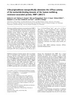
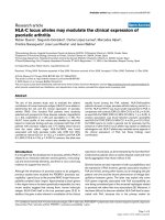
Figure 1 Schematic illustration of constructs with or without H69E mutation. (A) Organizatio n of structural domains in the Gag and Gag-
PR polyproteins: MA, matrix; CA, capsid (p24); NC, nucleocapsid; p6, late domain protein; TFR, transframe region; PR, protease. Straight arrows
indicate the protease cleavage sites. Amino acids that are different between NL4-3 and wt
pse
proteases are denoted. (B) Schematic summary on
H69E containing mutants and their relative Gag processing efficiencies.
Huang et al. Retrovirology 2010, 7:24
/>Page 2 of 8
HEK 293T cells for the study. A pproximately equal
amounts of total Gag proteins were detected in cell
lysates suggesting similar expression efficiencies
mediated by the pNL-PR pro viruses. Also, the amounts
of virus-like particle (VLP) released into the culture
medium were similar to each other, indicating these
mutations have minimal impact on virion production. In
the absence of any protease activity, as with D25N
mutant, the full length Gag polyprotein (p55) is the pre-
dominant product in transfected cells and the released
VLPs (Figure 2 lane 10). In the presence of mature pro-
teases as a result of effective autoprocessing, p24 was
detected as the predominant band with little p25 and
p55 (Figure 2 lanes 8 and 9). Consistent w ith our pre-
vious report, VLPs produced by wt
pse
H69E contained
predominantly the full length Gag polyprotein and no
processed p24, indicatin g lack of mature protease activ-
ity (Figure 2, lane 3). Interestingly, VLPs as well as cell
lysates made by NL4-3 H69E showed some p24
proteins, suggesting an ass ociation of mature protease
activity in both. We quantified the ratio of p24 to total
p24-cont aining proteins as a measure of relative Gag
processing efficiency to indirectly reflect auto processing
activity, and our data demonstrated that wt
pse
H69E
mutation had <5% of the wild type processing activity,
i.e. > 20-fold inhibition; while NL4-3 H69E showed
~25% of the wild type processing efficiency, i.e.~4-fold
inhibition (Figure 2B). Given that there are six point
mutations between NL4-3 and wt
pse
protease, our data
sugge sted that the inhibitory effect of H69E on protea se
autoprocessing is influenced by other residues.
C95 and other residues dampened the inhibitory effect of
H69E on protease autoprocessing
In order to define residues t hat rescued protease auto-
processing in the NL4-3 H69E construct, we engineered
a panel of H69E proviruses replacing the six point
mutat ions in the wt
pse
backbone with the corresponding
NL4-3 amino acids individually or in combination (Fig-
ure 1B) and tested their Gag processing efficiencies to
evaluate autoprocessing activities (Figure 2). The w t
pse
H69E mutants carrying NL4-3 Q7, L33/N 37 demon-
strated a phenotype very similar to the wt
pse
H69E, sug-
gesting that these residues contributed mini mall y to the
rescue effect. In contrast, wt
pse
H69E/A95C mutant,
which contains single amino acid reversion at residue
95, showed a relative Gag processing activity close to
NL4-3 H69E mutant, indicating that C95 could facilitate
autoprocessing. Interestingly, the double mutation I63L/
A67C also demonstrated rescued Gag processing to a
less extent (Figure 2 lane 6) . To further pinpoint the
contributing residue(s), we mutated each residue indivi-
dually, and the resulting constructs showed that both
rescued the activity similarly to the double mutation
(Figure 2A). Based on these observations, we suggested
that cysteine 95 is the primary residue facilitating
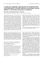
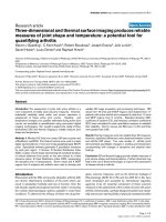
Figure 2 Cysteine 95 and other residues dampened the i nhibitory effect of H69E on protease autoprocessing in transfected
mammalian cells. (A) The indicated proviral DNAs were transfected into HEK 293T cells grown on 6-well plates with calcium phosphate. The
total cell lysates and VLPs were prepared as described (Material and Methods) and subjected to western blot analysis. Mouse monoclonal anti-
p24 antibody was used to detect proteins such as the full length Gag polyprotein (p55), CA-p2 intermediate (p25), and final processing product
(p24) in the transfected cells and the released VLPs. The cell lysates blot was stripped and reprobed for GAPDH as loading controls. (B) Relative
Gag processing efficiencies were quantified from three independent experiments and the bars represent standard deviations.
Huang et al. Retrovirology 2010, 7:24
/>Page 3 of 8
protease autoprocessing and the subsequent Gag proces-
sing; L63 and C67 can also rescue the H69E inhibitory
effect to a less extent probably because of the fact that
they are in the close proximity to H69 residue in pri-
mary sequence. The double mutations, L63/C67 and
C67/C95, only showed a slight enhancement on protease
activity compared to t he single mutations, indicating a
lack of synergistic effect. We interpreted that these resi-
dues are capable of facilitating autoprocessing indepen-
dently to a certain extent and t hese enhancements
might be parallel to each other and not additive.
H69D mutation abolishes protease autoprocessing even
in the context of NL4-3 PR backbone
In addition to H69E mutation, a previous study using
bacterially expressed Gag-Pol precursor d emonstrated
inhibition of p rotease autoprocess ing by H69D; whereas
changestoR,L,Y,N,andQ,individually,didnot
impair protease autoprocessing [29]. To compare H69E
with H69D for their effects on protease maturation
under the same context, we engineered a panel of muta-
tions changing the parental H69 to D, N and Q indivi-
dually in the pNL-PR backbone. As shown in Figure 3,
VLPs produced by H69Q mutant displayed a p24 pat-
tern similar to the wild-type control; and both H69N
and H69E showed partial Gag processing activities. In
contrast, H69D VLPs only contained the full length p55
precursor; no processed intermediates or p24 were
detected (Figure 3 lane 4), which resembled the D25N
negative control. This data further verified that aspartic
acid at position 69 significantly blocks protease matura-
tion even in the presence of L63, C67, and C95. It is
interesting that H69D mutation displays a more drastic
inhibitory effect than H69E considering the carboxyl
side chain of aspartic acid is only shorter by one methyl
group (-CH
2
) than that of glutamic acid. Quantitative
analysis demonstrated relative Gag processing efficien-
cies following an order of wt ≅ H69Q > H69N, H69E
>> H69D in VLPs produced from transfected mamma-
lian cells (Figure 3B). By examining structures of these
amino acids, it seemed that a combination of the carbo-
nyl group and its close distance to the Ca plays a role
in inhibiting protease maturation.
Steady state levels of mature protease detected in
VLPs (Figure 3A, the bottom panel) also qualitatively
correlated with the relative Gag processing activities
(Figure 3B). A rabbit polyclonal anti-PR antibody detects
bot h mature and precursor proteases, but the precursor
band overlaps with a non-specific background band
(Figure 3A lane 1), so we mainly focused on detection
of mature protease. In VLPs produced by the wild type
NL4-3 and wt
pse
, mature protease is the primary pro-
duct, consistent with t he high Gag processing efficien-
cies. The wt
pse
mature protease appeared to be more
than the NL4-3 mature protease probably due to its
higher stability because of the mutations engineered to
reduce autoproteolysis. In VLPs produced from H69Q,
mature protease was the primary form similar to the
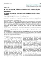
Figure 3 Different substitution s of H69 have differential effects on protease maturation. (A) HEK 293T cells grown on 6-well plates were
transfected with the indicated proviral DNAs by calcium phosphate. The total cell lysates and VLPs were prepared as described (Material and
Methods) and subjected to western blot analysis. Mouse monoclonal anti-p24 antibody was used to detect p24-containing proteins (p55, p25,
and p24) in the transfected cells and the released VLPs. The cell lysates blot was stripped and reprobed for GAPDH as loading controls. VLP
associated proteases were probed with polyclonal rabbit anti-PR antibodies. (B) Relative Gag processing efficiencies were quantified from three
independent experiments and the bars represent standard deviations.
Huang et al. Retrovirology 2010, 7:24
/>Page 4 of 8
wild type contr ol. Consistent with the partial Gag pro-
cessing activities, H69E and H69N VLPs contained
reduced amounts of mature protease as well as part ially
processed intermediates. In D25N, H69D, and wt
pse
H69E VLPs, minimal or no mature protease was
detected; and the full length Gag-PR precursor appeared
to be the predominant product.
Charge substitutions of several residues did not rescue
inhibition of H69E on protease maturation
Our mutagenesis analyses demonstrated that the nega-
tively charged carbonyl group at close proximity to the
C
a
of residue 69 inhibits protease maturation. Our pre-
vious study also suggested that H69E mutation inhibits
in vitro autoprocessing probably by affecting proper pre-
cursor folding. One speculation is that positively
charged side chains of the parental residues (H69 or
K69) interact with another negatively charged residue to
facilitate proper folding; and the carbonyl group of
H69E disrupts the electrostatic interaction. To test this
possibility,weperformedasmallscalescreeningfor
potential H69 interacting residues using a previously
reported precursor autoprocessing assay [33]. When
expressed in E. coli, GST-TF R-PR -FLA G fusion precur-
sor autoprocesses releasing mature protease that can be
detected in total lysates by Western blot (Figure 4
lane 1). H69E mutation significantly inhibits protease
maturation (lane 3). We chose to mutate five surface
residues (four acidic acids plus F99 that is in close
proximity to H69) individually in the H69E context to
examine whether a neutral or positivel y charge d residue
at these positions could rescue protease autoprocessing
by complementing mutations. Out of a total of 12 con-
structs (E21K, E 21Q, D31K, D31N, E34K, E35K, E34K/
E35K, F99K, F 99N, F99Q, F99H, F99A), none of them
reversed the inhibitory effect of H69E on protease
maturation (not all the mutants a re shown here) and
many of them fur ther suppressed autoprocess activity
(Figure 4, lanes 4 -9). Consequently, our limited screen-
ing was unable to define residues that might interact
with H69, and further exami nations would be necessary
to identify how H69 regulates protease maturation.
Discussion and Conclusions
Protease autoprocessing involv es precursor dimerization
and the N-terminal cleavage that releases mature pro-
tease. In the infected cell, this process is also temporally
correlated with the virion egress event. However, the
molecular and cellular mechanisms underlying this
highly regulated process are poorly understood. We pre-
viously reported that H69E mutation in a pseudo wild
type protease sequence abolishes protease autoproces-
sing in E. coli and in transfected mammalian cells [33].
The current study demonstrates that L63, C67, and C95
dampen the H69E inhibitory effect. The Levine group
also suggested a possible inter-play between H69 and
C67 using a model peptide spanning residues 59 to 75
more than a decade ago [35]. It is interesting to note
that highly conserved HIV-1 protease cysteines are not
requir ed for the catalytic activity, nor contributed to the
formation of intramolecular disulfide bonds. Instead,
they are thought to participate in redox regulation of
protease activity [36,37] via a yet-to-be -defined mechan-
ism. Both C67 and C95 appear to be sensitive to oxida-
tion with C95 seems more accessible than C67 [36,38].
Glutathionylation of C67 increases and stabilizes pro-
tease activity in vitro, whereas C95 glutathionylation
abolishes protease activity [38]. Using immature HIV
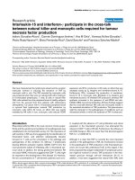
Figure 4 Charge substitutions of surface residues did not restore the inhibitory effect of H69E on protease autoprocessing. (A)
Schematic presentation of the mature protease dimer (PDB 2PK6) with the surface residues that were tested in this report highlighted in red or
green and histindine 69 in blue. (B) The pGEX-3X derived plasmids encoding for GST-TFR-PR-FLAG fusions bearing the indicated mutations were
introduced into E. coli BL21(DE3) and induced for protein expression. The total lysates were prepared as described (Materials and Methods) and
subjected to western blot analysis. A mouse anti-FLAG antibody was used to detect the full length precursor fusion, intermediates and mature
protease (PR-FLAG). The denoted protein markers are in kDa for reference.
Huang et al. Retrovirology 2010, 7:24
/>Page 5 of 8
virions produced in the presence of protease inhibitors
as a model system, Davis et al.demonstratedthat
immature virions made from a mutant lacking the two
cysteines undergo protease maturation at a higher rate
than the wild type immature virions following the
removal of inhibitors [37]. Reducing agent DTT
enhances protease maturation, and oxidizing agents
delay prote ase maturation o f the immature virions.
These results suggest an oxidation-and-reduction cycle
that is involved in regulation of protease autoprocess.
We envision that oxidation of cysteines prevents pro-
tease precu rsor from pre-maturation by locking it in an
inactive status in the infected cell. Upon virion release,
other factors trigger the reduction reaction that restores
free cysteines rendering protease activity. This cysteine
modification cycle seems unnecessary for protease
autoprocessing and mature protease activity as the
pseudo wild type protease containing mutations C67A/
C95A is able to process Gag polyprote in at levels com-
parable, yet slightly lower, to NL4-3 protease (Figure 2
and 3). However, in the context of H69E pseudo wild
type protease, cysteine containing protease demon-
strated a relative Gag processing activity higher than
that lacking cysteines (Figure 2A). Therefore, the modifi-
cation cycle might play an auxiliary role in concert with
other regulation mechanisms to modulate protease
autoprocessing.
Amino acid sequence alignment of HIV-1 proteases
(HIV database - ) indicates that
residue 69 is mostly histidine or lysine and occasionally
glutamine or tyrosine, which are neutral or positively
charged. Previous studies [29,33] and current report also
suppor t the notion that a carbonyl group at close proxi-
mity to the Ca positi on of this residue inhibits protease
autoprocessing. The H69 r esidue is exposed on the sur-
face of mature protease dimer and is close to the C-ter-
minus. It is intriguing that charge properties of a surface
residue would have drastic effects on protease autopro-
cessing. Previous biochemical analyses demonstrated
that H69E mutation significantly delays the TFR-PR pre-
cursor from autoprocessing in vitro; wherea s the appro-
priately folded H69E mature protease only showed a
slightly decreased catalytic activity [33]. This has led us
to speculate that residue 69 is involved in autoproces-
sing by influencing precursor structure. We hypothesize
that protease precursor undergoes conformational
changes during autoprocessing and a carbonyl group
close to the Ca of position 69 interferes with this path-
way. It would be critical to identify residues that transi-
ently interact with H69 during this process.
Unfortunately, our limited screening was unable to
define any of them. Extensive structural and biochemical
analyses on the wild type and H69D precursor would be
essential to provide insights into protease autoproces-
sing mechanisms.
Methods
DNA mutagenesis
Plasmids that were used in this report were generated
with the standard molecular cloning procedures and the
detailed sequence information is available upon request.
Construction of pNL-PR was described previously [33],
and all the pNL-PR mutants were derived from this vec-
tor by site-directed mutagenesis. Multiple D21, D30,
E34, E35 and F99 substitutions were introduced into a
pGEX-3X derived plasmid expressing GST-p6
pol
-PR
pse
-
FLAG H69E was generated in a previous report [33]. All
the plasmids were purified with QIAEX plasmid kits
and verified by DNA sequencing.
Cell culture, transfection and western blotting
Human embryonic kidney derived 293T cells (ATCC,
Manassas, VA) were maintained in DMEM with 10%
fetal bovine serum and transfected by calcium phos-
phate as previously described [33]. In brief, 293T cells
were plated in 6-we ll plates the night before to give 50-
60% confluence at the time of transfection. One hour
prior to the transfection, chloroquine was added to each
well to a final concentration of 25 uM. A total of 1 μg
DNA in 131.4 μLofddH
2
Owasmixedwith18.6μl2
MCaCl
2
to give a final volume of 150 μl. Then, 150 μl
of 2 × HBS was added dropwise to the DNA solution
whilemixingbyvortex.Theresultingmixturewas
directly added to the culture cells. After 7-11 h of incu-
bation, the c ulture medium was replaced with chloro-
quine-free DMEM.
Total cell lysates were pre pared as described pre-
viously [33,39,40] to examine proteins in transfected
cells. To examine proteins associated with the released
virions, culture media collected from 11 h to 4 8 h post
transfection was clarified of cell debri s by a brief centri-
fugation (20,800 × g for 2 min at ambient temperature)
and the supernatant was transferred to another tube and
centrifuged at 20,800 × g for 3 h at 4°C to pellet virions.
Virion pellets were resuspended in 40 μlofPBSfor
further analysis. About 1/6 of cell lysate made from
each well was resolved through 10% SDS-PAGE and the
proteins were transferred to a PVDF (Polyvinylidene
Fluoride) membrane followed by western blot. Approxi-
mately one half of virus-like particles (VLPs) collected
from each well were analyzed for p24 contents, and all
theVLPsmadefromonewellofa6-wellplatewere
used for protease detection. Mouse anti-HIV p24 anti-
bodies (Cat# 3537) and rabbit anti HIV-1 protease
serum (Cat# 4105) were obtained from t he NIH AIDS
research and reference program. Mouse anti-GAPDH
Huang et al. Retrovirology 2010, 7:24
/>Page 6 of 8
(clone 6C5) antibodies (Fisher Scientific, Pittsburgh, PA)
were used to reflect cell numbers. IR800 labelled goat
anti mouse or rabbit secondary antibodies were pur-
chased from Rockland Immunochemicals Inc (Gilberts-
ville, PA) for western detection with an Odyssey
infrared dual laser scanning unit.
Quantification of relative Gag processing activity
Western blot images that were captured by an Odyssey
infrared dual laser scanning unit in tiff format were ana-
lyzed by Totallab software (Nonlinear Dynamics Inc.,
Newcastle upon Tyne, UK). Total pixel volume (less than
the saturation threshold) of each band was quantified to
represent band intensity that is assumed to be propor-
tional to protein amounts as the blot wa s detected by
monoclonal antibodies. The anti-p24 antibody is able to
detect the full length (p55) Gag polyprotein as well as
p25 (CA-p2), a processing intermediate, and p24, the
final cleavage protein. Because the produc tion of p24
from p25 is solely dependent on mature protease, the
amounts of p24 in VLPs quantitat ively correlate with the
amounts of mature protease that indirectly reflect pre-
cursor maturation efficiencies. In this report, we calcu-
lated the ratio of p 24/(p24+p25+p55) as a measure of
Gag processing efficiency to indirectly represent autopro-
cessing activities with the value obtained from the wild
type pNL-PR VLPs set as 100% for normalization.
Protease autoprocessing in E. coli
The pGEX-3X derived plasmids were transformed into
BL21 cells (Novagen, San Diego, CA) and the individual
colony was grown in LB medium at 37°C overnight. The
overnightculturewasthendiluted100-foldinto2xYT
and incubated at 37°C for another 2.5~3 h prior to the
addition of IPTG (40 μM) to induce protein expression.
After IPTG induction at 30°C for 4 h, cells (~30 μL)
were directly mixed with 6× SDS loading buffer (6 μL)
and subsequently analyzed by 10% SDS-PAGE and Wes-
tern blot. The full length GST-TFR-PR- FLAG precursor
and mature protease (PR-FALG) along with processing
intermediates were detected with mouse anti-FLAG
antibody (Sigma, St. Luis, MO).
Acknowledgements
This work was supported in part by NIH, NIAID grant R21A1080351 to C.
Chen. The following reagents were obtained through the AIDS Research and
Reference Reagent Program, Division of AIDS, NIAID, NIH: HIV-1 p24
monoclonal antibody from Drs. Bruce Chesebro and Kathy Wehrly; HIV-1
protease antiserum from BioMolecular Technology (DAIDS, NIAID).
Authors’ contributions
CC designed the project and wrote the manuscript. LH constructed the
plasmids used in this study, performed 293T transfection and western blot
analyses. AH carried out the E. coli protease maturation assay and
participated in sequencing analysis of the constructs. All authors read and
approved the final manuscript.
Competing interests
The authors declare that they have no competing interests.
Received: 10 November 2009 Accepted: 23 March 2010
Published: 23 March 2010
References
1. Barre-Sinoussi F, Chermann JC, Rey F, Nugeyre MT, Chamaret S, Gruest J,
Dauguet C, Axler-Blin C, Vezinet-Brun F, Rouzioux C, Rozenbaum W,
Montagnier L: Isolation of a T-lymphotropic retrovirus from a patient at
risk for acquired immune deficiency syndrome (AIDS). Science 1983,
220(4599):868-871.
2. Gallo RC, Salahuddin SZ, Popovic M, Shearer GM, Kaplan M, Haynes BF,
Palker TJ, Redfield R, Oleske J, Safai B, et al: Frequent detection and
isolation of cytopathic retroviruses (HTLV-III) from patients with AIDS
and at risk for AIDS. Science 1984, 224(4648):500-503.
3. Scarlata S, Carter C: Role of HIV-1 Gag domains in viral assembly. Biochim
Biophys Acta 2003, 1614(1):62-72.
4. Freed EO: HIV-1 gag proteins: diverse functions in the virus life cycle.
Virology 1998, 251:1-15.
5. Louis JM, Weber IT, Tozser J, Clore GM, Gronenborn AM: HIV-1 protease:
maturation, enzyme specificity, and drug resistance. Adv Pharmacol 2000,
49:111-146.
6. Chen C, Montelaro RC: Characterization of RNA elements that regulate
gag-pol ribosomal frameshifting in equine infectious anemia virus. J Virol
2003, 77(19):10280-10287.
7. Oroszlan S, Luftig RB: Retroviral proteinases. Curr Top Microbiol Immunol
1990, 157:153-185.
8. Louis JM, Nashed NT, Parris KD, Kimmel AR, Jerina DM: Kinetics and
mechanism of autoprocessing of human immunodeficiency virus type 1
protease from an analog of the Gag-Pol polyprotein. Proc Natl Acad Sci
USA 1994, 91(17):7970-7974.
9. Pettit SC, Clemente JC, Jeung JA, Dunn BM, Kaplan AH: Ordered
processing of the human immunodeficiency virus type 1 GagPol
precursor is influenced by the context of the embedded viral protease. J
Virol 2005, 79(16):10601-10607.
10. Ludwig C, Leiherer A, Wagner R: Importance of protease cleavage sites
within and flanking human immunodeficiency virus type 1 transframe
protein p6* for spatiotemporal regulation of protease activation. J Virol
2008, 82(9):4573-4584.
11. Louis JM, Ishima R, Torchia DA, Weber IT: HIV-1 protease: structure,
dynamics, and inhibition. Adv Pharmacol 2007, 55:261-298.
12. Tessmer U, Krausslich HG: Cleavage of human immunodeficiency virus
type 1 proteinase from the N-terminally adjacent p6* protein is essential
for efficient Gag polyprotein processing and viral infectivity. J Virol 1998,
72(4):3459-3463.
13. Wondrak EM, Nashed NT, Haber MT, Jerina DM, Louis JM: A transient
precursor of the HIV-1 protease. Isolation, characterization, and kinetics
of maturation. J Biol Chem 1996, 271(8):4477-4481.
14. Cherry E, Liang C, Rong L, Quan Y, Inouye P, Li X, Morin N, Kotler M,
Wainberg MA: Characterization of human immunodeficiency virus type-1
(HIV-1) particles that express protease-reverse transcriptase fusion
proteins. J Mol Biol 1998, 284(1):43-56.
15. Paulus C, Ludwig C, Wagner R: Contribution of the Gag-Pol transframe
domain p6* and its coding sequence to morphogenesis and replication
of human immunodeficiency virus type 1. Virology 2004, 330(1):271-283.
16. Zybarth G, Carter C: Domains upstream of the protease (PR) in human
immunodeficiency virus type 1 Gag-Pol influence PR autoprocessing. J
Virol 1995, 69(6):3878-3884.
17. Pettit SC, Moody MD, Wehbie RS, Kaplan AH, Nantermet PV, Klein CA,
Swanstrom R: The p2 domain of human immunodeficiency virus type 1
Gag regulates sequential proteolytic processing and is required to
produce fully infectious virions. J Virol 1994, 68(12):8017-8027.
18. Pettit SC, Henderson GJ, Schiffer CA, Swanstrom R: Replacement of the P1
amino acid of human immunodeficiency virus type 1 Gag processing
sites can inhibit or enhance the rate of cleavage by the viral protease. J
Virol 2002, 76(20):10226-10233.
19. Eizert H, Bander P, Bagossi P, Sperka T, Miklossy G, Boross P, Weber IT,
Tozser J: Amino acid preferences of retroviral proteases for amino-
terminal positions in a type 1 cleavage site. J Virol 2008,
82(20):10111-10117.
Huang et al. Retrovirology 2010, 7:24
/>Page 7 of 8
20. Pettit SC, Lindquist JN, Kaplan AH, Swanstrom R: Processing sites in the
human immunodeficiency virus type 1 (HIV-1) Gag-Pro-Pol precursor are
cleaved by the viral protease at different rates. Retrovirology 2005, 2:66.
21. Liang C, Hu J, Russell RS, Roldan A, Kleiman L, Wainberg MA:
Characterization of a putative alpha-helix across the capsid-SP1
boundary that is critical for the multimerization of human
immunodeficiency virus type 1 gag. 2005, 76(22):11729-11737.
22. Kaplan AH, Zack JA, Knigge M, Paul DA, Kempf DJ, Norbeck DW,
Swanstrom R: Partial inhibition of the human immunodeficiency virus
type 1 protease results in aberrant virus assembly and the formation of
noninfectious particles. J Virol 1993, 67(7):4050-4055.
23. Wiegers K, Rutter G, Kottler H, Tessmer U, Hohenberg H, Krausslich HG:
Sequential steps in human immunodeficiency virus particle maturation
revealed by alterations of individual Gag polyprotein cleavage sites. J
Virol 1998, 72(4):2846-2854.
24. Li F, Goila-Gaur R, Salzwedel K, Kilgore NR, Reddick M, Matallana C,
Castillo A, Zoumplis D, Martin DE, Orenstein JM, Allaway GP, Freed EO,
Wild CT: PA-457: A potent HIV inhibitor that disrupts core condensation
by targeting a late step in Gag processing. ProcNatlAcadSciUSA 2003,
100(23):13555-13560.
25. Koh Y, Matsumi S, Das D, Amano M, Davis DA, Li J, Leschenko S,
Baldridge A, Shioda T, Yarchoan R, Ghosh AK, Mitsuya H: Potent Inhibition
of HIV-1 Replication by Novel Non-peptidyl Small Molecule Inhibitors of
Protease Dimerization. J Biol Chem 2007, 282(39):28709-28720.
26. Pearl LH, Taylor WR: A structural model for the retroviral proteases.
Nature 1987, 329(6137):351-354.
27. Sayer JM, Liu F, Ishima R, Weber IT, Louis JM: Effect of the active site
D25N mutation on the structure, stability, and ligand binding of the
mature HIV-1 protease. J Biol Chem 2008, 283(19):13459-13470.
28. Kohl NE, Emini EA, Schleif WA, Davis LJ, Heimbach JC, Dixon RA,
Scolnick EM, Sigal IS: Active human immunodeficiency virus protease is
required for viral infectivity. Proc Natl Acad Sci USA 1988,
85(13):4686-4690.
29. Loeb DD, Swanstrom R, Everitt L, Manchester M, Stamper SE, Hutchison CA:
Complete mutagenesis of the HIV-1 protease. Nature 1989,
340(6232):397-400.
30. Huang M, Orenstein JM, Martin MA, Freed EO: p6Gag is required for
particle production from full-length human immunodeficiency virus type
1 molecular clones expressing protease. JVirol 1995, 69:6810-6818.
31. Louis JM, Wondrak EM, Kimmel AR, Wingfield PT, Nashed NT: Proteolytic
processing of HIV-1 protease precursor, kinetics and mechanism. J Biol
Chem 1999, 274(33):23437-23442.
32. Tang C, Louis JM, Aniana A, Suh JY, Clore GM: Visualizing transient events
in amino-terminal autoprocessing of HIV-1 protease. Nature 2008,
455(7213):693-696.
33. Huang L, Sayer JM, Swinford M, Louis JM, Chen C: Modulation of human
immunodeficiency virus type 1 protease autoprocessing by charge
properties of surface residue 69. J Virol 2009,
83(15):7789-7793.
34. Louis JM, Clore GM, Gronenborn AM: Autoprocessing of HIV-1 protease is
tightly coupled to protein folding. Nat Struct Biol 1999, 6:868-875.
35. D’Ettorre C, Levine RL: Reactivity of cysteine-67 of the human
immunodeficiency virus-1 protease: studies on a peptide spanning
residues 59 to 75. Arch Biochem Biophys 1994, 313(1):71-76.
36. Davis DA, Newcomb FM, Starke DW, Ott DE, Mieyal JJ, Yarchoan R:
Thioltransferase (glutaredoxin) is detected within HIV-1 and can regulate
the activity of glutathionylated HIV-1 protease in vitro. J Biol Chem 1997,
272(41):25935-25940.
37. Davis DA, Yusa K, Gillim LA, Newcomb FM, Mitsuya H, Yarchoan R:
Conserved cysteines of the human immunodeficiency virus type 1
protease are involved in regulation of polyprotein processing and viral
maturation of immature virions. J Virol 1999, 73:1156-1164.
38. Karlstrom AR, Shames BD, Levine RL: Reactivity of cysteine residues in the
protease from human immunodeficiency virus: identification of a
surface-exposed region which affects enzyme function. Arch Biochem
Biophys 1993, 304(1):163-169.
39. Chen C, Li F, Montelaro RC: Functional roles of equine infectious anemia
virus Gag p9 in viral budding and infection. J Virol 2001,
75(20):9762-9770.
40. Chen C, Vincent O, Jin J, Weisz OA, Montelaro RC: Functions of early (AP-
2) and late (AIP1/ALIX) endocytic proteins in equine infectious anemia
virus budding. J Biol Chem 2005, 280(49):40474.
doi:10.1186/1742-4690-7-24
Cite this article as: Huang et al.: Cysteine 95 and other residues
influence the regulatory effects of Histidine 69 mutations on Human
Immunodeficiency Virus Type 1 protease autoprocessing. Retrovirology
2010 7:24.
Submit your next manuscript to BioMed Central
and take full advantage of:
• Convenient online submission
• Thorough peer review
• No space constraints or color figure charges
• Immediate publication on acceptance
• Inclusion in PubMed, CAS, Scopus and Google Scholar
• Research which is freely available for redistribution
Submit your manuscript at
www.biomedcentral.com/submit
Huang et al. Retrovirology 2010, 7:24
/>Page 8 of 8