Báo cáo y học: " Role of the C-terminal domain of the HIV-1 glycoprotein in cell-to-cell viral transmission between T lymphocytes" potx
Bạn đang xem bản rút gọn của tài liệu. Xem và tải ngay bản đầy đủ của tài liệu tại đây (1.89 MB, 11 trang )
Emerson et al. Retrovirology 2010, 7:43
/>Open Access
RESEARCH
BioMed Central
© 2010 Emerson et al; licensee BioMed Central Ltd. This is an Open Access article distributed under the terms of the Creative Commons
Attribution License ( which permits unrestricted use, distribution, and reproduction in
any medium, provided the original work is properly cited.
Research
Role of the C-terminal domain of the HIV-1
glycoprotein in cell-to-cell viral transmission
between T lymphocytes
Vanessa Emerson
1
, Claudia Haller
2
, Tanya Pfeiffer
1
, Oliver T Fackler
2
and Valerie Bosch*
1
Abstract
Background: Mutant HIV (HIV-Env-Tr712) lacking the cytoplasmic tail of the viral glycoprotein (Env-CT) exhibits a cell-
type specific replication phenotype such that replicative spread occurs in some T-cell lines (referred to as permissive
cells) but fails to do so in most T-cell lines or in PBMCs (referred to as non-permissive cells). We aim to gain insight on
the underlying requirement for the Env-CT for viral spread in non-permissive cells.
Results: We established that in comparison to HIV-Wt, both cell-free and cell-to-cell transmission of mutant HIV-Env-
Tr712 from non-permissive cells were severely impaired under naturally low infection conditions. This requirement for
Env-CT could be largely overcome by using saturating amounts of virus for infection. We further observed that in
permissive cells, which supported both routes of mutant virus transmission, viral gene expression levels, Gag
processing and particle release were inherently higher than in non-permissive cells, a factor which may be significantly
contributing to their permissivity phenotype. Additionally, and correlating with viral transfer efficiencies in these cell
types, HIV-Gag accumulation at the virological synapse (VS) was reduced to background levels in the absence of the
Env-CT in conjugates of non-permissive cells but not in permissive cells.
Conclusions: During natural infection conditions, the HIV-Env-CT is critically required for viral transmission in cultures
of non-permissive cells by both cell-free and cell-to-cell routes and is instrumental for Gag accumulation to the VS. The
requirement of the Env-CT for these related processes is abrogated in permissive cells, which exhibit higher HIV gene
expression levels.
Background
Infectious spread of viruses to new target cells in vitro
and in vivo occurs either via infection with released cell-
free virions or by direct transmission of virions from cell
to cell. Some viruses e.g. human T-cell leukemia virus
type 1 (HTLV-1) or Spuma retroviruses employ solely the
cell-to-cell route and cell-free viral infection is negligible
[1]. In the case of human immunodeficiency virus type 1
(HIV-1), both routes of viral spread are possible, but
already very early reports documented that transmission
by the cell-to-cell route was far more efficient [2-4]. A
series of more recent studies have now established cell-
to-cell transmission as the predominant mode of HIV-1
spread in T lymphocyte cultures [5-9]. Analogous to the
situation with HTLV-1 [10], confocal microscopic analy-
ses of infected T lymphocyte cultures revealed close con-
jugates of infected donor cells and uninfected target cells
and cell-to-cell transmission of virus particles across the
cell contact site referred to as the virological synapse
(VS). In addition, several types of membrane bridges have
also been observed to mediate transport and infection of
HIV-1 particles between T lymphocytes [11,12]. The
term cell-to-cell transmission thus summarizes all types
of HIV-1 spread between physically connected infected
donor and uninfected target cells, including spread via
short distance transmission of cell-free virions and direc-
tional transport along cellular protrusions [13]. Although
the relative contribution of these transmission modes still
remains to be determined, accumulation of both cellular
and viral proteins at these cell contacts has been estab-
lished as a hallmark of efficient HIV-1 cell-to-cell spread.
* Correspondence:
1
Forschungsschwerpunkt Infektion und Krebs, F020, Deutsches
Krebsforschungszentrum, Im Neuenheimer Feld 280, 69120 Heidelberg,
Germany
Full list of author information is available at the end of the article
Emerson et al. Retrovirology 2010, 7:43
/>Page 2 of 11
Such polarisation includes accumulation of the viral
structural proteins Gag and Env as well as the microtu-
bule organising centre (MTOC) at the donor cell contact,
while cellular receptors (CD4, coreceptor) and cytoskele-
tal proteins (F-actin, talin) typically accumulate at the tar-
get cell contact [12,14-18]. Even though some host cell
signalling cascades that govern polarisation of HIV-1 Gag
to the VS have been identified [18], it remains unclear
which domains of viral Env and Gag proteins are opera-
tional in mediating transport and functional accumula-
tion of HIV-1 structural proteins to the cell contact site.
The HIV-1 glycoprotein carries a very long cytoplasmic
C-terminal tail (CT, 151 amino acids (aa) long) which is
absolutely required for replication in vivo. Mutant virions
lacking this region exhibit a cell-type dependent pheno-
type in vitro such that replicative virus spread occurs in
some cell lines (termed permissive cells, e.g. MT-4 cells)
but not in the majority of T-cell lines (termed non-per-
missive cells, e.g. H9 cells) nor in PBMCs [19-22]. The
basis for the requirement for the Env-CT for viral spread
in non-permissive cells, and the reason(s) underlying the
permissivity phenotypes of different T-cell lines are pres-
ently unclear and are the focus of this study.
Genetic [23,24] and protein association data [25,26]
support the view that there is a functional interaction
between the Env-CT and the viral matrix protein (MA).
This interaction appears to be involved in a number of
processes. Thus, in released immature virions, Env-CT
interaction with the unprocessed Gag precursor prevents
premature fusion activity of Env [27]. The Env-CT
domain has also been shown to impact intracellular local-
isation of Gag and the subcellular localisation of particle
assembly. In the absence of Wt-Env, HIV particle release
from polarised epithelial cells was shown to occur at both
apical and basolateral membrane surfaces, yet in its pres-
ence release occurred exclusively at the basolateral mem-
brane [28,29]. The Env-CT domain and in particular a
membrane-proximal tyrosine-based signal within it were
shown to be instrumental in this. Furthermore, removal
of the same Env-CT tyrosine-based signal has been
reported to inhibit polarised budding of HIV in T-lym-
phocytes and to reduce cell-to-cell viral transmission
[30]. A further event, which for many years has been dis-
cussed to involve the Env-CT and its interaction with
Gag, concerns Env incorporation into released virions.
Nevertheless, HIV-Env-Tr712 virions, encoding Env lack-
ing the CT domain, when produced in transfected adher-
ent cells or in infected permissive cells did incorporate
truncated glycoprotein and were infectious [20,31,32].
On the other hand, it was reported several years ago that
cell-free mutant HIV-Env-Tr712 virions, released from
non-permissive cells, were non-infectious and that this
correlated with a lack of mutant glycoprotein incorpora-
tion [19,21]. In a previous study, aimed at further study-
ing the defective phenotype of HIV-Env-Tr712, we had
also analysed the infectivities of cell-free mutant virus
particles. These were generated by efficiently infecting
non-permissive H9 producer cells with VSV-G pseudo-
typed derivatives and collecting the newly generated
(unpseudotyped) virions 48 h later. However, under these
experimental conditions, cell-free HIV-Env-Tr712 virions
were only marginally reduced in their infectivity [33,34],
an observation which was difficult to reconcile with the
total lack of spread of mutant virus in these cells. The rea-
son for these discordant observations has remained
unclear until now. In this report, we considered the possi-
bility that, in contrast to cell-free infectivity, cell-to-cell
virus transmission of HIV-Env-Tr712 in non-permissive
T-cells could be more severely impacted and that this
could be the reason for the block in viral spread.
Methods
Constructs
Proviral plasmids were based on pNL4-3
BH10 env
(referred
to here as pNL-Wt) [35]. pNL-Env-Tr712 encodes Env
with a stop codon at position 713, i.e. lacking 144 aa [32].
pNL-Env
Fus-
is fusion-defective due to exchange of the
second aa (V513E) within the fusion peptide of gp41 [36]
and pNL-ΔEnv fails to synthesise Env due to an intro-
duced frame-shift mutation [37]. pMD.G is an expression
vector for the G glycoprotein of vesicular stomatis virus
(VSV) [38].
Cell lines, transfections, analysis of cell-free virion
infectivities
293T cells were cultivated in DMEM medium, 10% foetal
calf serum (FCS) and all T-cell lines, namely MT-4, MT-2,
C8166, H9, CEM-SS and Jurkat cells, in RPM-I medium,
10% FCS. H9 cells constitutively expressing GFP (H9-
GFP) have been previously described [39]. Procedures for
the infection of T-cells with VSV-G pseudotyped virions
leading to the generation of T-cell-produced cell-free
progeny virions have been previously described [33,34].
Briefly, VSV-G pseudotypes, released into the superna-
tant of 293T cells co-transfected with proviral pNL4-3
BH10 env
plasmids [32] and pMD.G, were quantified by
HIV-CA ELISA (Innogenetics, Belgium) and employed to
infect fresh MT-4 or H9 T-cells. At 5 h p.i., input virions
were removed,;the cells were washed three times with
medium and then further incubated for 43 h. In initial
experiments, we aimed to efficiently infect T-cells and
thus employed saturating amounts of 293T cell superna-
tants containing VSV-G pseudotyped viruses (15-25 μg
virus-associated p24 per 10
6
cells, infection level 50-
100%) [33,34]. In later experiments, limiting amounts of
supernatants (0.5-3 μg virus-associated p24 per 10
6
cells),
which resulted in only a fraction of the cells (<20%)
becoming infected, were employed. At 48 h p.i., the num-
Emerson et al. Retrovirology 2010, 7:43
/>Page 3 of 11
bers of infected cells were quantified by intracellular p24
FACS using PE-labeled HIV-p24 antibody (KC57-RD1
from Coulter, Florida) and the single-round infectivities
of newly produced cell-free virions in the culture super-
natants were assessed in MT-4 target cells as described
previously [33,34]. MT-4 target cells allow efficient cell-
free infection i.e. a high percentage (up to 100%) of the
cells becomes infected. This allows better discrimination
between the infectivities of different viruses than in H9
cells in which cell-free infection with the same amounts
of virions results in only a small percentage (<5%) of the
cells initially becoming infected [33].
Cell-to-cell viral transmission
In addition to analysis of the cell-free infectivities of
released virions, at 48 h p.i. the abilities of the infected
cells to transmit virus to new target cells by the cell-to-
cell route were assessed. Stable GFP expressing H9 cells
(H9-GFP) or dye-loaded MT-4 cells were employed as
targets. Dye loading of MT-4 cells was achieved by incu-
bation with 10 μM CellTracker Green CMFDA (Molecu-
lar Probes, Eugene, USA) in RPM-I medium without
additives for 30 min at 37°C. In an initial protocol, donor
cells were efficiently infected with saturating amounts of
VSV-G pseudotyped virus (equivalent to 15-25 μg virus-
associated p24 per 10
6
cells). The number of infected
donor T-cells was determined by intracellular p24 FACS
(and was >50%) and then adjusted to 50% with uninfected
cells. These infected cells were then mixed and incubated
with a 4-fold excess of labelled target cells i.e. there were
10% infected donor cells present in the coculture contain-
ing 4 × 10
6
cells in a volume of 10 ml. In later experiments,
donor T-cells were infected with limiting amounts of
VSV-G pseudotyped virus (equivalent to 0.5-3 μg virus-
associated p24 per 10
6
cells). The number of infected
cells, as determined by intracellular p24 FACS, was in the
range of 10-20% and was adjusted to 10% with unifected
cells and then mixed and incubated with a 9-fold excess
of labelled target cells i.e. there were 1% infected donor
cells present in the coculture containing 4 × 10
6
cells in a
volume of 10 ml. 5 h post-mixing, the CXCR4 antagonist,
AMD3100 (1 μg/ml), was added to the mixtures and 43 h
later, the percentage of labelled target cells infected, and
thus expressing HIV-CA, was established by intracellular
p24 FACS (10,000 gated cells were analysed in each case).
The value obtained for pNL-Wt was set at 100% and the
values for pNL-Tr712 and pNL-Env
Fus-
calculated relative
to this.
HIV gene expression in permissive and non-permissive T-
cell lines
Non-permissive or permissive T-cells were infected with
limiting amounts of VSV-G pseudotyped HIV-Wt or
HIV-Env-Tr712 virions (0.5-3 μg virus-associated p24 per
10
6
cells resulting in 10-20% infection level). At 5 h p. i.,
input virions were removed, and cells washed three times
with medium before being further cultivated in fresh
medium containing 1 μg/ml AMD 3100 to prevent viral
spread. At 48 h p. i., an aliquot of the infected cells was
subjected to intracellular p24 FACS to establish the per-
centage infected cells. The total cell densities and the per-
centages of cells infected in the respective cultures were
equalised by addition of uninfected cells and culture
medium. Lysates of equal numbers of cells, now contain-
ing equal percentages of infected cells, were prepared,
and protein determination using standard procedures
showed that these did not differ significantly between the
different T-cell lines. Equal aliquots were subjected to
Western blot analyses employing anti-p24 mAb 183-H12-
5C [40], anti-tubulin mAb (Sigma) and rabbit anti-gp120
serum. Comparative densitometric quantitation of spe-
cific bands on different exposures of the blots to film was
carried out using the Image J software from the NIH. The
amounts of virions released into the respective culture
supernatants were determined by HIV-CA ELISA (Inno-
genetics, Belgium).
Env incorporation into virus particles
HIV-Wt and HIV-Env-Tr712 virions were enriched from
culture supernatants of H9 (np) cells, weakly infected (<
20%) as described above. At 48 h p.i., supernatants were
filtered (0.450 μm filter) and subjected to ultracentrifuga-
tion through a 20% sucrose cushion in PBS. Equal
amounts of pelleted virions (as determined by HIV-CA
ELISA) were subjected to Western blot analysis employ-
ing rabbit anti-gp120 serum, gp41 mAb Chessie 8 [41] or
anti-p24 mAb 183-H12-5C [40]. Densitometric quantita-
tion of specific bands was carried out as above. The
amount of gp120 normalised to p24 amount was set at
100% for HIV-Wt and the relative gp120 incorporation
into HIV-Env-Tr712 virions calculated relative to this.
HIV-Gag localisation in conjugates between infected and
non-infected cells
In order to identify newly formed cell conjugates, unin-
fected target cells were labelled with 5 μM CellTracker
Blue CMAC (Molecular Probes, Eugene, USA) prior to
mixing. For this, cells were incubated in 5 μM dye in
RPM-I medium without additives for 30 min at 37°C.
Infected donor H9 or MT-4 cells were prepared by infec-
tion with VSV-G pseudotyped Wt or mutant HIV (see
above). For conjugate formation 2.5 × 10
5
infected cells
were mixed with 2.5 × 10
5
uninfected labelled target cells
in a volume of 100 μl RPM-I, 1% FCS in an Eppendorf
tube, centrifuged for 5 minutes at 200 g and incubated for
15 min at 37°C. The cell mixture was then gently resus-
pended and transferred onto poly-L-lysine coated cover-
slips. For poly-L-lysine coating, cover-slips were cleaned
Emerson et al. Retrovirology 2010, 7:43
/>Page 4 of 11
with 1 M HCl/70% ethanol for 30 min, dried at 60°C for
30 min, treated with 0.01% poly-L-lysine solution for 10
min and dried again at 60°C for 30 min before adding PBS
for storage. Once added to the coated cover-slips, cells
were incubated for a further 10 min at 37°C, and cells
attached to the cover-slips were fixed with 3% paraform-
aldehyde in PBS for 1.5 h. HIV-Gag was stained with rab-
bit-anti-CA [42] and cellular actin was stained with
TRITC-labelled phalloidin, cover-slips were mounted in
Elvanol and analyzed with a Leitz DMRBE fluorescence
microscope (Leica, Germany) using a 100× oil immersion
objective. The localisation phenotypes of actin and HIV-
Gag were evaluated and quantitated by three different
investigators, two of whom were not aware of the nature
of the different samples. Images were taken at a LSM 510
confocal laser-scanning microscope (Zeiss) using a 100×
oil immersion objective and processed by Adobe Photo-
shop.
Results and Discussion
Transmission of HIV with C-terminally truncated Env
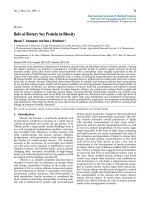
Fig 1A schematically depicts the Env proteins of HIV-Wt
and HIV-Env-Tr712. As detailed in the Introduction and
shown in Fig. 1B, HIV-Env-Tr712 virions exhibit a cell-
type specific defect such that replicative spread occurs in
some T-cell lines (here MT-4 cells, termed permissive (p)
cells) but fails to occur in the majority of T-cell lines (here
H9 cells, referred to as non-permissive (np) cells). In this
report, our emphasis was to analyse the possible impact
of Env-CT truncation on cell-to-cell viral transmission in
both H9 (np) and MT-4 (p) cells. Infected donor cells
were generated by infection with VSV-G pseudotyped Wt
or mutant viruses and, in the course of our studies, we
observed that their initial infection level markedly influ-
enced experimental outcome. Thus, in this report, we
describe cell-to-cell transmission experiments employ-
ing highly or weakly infected donor cells and have also
analysed cell-free viral infectivities in the context of both
of these scenarios.
Donor cells were infected with either saturating or lim-
iting amounts (on average 20 times less than used for sat-
urating infection, see Methods section) of VSV-G
pseudotyped HIV-Wt, HIV-Env-Tr712 or, as negative
control, HIV-Env
Fus-
virions. Input pseudotyped virions
were thoroughly removed at 5 h p.i.; and 43 h later,
infected cells were employed as donors for viral transmis-
Figure 1 Replicative spread of HIV-Wt and HIV-Env-Tr712 in different T-cells. A. Schematic depiction of the Env proteins from HIV-Wt and HIV-
Env-Tr712 B. Replication kinetics of HIV-Wt (circles) and HIV-Env-Tr712 (triangles) in MT-4 cells (p) and H9 cells (np) below. Cells were infected with
equal amounts of virus (equivalent to 100 ng p24 per 10
6
cells) and washed at 5 h p.i. Newly produced virions released into the culture supernatants
at the times indicated were quantified by CA-ELISA. Note that at 3-4 d post infection, MT-4 cells were infected to 100% with both viruses (as established
by indirect immunofluorescence) and succumbed to HIV induced cytotoxicity. Infection of H9 cells with HIV-Wt reached 100% at 4-5 d post-infection
whereas infection with HIV-Env-Tr712 virions (produced in permissive 293T cells) resulted in initial infection of only a low percentage (< 5%) of cells
which subsequently vanished from the culture. The cut-off of the assay lies at 0.01 ng/ml.
0.1
1
10
100
1000
10000
024
ng p24 / ml
51015
0.1
1
10
100
1000
10000
Days p. i. Days p. i.
MT-4 (p) H9 (np)
gp120
gp120
gp41
gp41
TMD
Wt
Tr712
151 aa
7 aa
856
712
512 684 705
gp120
gp120
gp41
gp41
TMD
Wt
Tr712
151 aa
7 aa
856
712
512 684 705
A.
B.
0.01 0.01
0
Emerson et al. Retrovirology 2010, 7:43
/>Page 5 of 11
sion to target cells constitutively expressing GFP (H9
cells) or labelled with a green dye (MT-4 cells). As shown
in Fig. 2A, labelled target cells could clearly be distin-
guished from unlabelled donor cells by FACS analysis.
Donor cells infected with saturating amounts of VSV-G
pseudotyped virions, and thus highly infected (50-90%
level), were adjusted to 50% infection level with unin-
fected H9 donor cells and then mixed with a 4-fold excess
of uninfected labelled target cells (i.e. 10% of the cells in
the mixture were infected) (Fig. 2B). Donor cells infected
with only limiting amounts of VSV-G pseudotyped virus,
and thus weakly infected, (less than 20% infection) were
adjusted to 10% infection level and mixed with a 9-fold
excess of target cells, (i.e. 1% of the cells in the mixture
were infected) (Figs. 2C, D). Five hours post-mixing, the
CXCR4 antagonist, AMD3100, was added to the mixture
and 43 h later, the number of labelled target cells express-
ing HIV-CA and thus being productively infected, was
established by intracellular p24 staining. In line with pre-
vious reports [7,43], we confirmed that addition of
AMD3100 prior to mixing of infected and uninfected
cells completely inhibited p24 detection in target cells.
This means that the assay is not measuring endocytosis of
virus particles, but rather only productive viral transmis-
sion in conjugates, formed within the 5 h incubation prior
to drug addition. Lack of significant detection of trans-
ferred "input" virus in the assay is also supported by the
fact that, when analysed at early time points post-mixing
(e.g. at 6 h), intracellular p24 staining of target cells was
negligible.
The fraction of target cells infected by HIV-Wt was set
at 100% and, relative to this, the transmission efficiencies
of HIV-Env-Tr712 or HIV-Env
Fus-
were calculated (Fig.
2B, C, upper panels). Additionally, newly synthesised viri-
ons in the media of the originally infected H9 T-cells at 48
h p.i. were collected and their infectivities analysed
employing susceptible MT-4 cells as targets (Fig. 2B, C,
lower panels).
Examples of cell-to-cell and cell-free viral transmission
experiments from either highly (Fig. 2B) or weakly (Fig.
2C) infected H9 (np) donor cells or cell-to-cell transmis-
sion from weakly infected MT-4 (p) cells (Fig 2D) are
shown. The respective mean percentage levels of cell-to-
cell transmission of HIV-Env-Tr712 in comparison to
HIV-Wt from several experiments employing these dif-
ferent experimental set-ups are shown in Fig. 2E. In line
with our previous report [33], when H9 (np) donor cells
were highly infected, HIV-Env-Tr712 cell-free virions
exhibited only moderately reduced infectivity in compari-
son to HIV-Wt (to 80%, Fig. 2B, lower panels). Cell-to-cell
transmission was somewhat more affected, but this
reduction (on average to 36% of HIV-Wt, (Fig. 2B, upper
panels, Fig. 2E)) was still relatively moderate considering
the total lack of productive viral spread of mutant virions
in H9 T-cells (Fig. 1B). As to be expected, transmission of
HIV-Env
Fus-
by both cell-free or cell-to-cell routes was
only at background levels.
In contrast to the situation in which the donor cells
were highly infected with saturating amounts of VSV-G
pseudotypes, when the H9 (np) donor cells were only
weakly infected (employing on average 20-fold less VSV-
G pseudotypes), the reductions in HIV-Env-Tr712 trans-
mission by both the cell-to-cell route (Fig. 2C, upper pan-
els to 7% of HIV-Wt, mean 8% (Fig. 2E)) or by cell-free
virus (Fig. 2C, lower panels to 15% of HIV-Wt) were
markedly more pronounced. Under these conditions,
these impairments together probably completely account
for the observed abrogated spread of HIV-Env-Tr712 in
H9 (np) cells (Fig. 1B). Finally, in congruence with their
ability to support replicative spread of mutant virus,
when permissive donor cells were employed, regardless of
their infection level (Figs. 2D, E and not shown), cell-to-
cell transmission of HIV-Env-Tr712 was not reduced.
Note that in Fig. 2D, due to the low percentage of infected
donors (1%), transfer efficiencies of both viruses are low
despite both donor and target cells being permissive.
In summary, the results obtained indicate that under
low infection conditions of non-permissive cells, which
likely reflect the situation in natural infection, the Env-
CT appears to play a pivotal role in both cell-free and
cell-to-cell infection routes. It is plausible that these two
phenotypes may be at least partially related and that
reduced infectivity of HIV-Env-Tr712 particles, released
locally into the cleft of the VS, may contribute signifi-
cantly to defective cell-to-cell spread.
Additionally, it is likely that differences in infection lev-
els of producer cells also account for the fact that, in con-
trast to our earlier report [33], others had previously
reported reduction in the cell-free infectivity of HIV-Env-
Tr712 virions produced in non-permissive cells [19,21].
In these latter studies, reduced infectivity had been
reported to correlate with a defect in Env-Tr712 incorpo-
ration [19,21]. Thus, in order to study this here, HIV-Wt
and HIV-Env-Tr712 virions were produced in weakly
infected H9 (np) cells (less than 20% infected) and their
protein content analysed by Western blot. Three inde-
pendent experiments were evaluated by quantifying the
amount of incorporated gp120 relative to viral p24 con-
tent for each virus preparation. As seen in Fig. 3A, gp120
incorporation into HIV-Env-Tr712 virions can clearly be
seen and, as shown in Fig. 3B, the amount is, on average
79% of that in HIV-Wt. The reason for this discrepancy
between these Env incorporation results and those previ-
ously published is presently not known. At any rate, this
modest reduction in Env incorporation appears unlikely
to account for the strongly reduced infectivity of cell-free
HIV-Env-Tr712 virions.
Emerson et al. Retrovirology 2010, 7:43
/>Page 6 of 11
Figure 2 Cell-to-cell and cell-free transmission of HIV-Wt and mutant virions. A. FACS analysis of uninfected donor cells and GFP-labelled H9 (np)
or dye-labelled MT-4 (p) uninfected target cells. B. Virus transmissions from H9 (np) donor cells highly infected (high) with VSV-G pseudotyped HIV-
Wt, HIV-Env-Tr712 or HIV-Env
Fus-
. Top panels: cell-to-cell transmission. Washed donor cells were adjusted to 50% infection level with uninfected cells
and then mixed with a 4-fold excess of H9 target cells. FACS analysis was performed as in A. The percentage target cells infected with HIV-Wt was set
at 100% and the levels of transmission of HIV-Env-Tr712 or HIV-Env
Fus-
, calculated relative to this. Bottom panels: cell-free infection. Equal amounts of
released virions from highly infected donor cells were employed to infect susceptible MT-4 cells as described in the Materials and Methods section.
At 48 h p.i., the cells were analysed by intracellular p24 FACS. The percentage of cells infected by HIV-Wt (right peak) was set at 100% and the infec-
tivities of HIV-Env-Tr712 and control HIV-Env
Fus-
calculated relative to this. C. Virus transmissions from H9 (np) donor cells weakly infected (low) with
VSV-G pseudotyped HIV-Wt or HIV-Env-Tr712. Washed donor cells were adjusted to 10% infection level with uninfected cells and then mixed with a
9-fold excess of H9 target cells. Further procedures were as in B. D. As in C. except that MT-4 cells (p) were employed both as donor and target cells.
E. Mean percentage transmission levels, relative to that of HIV-Wt, of HIV-Env-Tr712 from H9 (np) donor cells infected to high levels (left panel) (12
experiments), H9 (np) donor cells infected to low levels (middle panel) (4 experiments) or MT-4 (p) donor cells infected to low levels (right panel) (4
experiments). The statistical significance of the respective differences is shown (Student's t-test).
A.
10
0
10
1
10
2
10
3
10
4
H9 target cells
GFP
10
0
10
1
10
2
10
3
10
4
H9 target cells
GFP
10
0
10
1
10
2
10
3
10
4
10
1
10
2
10
3
10
4
H9 donors
p24
GFP
10
0
10
1
10
2
10
3
10
4
10
1
10
2
10
3
10
4
H9 donors
p24
GFP
B.
10
0
10
1
10
2
10
3
10
4
np / Wt, high
GFP
10
1
10
2
10
3
10
4
p24
100%
10
0
10
1
10
2
10
3
10
4
np / Tr712, high
GFP
32%
10
0
10
1
10
2
10
3
10
4
GFP
np / Fus-, high
0.3%
10
0
10
1
10
2
10
3
10
4
GFP
10
1
10
2
10
3
10
4
p24
100%
np / Wt, low
10
0
10
1
10
2
10
3
10
4
GFP
7%
np / Tr712, low
D. E.
C.
np / high
% cell-to-cell transmission
Wt Tr712
0
36%
20
40
60
80
100
120
p=0.0084
np / low
Wt Tr712
0
8%
20
40
60
80
100
120
p=0.006
p / low
Wt Tr712
0
110%
20
40
60
80
100
120
10
0
10
1
10
2
10
3
10
4
p24
Cell
number
100%
np / Wt, high
10
0
10
1
10
2
10
3
10
4
p24
80%
np / Tr712, high
10
0
10
1
10
2
10
3
10
4
p24
0%
np / Fus-, high
10
0
10
1
10
2
10
3
10
p24
Cell
number
100%
np / Wt, low
4
10
0
10
1
10
2
10
3
10
4
p24
15%
np / Tr712, low
10
1
10
2
10
3
10
4
p24
10
0
10
1
10
2
10
3
10
4
Dye
100%
p / Wt, low
10
0
10
1
10
2
10
3
10
4
Dye
103%
p / Tr712, low
Cell-to-cell
Cell-to-cell
Cell-to-cell
Cell - free
Cell - free
10
0
10
1
10
2
10
3
10
4
Dye
MT- 4 donors
10
0
10
1
10
2
10
3
10
4
Dye
MT- 4 donors
MT- 4 target cells
10
0
10
1
10
2
10
3
10
4
Dye
MT- 4 target cells
10
0
10
1
10
2
10
3
10
4
Dye
p=0.005
Emerson et al. Retrovirology 2010, 7:43
/>Page 7 of 11
HIV gene expression in non-permissive and permissive cells
A plausible explanation for the partial "masking" of the
defective phenotype when H9 (np) donor cells are
infected at saturating levels with HIV-Env-Tr712 could be
that multiple integrated proviruses result in increased
HIV gene expression in the producer cell and by this
mechanism compensate for the Env-CT truncation. This
led to the idea that differences in viral gene expression
levels could be an underlying phenomenon contributing
to the differences in permissivity of H9 and MT-4 cells.
To test this, H9 (np) cells and MT-4 (p) cells were weakly
infected with limiting amounts of VSV-G pseudotyped
HIV-Wt or HIV-Env-Tr712 virions in principle as
reported for Fig. 2. Forty-three hours later, the percentage
of infected cells was determined by p24 FACS (these were
in the range of 10-20%) and the cultures adjusted (with
uninfected cells and medium) to equal total cell densities
of equally infected cells. That is, at the time of harvest,
the same number of equally infected cells was present in
each culture (see also Methods). As shown in Fig. 4A,
Western blot analysis revealed that there was a striking
difference in the HIV Gag protein profiles in cell lysates
of the respective H9 (np) and MT-4 (p) cultures indepen-
dent of infection being with HIV-Wt or HIV-Env-Tr712.
Proteolytic processing of Pr55
gag
to p24 (CA) was clearly
more efficient in MT-4 (p) cells. In these cells about 50%
of the total p24-reactive protein was present as p24
monomer while this was only about 15% in H9 (np) cells.
Moreover, total amounts of Gag protein, i.e. Pr55
gag
+p24,
in MT-4 cells were about twice as much as in H9 cells (Fig
4A). In accordance with this, at comparable levels of
infected cells, MT-4 (p) cells had released on average 2-3
times more virus into the supernatant as compared to H9
(np) cells (5 experiments performed). In order to rule out
that these observations were restricted to these two T-cell
lines, we have analysed a small panel of non-permissive
cells, namely H9, Jurkat, CEM-SS and MT-2 cells and, in
addition to MT-4 cells, the only further permissive T-cell
line known to us, namely C8166 cells. We (not shown)
and others [19,21] have confirmed the permissivity status
of these cell lines with respect to replication of HIV-Env-
Tr712 virus. As shown in Fig. 4B, in all of the non-per-
missive cells, total Gag and, additionally, gp120/gp160
amounts were lower. Pr55
gag
processing in the non-per-
missive cells was less efficient than in both the permissive
MT-4 and C8166 cell lines. The effect was less pro-
nounced in the case of non-permissive MT-2 cells but
Gag processing was still about 50% of that observed in
MT-4 (p) and C8166 (p) cells. However, in the cases of
Jurkat (np) and CEM-SS (np) cells, the observed
decreases were as pronounced as in H9 (np) cells.
Increased Gag expression in permissive cells presumably
leads to increased virus particle assembly and release
which is the stage at which Gag proteolytic processing
occurs. Thus, although it cannot be ruled out that spe-
cific cellular environments may affect Gag processing
efficiency per se, it is rather more likely that increased
Gag processing is a direct consequence of higher Gag
expression in permissive cells. In addition to the two per-
Figure 4 HIV gag gene expression in non-permissive (np) and
permissive (p) cells. A. Western blot analysis employing antibodies to
HIV-CA and cellular tubulin of equal amounts of lysates of the indicated
cell lines infected to equal levels with HIV-Wt or HIV-Env-Tr712. In this
experiment, the amounts of virus released into the culture superna-
tants (determined by HIV-CA ELISA) were 14 ng/ml and 9 ng/ml for
HIV-Wt and HIV-Env-Tr712, respectively, produced in H9 (np) cells and
43 ng/ml for both viruses produced in MT-4 (p) cells. B. Western blot
analysis as in A of the indicated cell lines infected to equal levels with
HIV-Wt. The positions of the detected gp120/gp160, Pr55
gag
, p24 and
cellular tubulin proteins are given on the right.
H9 (
np
)
Jurkat
(
np
)
CEM
-
SS (
np
)
MT
-
2 (
np
)
MT
-
4 (p)
C8166 (p)
Wt
p24
Pr55
tub
A
.
B.
gp160/
gp120
Wt
Wt
Tr712
Tr712
H9 (np)
MT- 4 (p)
p24
Pr55
tub
Figure 3 Env incorporation into Wt-HIV and HIV-Env-Tr712 virion.
Producer H9 (np) cells had been weakly infected (less than 20% infec-
tion level). A. Western blot: the top part of the filter has been probed
with gp120 antibodies, the middle part with gp41 mAb, Chessie 8
against the Env C-terminal tail (truncated in HIV-Env-Tr712) and the
bottom part with p24 mAb. B. Average gp120 incorporation into HIV-
Env-Tr712 virions in comparison to HIV-Wt (from 3 independent exper-
iments: individual values 37%, 118%, 83%).
Wt
Tr712
p24
gp41
gp120
B.
79%
np low
% gp120 incorporation relative to Wt
Wt Tr712
0
100%
20
40
60
80
100
A.
Emerson et al. Retrovirology 2010, 7:43
/>Page 8 of 11
missive T-cell lines employed here, several frequently
employed adherent cell lines e.g. 293T or HeLa, can be
regarded as being permissive inasmuch as HIV-Env-
Tr712 virions, produced after transient transfection of
proviral DNA, exhibit Wt levels of infectivity. This is
again likely to be a consequence of high HIV gene expres-
sion at the single cell level presumably overriding the
requirement for the Env-CT.
The basis for the observed increased Gag expression in
permissive MT-4 and C8166 is presently unknown. Con-
ceivably the HTLV-1 transformation status, and expres-
sion of Tax transactivator protein, in both of these cell
lines [44,45] may contribute to higher transcriptional
activity from the HIV-LTR. However, MT-2 cells are also
HTLV-1 transformed and express Tax [45], but are non-
permissive for spread of HIV-Env-Tr712. Perhaps in this
case, the observed less marked increase in gene expres-
sion is not sufficient to compensate for the Env-CT trun-
cation, or additional cellular factors underlying the
permissivity phenotype have to be invoked.
We had postulated that in non-permissive H9 donor
cells, infected with saturating amounts of VSV-G pseudo-
typed virions, the requirement for the Env-CT was par-
tially overcome due to enhanced overall gene expression
from multiple proviruses. Direct examination of HIV
protein amounts per infected cell did reveal a moderate
increase (about 20% more CA protein) when H9 (np) cells
were infected with saturating, rather than limiting,
amounts of HIV-Wt or HIV-Env-Tr712 virions (data not
shown).
CA distribution in cell-to-cell transmission conjugates of T
lymphocytes infected with HIV-Wt and mutant HIV
It has been reported that during cell-to-cell HIV trans-
mission, HIV-Gag protein accumulates at the VS and that
this is reduced in the absence of Env [46]. Since the Env-
CT has been shown to be able to influence Gag localisa-
tion in other cell systems [28,29], we were interested in
comparing the localisation of HIV-Gag in Wt and mutant
virion infected H9 (np) and MT-4 (p) cells. For this, cul-
tures were weakly infected (to about 10%) with VSV-G
pseudotyped HIV-Wt, HIV-Env-Tr712 or, as negative
control, HIV-ΔEnv virions. 48 hours p.i., cell conjugates
with non-infected cells were allowed to form and subse-
quently stained for HIV-CA and cellular F-actin. Analysis
by confocal microscopy indicated that both in cultures of
permissive or non-permissive cells, the absolute number
of contacts was similar when using only uninfected cells
as compared to mixtures of infected donors with unin-
fected targets. Furthermore, no major differences in the
frequency of conjugation with uninfected target cells
between HIV-Wt-, HIV-Env-Tr712- and HIV-ΔEnv-
infected cells, irrespective of the permissivity status of the
donor cell were observed (data not shown). This suggests
that, at any rate in this cell system, conjugate formation is
not necessarily driven by Env interaction with its cognate
cellular receptor on the target cell. Polarisation of F-actin
to cell-to-cell contacts was observed in some but not all
cases, a variability that again did not correlate with cell
permissivity or the Env variant used. In contrast, a large
fraction (30-60% depending on the experiment) of H9
(np) cell conjugates infected with HIV-Wt displayed a
marked accumulation of CA at the contact site (Fig. 5A,
left panel) that was distinct from the diffuse cytoplasmic
distribution observed in unconjugated cells (not shown).
This result is in line with reports on the rapid polariza-
tion of the VS following contact formation between
donor and target cell [7,14]. Notably, this polarisation of
CA to cell-to-cell contacts was significantly reduced in
conjugates with HIV-Env-Tr712- and HIV-ΔEnv-infected
H9 cells and exhibited a similar diffuse cytoplasmic dis-
tribution as observed in unconjugated cells (Fig. 5A, mid-
dle and right panels). Quantification revealed that these
reductions were to 43% and 51%, respectively, of HIV-Wt
that was set to 100% (Fig. 5C, left panel). These findings
presented here for HIV-1 are remarkably similar to a
recent report on murine leukemia virus (MuLV) for
which it has been demonstrated that polarised assembly
at cell-cell contacts and release, but not cell conjugation,
are mediated by the cytoplasmic tail of MuLV-Env [47].
For both, MuLV and HIV-1, the exact manner in which
Env-Wt, via its Env-CT domain, mediates Gag accumula-
tion at the VS remains unclear. It would however appear
likely that direct or indirect interaction between the Env-
CT and the Gag precursor, Pr55
gag
, could be an obvious
manner in which the Env-CT achieves this altered CA
(Gag) localisation. A simple hypothesis would thus be
that the CT region of Env protein, interacting with CD4
and coreceptor at the VS, undergoes inherent or induced
interaction with proximal Gag (MA) and results in Gag
localisation and assembly at that site. Unfortunately,
using the fixation procedures described here, we were not
able to convincingly and reproducibly stain Env protein
in conjugates. As an alternative to a direct or indirect
physical interaction to the Env-CT being the sole basis
for Gag accumulation at the VS, it is also conceivable that
localisation of the Env-CT domain to the VS could affect
signal transduction processes which, in turn, could medi-
ate preferential Gag transport to that site. Future mecha-
nistic studies are warranted to distinguish between these
different models.
It is likely that the increased CA accumulation observed
with HIV-Wt reflects the formation of functional VSs
capable of transmitting virus. It is, however, notable that
even in cultures of cells infected with HIV-Env-Tr712 or
HIV-ΔEnv, quite a high percentage of conjugates (15-
25%) still exhibited Gag accumulation at the VS. At least
in the case of Env-Tr712 these conjugates obviously rep-
Emerson et al. Retrovirology 2010, 7:43
/>Page 9 of 11
Figure 5 Analysis of CA accumulation at the VS. Confocal microscopic analysis of CA and F-actin localisation phenotypes in conjugates of non-
permissive H9 cells or permissive MT-4 cells infected with HIV-Wt, HIV-Env-Tr712 or HIV-ΔEnv. Conjugates between infected donor cells and freshly
added dye-labelled target cells were generated as described in the Material and Methods section and selected for analysis initially by widefield mi-
croscopy. A. Predominant CA localisation patterns in conjugates of H9 (np) cells weakly infected with either HIV-Wt (distinct accumulation at the cell
contact site), HIV-Env-Tr712 or HIV-ΔEnv (both diffuse cytoplasmic staining) as indicated. Dye-labelled cells, which were not visualised by our confocal
microcope, are marked with × in the merge. B: Predominant CA localisation patterns in conjugates of weakly infected MT-4 (p) cells (CA accumulation
at multiple sites at the cell periphery). Note that due to enhanced per cell CA expression levels in MT-4 cells, exposure times for taking the micrographs
in B were approximately half as long as those employed in A in order to allow detection of individual CA clusters in both cases. C. The percentages of
HIV-Wt conjugates exhibiting CA accumulation at the contact site (in the case of permissive MT-4 cells with or without accumulation elsewhere at the
cell periphery) was set at 100% and the percentages of HIV-Env-Tr712 and HIV-ΔEnv conjugates exhibiting this phenotype calculated relative to this.
The mean percentages from several experiments in weakly infected H9 cells (np, low) or MT-4 cells (p, low) are given.
A.
Wt
Tr712
ΔEnv Wt
CAF-actin
Merge
non-permissive cells (low) permissive cells (low)
Tr712 ΔEnv
10 μm
10 μm10 μm 10 μm10 μm 10 μm10 μm 10 μm10 μm10 μm10 μm
C. Summary
83%
43%
np low
% CA accumulation at contact site
relative to Wt
Wt Tr712 Wt Tr712 ΔEnv
p low
0
100% 100%
20
40
60
80
100
ΔEnv
51%
77%
p<0.0001
p=0.0027
C. Summary
83%
43%
np low
% CA accumulation at contact site
relative to Wt
Wt Tr712 Wt Tr712 ΔEnv
p low
0
100% 100%
20
40
60
80
100
ΔEnv
51%
77%
p<0.0001
p=0.0027
B.
x
x
x
x
x
x
x
x
x
Emerson et al. Retrovirology 2010, 7:43
/>Page 10 of 11
resent cell contacts which do not mediate efficient viral
transfer despite high local concentrations of Gag and the
presence of a fusion competent Env. This could mean that
the Env-CT may play roles in cell-to-cell transmission
beyond the recruitment of Gag. It will be of interest to
address whether host cell factors known to interact with
the Env-CT, such as the recently described heterodimer
of prohibitin 1 and 2 [48], are involved in this activity.
Finally, we assessed the localization of CA in conjugates
of permissive MT-4 donor cells weakly infected with
HIV-Wt or mutant virus (Fig. 5B). Due to the increased
per cell levels of CA expression relative to non-permissive
H9 cells, shorter exposure times were employed for this
analysis in order to allow detection of individual CA clus-
ters. In general, CA accumulated more in multiple unpo-
larised plasma membrane patches and less diffusely in the
cytoplasm than in H9 cells. CA accumulation at the cell-
cell contact sites (generally with additional peripheral
patches) was detected in approximately 50% of the conju-
gates. However, there was no statistically significant
reduction in the cases of HIV-Env-Tr712 and HIV-ΔEnv
conjugates in comparison to HIV-Wt (Fig. 5C, right
panel). It is tempting to postulate that it is the observed
increased Gag expression levels in permissive cells which
results in both increased CA accumulation at the cell
periphery and increased cell-to-cell transmission leading
to the Env-CT being dispensable for these processes in
this cell type.
Conclusions
This study reveals a critical role of the HIV-Env-CT for
virus spread in cultures of non-permissive cells by both
cell-free and cell-to-cell transmission routes. This
involvement in HIV-1 transmission correlates with a
requirement of HIV-Env-CT for Gag accumulation at the
VS and is overcome when donor cells have been infected
with saturating amounts of HIV-1. HIV-Env-CT is dis-
pensable for virus transmission and Gag polarization in
permissive cells, which inherently exhibit higher HIV
gene expression levels. Thus, intracellular concentrations
of Gag may dictate to which extent HIV-1 spread depends
on the Env-CT.
Competing interests
The authors declare that they have no competing interests.
Authors' contributions
VB, VE and OTF designed the study, VE, TP and CH performed the analyses and
VB, VE and OTF made contributions with drafting the manuscript.
Acknowledgements
We thank Denise Holtkotte for help and discussion and Oliver T. Keppler for
valuable comments on the manuscript. We are grateful to Hans-Georg Kräussl-
ich, Heidelberg for providing the anti-p24 antiserum. This project was sup-
ported by the Fazit Stiftung (to VE), the Deutsche Forschungsgemeinschaft
(SPP1150 to O.T.F.) and the Faculty of Medicine, Heidelberg University (post-
doctoral fellowship to C.H.). O.T.F is a member of the CellNetworks Cluster of
Excellence EXC81.
Author Details
1
Forschungsschwerpunkt Infektion und Krebs, F020, Deutsches
Krebsforschungszentrum, Im Neuenheimer Feld 280, 69120 Heidelberg,
Germany and
2
Department für Infektiologie, Virologie, Universitätsklinikum
Heidelberg, Im Neuenheimer Feld 324, 69120 Heidelberg, Germany
References
1. Jolly C, Sattentau QJ: Retroviral spread by induction of virological
synapses. Traffic 2004, 5:643-50.
2. Carr JM, Hocking H, Li P, Burrell CJ: Rapid and efficient cell-to-cell
transmission of human immunodeficiency virus infection from
monocyte-derived macrophages to peripheral blood lymphocytes.
Virology 1999, 265:319-29.
3. Dimitrov DS, Willey RL, Sato H, Chang LJ, Blumenthal R, Martin MA:
Quantitation of human immunodeficiency virus type 1 infection
kinetics. J Virol 1993, 67:2182-90.
4. Sato H, Orenstein J, Dimitrov D, Martin M: Cell-to-cell spread of HIV-1
occurs within minutes and may not involve the participation of virus
particles. J Virol 1992, 186:712-24.
5. Blanco J, Bosch B, Fernandez-Figueras MT, Barretina J, Clotet B, Este JA:
High level of coreceptor-independent HIV transfer induced by contacts
between primary CD4 T cells. J Biol Chem 2004, 279:51305-14.
6. Chen P, Hubner W, Spinelli MA, Chen BK: Predominant mode of human
immunodeficiency virus transfer between T cells is mediated by
sustained Env-dependent neutralization-resistant virological
synapses. J Virol 2007, 81:12582-95.
7. Hubner W, McNerney GP, Chen P, Dale BM, Gordon RE, Chuang FY, Li XD,
Asmuth DM, Huser T, Chen BK: Quantitative 3D video microscopy of HIV
transfer across T cell virological synapses. Science 2009, 323:1743-7.
8. Ruggiero E, Bona R, Muratori C, Federico M: Virological consequences of
early events following cell-cell contact between human
immunodeficiency virus type 1-infected and uninfected CD4+ cells. J
Virol 2008, 82:7773-89.
9. Sourisseau M, Sol-Foulon N, Porrot F, Blanchet F, Schwartz O: Inefficient
human immunodeficiency virus replication in mobile lymphocytes. J
Virol 2007, 81:1000-12.
10. Igakura T, Stinchcombe JC, Goon PK, Taylor GP, Weber JN, Griffiths GM,
Tanaka Y, Osame M, Bangham CR: Spread of HTLV-I between
lymphocytes by virus-induced polarization of the cytoskeleton.
Science 2003, 299:1713-6.
11. Sherer NM, Lehmann MJ, Jimenez-Soto LF, Horensavitz C, Pypaert M,
Mothes W: Retroviruses can establish filopodial bridges for efficient
cell-to-cell transmission. Nat Cell Biol 2007, 9:310-5.
12. Sowinski S, Jolly C, Berninghausen O, Purbhoo MA, Chauveau A, Köhler K,
Oddos S, Eissmann P, Brodsky FM, Hopkins C, Onfelt B, Sattentau Q, Davis
DM: Membrane nanotubes physically connect T cells over long
distances presenting a novel route for HIV-1 transmission. Nat Cell Biol
2008, 10:211-9.
13. Haller C, Fackler OT: HIV-1 at the immunological and T-lymphocytic
virological synapse. Biol Chem 2008, 389:1253-60.
14. Jolly C, Kashefi K, Hollinshead M, Sattentau QJ: HIV-1 cell to cell transfer
across an Env-induced, actin-dependent synapse. J Exp Med 2004,
199:283-93.
15. Jolly C, Mitar I, Sattentau QJ: Requirement for an intact T-cell actin and
tubulin cytoskeleton for efficient assembly and spread of human
immunodeficiency virus type 1. J Virol 2007, 81:5547-60.
16. Jolly C, Mitar I, Sattentau QJ: Adhesion molecule interactions facilitate
human immunodeficiency virus type 1-induced virological synapse
formation between T cells. J Virol 2007, 81:13916-21.
17. Jolly C, Sattentau QJ: Human immunodeficiency virus type 1 assembly,
budding, and cell-cell spread in T cells take place in tetraspanin-
enriched plasma membrane domains. J Virol 2007, 81:7873-84.
18. Sol-Foulon N, Sourisseau M, Porrot F, Thoulouze MI, Trouillet C, Nobile C,
Blanchet F, di Bartolo V, Noraz N, Taylor N, Alcover A, Hivroz C, Schwartz O:
Received: 11 February 2010 Accepted: 12 May 2010
Published: 12 May 2010
This article is available from: 2010 Emerson et al; licensee BioMed Central Ltd. This is an Open Access article distributed under the terms of the Creative Commons Attribution License ( which permits unrestricted use, distribution, and reproduction in any medium, provided the original work is properly cited.Retrovirology 2010, 7:43
Emerson et al. Retrovirology 2010, 7:43
/>Page 11 of 11
ZAP-70 kinase regulates HIV cell-to-cell spread and virological synapse
formation. EMBO J 2007, 26:516-26.
19. Akari H, Fukumori T, Adachi A: Cell-dependent requirement of human
immunodeficiency virus type 1 gp41 cytoplasmic tail for Env
incorporation into virions. J Virol 2000, 74:4891-3.
20. Dubay JW, Roberts SJ, Hahn BH, Hunter E: Truncation of the human
immunodeficiency virus type 1 transmembrane glycoprotein
cytoplasmic domain blocks virus infectivity. J Virol 1992, 66:6616-25.
21. Murakami T, Freed EO: The long cytoplasmic tail of gp41 is required in a
cell type-dependent manner for HIV-1 envelope glycoprotein
incorporation into virions. Proc Natl Acad Sci USA 2000, 97:343-8.
22. Yu X, Yuan X, McLane MF, Lee TH, Essex M: Mutations in the cytoplasmic
domain of human immunodeficiency virus type 1 transmembrane
protein impair the incorporation of Env proteins into mature virions. J
Virol 1993, 67:213-21.
23. Freed EO, Martin MA: Domains of the human immunodeficiency virus
type 1 matrix and gp41 cytoplasmic tail required for envelope
incorporation into virions. J Virol 1996, 70:341-51.
24. Murakami T, Freed EO: Genetic evidence for an interaction between
human immunodeficiency virus type 1 matrix and alpha-helix 2 of the
gp41 cytoplasmic tail. J Virol 2000, 74:3548-54.
25. Cosson P: Direct interaction between the envelope and matrix proteins
of HIV-1. Embo J 1996, 15:5783-8.
26. Wyma DJ, Kotov A, Aiken C: Evidence for a stable interaction of gp41
with Pr55(Gag) in immature human immunodeficiency virus type 1
particles. J Virol 2000, 74:9381-7.
27. Wyma DJ, Jiang J, Shi J, Zhou J, Lineberger JE, Miller MD, Aiken C:
Coupling of human immunodeficiency virus type 1 fusion to virion
maturation: a novel role of the gp41 cytoplasmic tail. J Virol 2004,
78:3429-35.
28. Lodge R, Gottlinger H, Gabuzda D, Cohen EA, Lemay G: The
intracytoplasmic domain of gp41 mediates polarized budding of
human immunodeficiency virus type 1 in MDCK cells. J Virol 1994,
68:4857-61.
29. Lodge R, Lalonde JP, Lemay G, Cohen EA: The membrane-proximal
intracytoplasmic tyrosine residue of HIV-1 envelope glycoprotein is
critical for basolateral targeting of viral budding in MDCK cells. Embo J
1997, 16:695-705.
30. Deschambeault J, Lalonde JP, Cervantes-Acosta G, Lodge R, Cohen EA,
Lemay G: Polarized human immunodeficiency virus budding in
lymphocytes involves a tyrosine-based signal and favors cell-to-cell
viral transmission. J Virol 1999, 73:5010-7.
31. Mammano F, Kondo E, Sodroski J, Bukovsky A, Gottlinger HG: Rescue of
human immunodeficiency virus type 1 matrix protein mutants by
envelope glycoproteins with short cytoplasmic domains. J Virol 1995,
69:3824-30.
32. Wilk T, Pfeiffer T, Bosch V: Retained in vitro infectivity and
cytopathogenicity of HIV-1 despite truncation of the C-terminal tail of
the env gene product. Virology 1992, 189:167-77.
33. Holtkotte D, Pfeiffer T, Bosch V: Cell-Free Infectivity of HIV Type 1
Produced in Nonpermissive Cells Is Only Moderately Impacted by C-
Terminal Env Truncation Despite Abrogation of Viral Spread. AIDS Res
Hum Retroviruses 2007, 23:729-40.
34. Holtkotte D, Pfeiffer T, Pisch T, Bosch V: Selection and characterization of
a replication-competent human immunodeficiency virus type 1
variant encoding C-terminally truncated env. AIDS Res Hum Retroviruses
2006, 22:57-65.
35. Bosch V, Pawlita M: Mutational analysis of the human
immunodeficiency virus type 1 env gene product proteolytic cleavage
site. J Virol 1990, 64:2337-44.
36. Freed EO, Myers DJ, Risser R: Characterization of the fusion domain of
the human immunodeficiency virus type 1 envelope glycoprotein
gp41. Proc Natl Acad Sci USA 1990, 87:4650-4.
37. Henriksson P, Bosch V: Inhibition of cellular glycoprotein incorporation
into human immunodeficiency virus-like particles by coexpression of
additional cellular interaction partner. Virology 1998, 251:16-21.
38. Naldini L, Blomer U, Gallay P, Ory D, Mulligan R, Gage FH, Verma IM, Trono
D: In vivo gene delivery and stable transduction of nondividing cells by
a lentiviral vector. Science 1996, 272:263-7.
39. Holtkotte D, Pfeiffer T, Bosch V: Generation of H9 T-cells stably
expressing a membrane-bound form of the cytoplasmic tail of the Env-
glycoprotein: lack of transcomplementation of defective HIV-1 virions
encoding C-terminally truncated Env. Retrovirology 2006, 3:27.
40. Chesebro B, Wehrly K, Nishio J, Perryman S: Macrophage-tropic human
immunodeficiency virus isolates from different patients exhibit
unusual V3 envelope sequence homogeneity in comparison with T-
cell-tropic isolates: definition of critical amino acids involved in cell
tropism. J Virol 1992, 66:6547-54.
41. Abacioglu YH, Fouts TR, Laman JD, Claassen E, Pincus SH, Moore JP, Roby
CA, Kamin-Lewis R, Lewis GK: Epitope mapping and topology of
baculovirus-expressed HIV-1 gp160 determined with a panel of
murine monoclonal antibodies. AIDS Res Hum Retroviruses 1994,
10:371-81.
42. Muller B, Daecke J, Fackler OT, Dittmar MT, Zentgraf H, Krausslich HG:
Construction and characterization of a fluorescently labeled infectious
human immunodeficiency virus type 1 derivative. J Virol 2004,
78:10803-13.
43. Martin N, Welsch S, Jolly C, Briggs JA, Vaux D, Sattentau QJ: Virological
synapse-mediated spread of human immunodeficiency virus type 1
between T cells is sensitive to entry inhibition. J Virol 84:3516-27.
44. Goh WC, Sodroski J, Rosen C, Essex M, Haseltine WA: Subcellular
localization of the product of the long open reading frame of human T-
cell leukemia virus type I. Science 1985, 227:1227-8.
45. Harada S, Koyanagi Y, Yamamoto N: Infection of HTLV-III/LAV in HTLV-I-
carrying cells MT-2 and MT-4 and application in a plaque assay. Science
1985, 229:563-6.
46. Rudnicka D, Feldmann J, Porrot F, Wietgrefe S, Guadagnini S, Prévost MC,
Estaquier J, Haase AT, Sol-Foulon N, Schwartz O: Simultaneous cell-to-cell
transmission of human immunodeficiency virus to multiple targets
through polysynapses. J Virol 2009, 83:6234-46.
47. Jin J, Sherer NM, Heidecker G, Derse D, Mothes W: Assembly of the
murine leukemia virus is directed towards sites of cell-cell contact.
PLoS Biol 2009, 7:e1000163.
48. Emerson V, Holtkotte D, Pfeiffer T, Wang IH, Schnolzer M, Kempf T, Bosch
V: Identification of the cellular prohibitin 1 and 2 heterodimer as
interaction partner of the C-terminal cytoplasmic domain of the HIV-1
glycoprotein. J Virol 2009, 84:1355-65.
doi: 10.1186/1742-4690-7-43
Cite this article as: Emerson et al., Role of the C-terminal domain of the HIV-
1 glycoprotein in cell-to-cell viral transmission between T lymphocytes Retro-
virology 2010, 7:43