Báo cáo khoa học: "Bench-to-bedside review: The role of activated protein C in maintaining endothelial tight junction function and its relationship to organ injury" pot
Bạn đang xem bản rút gọn của tài liệu. Xem và tải ngay bản đầy đủ của tài liệu tại đây (361.08 KB, 6 trang )
Page 1 of 6
(page number not for citation purposes)
Available online />Abstract
Activated protein C (APC) has emerged as a novel therapeutic
agent for use in selected patients with severe sepsis, even though
the mechanism of its benefit is not well established. APC has
anticoagulant, anti-inflammatory, antiapoptotic, and profibrinolytic
properties, but it is not clear through which of these mechanisms
APC exerts its benefit in severe sepsis. Focus has recently turned
to the role of APC in maintaining endothelial barrier function, and in
vitro and in vivo studies have examined this relationship. This
article critically reviews these studies, with a focus on potential
mechanisms of action.
Introduction
A defining feature of sepsis and the related acute respiratory
distress syndrome (ARDS) and acute lung injury (ALI) is
damage to the microvascular endothelium leading to altered
blood flow, oxygen extraction, and increased permeability to
protein and solutes [1-3]. Increased lung capillary permeability
leads to flooding of the alveolus with protein-rich pulmonary
edema fluid, with resulting hypoxemia and decreased lung
compliance. Much effort over recent years has focused on
elucidating the mechanisms responsible for maintaining the
integrity of the endothelium in sepsis and in ALI/ARDS, and
many potential mediators have been identified.
Activated protein C and sepsis
The major pathophysiologic processes involved in producing
organ dysfunction in severe sepsis include exuberant inflam-
mation, coagulation, and apoptosis. Over recent years much
effort has been devoted to targeting specific mediators of the
inflammatory cascade in sepsis and ALI/ARDS. Unfortunately,
these anti-inflammatory strategies, whether based on
anticytokine antibodies or systemic glucocorticoids, have
been unsuccessful in ameliorating organ injury [3]. Recently,
anticoagulants with anti-inflammatory properties have been
tested in clinical trials of sepsis with variable results.
The protein C pathway has been appreciated to be important
in experimental models of sepsis, and in a randomized clinical
trial of patients with severe sepsis activated protein C (APC)
significantly decreased mortality [4,5]. Protein C is activated
on the endothelial surface by the thrombin-thrombomodulin
complex to yield APC, a natural anticoagulant that limits
thrombin production [6]. The epithelial protein C receptor
(EPCR) plays a role in accelerating the activation of protein C
by binding protein C and moving it closer to the thrombin-
thrombomodulin complex [7]. APC appears to have
pleiotropic properties that may form the basis of its observed
benefit in sepsis models. In addition to its anticoagulant
properties, APC has anti-inflammatory effects through the
inhibition of nuclear factor-κB (NF-κB) activation [8] and it
inhibits neutrophil chemotaxis [9]. APC also has antiapoptotic
properties and is neuroprotective in stroke models through this
mechanism [10,11]. Finally, APC binds plasminogen activator
inhibitor-1, a potent antifibrinolytic factor, and is thus indirectly
profibrinolytic. Other anticoagulants that have been successful
in experimental models, but not clinical trials, may have a more
limited profile of actions as compared with APC [12,13].
Despite all of these potentially beneficial properties of APC in
the context of sepsis, it is not clear through which
mechanism(s) APC exerts its clinical effects. In studies
conducted in humans, the procoagulant effects of
intrapulmonary endotoxin were countered by pretreatment
with APC, and there was also evidence of decreased neutro-
phil migration into the air spaces [14,15]. However, in the
Review
Bench-to-bedside review: The role of activated protein C in
maintaining endothelial tight junction function and its
relationship to organ injury
Mark R Looney
1
and Michael A Matthay
1,2
1
Department of Medicine, Cardiovascular Research Institute, University of California, 505 Parnassus Avenue, San Francisco, California 94143-0130, USA
2
Department of Anesthesia, University of California, 505 Parnassus Avenue, San Francisco, California 94143-0130, USA
Corresponding author: Mark R Looney,
Published: 7 December 2006 Critical Care 2006, 10:239 (doi:10.1186/cc5099)
This article is online at />© 2006 BioMed Central Ltd
ALI = acute lung injury; APC = activated protein C; ARDS = acute respiratory distress syndrome; EPCR = epithelial protein C receptor; HUVEC =
human umbilical vein endothelial cell; NF-κB = nuclear factor-κB; PAR = protease-activated receptor; S1P = sphingosine 1-phosphate; siRNA =
small interfering RNA.
Page 2 of 6
(page number not for citation purposes)
Critical Care Vol 10 No 6 Looney and Matthay
human systemic endotoxin model, pretreatment with APC
does not lead to an anti-inflammatory, anticoagulant, or pro-
fibrinolytic response, although in one study the systemic
mean arterial blood pressure was better preserved in the
APC treatment group [16,17]. In the landmark PROWESS
(Recombinant Human Activated Protein C Worldwide
Evaluation in Severe Sepsis) study, patients with severe
sepsis receiving APC infusion also had an improvement in
cardiovascular outcomes with decreased vasopressor
requirements [18].
Direct and indirect modulation of
endothelium by activated protein C
Although sepsis often causes clinically apparent injury to
multiple organs, the major common denominator of injury is
the vascular endothelium. In the lung, this manifests as a
permeability pulmonary edema, which is the hallmark of
ALI/ARDS. Can APC protect against or help to repair injured
endothelium, and if so then through which of its mechanisms?
Evidence has been produced using in vitro models that
address mechanisms and more limited evidence exists from
in vivo models. We summarize the in vitro and in vivo
evidence and concentrate on potential mechanisms of
endothelial barrier preservation.
Experimental evidence supports a role for APC in maintaining
the integrity of the endothelium through both direct and
indirect mechanisms. APC can potentially limit the elabora-
tion of proinflammatory cytokines, such as tumor necrosis
factor-α [19], which can indirectly protect the endothelium
from cytokine-mediated apoptosis or upregulation of endo-
thelial adhesion molecules that could facilitate neutrophil-
endothelial interaction [20-22]. Also, via its anticoagulant
properties, APC inhibits thrombin generation, which can
reduce the protease-activated receptor (PAR)-mediated pro-
inflammatory effects of thrombin [23]. In addition to indirect
mechanisms through which APC maintains endothelial
integrity, there has been considerable work done on the
potential direct effects of APC on the endothelium. Direct
effects of APC on the vascular endothelium are biologically
plausible because this is the site of protein C activation, the
endothelium contains the receptor for APC (EPCR), and the
endothelium contains the PARs, which may also mediate
APC signaling [24].
Evidence for direct modulation of endothelial function has
been reported through a variety of experimental techniques.
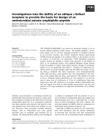
Using a gene expression approach, Joyce and colleagues
[25] identified modulation of proinflammatory and cell survival
pathways in primary cultured human umbilical vein endothelial
cells (HUVECs) exposed to APC. Human APC directly
suppressed the expression of NF-κB subunits and blocked
the expression of NF-κB regulated genes following TNF-α
challenge. Antiapoptotic transcripts, such as survivin
(inhibitor of apoptosis protein) and BCL-2, were upregulated
by APC, whereas there was suppression of the apoptotic
genes calreticulin and TRMP-2. Furthermore, when endo-
thelial cells were challenged with a potent inducer of
apoptosis, the APC-treated cells were protected in a dose-
dependent manner. The potential direct anti-inflammatory and
antiapoptotic effects of APC are summarized in Figure 1.
Other investigators have also documented a direct anti-
apoptotic effect of APC. Using human brain endothelium in a
stroke model, Cheng and coworkers [10] reported that APC
had a direct antiapoptotic effect on hypoxic brain endothelium
that required binding to EPCR and PAR1 activation. The
mechanism of neuroprotection in this model was attributed to
inhibition of the proapoptotic transcription factor p53,
normalization of the proapoptotic Bax/Bcl-2 ratio, and
reduction of caspase-3 signaling, all of which decreased
apoptosis. Using an in vivo murine model of focal ischemic
stroke, administration of mouse APC significantly decreased
brain infarct size and edema, and was dependent on EPCR
and PAR1. Furthermore, low-dose mouse APC produced in
vivo neuroprotection, independent of its anticoagulant
activity.
Activated protein C and endothelial barrier
protection
Another direct mechanism of action of APC on the endo-
thelium is modulation of the endothelial monolayer, leading to
increased cell-cell contact and decreased permeability. Two
investigations have documented this phenomenon and
explored its mechanisms. Feistritzer and Riewald [26] used
HUVECs grown in a transwell with a dual chamber liquid
interface to explore the permeability effects of APC and other
agents. Thrombin and the PAR1 agonist peptide both greatly
increased the permeability of the HUVECs to Evans blue
labeled albumin. The thrombin-mediated hyperpermeability
was reduced by pretreatment with human APC. Also, when
subconfluent endothelial monolayers were incubated with
control or APC, there was less permeability in the APC-
treated cells, implying that APC somehow sealed cell-cell
contacts. Using a cleavage site specific antibody to PAR1,
the endothelial protective effects of APC and the endothelial
disruptive effects of thrombin could both be blocked, which
suggests that the opposing effects of the two proteases were
operating through the same receptor.
It seems paradoxical that thrombin and APC, both operating
through PAR1, can have opposing biologic effects on
endothelial permeability. A potential explanation for this
paradox was explored by targeting the sphingosine
1-phosphate (S1P) pathway, which is known to enhance
endothelial barrier integrity via cytoskeletal rearrangement
[27]. Transfection of the endothelial cells with small
interfering RNA (siRNA) targeting the enzyme responsible for
S1P production, sphingosine kinase-1, blocked the barrier-
enhancing signaling of APC. In addition, siRNA targeting the
S1P receptor S1P
1
also blocked barrier enhancement by
APC. Feistritzer and Riewald [26] concluded that the
Page 3 of 6
(page number not for citation purposes)
endothelial barrier protection produced by APC is mediated
through PAR1 and by crosstalk with the S1P pathway.
In another investigation, Finigan and colleagues [28] also
explored the endothelial barrier enhancement properties of
APC. Those investigators used human pulmonary artery
endothelial cells and measured transendothelial electrical
resistance in response to thrombin in the presence or
absence of APC. Using this in vitro system, APC attenuated
thrombin-induced endothelial cell disruption at concentra-
tions as low as 0.1 to 1.0 µg/ml. Additionally, APC reversed
the formation of transcellular actin stress fibers by thrombin
and produced peripheral cortical actin distribution, which
promotes cell-cell tethering and barrier protection. This
peripheral cytoskeletal arrangement is similar to the effects of
S1P, and indeed using siRNA against S1P
1
this effect of
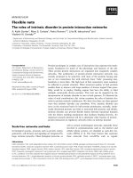
APC was also S1P dependent. Using immunoprecipitation
studies the APC-mediated phosphorylation of S1P
1
was also
documented, as was the co-immunoprecipitation of EPCR
and S1P
1
. The proposed schema for endothelial barrier
protection by APC and its involvement with the S1P pathway
is summarized in Figure 2. In summary, in two different in vitro
investigations, APC promoted endothelial barrier protection in
a PAR1- and S1P
1
-dependent mechanism.
Very low (picomolar) concentrations of thrombin and PAR1
agonist peptide can actually be barrier protective, analogous
to the effects of APC. Also, supraphysiologic concentrations
of APC can be barrier disruptive, which suggests that the
level of PAR1 activation may determine the cellular response
[29]. Thrombin is an excellent activator of PAR1, and pico-
molar concentrations of thrombin may produce similar PAR1
activation as pharmacologic concentrations of APC, which is
a poor activator of PAR1. Furthermore, thrombin can locally
generate APC that may potentially exert its own barrier
enhancing effects [30].
In vivo
endothelial barrier protection by
activated protein C
The in vivo significance of APC signaling through PAR1 is
not entirely clear. It is clear, however, that thrombin is much
more potent (approximately 10
4
-fold) at cleaving PAR1 than
is APC [31]. The concentrations of APC used in the in vitro
studies showing endothelial barrier protection were within the
pharmacologic range of APC in the PROWESS study in one
investigation [26], but another investigation failed to show
significant PAR1 cleavage at concentrations of APC that
were approximately 10-fold higher than the plasma concen-
trations in the PROWESS study [31]. Also, PAR1
-/-
mice
have the same rate of death as wild-type mice in a model of
endotoxemia, arguing that PAR1 activation by endogenous
mediators in vivo does not play a role in a standard model of
sepsis [32,33]. Methodologic differences between in vitro
models and the inherent limitations of in vitro modeling may
explain the discordant results on the significance of APC
signaling through PAR1.
Other in vivo models have yielded conflicting results that may
have tempered the enthusiasm surrounding an endothelial
protective effect of APC. Robriquet and colleagues [34]
Available online />Figure 1
The role of the protein C pathway in the endothelial cell. APC modulates endothelial phenotype by inhibiting thrombin production, direct
antiapoptotic effects, and suppression of NF-κB subunits and therefore decreased inflammatory cell adhesion. APC, activated protein C; ICAM,
intercellular adhesion molecule; NF-κB, nuclear factor-κB; TNF, tumor necrosis factor; VCAM, vascular cell adhesion molecule. Reprinted with
permission from the American Society for Biochemistry and Molecular Biology [25].
reported their experience with a rat model of Pseudomonas
aeruginosa induced lung injury and continuous intravenous
human APC. Rats that received APC exhibited trends toward
increased vascular permeability to radiolabeled albumin and
increased lung edema. The authors postulated that early fibrin
formation in this pneumonia model was potentially beneficial,
and that disruption of this fibrin response by intravenous APC
was possibly deleterious. Of note, human APC was used in
this investigation at a dose of 300 µg/kg per hour, which is a
much higher dose than used in humans but may be appro-
priate given the activity of human APC in rats. In another
investigation of systemic endotoxin in rats, Murakami and
coworkers [35] showed that APC prevented lipopoly-
saccharide-induced pulmonary vascular permeability.
We have preliminary data from a noninfectious model of ALI
(intratracheal acid) on the potential role of APC in endothelial
permeability. Acid-induced lung injury produces damage to
the alveolar epithelium and prominent lung vascular
permeability to protein [36]. This model of lung injury is also
very neutrophil dependent and is therefore a good choice for
testing the direct and indirect effects of APC on the lung
microvasculature. Mice were given acid intratracheally and
were then treated with murine APC. In the APC-treated mice
lung injury was worsened, with increased pulmonary edema
and lung vascular permeability to protein (unpublished data).
The reason for the conflicting results of endothelial barrier
protection in the in vivo studies is not clear, but these
findings reinforce the need to cautiously interpret cell culture
experiments and their relationship to in vivo experimental or
human conditions.
Potential additional clinical applications
beyond sepsis
The PROWESS trial showed a 6% mortality benefit in severe
sepsis from APC in a large, multicenter, placebo-controlled
trial of 1640 patients [4]. Most of the patients had a
pulmonary source of sepsis and 75% were intubated and
ventilated. Because patients were not required to have a
chest radiograph and arterial blood gas assessment at the
time of study enrollment, we do not know how many of these
severe sepsis patients had ALI. Thus, it is plausible that APC
was beneficial in sepsis-induced lung injury, although the
data cannot be obtained from the PROWESS study. The
pathogenesis of organ injury in ALI/ARDS is similar to the
proposed mechanisms for septic-induced injury, and so it is
conceivable that APC may exert anticoagulant, anti-
inflammatory, antiapoptotic, or barrier-enhancing effects that
Critical Care Vol 10 No 6 Looney and Matthay
Page 4 of 6
(page number not for citation purposes)
Figure 2
Proposed schema for APC signaling in the endothelial cell. APC binds to EPCR, which then interacts with the S1P
1
receptor leading to its
phosphorylation by PI3-kinase. S1P
1
signaling through Rac1 leads to cortical cytoskeletal rearrangement and endothelial barrier protection. APC,
activated protein C; EPCR, epithelial protein C receptor; PI3-kinase, phosphatidyl-inositol-3 kinase; S1P, sphingosine 1-phosphate. Reprinted with
permission from the American Society for Biochemistry and Molecular Biology [28].
might benefit patients with ALI from a variety of risk factors
besides sepsis. Also, some studies in patients with ALI from
nonseptic causes demonstrated reduced plasma protein C
and elevated plasminogen activator inhibitor-1 levels, which
correlate with worse clinical outcomes [37,38]. Therefore, we
hypothesized that APC may be of therapeutic value in
patients with ALI. Accordingly, we are currently conducting a
randomized, double blind phase II clinical trial of APC for
early ALI. This multicenter trial is supported by the US
National Heart, Lung, and Blood Institute and will enroll 90
patients to test for several biologic and clinical end-points. If
the results are encouraging, then a phase III randomized trial
could be conducted to test the potential value of APC in ALI
in a large number of patients.
Conclusion
APC has important indirect effects on the integrity of the
vascular endothelium that are both thrombin dependent and
independent, but it also has emerging direct effects on
endothelial function. Apoptosis appears to be a significant
mechanism contributing to endothelial dysfunction in sepsis,
and APC has well described direct antiapoptotic properties
that are independent of its anticoagulant activity. APC also
has a direct effect on endothelial cytoskeletal rearrangement
that strengthens endothelial tight junctions. This mechanism
appears to operate in a PAR1 and SIP
1
dependent manner.
The lack of significant anticoagulant or anti-inflammatory
responses in the human systemic endotoxin-APC model
lends credence to the benefits of APC in sepsis operating
through alternative mechanisms, such as antiapoptosis and
SIP-mediated endothelial protection. APC remains an
important therapy for patients with severe sepsis with major
organ dysfunction, and the mechanism of its benefit in these
patients appears to be in part through direct interactions with
the endothelium.
Competing interests
The authors declare that they have no competing interests.
References
1. Bateman RM, Walley KR: Microvascular resuscitation as a thera-
peutic goal in severe sepsis. Crit Care 2005, Suppl 4:S27-S32.
2. Ware LB, Matthay MA: The acute respiratory distress syn-
drome. N Engl J Med 2000, 342:1334-1349.
3. Hotchkiss RS, Karl IE: The pathophysiology and treatment of
sepsis. N Engl J Med 2003, 348:138-150.
4. Bernard GR, Vincent JL, Laterre PF, LaRosa SP, Dhainaut JF,
Lopez-Rodriguez A, Steingrub JS, Garber GE, Helterbrand JD, Ely
EW, et al.: Efficacy and safety of recombinant human activated
protein C for severe sepsis. N Engl J Med 2001, 344:699-709.
5. Looney MR, Matthay MA: The role of protein C in sepsis. Curr
Infect Dis Rep 2001, 3:413-418.
6. Esmon C: The protein C pathway. Crit Care Med 2000,
Suppl:S44-S48.
7. Stearns-Kurosawa DJ, Kurosawa S, Mollica JS, Ferrell GL, Esmon
CT: The endothelial cell protein C receptor augments protein
C activation by the thrombin-thrombomodulin complex. Proc
Natl Acad Sci USA 1996, 93:10212-10216.
8. White B, Schmidt M, Murphy C, Livingstone W, O’Toole D, Lawler
M, O’Neill L, Kelleher D, Schwarz HP, Smith OP: Activated
protein C inhibits lipopolysaccharide-induced nuclear translo-
cation of nuclear factor kappaB (NF-kappaB) and tumour
necrosis factor alpha (TNF-alpha) production in the THP-1
monocytic cell line. Br J Haematol 2000, 110:130-134.
9. Sturn DH, Kaneider NC, Feistritzer C, Djanani A, Fukudome K,
Wiedermann CJ: Expression and function of the endothelial
protein C receptor in human neutrophils. Blood 2003, 102:
1499-1505.
10. Cheng T, Liu D, Griffin JH, Fernandez JA, Castellino F, Rosen ED,
Fukudome K, Zlokovic BV: Activated protein C blocks p53-
mediated apoptosis in ischemic human brain endothelium
and is neuroprotective. Nat Med 2003, 9:338-342.
11. Guo H, Liu D, Gelbard H, Cheng T, Insalaco R, Fernandez JA,
Griffin JH, Zlokovic BV: Activated protein C prevents neuronal
apoptosis via protease activated receptors 1 and 3. Neuron
2004, 41:563-572.
12. Abraham E, Reinhart K, Opal S, Demeyer I, Doig C, Rodriguez AL,
Beale R, Svoboda P, Laterre PF, Simon S, et al.: Efficacy and
safety of tifacogin (recombinant tissue factor pathway
inhibitor) in severe sepsis: a randomized controlled trial.
JAMA 2003, 290:238-247.
13. Warren BL, Eid A, Singer P, Pillay SS, Carl P, Novak I, Chalupa P,
Atherstone A, Penzes I, Kubler A, et al.: Caring for the critically ill
patient. High-dose antithrombin III in severe sepsis: a ran-
domized controlled trial. JAMA 2001, 286:1869-1878.
14. Nick JA, Coldren CD, Geraci MW, Poch KR, Fouty BW, O’Brien J,
Gruber M, Zarini S, Murphy RC, Kuhn K, et al.: Recombinant
human activated protein C reduces human endotoxin-induced
pulmonary inflammation via inhibition of neutrophil chemo-
taxis. Blood 2004, 104:3878-3885.
15. van der Poll T, Levi M, Nick JA, Abraham E: Activated protein C
inhibits local coagulation after intrapulmonary delivery of
endotoxin in humans. Am J Respir Crit Care Med 2005, 171:
1125-1128.
16. Derhaschnig U, Reiter R, Knobl P, Baumgartner M, Keen P, Jilma
B: Recombinant human activated protein C (rhAPC; drotreco-
gin alfa [activated]) has minimal effect on markers of coagu-
lation, fibrinolysis, and inflammation in acute human
endotoxemia. Blood 2003, 102:2093-2098.
17. Kalil AC, Coyle SM, Um JY, LaRosa SP, Turlo MA, Calvano SE,
Sundin DP, Nelson DR, Lowry SF: Effects of drotrecogin alfa
(activated) in human endotoxemia. Shock 2004, 21:222-229.
18. Vincent JL, Angus DC, Artigas A, Kalil A, Basson BR, Jamal HH,
Johnson G III, Bernard GR: Effects of drotrecogin alfa (acti-
vated) on organ dysfunction in the PROWESS trial. Crit Care
Med 2003, 31:834-840.
19. Grey ST, Tsuchida A, Hau H, Orthner CL, Salem HH, Hancock
WW: Selective inhibitory effects of the anticoagulant acti-
vated protein C on the responses of human mononuclear
phagocytes to LPS, IFN-gamma, or phorbol ester. J Immunol
1994, 153:3664-3672.
20. Joyce DE, Nelson DR, Grinnell BW: Leukocyte and endothelial
cell interactions in sepsis: relevance of the protein C pathway.
Crit Care Med 2004, Suppl:S280-S286.
21. Joyce DE, Grinnell BW: Recombinant human activated protein
C attenuates the inflammatory response in endothelium and
monocytes by modulating nuclear factor-kappaB. Crit Care
Med 2002, Suppl:S288-S293.
22. Iba T, Kidokoro A, Fukunaga M, Nagakari K, Shirahama A, Ida Y:
Activated protein C improves the visceral microcirculation by
attenuating the leukocyte-endothelial interaction in a rat
lipopolysaccharide model. Crit Care Med 2005, 33:368-372.
23. Coughlin SR: Thrombin signalling and protease-activated
receptors. Nature 2000, 407:258-264.
24. Riewald M, Petrovan RJ, Donner A, Mueller BM, Ruf W: Activa-
tion of endothelial cell protease activated receptor 1 by the
protein C pathway. Science 2002, 296:1880-1882.
25. Joyce DE, Gelbert L, Ciaccia A, DeHoff B, Grinnell BW: Gene
expression profile of antithrombotic protein c defines new
mechanisms modulating inflammation and apoptosis. J Biol
Chem 2001, 276:11199-11203.
26. Feistritzer C, Riewald M: Endothelial barrier protection by acti-
vated protein C through PAR1-dependent sphingosine 1-phos-
phate receptor-1 crossactivation. Blood 2005, 105:3178-3184.
27. Garcia JG, Liu F, Verin AD, Birukova A, Dechert MA, Gerthoffer
WT, Bamberg JR, English D: Sphingosine 1-phosphate pro-
motes endothelial cell barrier integrity by Edg-dependent
cytoskeletal rearrangement. J Clin Invest 2001, 108:689-701.
28. Finigan JH, Dudek SM, Singleton PA, Chiang ET, Jacobson JR,
Available online />Page 5 of 6
(page number not for citation purposes)
Camp SM, Ye SQ, Garcia JG: Activated protein C mediates
novel lung endothelial barrier enhancement: role of sphingo-
sine 1-phosphate receptor transactivation. J Biol Chem 2005,
280:17286-17293.
29. Camerer E, Coughlin SR: APC signaling: tickling PAR1 for
barrier protection? Blood 2005, 105:3004-3005.
30. Feistritzer C, Schuepbach RA, Mosnier LO, Bush LA, Di Cera E,
Griffin JH, Riewald M: Protective signaling by activated protein
C is mechanistically linked to protein C activation on endothe-
lial cells. J Biol Chem 2006, 281:20077-20084.
31. Ludeman MJ, Kataoka H, Srinivasan Y, Esmon NL, Esmon CT,
Coughlin SR: PAR1 cleavage and signaling in response to acti-
vated protein C and thrombin. J Biol Chem 2005, 280:13122-
13128.
32. Pawlinski R, Pedersen B, Schabbauer G, Tencati M, Holscher T,
Boisvert W, Andrade-Gordon P, Frank RD, Mackman N: Role of
tissue factor and protease-activated receptors in a mouse
model of endotoxemia. Blood 2004, 103:1342-1347.
33. Camerer E, Cornelissen I, Kataoka H, Duong DN, Zheng YW,
Coughlin SR: Roles of protease-activated receptors in a
mouse model of endotoxemia. Blood 2006, 107:3912-3921.
34. Robriquet L, Collet F, Tournoys A, Prangere T, Neviere R, Fourrier
F, Guery BP: Intravenous administration of activated protein C
in Pseudomonas-induced lung injury: impact on lung fluid
balance and the inflammatory response. Respir Res 2006, 7:
41.
35. Murakami K, Okajima K, Uchiba M, Johno M, Nakagaki T, Okabe
H, Takatsuki K: Activated protein C attenuates endotoxin-
induced pulmonary vascular injury by inhibiting activated
leukocytes in rats. Blood 1996, 87:642-647.
36. Folkesson HG, Matthay MA, Hebert CA, Broaddus VC: Acid
aspiration-induced lung injury in rabbits is mediated by inter-
leukin-8-dependent mechanisms. J Clin Invest 1995, 96:107-
116.
37. Ware LB, Fang X, Matthay MA: Protein C and thrombomodulin
in human acute lung injury. Am J Physiol Lung Cell Mol Physiol
2003, 285:L514-L521.
38. Prabhakaran P, Ware LB, White KE, Cross MT, Matthay MA,
Olman MA: Elevated levels of plasminogen activator inhibitor-
1 in pulmonary edema fluid are associated with mortality in
acute lung injury. Am J Physiol Lung Cell Mol Physiol 2003,
285:L20-L28.
Critical Care Vol 10 No 6 Looney and Matthay
Page 6 of 6
(page number not for citation purposes)