Báo cáo khoa học: " eal-time ultrasound-guided catheterisation of the internal jugular vein: a prospective comparison with the landmark technique in critical care patients" pot
Bạn đang xem bản rút gọn của tài liệu. Xem và tải ngay bản đầy đủ của tài liệu tại đây (348.02 KB, 8 trang )
Open Access
Available online />Page 1 of 8
(page number not for citation purposes)
Vol 10 No 6
Research
eal-time ultrasound-guided catheterisation of the internal jugular
vein: a prospective comparison with the landmark technique in
critical care patients
Dimitrios Karakitsos
1
, Nicolaos Labropoulos
2
, Eric De Groot
3
, Alexandros P Patrianakos
4
,
Gregorios Kouraklis
5
, John Poularas
1
, George Samonis
6
, Dimosthenis A Tsoutsos
7
,
Manousos M Konstadoulakis
8
and Andreas Karabinis
1
1
Department of Intensive Care, General State Hospital of Athens, 154 Mesogeion Avenue, 11527 Athens, Greece
2
Division of Vascular Surgery, University of Medicine and Dentistry of New Jersey, The University Hospital-150 Bergen Street Newark, NJ 07103 USA
3
Academic Medical Center, Department of Vascular Medicine, University of Amsterdam Tafelbergweg 51 .1105 BD Amsterdam, The Netherlands
4
Department of Cardiology, University Hospital of Heraklion, PO Box 1352 Stavrakia, Heraklion, Crete, Greece
5
2nd Department of Propedeutic Surgery, University of Athens School of Medicine, Laiko General Hospital, 17 Agiou Thoma street-11527 Athens,
Greece
6
Department of Internal Medicine and Infectious Diseases, University of Crete, P. O. Box 2203, 71003 Heraklion, Greece
7
'J. Ioannovic' Burn Center, General State Hospital of Athens, 154 Mesogeion Avenue, 11527 Athens, Greece
8
1st Department of Propedeutic Surgery, University of Athens School of Medicine, Hipokrateion University Hospital,114 Vasilis Sofias Avenue 11527
Athens, Greece
Corresponding author: Dimitrios Karakitsos,
Received: 23 May 2006 Revisions requested: 15 Jun 2006 Revisions received: 8 Sep 2006 Accepted: 10 Nov 2006 Published: 17 Nov 2006
Critical Care 2006, 10:R162 (doi:10.1186/cc5101)
This article is online at: />© 2006 Karakitsos et al.; licensee BioMed Central Ltd.
This is an open access article distributed under the terms of the Creative Commons Attribution License ( />),
which permits unrestricted use, distribution, and reproduction in any medium, provided the original work is properly cited.
See related commentary by Bodenham, />Abstract
Introduction Central venous cannulation is crucial in the
management of the critical care patient. This study was
designed to evaluate whether real-time ultrasound-guided
cannulation of the internal jugular vein is superior to the standard
landmark method.
Methods In this randomised study, 450 critical care patients
who underwent real-time ultrasound-guided cannulation of the
internal jugular vein were prospectively compared with 450
critical care patients in whom the landmark technique was used.
Randomisation was performed by means of a computer-
generated random-numbers table, and patients were stratified
with regard to age, gender, and body mass index.
Results There were no significant differences in gender, age,
body mass index, or side of cannulation (left or right) or in the
presence of risk factors for difficult venous cannulation such as
prior catheterisation, limited sites for access attempts, previous
difficulties during catheterisation, previous mechanical
complication, known vascular abnormality, untreated
coagulopathy, skeletal deformity, and cannulation during cardiac
arrest between the two groups of patients. Furthermore, the
physicians who performed the procedures had comparable
experience in the placement of central venous catheters (p =
non-significant). Cannulation of the internal jugular vein was
achieved in all patients by using ultrasound and in 425 of the
patients (94.4%) by using the landmark technique (p < 0.001).
Average access time (skin to vein) and number of attempts were
significantly reduced in the ultrasound group of patients
compared with the landmark group (p < 0.001). In the landmark
group, puncture of the carotid artery occurred in 10.6% of
patients, haematoma in 8.4%, haemothorax in 1.7%,
pneumothorax in 2.4%, and central venous catheter-associated
blood stream infection in 16%, which were all significantly
increased compared with the ultrasound group (p < 0.001).
Conclusion The present data suggest that ultrasound-guided
catheterisation of the internal jugular vein in critical care patients
is superior to the landmark technique and therefore should be
the method of choice in these patients.
BMI = body mass index; CVC = central venous catheter; CVC-BSI = central venous catheter-associated blood stream infection; IJV = internal jugular
vein; 2D = two-dimensional.
Critical Care Vol 10 No 6 Karakitsos et al.
Page 2 of 8
(page number not for citation purposes)
Introduction
Catheterisation of the internal jugular vein (IJV) is commonly
attempted to obtain central venous access for haemodynamic
monitoring, long-term administration of fluids, antibiotics, total
parenteral nutrition, and haemodialysis in critical care patients.
The safe puncture of the IJV is achieved by using anatomical
landmarks on the skin's surface and thus passing the needle
along the anticipated line of the vein. Many anatomic landmark-
guided techniques for IJV puncture have been described since
1966 [1-4]. Complications, including death, are influenced by
patient factors such as body mass index (BMI), site of
attempted access, and operator experience [5-7]. Further-
more, inability to cannulate the IJV may occur in up to 19.4%
of cases [6].
It has been suggested that ultrasound guidance could be ben-
eficial in placing central venous catheters (CVCs) by improv-
ing the success rate, reducing the number of needle passes,
and decreasing complications [8-12]. Also, employment of
ultrasound imaging may identify patients in whom central
venous access may be more difficult and/or in whom conse-
quences of complications could be more serious [13].
Although the ultrasound method has compared favourably
with the landmark technique, its widespread use has been
hampered by the impracticality of specially designed ultra-
sound devices or sterile scanner manipulation, unavailability of
equipment, and lack of trained personnel. Furthermore, previ-
ous studies of ultrasound location of vessels followed by sub-
sequent catheter placement with landmark techniques found
no advantages over standard landmark techniques [7]. How-
ever, few prospective studies exist comparing the technique
itself of ultrasound-guided central venous cannulation versus
the landmark method in critical care patients [14]. This pro-
spective study was designed to compare the real-time ultra-
sound-guided approach with the landmark technique in the
cannulation of the IJV in critical care patients.
Materials and methods
Patients
This prospective study was conducted from January 2000 to
December 2006 in 900 mechanically ventilated critical care
patients (the average number of patients hospitalised per year
in our unit is 170). The patients were randomly assigned on a
one-to-one ratio. Randomisation was performed by means of a
computer-generated random-numbers table, and patients
were stratified with regard to age, gender, and BMI. Block ran-
domisation was used to ensure equal numbers of patients in
the above groups [15]. All physicians and other research per-
sonnel were blinded to the randomisation schedule and the
block size. Family members provided written, informed con-
sent for all patients. The study was conducted in accordance
with the principles outlined in the Declaration of Helsinki and
was approved by the Institutional Ethics Committee.
Successful placement of the CVC was assessed by a chest x-
ray obtained after the procedure. Mechanical complications
were defined as carotid artery puncture, skin haematoma,
pneumothorax, haemothorax, and catheter malposition.
Carotid artery puncture was noted by forceful pulsatile expul-
sion of bright red blood from the needle. All mechanical com-
plications were evaluated clinically, by a chest x-ray, and by
means of ultrasonography where appropriate. In most patients
in whom the first attempt (one pass of the introducing needle)
at catheterisation failed, another physician performed the next
attempt. If a catheter was misplaced, the position was cor-
rected either by a 'power flash' (a rapid infusion of 10 ml of
saline solution pushed through the catheter with a syringe) or
by manipulation of the catheter under fluoroscopic guidance.
Pneumothorax was treated with tube thoracostomy if it was
symptomatic or progressive or if more than 20 percent of the
interface between the lung and the chest wall was separated.
Methods
Landmark technique
For the landmark technique, the patient was placed in a supine
position. The skin at the top of the triangle between the sternal
and clavicular head of the sternocleidomastoid muscle was
degreased with acetone and prepared in a sterile fashion with
povidone-iodine. Then, the area was anaesthetised with a 1%
xylocaine solution with a 22-gauge needle. Physicians were
encouraged to locate the IJV with this 'finder' needle con-
nected to a 2-ml syringe as the needle was advanced through
the skin at a 45° angle in the direction of the right or the left
nipple (for cannulation of the right or the left IJV, respectively).
The return of venous blood into the syringe attached to the
needle confirmed entry into the vessel, and the finder needle
was used to guide a 19-gauge, 10-cm needle connected to a
10-ml syringe (Arrow Howes; Arrow International, Inc., Read-
ing, PA, USA) [16]. A guidewire was then placed through the
needle into the vein, and the needle was removed. A catheter
or sheath was placed over the wire and advanced into the IJV.
Real-time ultrasound-guided method
The neck area was prepared and draped sterilely with the
patient supine as described above. A 7.5-MHz linear-array
ultrasound probe connected to a real-time ultrasound unit
(ATL 3500; Philips Medical Systems, Andover, MA, USA), and
focused at 6.5-cm depth, was covered with ultrasonic gel and
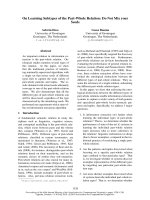
wrapped in a sterile plastic sheath. By wrapping the trans-
ducer in a sterile sheath, its use in consecutive patients is facil-
itated (Figure 1). Standard ultrasound two-dimensional (2D)
imaging was used to measure the depth and calibre of the IJV,
evaluate its patency and compressibility, and identify whether
there were any thrombi in the vein. In cases of pre-existing
thrombus formation and/or failure to gain access due to
trauma or other anatomical anomalies, the IJV on the contralat-
eral side was catheterised. Catheterisation was performed
under continuous dynamic observation of real-time 2D images
obtained by placing the transducer parallel and superior to the
Available online />Page 3 of 8
(page number not for citation purposes)
clavicle, over the groove between the sternal and clavicular
heads of the sternocleidomastoid muscle. This readily visual-
ised the IJV, the external jugular vein, and the carotid artery
(Figure 2). A 19-gauge, 10-cm needle (Arrow Howes; Arrow
International, Inc.) was advanced through the skin under ultra-
sound guidance into the IJV. A guidewire was then placed
through the needle into the vein, and the needle was removed.
A catheter or sheath was placed over the wire and advanced
into the IJV (Figure 2). The needles and guidewires used were
all standard components of catheterisation kits and were not
modified versions for use with ultrasound. All ultrasound-
guided and landmark-guided catheterisations were performed
by well-trained attending cardiologists, intensivists, and sur-
geons with similar experience (10 years of experience in IJV
catheter placements, p = non-significant) to minimise the
effect of operator experience on the success rate and the rate
of mechanical complications. Furthermore, the physicians who
performed the ultrasound-guided method were well trained
and had at least 5 years of experience in performing this
method.
Data collection and statistical analysis
Forms containing patients' characteristics and all the pertinent
fields for each technique were filled out in a timely fashion, and
data were entered in a customised database. The following
data were also recorded: side of catheterisation (either left or
right) and the presence of risk factors for difficult venous can-
nulation such as prior catheterisation, limited sites for access
attempts (other catheters, pacemaker, and local surgery or
infection), previous difficulties during catheterisation (more
than three punctures at one site, two sites attempted, and fail-
ure to gain access), previous mechanical complication, known
vascular abnormality, untreated coagulopathy (international
normalisation ratio >2, activated partial thromboplastin time
>1.5, and platelets <50 × 10
9
per litre), skeletal deformity, and
cannulation during cardiac arrest [5,13]. The outcomes
assessed were the access time, the average number of
attempts before successful placement (defined as separate
skin punctures), the success of placement, the rate of
mechanical complications, and the incidence of CVC-associ-
ated blood stream infection (CVC-BSI). Access time was
defined as the time between penetration of skin and aspiration
of venous blood into the syringe. When a multiple pass was
performed, only the time from skin contact of the first needle
to IJV cannulation was taken into account. This was made to
ensure an objective comparison between the two methods.
Counting the entire procedural time would have clouded the
issue because other parameters such as nursing performance
could affect the measurement. Preparation times for both
Figure 1
The transducer is placed over the groove parallel and superior to the right clavicle (arrow)The transducer is placed over the groove parallel and superior to the
right clavicle (arrow).
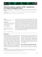
Figure 2
(Top left): Visualisation of the needle entering the anterior wall of the right internal jugular vein (RIJV) (longitudinal axis) (arrow)(Top left): Visualisation of the needle entering the anterior wall of the
right internal jugular vein (RIJV) (longitudinal axis) (arrow). (Bottom
left): Visualisation of the guidewire entering the venous lumen (arrow).
(Top right): Visualisation of the needle entering the venous lumen
(transverse axis). The black line behind the needle is the echo shadow
(arrow). (Bottom right): Sagittal view of the neck, showing the catheter
placed within the lumen (arrow). RCCA, right common carotid artery;
REJV, right external jugular vein.
Critical Care Vol 10 No 6 Karakitsos et al.
Page 4 of 8
(page number not for citation purposes)
techniques were quite similar. The access time was measured
in seconds by stopwatch by other physicians, and the number
of attempts and complications were recorded. It is of note that
every effort was made to ensure the application of evidence-
based catheter insertion practices in both methods [17]. All
patients were receiving antibiotic treatment during the study
period. CVC-BSIs were defined as only those blood stream
infections for which other sources were excluded by careful
examination of the patient record and in which a culture of the
catheter tip demonstrated substantial colonies of an organism
identical to those found in the bloodstream [17].
Data were expressed as mean ± standard deviation. The Stu-
dent t test for independent means, χ
2
analysis, or Fisher exact
test where appropriate were used to identify differences
between the two groups. Correlations between continuous
variables were assessed using the Pearson correlation coeffi-
cient. For ordinal data, the Spearman rank correlation was
used. A p value (two-sided in all tests) of <0.05 was consid-
ered significant. SPSS software, version 11.0, was used
(SPSS Inc., Chicago, IL, USA).
Results
Baseline characteristics of the study population are presented
in Table 1. There were no significant differences between the
two groups of patients in gender ratio, age, BMI, or side of
catheterisation or in the presence of risk factors for difficult
venous cannulation such as prior catheterisation, limited sites
for access attempts, previous difficulties during catheterisa-
tion, previous mechanical complication, known vascular abnor-
mality, untreated coagulopathy, skeletal deformity, and
cannulation during cardiac arrest (Table 1).
In all except 34 patients in the ultrasound group, the IJV was
visualised and cannulated. In these 34 patients who had had
prior surgery and/or prior cannulations, ultrasound imaging
clearly detected the presence of thrombus (Figure 3); thus,
during the same session, the IJV on the contralateral side of
the neck was catheterised instead. Furthermore, 25 patients in
the landmark group in whom catheterisation was unsuccessful
were converted to the ultrasound method. Thrombosis was
identified in 20 cases (which led to formal anticoagulation of
these patients) and anatomical variation of the IJV in five
patients, and these were very likely the reasons for which the
landmark method failed. During the ultrasound-guided proce-
dure, the IJV can be compressed completely by the needle
before the vessel is actually penetrated. Then, the needle must
be advanced a little deeper and retracted slightly to be posi-
tioned in the center of the lumen. In accordance with this, we
have used 2D ultrasound images recorded on both transverse
and longitudinal axes during the same session (Figure 2). The
2D image provides important information about venous loca-
tion and size. Visualisation of the IJV on the transverse axis was
particularly useful for catheterisation, especially when the vein
diameter was small, whereas visualisation of the vein on the
longitudinal axis provided a clear image of both walls of the
vessel (the actual vein puncture using either the longitudinal or
the transverse axis of the 2D image was left to the discretion
of the operator). Also, using this approach, a single-wall punc-
ture can be made by observing the point at which the needle
first indents the anterior wall of the IJV. A short stabbing
motion of the needle at this point will tend to puncture the
anterior wall without opposing it to the posterior wall, thereby
avoiding a double-wall puncture (Figure 2). Single-wall punc-
tures were achieved in all cases using ultrasound guidance.
Table 1
Characteristics of the total study population
Characteristics Ultrasound group (n = 450) Landmark group (n = 450)
Age (years)
a
58.3 ± 10.3 59 ± 9.5
Gender (male/female ratio)
a
0.56 ± 0.4 0.6 ± 0.4
Side of catheterisation (left/right) 222/228 218/232
Body mass index (kg/m
2
)
a
24.1 ± 5.3 23.7 ± 5.9
Prior catheterisation 85 (18.8%) 76 (16.8%)
Limited sites for access attempts 51 (11.3%) 55 (12.2%)
Previous difficulties during catheterisation 44 (9.7%) 40 (8.8%)
Previous mechanical complication 18 (4%) 20 (4.4%)
Known vascular abnormality 4 (0.8%) 3 (0.6%)
Untreated coagulopathy 25 (5.5%) 24 (5.3%)
Skeletal deformity 15 (3.3%) 13 (2.8%)
Cannulation during cardiac arrest 31 (6.8%) 35 (7.7%)
a
Values are presented as mean ± standard deviation.
Available online />Page 5 of 8
(page number not for citation purposes)
Interestingly, five significant anatomical variants between the
IJV and common carotid were observed in the ultrasound
group. In 188 (41.7%) cases, the IJV was anterior and lateral
to the artery; in 120 (26.6%) cases, it was laterally located;
and in 72 (16%) cases, it was directly anterior to the common
carotid artery. In the remaining cases, the IJV was anterior and
medial to the common carotid artery in 53 (12.6%) cases and
directly medial to the artery in 17 (3.7%) cases.
Results using the landmark technique are in sharp contrast to
those obtained by the ultrasound method and are presented in
Table 2. Average access time and number of attempts were
both significantly reduced using ultrasound compared with the
landmark technique (p < 0.001) (Table 2). The success rate
was significantly lower and the rate of mechanical complica-
tions was significantly higher in the landmark group of patients
as compared with the ultrasound group (p < 0.001) (Table 2).
Furthermore, in the landmark group, four cases of haemotho-
rax and four cases of pneumothorax which required therapeu-
tic intervention occurred, but no such complication was
observed in the ultrasound group. Interestingly, the present
data showed a significantly increased number of CVC-BSIs in
the landmark group compared with those documented in the
ultrasound group (p < 0.001) (Table 2). It is of note that the
number of CVC-BSIs was positively correlated to the number
of needle passes in the total study population (r = 0.65, p <
0.001). The type of microorganisms responsible for the CVC-
BSIs in the ultrasound group of patients versus those respon-
sible for the CVC-BSIs in the landmark group was similar:
coagulase-negative Staphylococci (48.6% versus 56.8%,
respectively), Staphylococcus aureus (27.02% versus
24.1%), Enterococus species (13.5% versus 10.3%),
Escherichia coli (2.7% versus 3.4%), Enterobacter (2.7% ver-
sus 1.7%), Pseudomonas aeruginosa (2.7% versus 1.7%),
and Candida species (2.7% versus 1.7%). However, the inci-
dence of coagulase-negative Staphylococci was significantly
higher in the landmark group of patients compared with the
ultrasound group (p < 0.05). Finally, the incidence of co-mor-
bidities, including cancer, that might have affected the
patients' immune status was similar in the ultrasound group as
compared with the landmark group (10% versus 11%, p =
non-significant, respectively).
Discussion
The use of CVCs may be associated with adverse effects that
are both hazardous to patients and expensive to treat [18].
Mechanical complications are reported to occur in 5% to 19%
of patients, infectious complications in 5% to 26%, and throm-
botic complications in 2% to 26% [19,20]. These complica-
tions increase in association with several characteristics,
including patient anatomy (for example, morbid obesity,
Figure 3
Thrombus visualised within the right internal jugular vein (RIJV) (arrow)Thrombus visualised within the right internal jugular vein (RIJV) (arrow).
The vessel could not be compressed. RCCA, right common carotid
artery.
Table 2
Outcome measures in the ultrasound group versus the landmark group of patients
Outcome measures Ultrasound group (n = 450) Landmark group (n = 450)
Access time (seconds) 17.1 ± 16.5 (11.5 to 41.4)
a
44 ± 95.4 (33.2 to 77.5)
Success rate 450 (100%)
a
425 (94.4%)
Carotid puncture 5 (1.1%)
a
48 (10.6%)
Haematoma 2 (0.4%)
a
38 (8.4%)
Haemothorax 0 (0%)
a
8 (1.7%)
Pneumothorax 0 (0%)
a
11 (2.4%)
Average number of attempts 1.1 ± 0.6 (1.1 to 1.9)
a
2.6 ± 2.9 (1.5 to 6.3)
CVC-BSI 47 (10.4%)
a
72 (16%)
a
Comparison of the outcome measures between the ultrasound group and the landmark group of patients (p < 0.001). Access time and average
number of attempts are expressed as mean ± standard deviation (95% confidence interval). Success rate, carotid puncture, haematoma,
haemothorax, pneumothorax, and CVC-BSI are expressed as the absolute number of patients and percentage of their group. CVC-BSI, central
venous catheter-associated blood stream infection.
Critical Care Vol 10 No 6 Karakitsos et al.
Page 6 of 8
(page number not for citation purposes)
cachexia, or local scarring from surgery or radiation treatment),
patient setting (for example, patients receiving mechanical
ventilation or during emergencies such as cardiac arrest), co-
morbidities, and operator's experience [6-8,13]. Real-time
ultrasound guidance of CVC insertion provides the operator
with visualisation of the desired vein and the surrounding ana-
tomic structures prior to and during the insertion of the cathe-
ter. This method appears to improve the success rate and
decrease the complication rate associated with CVC place-
ment [8,11,12]. The present data further support the superior-
ity of real-time ultrasound-guided IJV cannulation as compared
with the landmark technique in mechanically ventilated, critical
care patients.
Using the landmark method, we showed a successful IJV can-
nulation rate of 94.4%, which is in accordance with success
rates documented in previous reports ranging from 85% to
99% [2,3,6,12,21,22]. The incidence of carotid puncture
using the landmark method (10.6%) was comparable with
larger studies [12,21] but higher than those reported (3% to
6%) in smaller series [2,6]. Also, the incidence of haematoma
and pneumothorax (8.4% and 2.4%, respectively) using the
landmark method was in the range of previous studies
[2,6,12,21].
The incidence of mechanical complications using the ultra-
sound-guided technique was negligible, which is in agreement
with previous reports [12,14,22]. Using ultrasound guidance,
the incidence of carotid puncture and haematoma was very
low, and (as shown before) no cases of haemothorax and/or
pneumothorax were observed [12,22]. Interestingly, in several
patients in whom carotid puncture occurred, it was noted that
the IJV was overlying the carotid artery rather than being more
lateral. To avoid the carotid artery in these cases, a sideway
approach of puncturing the IJV was used instead of the per-
pendicular approach.
It is of note that the most favourable outcomes associated with
real-time ultrasound guidance as compared with the landmark
technique were found in studies of inexperienced observers
[22-24]. We showed the superiority of the ultrasound method
in a study in which all observers had comparable experience in
CVC placement. The patients who underwent the ultrasound-
guided cannulation and those who underwent the landmark-
guided cannulation of the IJV were comparable in everything
that is pertinent to this procedure, including the presence of
risk factors for difficult venous cannulation. To the best of our
knowledge, this is the first time that such a controlled compar-
ison has been done between the two methods of IJV cannula-
tion. This is important because no other factors should have
affected the results and therefore the only determining factor
was the technique itself.
The clinical notion that the additional equipment and manipu-
lation associated with the ultrasound method might have
increased the rate of catheter-related infection was not con-
firmed by the present data. We found that the incidence of
CVC-BSI in the ultrasound group of patients was significantly
lower compared with that documented in the landmark group.
The number of CVC-BSIs was significantly correlated to the
number of needle passes in the total study population. We
could speculate that repeated attempts might lead to a break-
down of aseptic technique and more colonisation of skin-
related pathogens [17]. The above findings may be of clinical
importance for two reasons. First, it is well documented that
CVC-BSI is a common problem in the management of the crit-
ical care patient [17]. Second, catheters inserted into the IJV
have been associated with higher risk for infection than those
inserted into the subclavian or femoral vein [25,26]. In this
study, the incidence of bacterial strains implicated in CVC-BSI
was similar in patients of the landmark group as compared
with patients of the ultrasound group, except for a significantly
higher incidence of coagulase-negative Staphylococci which
was documented in the landmark group. A possible explana-
tion for the above observation may be the increased access
time and number of average attempts which were docu-
mented in the landmark group compared with the ultrasound
group. Furthermore, the density of skin flora at the catheter
insertion site is a major risk factor for CVC-BSI [25]. Some
authors recommend that, to reduce the risk for infection, CVCs
be placed in a subclavian site instead of a jugular or femoral
site [27,28]. However, no randomised trial has satisfactorily
compared infection rates for catheters placed in jugular, sub-
clavian, and femoral sites [17,19].
Thrombosis was detected in 54 patients, 20 of whom were in
the landmark group and were converted to the ultrasound
group. Hence, ultrasound imaging is an important tool in iden-
tifying cases of pre-existing thrombus formation and anatomic
variations in the IJV location, thus facilitating safer and more
successful catheterisation of the vessel. In a previous study,
ultrasonography imaging detected venous thrombosis in 33%
of critical care patients; in approximately 15% of these
patients, the thrombosis was catheter-related [29]. Also,
attempts to cannulate thrombotic veins usually are unsuccess-
ful even when the anatomy is normal [13]. It was suggested
that the risk of catheter-related thrombosis varies according to
the site of insertion. In one report, catheter-related thrombosis
occurred in 21.5% of the patients with femoral venous cathe-
ters and in 1.9% of those with subclavian venous catheters
[19]. In an observational study, the risk of thrombosis associ-
ated with internal jugular insertion was approximately four
times the risk associated with subclavian insertion [30].
We used only standard components of catheterisation kits
and not modified versions for use with ultrasound. Other
groups have used more sophisticated, and thus more expen-
sive, ultrasound-guided cannulation devices [31]. The major
impediments to the widespread implementation of the above
method are the purchase costs of the ultrasound machines.
Available online />Page 7 of 8
(page number not for citation purposes)
However, past studies have provided sufficient economic
arguments supporting the notion that ultrasound-guided cen-
tral venous cannulation is cost-effective [31,32].
Technical considerations and study limitations
The ultrasound method is technically demanding, requiring
well-trained operators and adequate experience in performing
it [18]. The benefits of this method may not accrue until after
an initial learning period for operators already experienced in
the landmark technique [31]. We employed the visualisation of
the IJV and of neighboring anatomical structures on both the
longitudinal and transverse axes during real-rime ultrasound-
guided cannulation. This approach offers the advantage of
better positioning of the needle, clear visualisation of the pro-
cedure, avoidance of double-wall puncture, and precise place-
ment of the catheter in the vessel lumen [33].
Perspectives and conclusions
In addition to real-time ultrasound guidance, other approaches
may reduce the risks associated with CVC insertion. Periph-
eral venous cannulation under ultrasound may be an accepta-
ble substitute for CVC placement for certain indications (long-
term i.v. access or parenteral nutrition) [34,35]. Alternative
methods for teaching CVC insertion may employ computer-
ised technologies for simulations. Haptic techniques use vir-
tual reality models to create immersive simulated environments
that recreate the sensation of performing a procedure [36].
However, we believe that ultrasound imaging is a readily avail-
able technology and may be employed by inexperienced oper-
ators to facilitate the placement of a CVC and by experienced
operators to improve the safety of the procedure.
Conclusion
This study showed that real-time ultrasound-guided catheteri-
sation of the IJV offers the advantage of a shorter access time
and a reduced number of successful attempts compared with
the landmark-guided technique. Also, this method has a lower
mechanical complication rate and may result in a lower inci-
dence of CVC-BSI compared with the landmark technique.
There is no doubt that critically ill patients benefit most from
the above advantages of the ultrasound method
[8,11,14,18,19,30].
Competing interests
The authors declare that they have no competing interests.
Authors' contributions
DK conceived of this study, participated in the design of the
study, performed both methods in the intensive care unit set-
ting, and drafted the manuscript. NL participated in the design
of the study, provided expert advice concerning both methods,
and performed the statistical analysis. EDG participated in the
design of the study, provided expert advice concerning the
ultrasound method, and helped to draft the manuscript. APP
participated in the design of the study, carried out both meth-
ods in the intensive care unit setting, and helped to draft the
manuscript. GK performed both methods in the intensive care
unit setting and helped to draft the manuscript. JP performed
both methods in the intensive care unit setting and helped to
draft the manuscript. GS participated in the design of this
study and provided expert advice concerning catheter associ-
ated blood stream infections. DAT participated in the design
of this study and performed both methods in the intensive care
unit setting. MMK participated in the design of the study, per-
formed both methods in the intensive care unit setting, and
helped in the statistical analysis. AK participated in the design
of the study and coordination and helped to draft the manu-
script. All authors read and approved the final manuscript.
References
1. Hermosura B, Vanags L, Dickey MW: Measurement of pressure
during intravenous therapy. JAMA 1966, 195:181.
2. Daily PO, Griep RB, Shumway NE: Percutaneous internal jugu-
lar vein cannulation. Arch Surg 1970, 101:534-536.
3. Rao TLK, Wong AY, Salem MR: A new approach to percutane-
ous catheterization of the internal jugular vein. Anesthesiology
1977, 46:362-364.
4. Hayashi H, Ootaki C, Tsuzuku M, Amano M: Respiratory jugular
vasodilation: a new landmark for right internal jugular vein
puncture in ventilated patients. J Cardiothorac Vasc Anesth
2000, 14:40-44.
5. Digby S: Fatal respiratory obstruction following insertion of a
central venous line. Anaesthesia 1994, 49:1013-1014.
6. Sznajder JI, Zveibil FR, Bitterman H, Weiner P, Bursztein S: Cen-
tral vein catheterization: failure and complication rates by
three percutaneous approaches. Arch Intern Med 1986,
146:259-261.
7. Mansfield PF, Hohn DC, Fornage BD, Gregurich MA, Ota DM:
Complication and failures of subclavian-vein catheterization.
N Engl J Med 1994, 331:1735-1738.
8. Randolph AG, Cook DJ, Gonzales CA, Pribble CG: Ultrasound
guidance for placement of central venous catheters: a meta-
analysis of the literature. Crit Care Med 1996, 24:2053-2058.
9. Machi J, Takeda J, Kakegawa T: Safe jugular and subclavian ven-
ipuncture under ultrasonographic guidance. Am J Surg 1987,
153:321-323.
10. Bond DM, Champion LK, Nolan R: Real-time ultrasound imaging
aids jugular venipuncture. Anesth Analg 1989, 68:700-701.
11. Malloy DL, McGee WT, Shawker TH, Brenner M, Bailey KR, Evans
RG, Parker MM, Farmer JC, Parillo JE: Ultrasound guidance
improves the success rate of internal jugular vein cannulation:
a prospective, randomized trial. Chest 1990, 98:157-160.
12. Dennys BG, Uretsky BF, Reddy S: Ultrasound-assisted cannu-
lation of the internal jugular vein a prospective comparison to
the external landmark-guided technique. Circulation 1993,
87:
1557-1562.
Key messages
• The visualisation of the IJV and of neighboring anatomi-
cal structures on both the longitudinal and transverse
axes ensures the avoidance of a double-wall puncture
during catheterisation.
• Ultrasound-guided cannulation of the IJV offers the
advantage of a shorter access time and a reduced
number of successful attempts.
• Ultrasound-guided cannulation of the IJV has a lower
mechanical complication rate and may result in a lower
incidence of CVC-BSI compared with the landmark
technique.
Critical Care Vol 10 No 6 Karakitsos et al.
Page 8 of 8
(page number not for citation purposes)
13. Hatfield A, Bodenham A: Portable ultrasound for difficult central
venous access. Br J Anaesth 1999, 82:822-826.
14. Hayashi H, Amano M: Does ultrasound imaging before punc-
ture facilitate internal jugular vein cannulation? Prospective
randomized comparison with landmark-guided puncture in
ventilated patients. J Cardiothorac Vasc Anesth 2002,
16:572-575.
15. Lee ET: Statistical Methods for Survival Data Analysis 2nd edition.
New York: John Wiley; 1992:355-357.
16. Jobes DR, Schwartz AJ, Greenhow DE, Stephenson LW, Ellison N:
Safer jugular vein cannulation: recognition of arterial puncture
and preferential use of the external jugular route. Anesthesiol-
ogy 1983, 59:353-355.
17. Center for Disease Control and PreventionDC: National Nosoco-
mial Infections Surveillance (NNIS) System Report, Data Sum-
mary from January 1992–June issued August 2001. Am J Infect
Control 2001, 29:404-421.
18. McGee DC, Gould MK: Preventing complications of central
venous catheterization. N Engl J Med 2003, 348:1123-1133.
19. Merrer J, De Jonghe B, Golliot F, Lefrant JY, Raffy B, Barre E,
Rigaud JP, Casciani D, Misset B, Bosquet C, et al.: French Cath-
eter Study Group in Intensive Care. Complications of femoral
and subclavian venous catheterization in critically ill patients:
a randomized control trial. JAMA 2001, 286:700-707.
20. Richards MJ, Edwards JR, Culver DH, Gaynes RP: Nosocomial
infections in medical intensive care units in the United States.
National Nosocomial Infections Surveillance System. Crit
Care Med 1999, 27:887-892.
21. Schwartz AJ, Jobes DR, Greenhow DE, Stephenson LW, Ellison N:
Carotid artery puncture with internal jugular cannulation using
the Seldinger technique: incidence, recognition, treatment,
and prevention. Anesthesiology 1979, 51:160.
22. Gordon AC, Saliken JC, Johns D, Owen R, Gray RR: US-guided
puncture of the internal jugular vein: complications and ana-
tomic considerations. J Vasc Interv Radiol 1998, 9:333-338.
23. Gilbert TB, Seneff MG, Becker RB: Facilitation of internal jugu-
lar venous cannulation using an audio-guided Doppler ultra-
sound vascular access device: results from a prospective,
dual-center, randomized, crossover clinical study. Crit Care
Med 1995, 23:60-65.
24. Gualtieri E, Deppe SA, Sipperly ME, Thompson DR: Subclavian
venous catheterization: greater success rate for less experi-
enced operators using ultrasound guidance. Crit Care Med
1995, 23:692-697.
25. Heard SO, Wagle M, Vijayakumar E, McLean S, Brueggemann A,
Napolitano LM, Edwards LP, O'Connell FM, Puyana JC, Doern GV:
Influence of triple-lumen central venous catheters coated with
chlorhexidine and silver sulfadiazine on the incidence of cath-
eter-related bacteremia. Arch Intern Med 1998, 158:81-87.
26. Richet H, Hubert B, Nitemberg G, Andremont A, Buu-Hoi A, Our-
bak P, Galicier C, Veron M, Boisivon A, Bouvier AM, et al.: Pro-
spective multicenter study of vascular-catheter-related
complications and risk factors for positive central-catheter
cultures in intensive care unit patients. J Clin Microbiol 1990,
28:2520-2525.
27. Raad I, Darouiche R, Dupuis J, Abi-Said D, Gabrielli A, Hachem R,
Wall M, Harris R, Jones J, Buzaid A, et al.: Central venous cathe-
ters coated with minocycline and rifampin for the prevention of
catheter-related colonization and bloodstream infections: a
randomized, double-blind trial. Ann Intern Med 1997,
127:267-274.
28. Centers for Disease Control and Prevention (CDC): Reduction in
central line-associated bloodstream infections among
patients in intensive care units-Pennsylvania, April 2001–
March 2005. MMWR Morb Mortal Wkly Rep 2005,
54:1013-1016.
29. Hirsch DR, Ingenito EP, Goldhaber SZ: Prevalence of deep
venous thrombosis among patients in medical intensive care.
JAMA 1995, 274:335-337.
30. Timsit JF, Farkas JC, Boyer JM, Martin JB, Misset B, Renaud B,
Carlet J: Central vein catheter-related thrombosis in intensive
care patients: incidence, risk factors and relationship with
catheter-related sepsis. Chest 1998, 114:207-213.
31. Bold RJ, Winchester DJ, Madary AR, Gregurich MA, Mansfield PF:
Prospective, randomized trial of Doppler-asisted subclavian
vein catheterization. Arch Surg 1998, 133:1089-1093.
32. Calvert N, Hind D, McWilliams R, Davidson A, Beverley CA, Tho-
mas SM: Ultrasound for central venous cannulation: economic
evaluation of cost-effectiveness. Anaesthesia 2004,
59:1116-1120.
33. Augoustides JG, Diaz D, Weiner J, Clarke C, Jobes DR: Current
practice of internal jugular venous cannulation in a university
anesthesia department: influence of operator experience on
success of cannulation and arterial injury. J Cardiothorac Vasc
Anesth 2002, 16:567-571.
34. Lam S, Scannell R, Roessler D, Smith MA: Peripherally inserted
central catheters in an acute–scare hospital. Arch Intern Med
1994, 154:1833-1837.
35. Chrisman HBM, Omary RAM, Nemcek AA, Ryu RK, Saker MB,
Vogeltzang RL: Peripherally inserted central catheters: guid-
ance with use of ultrasound versus venography in 2650
patients. J Vasc Interv Radiol 1999, 10:473-475.
36. Kaufmann C, Rhee P, Burris D: Telepresence surgery system
enhances medical student surgery training. Stud Health Tech-
nol Inform 1999, 62:174-178.