Báo cáo khoa học: "High mobility group box-1 protein in patients with suspected community-acquired infections and sepsis: a prospective study" pot
Bạn đang xem bản rút gọn của tài liệu. Xem và tải ngay bản đầy đủ của tài liệu tại đây (420.62 KB, 10 trang )
Open Access
Available online />Page 1 of 10
(page number not for citation purposes)
Vol 11 No 2
Research
High mobility group box-1 protein in patients with suspected
community-acquired infections and sepsis: a prospective study
Shahin Gaïni
1
, Svend Stenvang Pedersen
1
, Ole Græsbøll Koldkjær
2
, Court Pedersen
1
and
Holger Jon Møller
3
1
Department of Infectious Diseases, Odense University Hospital, Søndre Boulevard 29, DK-5000 Odense C, Denmark
2
Department of Clinical Biochemistry, Sønderborg Hospital, Sydvang 1, DK-6400 Sønderborg, Denmark
3
Department of Clinical Biochemistry, Aarhus University Hospital, Nørrebrogade 44, DK-8000 Aarhus C, Denmark
Corresponding author: Shahin Gaïni,
Received: 29 Dec 2006 Revisions requested: 7 Feb 2007 Revisions received: 16 Feb 2007 Accepted: 8 Mar 2007 Published: 8 Mar 2007
Critical Care 2007, 11:R32 (doi:10.1186/cc5715)
This article is online at: />© 2007 Gaïni et al.; licensee BioMed Central Ltd.
This is an open access article distributed under the terms of the Creative Commons Attribution License ( />),
which permits unrestricted use, distribution, and reproduction in any medium, provided the original work is properly cited.
Abstract
Introduction Sepsis is a serious condition with a significant
morbidity and mortality. New insight into the
immunopathogenesis of sepsis could promote the development
of new strategies for diagnosis and therapy. High mobility group
box-1 protein (HMGB1) has been known for many years as a
nuclear chromosomal protein. Its role as a pro-inflammatory
cytokine in sepsis and rheumatoid arthritis has been described
recently. The aim of our study was to evaluate HMGB1 as a
molecular marker in patients with community-acquired
infections.
Methods Patients suspected of having infections/sepsis and
admitted to a department of internal medicine were included in
the study in a prospective manner. Demographic data,
comorbidity, routine biochemistry, microbiological data,
infection focus, severity score, and mortality on day 28 were
recorded. Plasma and serum were sampled at the time of
admission. HMGB1 levels were measured with a commercially
available enzyme-linked immunosorbent assay (ELISA).
Procalcitonin levels were measured with a TRACE (time-
resolved amplified cryptate emission) assay.
Lipopolysaccharide-binding protein and interleukin-6 were
measured with a chemiluminiscent immunometric assay.
Soluble haemoglobin scavenger receptor (sCD163) levels were
measured with an in-house ELISA.
Results One hundred and ninety-four patients were included in
the study. Levels of HMGB1 are presented as medians and
interquartile ranges: healthy controls (0.77 ng/ml, 0.6 to 1.46),
non-infected patients (1.54 ng/ml, 0.79 to 2.88), infected
patients without systemic inflammatory response syndrome
(2.41 ng/ml, 0.63 to 3.44), patients with sepsis (2.24 ng/ml,
1.30 to 3.75), and patients with severe sepsis (2.18 ng/ml, 0.91
to 3.85). In a receiver operator characteristic curve analysis
discriminating between non-infected patients and all infected
patients, the area under the curve for HMGB1 was 0.59 (P <
0.0001). HMGB1 correlated only weakly to levels of white blood
cell count, neutrophils, C-reactive protein, interleukin-6,
procalcitonin, and lipopolysaccharide-binding protein (P <
0.001). HMGB1 did not correlate to sCD163.
Conclusion In a cohort of patients with suspected community-
acquired infections and sepsis, HMGB1 levels were statistically
significantly higher in patients compared to the healthy controls.
There was no statistically significant difference between the
infected and the non-infected patients. Levels of HMGB1
correlated only very weakly to other pro-inflammatory markers
and did not correlate to the anti-inflammatory marker sCD163.
Introduction
Sepsis is a serious condition with a significant morbidity and
mortality [1]. Clinicians treating patients with infections and
sepsis are in need of better diagnostic, prognostic, and immu-
nological molecular markers. Better markers for the presence
of infection and the degree of inflammation would enable the
AUC = area under the curve; CI = confidence interval; CRP = C-reactive protein; ELISA = enzyme-linked immunosorbent assay; HMGB1 = high
mobility group box-1 protein; IL = interleukin; IQR = interquartile range; LBP = lipopolysaccharide-binding protein; PaO
2
= partial pressure of oxygen,
arterial; PCT = procalcitonin; ROC = receiver operating characteristic; sCD163 = soluble haemoglobin scavenger receptor; SIRS = systemic inflam-
matory response syndrome; SOFA = Sepsis-related Organ Failure Assessment; WBC = white blood cell.
Critical Care Vol 11 No 2 Gaïni et al.
Page 2 of 10
(page number not for citation purposes)
clinician to start relevant therapy as early as possible.
Increased insight into the immunopathogenesis of sepsis
would offer the potential to generate new treatment options.
Sepsis is characterised by an activation of the innate immune
system when the immune system is challenged by an invading
pathogenic microorganism [2]. This results in the production
of pro-inflammatory and anti-inflammatory cytokines [2].
A lot of attention has been given to several pro-inflammatory
cytokines involved in sepsis. Cytokines like interleukin (IL)-1,
IL-6, and tumour necrosis factor-alpha have been studied in
animal models and in clinical sepsis cohorts [3-9]. These
cytokines have an important role in initiating the systemic
inflammatory response syndrome (SIRS) in the early phases of
sepsis. In a laboratory model with cultured macrophages stim-
ulated with endotoxin, high mobility group box-1 protein
(HMGB1) was identified as a potential 'late-onset' pro-inflam-
matory cytokine [10]. It was also observed that mice had
increased levels of HMGB1 8 to 32 hours after exposure to
endotoxin [10]. Treatment with antibodies against HMGB1
reduced mortality in endotoxin-exposed mice, and administra-
tion of HMGB1 increased mortality [10].
HMGB1 levels have been studied in critically ill patients. How-
ever, the studies were characterised by few patients in heter-
ogenic patient populations. Measuring HMGB1 has been
quite challenging because no enzyme-linked immunosorbent
assay (ELISA) was available until recently. Earlier studies used
blotting methods for measuring HMGB1. We have previously
shown that C-reactive protein (CRP) and IL-6 were better
markers for infection than soluble haemoglobin scavenger
receptor (sCD163) in a population of patients prospectively
admitted to a department of internal medicine [11]. We have
also shown that CRP, lipopolysaccharide-binding protein
(LBP), and IL-6 were better diagnostic markers for infection
and sepsis than procalcitonin (PCT) in the same cohort of
patients [12]. The purpose of the present study was to
describe levels of HMGB1 in a non-critically ill population of
patients suspected of having sepsis. To perform the study, we
used the patient cohort used in two previous studies [11,12].
Materials and methods
Patients
Patients admitted to the department of internal medicine were
consecutively included in our study in a five month period in
2003. Odense University Hospital (Odense, Denmark) has
1,200 beds and serves a local population of 185,000 inhabit-
ants. Inclusion criteria were suspected diagnosis of infection
as judged by the referring physician and blood cultures drawn
at the time of admission. Exclusion criteria were age below 18
years, earlier participation in the study, or prior hospitalisation
within seven days before admission. Plasma and serum were
sampled immediately after admission. The samples were proc-
essed and frozen at -80°C within 1.5 hours after sampling.
Sampling was performed before the administration of any anti-
biotics was started at the hospital. Informed consent was
obtained from all patients or from their close relatives. The
project was approved by the Ethics Committee of Vejle and
Fyns counties.
Baseline characteristics, demographic characteristics, routine
biochemical data, SIRS criteria, and severity score were
obtained at the time of inclusion. Comorbidity was assessed
with the Charlson Index [13]. Severity was assessed with the
Sepsis-related Organ Failure Assessment (SOFA) score [14].
Patients were assessed with the SIRS criteria at the time of
admission [15]. Severe sepsis was defined as the presence of
sepsis combined with one or more of the following: Glasgow
Coma Scale of less than or equal to 14, PaO
2
(partial pressure
of oxygen, arterial) of less than or equal to 9.75 kPa, oxygen
saturation of less than or equal to 92%, PaO
2
/FiO
2
(fraction of
inspired oxygen) of less than or equal to 250, systolic blood
pressure of less than or equal to 90 mm Hg, systolic blood
pressure decrease of more than or equal to 40 mm Hg from
baseline, pH of less than or equal to 7.3, lactate of more than
or equal to 2.5 mmol/l, creatinine of more than or equal to 177
μmol/l, 100% increase of creatinine in patients with known
kidney disease, oliguria of less than or equal to 30 ml/hour in
more than three hours or of less than or equal to 0.7 litres per
24 hours, prothrombin time of less than or equal to 0.6 (refer-
ence: 0.70–1.30), platelets of less than or equal to 100 × 10
9
/
l, bilirubin of more than or equal to 43 μmol/l, and paralytic
ileus. Septic shock was defined as hypotension persisting
despite adequate fluid resuscitation for at least one hour. The
criteria were not valid if any comorbidity could more relevantly
explain them. The presence of infection was defined by at least
one of the following: identification of a pathogenic microorgan-
ism by cultures or polymerase chain reaction, pneumonia veri-
fied by chest x-ray, infection documented by other imaging
technique, serologically documented infection, and obvious
clinical infection (for instance, erysipelas and wound infection).
The physician classifying the infection status of the included
patients was blinded to all biochemical laboratory results. The
patients were divided into the following groups for analyses:
non-infected patients, infected patients without SIRS, sepsis
patients, and patients with severe sepsis/septic shock.
Patients who could not be classified were excluded from the
analyses.
Laboratory assays
HMGB1 was measured with a commercially available ELISA
(HMGB1 ELISA kit; Shino-Test Corporation, Tokyo, Japan).
The measuring range was 0.6 to 93.8 ng/ml. The range could
be broadened by dilution of high samples. The coefficients of
variation were 5% for samples above 10 ng/ml and 10% for
samples from 2 to 5 ng/ml. Recovery of HMGB1 in this ELISA
was 92% to 111% [16]. PCT was measured with a TRACE
(time-resolved amplified cryptate emission) technology assay
(Kryptor PCT
®
; B·R·A·H·M·S Aktiengesellschaft, Hennigsdorf,
Germany). The functional assay sensitivity was 0.06 ng/ml.
Available online />Page 3 of 10
(page number not for citation purposes)
LBP and IL-6 were measured with a chemiluminiscent immu-
nometric assay (Immulite-1000
®
; Diagnostic Products Corpo-
ration, Los Angeles, CA, USA). The detection limit of LBP was
0.2 μg/ml. The detection limit of IL-6 was 2 pg/ml. sCD163
was measured with an in-house ELISA as described previ-
ously [17]. CRP was measured with an immunoturbidimetric
principle (Modular P
®
; Hitachi, Ltd., Tokyo, Japan). White
blood cells (WBCs) and neutrophils were counted on a Sys-
mex SE 9000
®
(TOA Corporation, Kobe, Japan).
Statistical analysis
Data are presented as medians and interquartile ranges
(IQRs) and as means ± standard deviations. Significance test-
ing was performed using the Kruskal-Wallis test and the Wil-
coxon two-sample test. A two-tailed P value of less than 0.05
was considered statistically significant. Receiver operator
characteristic (ROC) curves and the area under the curve
(AUC) were calculated for all the examined inflammatory mark-
ers. Ninety-five percent confidence intervals (CIs) were
reported for the AUC. The method described by DeLong and
colleagues [18] was used as the significance test for ROC
curve and AUC comparison. The Spearman rank correlation
test was used to determine correlations. At the time of the
study, our clinical biochemistry department did not report CRP
levels below 10 mg/l. CRP levels below 10 mg/l were there-
fore assigned a value of 10 mg/l for calculations. The detection
limit of the HMGB1 ELISA was 0.6 ng/ml. HMGB1 levels
below 0.6 ng/ml were therefore assigned a value of 0.6 ng/ml
for calculations. The detection limit of the IL-6 assay was 2 pg/
ml. IL-6 measurements below 2 pg/ml were therefore assigned
a value of 2 pg/ml for calculations. All statistical calculations
were performed with the STATA 8
®
statistical software pack-
age (StataCorp LP, College Station, TX, USA).
Results
Patient characteristics
One hundred and ninety-four patients were included in the
study. The patients were divided according to our plan for
analyses into the following groups: non-infected patients (n =
67), infected patients without SIRS (n = 32), patients with
sepsis (n = 47), and patients with severe sepsis (n = 27).
Twenty-one patients could not be classified and were
excluded from analyses. Only one patient had septic shock.
This patient was included in the severe sepsis group. The
diagnoses of the non-infected patients were respiratory dis-
ease (n = 22), cardiovascular disease (n = 10), rheumatologic
disease (n = 8), central nervous system disease (n = 5), and
various other diseases (n = 22). Sixteen patients in the non-
infected group were treated with immunosuppressive drugs at
the time of admission (15 with prednisolone and 1 with meth-
otrexate). Fifteen of the infected patients (with or without
SIRS) were treated with prednisolone at the time of admission.
All but one of these patients continued on their immunosup-
pressive treatment during their hospital stay. The mortality rate
among all the infected patients was 3.8%. Thirty-two healthy
hospital workers served as a healthy control group for HMGB1
analyses. Baseline characteristics and mortality at day 28 are
presented in Table 1. The microbiology and infectious focus
are presented in Table 2.
Levels of high mobility group box-1 protein
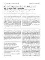
The levels of HMGB1 were statistically significantly higher
among patients compared to the healthy control group (P <
0.001) (Table 3). However, there were no statistically signifi-
cant differences in HMGB1 levels between the following
groups: non-infected patients, infected patients without SIRS,
patients with sepsis, and patients with severe sepsis (Table 3;
Figure 1). When all infected patients (infection without SIRS,
sepsis, and severe sepsis) as a group were compared with the
non-infected patients, the difference was marginally significant
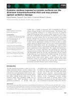
(P = 0.054) (Figure 2). Levels of HMGB1 were significantly
higher in infected patients (infection without SIRS, sepsis, and
severe sepsis) without bacteraemia (n = 94) and in patients
with bacteraemia (n = 12) compared to healthy controls
(Figure 3). Levels of HMGB1 were higher among non-infected
patients treated with immunosuppressive drugs (median 2.8
ng/ml) compared to non-infected patients not treated with
immunosuppressive drugs (median 1.5 ng/ml) (P < 0.05).
There were no statistically significant differences in HMGB1
levels between infected patients treated with immunosuppres-
sive drugs and infected patients not treated with immunosup-
pressive drugs. Levels of CRP, PCT, LBP, and IL-6 were
statistically significantly higher among all infected patients
compared to the non-infected patients (Table 3). Levels of
sCD163 were statistically significantly higher only in the group
with severe sepsis compared to the non-infected patients
(Table 3). Levels of WBC count and neutrophils were statisti-
cally significantly higher among patients with sepsis and
severe sepsis compared to the non-infected patients (Table
3).
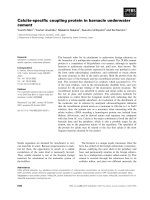
Receiver operating characteristic curve
In an ROC curve analysis to distinguish between the non-
infected patients and all infected patients (infection without
SIRS, sepsis, and severe sepsis), the markers performed with
the following AUCs: HMGB1 0.59 (95% CI 0.5 to 0.68), CRP
0.83 (95% CI 0.77 to 0.89), PCT 0.76 (95% CI 0.69 to 0.84),
LBP 0.79 (95% CI 0.72 to 0.86), IL-6 0.82 (95% CI 0.76 to
0.88), sCD163 0.59 (95% CI 0.5 to 0.68), WBC 0.70 (95%
CI 0.62 to 0.78), and neutrophils 0.69 (95% CI 0.62 to 0.77).
HMGB1 and sCD163 thus performed poorest in this compar-
ative ROC curve analysis (Figure 4).
Correlations
Weak correlations were found between HMGB1 and CRP
(Spearman rank correlation coefficient r = 0.27, P < 0.001),
HMGB1 and PCT (Spearman rank correlation coefficient r =
0.17, P < 0.05), HMGB1 and LBP (Spearman rank correlation
coefficient r = 0.26, P < 0.001), and HMGB1 and IL-6
(Spearman rank correlation coefficient r = 0.21, P < 0.01).
Critical Care Vol 11 No 2 Gaïni et al.
Page 4 of 10
(page number not for citation purposes)
Correlations of moderate strength were found between
HMGB1 and WBC (Spearman rank correlation coefficient r =
0.36, P < 0.0001) and HMGB1 and neutrophils (Spearman
rank correlation coefficient r = 0.42, P < 0.0001). No correla-
tion was found between HMGB1 and sCD163.
Discussion
The patients included in our study were representative of
patients admitted to a department of internal medicine with the
diagnosis of suspected infection. They were elderly patients
with a considerable burden of comorbidity. Compared to
patients in previous clinical studies focusing on markers of
infection and sepsis, our patients were not as ill. This is shown
by the relatively low mortality rate, low SOFA scores, and the
fact that only one patient had septic shock. Our cohort was
therefore dominated by the milder end of the sepsis spectrum.
Most previous studies investigating different immunological
aspects of sepsis have focused on patients admitted to inten-
sive care units and thus on the more severely ill. We believe
that focusing on patients in the milder end of the sepsis spec-
trum is a strength. In the early phase of infectious/sepsis
disease, it is critical to have good diagnostic markers to iden-
tify patients in need of effective antibiotic therapy and other
supportive care. It is also important to have good prognostic
and immunological markers in these patients. If clinicians want
to use clinical research results on their patients, the validation
of (for instance) sepsis markers will ideally have been
performed on a patient sample representative of the clinical
reality that faces the clinician. Our study cohort was well
characterised, and the sampling and processing of plasma/
serum were optimised. We avoided work-up bias in the classi-
fication of the infectious status of the patients by blinding the
clinicians and laboratory technicians. Drawbacks of this study
(as of most other clinical sepsis studies) were heterogeneity
among included patients, a heavy burden of comorbidity, and
different length of disease prior to hospital admission. Another
drawback was the risk of imperfect gold-standard bias in clas-
sifying the patients.
HMGB1 is a 215-amino acid protein that has been shown to
be highly conserved among different species [19]. It has been
known for approximately 30 years as a nuclear chromosomal
protein [19,20]. In recent years, there has been a focus on a
new role for HMGB1. It has been suggested that HMGB1 has
Table 1
Baseline characteristics and outcome of the patients
Variable Non-infected patients
(n = 67)
Infected patients without SIRS
(n = 32)
Patients with sepsis
(n = 47)
Patients with severe sepsis
(n = 27)
Gender, number (percentage)
Male 23 (34.3) 18 (56.3) 20 (42.6) 18 (66.7)
Female 44 (65.7) 14 (43.7) 27 (57.4) 9 (33.3)
Age in years, mean ± SD 67.3 ± 17.1 60.8 ± 16.6 60.4 ± 19.9 66.4 ± 17.8
Length of hospitalisation in days, mean ± SD 8.5 ± 6.9 10.3 ± 11.5 7.8 ± 6.7 10.8 ± 10.5
Mortality on day 28, number (percentage) 6 (8.9) 0 0 4 (14.8)
Severity of disease, mean ± SD
SOFA score 1.4 ± 1.1 1.6 ± 1.5 1.6 ± 1.2 3.0 ± 1.9
Comorbidity, mean ± SD
Charlson Index 1.6 ± 1.3 1.3 ± 1.3 1.1 ± 1.3 1.2 ± 1.3
Laboratory findings, mean ± SD
Haemoglobin, mmol/l 8.1 ± 1.1 8.2 ± 1.2 8.2 ± 1.2 8.2 ± 1.1
Platelet count, 10
9
/l 288.5 ± 108.2 324.5 ± 210.6 254.4 ± 107.3 268.0 ± 184.4
Bilirubin, μmol/l 9.3 ± 6.9 21.9 ± 36.6 10.6 ± 6.8 13.6 ± 5.5
Prothrombin time (reference: 0.70–1.30) 1.0 ± 0.3 0.9 ± 0.4 0.9 ± 0.3 0.9 ± 0.3
Creatinine, μmol/l 96.7 ± 27.3 100.6 ± 31.2 100.4 ± 31.7 140.3 ± 79.5
SD, standard deviation; SIRS, systemic inflammatory response syndrome; SOFA, Sepsis-related Organ Failure Assessment.
Available online />Page 5 of 10
(page number not for citation purposes)
a role as a pro-inflammatory cytokine [21], and HMGB1 has
been shown to have many organ-specific biological functions,
including inducing fever, anorexia, weight loss, and cytokine
production in the brain; inducing acute lung injury and produc-
tion of pro-inflammatory cytokines/mediators in the lungs; pro-
moting translocation in the gut; inducing arthritis and joint
inflammation; affecting heart rhythm; and having bactericidal
effects [22].
To our knowledge, only three clinical studies with data on
HMGB1 levels in infections and sepsis have been published
[10,23,24]. In one small study with 8 healthy people and 25
patients with sepsis, the highest levels of HMGB1 (median 84
ng/ml) were observed in sepsis patients with a fatal outcome
[10]. Surviving patients had a median level of HMGB1 of 25
ng/ml, and healthy controls had undetectable levels of
HMGB1 [10]. In that study, HMGB1 levels were measured
with an immunoblotting method [10]. In a prospective obser-
vational study, HMGB1 and several other cytokines were
measured over several days in patients who had different
degrees of sepsis and who were admitted to intensive care
units [23]. In that study, HMGB1 was measured with two dif-
ferent immunoblotting methods [23]. Levels of HMGB1
remained elevated in this cohort of critically ill patients up to
Table 2
Microbiological and infection characteristics of the patients
Variable Infected patients without SIRS
(n = 32)
Patients with sepsis
(n = 47)
Patients with severe sepsis
(n = 27)
Assessment of infection, number
Gram-positive bacteria 6 12 10
Gram-negative bacteria 7 10 7
Other bacteria 0 2
a
0
Bacteraemia 147
Virus 3
b
4
c
1
d
CXR verified pneumonia
e
9137
Radiological evidence
f
010
Obvious clinical infection
g
752
Focus of infection, number
Upper respiratory tract
infection
101
Lower respiratory tract
infection
12 25 15
Endocarditis 1 0 1
Gastroenteritis 5 1 0
Pyelonephritis 2 2 1
Cystitis 033
Skin/Soft tissue infection 1 4 3
Bone/Joints 2 1 0
Other 8 11 3
a
Mycoplasma pneumoniae (n = 2);
b
Epstein-Barr virus (n = 1), influenza A virus (n = 2);
c
Epstein-Barr virus (n = 2), influenza A virus (n = 2);
d
Puumala virus (n = 1);
e
CXR verified pneumonia with no identified pathogen;
f
infection
documented by imaging techniques (other than CXR) with no identified pathogen;
g
clinical infection (for instance, erysipelas, wound infection).
CXR, chest x-ray; SIRS, systemic inflammatory response syndrome.
Critical Care Vol 11 No 2 Gaïni et al.
Page 6 of 10
(page number not for citation purposes)
Table 3
Levels of HMGB1, PCT, LBP, CRP, IL-6, WBC, and neutrophils in different groups
Variable Healthy controls
(n = 32)
Non-infected patients
(n = 67)
Infected patients without SIRS
(n = 32)
Patients with sepsis
(n = 47)
Patients with severe sepsis
(n = 27)
P value
a
HMGB1 (ng/ml) < 0.001
Median 0.77 1.54 2.41 2.24 2.18
IQR 0.6–1.46 0.79–2.88 0.63–3.44 1.30–3.75 0.91–3.85
PCT (ng/ml) < 0.001
Median 0.09 0.16 0.2 1.9
IQR 0.05–0.13 0.07–0.34 0.08–0.65 0.22–14.6
LBP (μg/ml) < 0.001
Median 16.3 27.4 33.5 40.4
IQR 12.05–25.3 18.3–41.2 25.0–43.2 18.0–63.6
CRP (mg/l) < 0.001
Median 18 122.0 120.0 217.0
IQR 10–42 54.0–215.0 41.0–190.0 78.0–414.0
IL-6 (pg/ml) < 0.001
Median 9.8 20.6 72.6 199.3
IQR 2.85–21.65 9.8–99.4 25.9–274.5 67.5–2,833.0
WBC count (10
9
/l) < 0.001
Median 8.2 9.5 13.0 12.2
IQR 6.7–10.3 7.7–11.9 9.2–17.1 7.0–17.5
Neutrophils (10
9
/l) < 0.001
Median 6.19 7.1 10.1 10.3
IQR 4.73–7.9 5.1–9.7 7.1–14.8 5.5–15.4
sCD163 (mg/l) 0.06
Median 2.99 3.62 3.17 3.63
IQR 2.21–4.05 2.44–4.54 2.27–4.64 2.67–5.72
a
Kruskal-Wallis test. CRP, C-reactive protein; HMGB1, high mobility group box-1 protein; IL-6, interleukin-6; IQR, interquartile range; LBP,
lipopolysaccharide-binding protein; PCT, procalcitonin; sCD163; soluble haemoglobin scavenger receptor; SIRS, systemic inflammatory
response syndrome; WBC, white blood cell.
Available online />Page 7 of 10
(page number not for citation purposes)
one week after inclusion [23]. Levels of HMGB1, in general,
remained elevated for a longer time compared to other
cytokines measured in the same cohort [23]. In a recent study,
HMGB1 levels were measured with an ELISA in several
groups of patients, some of them infected [24]. Mean levels of
HMGB1 were as follows: undetectable in healthy controls,
4.54 ng/ml in infected patients, 2.15 ng/ml in patients with
malignant disease, 6.47 ng/ml in trauma patients, and 14.05
ng/ml in patients with disseminated intravascular coagulation
[24]. High levels were observed in patients with organ failure
(mean 8.29 ng/ml) and in fatal cases (mean 16.58 ng/ml) [24].
It is difficult to compare the abovementioned studies; two of
them used immunoblotting methods for measuring HMGB1
[10,23] and the other used an ELISA [24]. Our study data sug-
gest that HMGB1 levels are much lower in patients in the
milder end of the sepsis spectrum. It is possible that the low
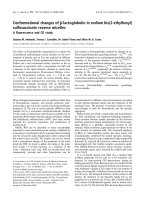
Figure 1
Boxplot of HMGB1 levels in healthy controls, non-infected patients (P < 0.001 compared to healthy controls), infected patients without systemic inflammatory response syndrome (SIRS) (P = 0.32 compared to non-infected patients), patients with sepsis (P = 0.48 compared to infected patients without SIRS), and patients with severe sepsis (P = 0.37 compared to patients with sepsis)Boxplot of HMGB1 levels in healthy controls, non-infected patients (P < 0.001 compared to healthy controls), infected patients without systemic
inflammatory response syndrome (SIRS) (P = 0.32 compared to non-infected patients), patients with sepsis (P = 0.48 compared to infected
patients without SIRS), and patients with severe sepsis (P = 0.37 compared to patients with sepsis). HMGB1, high mobility group box-1 protein.
Figure 2
Boxplot of HMGB1 levels in healthy controls, non-infected patients (P < 0.001 compared to healthy controls), and all infected patients (P = 0.054 compared to non-infected patients)Boxplot of HMGB1 levels in healthy controls, non-infected patients (P < 0.001 compared to healthy controls), and all infected patients (P = 0.054
compared to non-infected patients). HMGB1, high mobility group box-1 protein.
Critical Care Vol 11 No 2 Gaïni et al.
Page 8 of 10
(page number not for citation purposes)
levels of HMGB1 in our study could be explained either by the
fact that the patients were less ill or by the laboratory method
we used to measure HMGB1. The presence of interfering
inhibiting factors/autoantibodies to HMGB1 in human serum
could affect the results of HMGB1 measurements with ELISA
techniques [25]. It is still unknown whether the currently used
assays detect biologically active HMGB1. This is an important
issue in studies focusing on HMGB1 levels and disease activ-
ity. Another explanation could be that we sampled our patients
early in their disease course, and this could explain why levels
of a 'late-onset' cytokine would be low early after admission.
As mentioned earlier, a drawback of the study design was the
lack of data on the length of illness before admission. Our data
showed no statistically significant difference between the non-
infected patients and the infected patients. Our ROC curve
analysis confirmed the abovementioned observation showing
Figure 3
Boxplot of HMGB1 levels in healthy controls, infected patients without bacteraemia (P < 0.0001 compared to healthy controls), and patients with bacteraemia (P < 0.05 compared to healthy controls; P = 0.38 compared to infected patients without bacteraemia)Boxplot of HMGB1 levels in healthy controls, infected patients without bacteraemia (P < 0.0001 compared to healthy controls), and patients with
bacteraemia (P < 0.05 compared to healthy controls; P = 0.38 compared to infected patients without bacteraemia). HMGB1, high mobility group
box-1 protein.
Figure 4
Receiver operating characteristic (ROC) curves comparing the discriminating capabilities of high mobility group box-1 protein (HMGB1), C-reactive protein (CRP), procalcitonin (PCT), lipopolysaccharide-binding protein (LBP), interleukin-6 (IL-6), white blood cell (WBC), neutrophils (neutro), and soluble haemoglobin scavenger receptor (sCD163) between non-infected patients and all infected patients (P < 0.0001)Receiver operating characteristic (ROC) curves comparing the discriminating capabilities of high mobility group box-1 protein (HMGB1), C-reactive
protein (CRP), procalcitonin (PCT), lipopolysaccharide-binding protein (LBP), interleukin-6 (IL-6), white blood cell (WBC), neutrophils (neutro), and
soluble haemoglobin scavenger receptor (sCD163) between non-infected patients and all infected patients (P < 0.0001).
Available online />Page 9 of 10
(page number not for citation purposes)
that HMGB1, in common with sCD163, performed poorly in
ROC curve analysis (with an AUC of only 0.59). The trend of
lower HMGB1 levels which we observed in the most severely
ill patients (severe sepsis and bacteraemia) was observed ear-
lier in one of the abovementioned studies [23]. Given the
increasing focus on immune paresis as a possible mechanism
in severe sepsis and septic shock, this is an interesting obser-
vation [26]. If HMGB1 is considered a strong pro-inflammatory
cytokine involved in the pro-inflammatory phase of SIRS/sep-
sis, it could be hypothesised that lower levels could be
observed when the patient with severe sepsis/septic shock
moves from a pro-inflammatory state to a state with immune
paresis. In our study, there was no correlation between an anti-
inflammatory marker of sepsis (sCD163) and HMGB1.
Conclusion
This is the first study focusing on HMGB1 levels in a cohort of
patients suspected of having community-acquired infections
and sepsis and admitted to a department of internal medicine.
This cohort was dominated by patients with infections without
SIRS, patients with sepsis, and patients with severe sepsis.
These sepsis patients were in the mild end of the sepsis spec-
trum and had low SOFA scores and a low mortality rate. Sixty-
seven of the patients were classified as non-infected patients
and served as our main control group. Levels of HMGB1 were
significantly higher in patients compared to healthy controls.
There was no significant difference in levels between the non-
infected patients and the infected patients (infection without
SIRS, sepsis, and severe sepsis) (P = 0.054). HMGB1 levels
correlated only very weakly to other pro-inflammatory markers
(CRP, IL-6, PCT, LBP, WBC, and neutrophils). HMGB1 did
not correlate to the anti-inflammatory marker sCD163. Our
data do not suggest that HMGB1 has a role in differentiating
between infected and non-infected patients admitted to a
department of internal medicine. Further studies are needed to
elucidate the role of HMGB1 in the immunopathogenesis of
sepsis. Studies focusing on the kinetics of HMGB1 and con-
secutive measurements of HMGB1 should also be
encouraged.
Competing interests
The authors declare that they have no competing interests.
Authors' contributions
SG planned the study, wrote the protocol, collected and ana-
lysed data, and wrote the report. OGK was responsible for
PCT, IL-6, and LBP analyses. HJM developed the sCD163 in-
house ELISA and was responsible for HMGB1 and sCD163
analyses. SSP and CP were involved in planning the study,
revising the manuscript, and practical clinical aspects. All
authors read and approved the final manuscript.
Acknowledgements
The study was financially supported by the University of Southern Den-
mark, the M.L. Jørgensen and G. Hansens Foundation, the J. Boserup
Foundation, the Odense University Hospital Consultant Foundation, the
Foundation for Medical Research in the County of Fyn, the C. Larsen
and Judge E. Larsens Foundation, the P.A. Messerschmidt and Wife
Foundation, the County of Southern Jutland Research Foundation, the J.
Madsen and O. Madsen Foundation, and the Toyota Foundation in Den-
mark. We thank Joan Clausen and Kirsten Bank Petersen for excellent
technical assistance. We also thank study nurses Lene Hergens, Anita
Nymark, and Nete Bülow for excellent clinical assistance.
References
1. Wheeler AP, Bernard GR: Treating patients with severe sepsis.
N Engl J Med 1999, 340:207-214.
2. Cohen J: The immunopathogenesis of sepsis. Nature 2002,
420:885-891.
3. Waage A, Halstensen A, Espevik T: Association between tumour
necrosis factor in serum and fatal outcome in patients with
meningococcal disease. Lancet 1987, 1:355-357.
4. Girardin E, Grau GE, Dayer JM, Roux-Lombard P, Lambert PH:
Tumor necrosis factor and interleukin-1 in the serum of chil-
dren with severe infectious purpura. N Engl J Med 1988,
319:397-400.
5. Waage A, Brandtzaeg P, Halstensen A, Kierulf P, Espevik T: The
complex pattern of cytokines in serum from patients with
meningococcal septic shock. Association between interleukin
6, interleukin 1, and fatal outcome. J Exp Med 1989,
169:333-338.
6. Offner F, Philippe J, Vogelaers D, Colardyn F, Baele G, Baudrihaye
M, Vermeulen A, Leroux-Roels G: Serum tumor necrosis factor
levels in patients with infectious disease and septic shock. J
Lab Clin Med 1990, 116:100-105.
7. Calandra T, Baumgartner JD, Grau GE, Wu MM, Lambert PH,
Schellekens J, Verhoef J, Glauser MP: Prognostic values of
tumor necrosis factor/cachectin, interleukin-1, interferon-
alpha, and interferon-gamma in the serum of patients with
septic shock. Swiss-Dutch J5 Immunoglobulin Study Group. J
Infect Dis 1990, 161:982-987.
8. Munoz C, Misset B, Fitting C, Bleriot JP, Carlet J, Cavaillon JM:
Dissociation between plasma and monocyte-associated
cytokines during sepsis. Eur J Immunol 1991, 21:2177-2184.
9. Calandra T, Gerain J, Heumann D, Baumgartner JD, Glauser MP:
High circulating levels of interleukin-6 in patients with septic
shock: evolution during sepsis, prognostic value, and interplay
with other cytokines. The Swiss-Dutch J5 Immunoglobulin
Study Group. Am J Med 1991, 91:23-29.
10. Wang H, Bloom O, Zhang M, Vishnubhakat JM, Ombrellino M, Che
J, Frasier A, Yang H, Ivanova S, Borovikova L, et al.: HMG-1 as a
late mediator of endotoxin lethality in mice. Science 1999,
285:248-251.
11. Gaini S, Koldkjaer OG, Pedersen SS, Pedersen C, Moestrup SK,
Moller HJ: Soluble haemoglobin scavenger receptor (sCD163)
in patients with suspected community-acquired infections.
APMIS 2006, 114:103-111.
12. Gaini S, Koldkjaer O, Pedersen C, Pedersen S: Procalcitonin,
lipopolysaccharide-binding protein, interleukin-6 and C-reac-
tive protein in community-acquired infections and sepsis: a
prospective study. Critical Care 2006, 10:R53.
Key messages
• The role of HMGB1 as a nuclear chromosomal protein
has been known for many years.
• In recent years, the role of HMGB1 as an inflammatory
cytokine has been explored.
• In a cohort of patients suspected of having community-
acquired infections and sepsis, levels of HMGB1 were
statistically significantly higher in patients compared to
the healthy controls.
Critical Care Vol 11 No 2 Gaïni et al.
Page 10 of 10
(page number not for citation purposes)
13. Charlson ME, Pompei P, Ales KL, MacKenzie CR: A new method
of classifying prognostic comorbidity in longitudinal studies:
development and validation. J Chronic Dis 1987, 40:373-383.
14. Vincent JL, Moreno R, Takala J, Willatts S, De MA, Bruining H,
Reinhart CK, Suter PM, Thijs LG: The SOFA (Sepsis-related
Organ Failure Assessment) score to describe organ dysfunc-
tion/failure. On behalf of the Working Group on Sepsis-
Related Problems of the European Society of Intensive Care
Medicine. Intensive Care Med 1996, 22:707-710.
15. Bone RC, Sibbald WJ, Sprung CL: The ACCP-SCCM consensus
conference on sepsis and organ failure. Chest 1992,
101:1481-1483.
16. Yamada S, Yakabe K, Ishii J, Imaizumi H, Maruyama I: New high
mobility group box 1 assay system. Clin Chim Acta 2006,
372:173-178.
17. Moller HJ, Hald K, Moestrup SK: Characterization of an enzyme-
linked immunosorbent assay for soluble CD163. Scand J Clin
Lab Invest 2002, 62:293-299.
18. DeLong ER, DeLong DM, Clarke-Pearson DL: Comparing the
areas under two or more correlated receiver operating charac-
teristic curves: a nonparametric approach. Biometrics 1988,
44:837-845.
19. Wang H, Yang H, Czura CJ, Sama AE, Tracey KJ: HMGB1 as a
late mediator of lethal systemic inflammation. Am J Respir Crit
Care Med 2001, 164:1768-1773.
20. Bustin M: Regulation of DNA-dependent activities by the func-
tional motifs of the high-mobility-group chromosomal
proteins. Mol Cell Biol 1999, 19:5237-5246.
21. Andersson U, Tracey KJ: HMGB1 in sepsis. Scand J Infect Dis
2003, 35:577-584.
22. Yang H, Wang H, Czura CJ, Tracey KJ: The cytokine activity of
HMGB1. J Leukoc Biol 2005, 78:1-8.
23. Sunden-Cullberg J, Norrby-Teglund A, Rouhiainen A, Rauvala H,
Herman G, Tracey KJ, Lee ML, Andersson J, Tokics L, Treutiger CJ:
Persistent elevation of high mobility group box-1 protein
(HMGB1) in patients with severe sepsis and septic shock. Crit
Care Med 2005, 33:564-573.
24. Hatada T, Wada H, Nobori T, Okabayashi K, Maruyama K, Abe Y,
Uemoto S, Yamada S, Maruyama I:
Plasma concentrations and
importance of high mobility group box protein in the prognosis
of organ failure in patients with disseminated intravascular
coagulation. Thromb Haemost 2005, 94:975-979.
25. Urbonaviciute V, Furnrohr BG, Weber C, Haslbeck M, Wilhelm S,
Herrmann M, Voll RE: Factors masking HMGB1 in human serum
and plasma. J Leukoc Biol 2007, 81:67-74.
26. Hotchkiss RS, Karl IE: The pathophysiology and treatment of
sepsis. N Engl J Med 2003, 348:138-150.