Anaesthesia, Pain, Intensive Care and Emergency - Part 5 pptx
Bạn đang xem bản rút gọn của tài liệu. Xem và tải ngay bản đầy đủ của tài liệu tại đây (824.67 KB, 47 trang )
ciency of the treatments appears to be tied to removal of inflammatory mediators,
even though no difference in mortality between specific treatments has been
confirmed in the literature.
More specific approaches have been proposed, such as high-volume haemofil-
tration and continuous plasma filtration [16, 17], in order to remove several pro-
and anti-inflammatory mediators and to overcome the limitations of conventional
continuous renal replacement therapy (CRRT) (i.e., low volume exchange and low
sieving coefficients for sepsis-associated mediators).
In order to improve the efficacy of a blood purification system in the critically
ill septic patients, unselective adsorption onto a cartridge was added to plasma
filtration and conventional diffusion/convection in a newly designed extracorpo-
real device called coupled plasma filtration–adsorption (CPFA) (Fig. 1).
CPFA is a specific method for the treatment of sepsis. The equipment it requires
is as follows (Fig. 2):
1. A plasma-filter (polyethersulfone 0.45 m
2
with a cut-off of approx 800 kDa)
2. A haemofilter (polyethersulfone 1.4 m
2
)
3. A cartridge (containing approximately 140 ml of hydrophobic styrenic resin)
The kit is lodged in the Bellco ‘Lynda’ machine (Bellco, Mirandola, Italy).
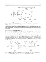
The treatment consists in separation of plasma from the whole blood with
adsorption of the inflammatory mediators and cytokines from the plasma, and a
subsequent purification step accomplished by way of a haemofilter.
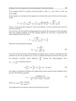
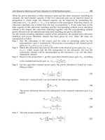
Fig. 1. The peak concentration hypothesis suggests that nonselective control of the peaks of
inflammation and immunoparalysis may help to restore immunohomeostasis
Plasma
“bad molecules”
“good molecules”
UF out
Reinfusate in
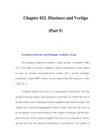
182 S. Livigni, M. Maio, G. Bertolini
The life of the cartridge, as demonstrated by in vitro experiments, is 10 h, which
corresponds to the mean expected treatment duration.
In recent years resins and charcoals have been used because of their capacity
and ability to remove toxic substances from blood, but the medical applications
were often counterbalanced by safety concerns, such as leaching of metals, release
of small microparticles and poor homogeneity and biocompatibility. Haemoper-
fusion through ion/cation exchange resins was first proposed in 1948 for the
treatment of renal failure, but several variations followed. Early experience and
treatments were complicated by pyrogenic reactions, electrolyte disturbances and
haemolysis.
In fact, the use of more sophisticated technologies to coat resins reduces the
problems that result from loss of efficiency, poor reproducibility and mixed out-
comes. Extracorporeal applications require that resin is defined in terms of the
chemical nature of the resin, particle size, porosity and surface area. Resins must
be also tested for the release of microparticles, heavy metals and other toxic
substances. The resin test is done in real conditions similar to those obtaining
during a patient application. The optimisation of flow and column geometry is a
parameter that also greatly influences adsorption efficacy. There is a balance
between the volume of plasma being treated and the time plasma is in contact with
the resin [18].
Using an experimental model of acute endotoxaemia in rabbits, Tetta et al.
Fig. 2. Scheme of coupled plasma filtration–adsorption
Infusion
Haemofilter
Cartridge
Plasma
Plasmafilter
Plasma filtration in sepsis: a research protocol 183
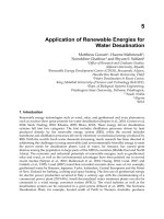
studied whether nonselective adsorption from plasma of cytokines and other
pro-inflammator y mediators known to be produced in excess during sepsis could
reduce 72-h mortality. Cumulative survival was significantly impr oved in rabbits
treated with CPFA, and cumulative su rviv al of the resin with the lipopolysaccha-
ride (LPS) group was not significantly different from that of the control group
(Fig. 3) [19].
Human studies are limited, but promising:Roncoetal.compared CPFA against
haemodiafiltration by measuring homodynamic and immune responsiveness in
ARF patients in septic shock. These authors observed that the haemodynamic was
significantly better with the use of CPFA than with haemodiafiltration. They also
observed significantly higher leucocyte responsiveness after CPFA treatment [20].
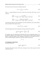
Another clinical study was conducted by Formica et al. The authors examined
the effect of repeated applications of CPFA on haemodynamic response in septic
patients with and without renal failure.In this long-term study, theauthors showed
CPFA to be a safe and feasible treatment leading to significant improvements in
haemodynamic stability, vasopressor requirement, pulmonary function, and 28-
and 90-day survival (Fig. 4). The 28-day survival rate was 90%, which was quite
unexpected considering an APACHE II-predicted mortality of about 40% for these
patients [21].
On the grounds of these experiences it was also expected that early therapy
would hamper the inflammatory cascade.
In the light of these remarks, GiViTI decided to launch a collaborative rando-
mised controlled trial for formal evaluation of the efficacy and clinical safety of
CPFA in septic shock. The main study objective is to clarify whether the implemen-
tation of CPFA in addition to the current clinical practice can reduce mortality of
septic shock patients in ICU. The second objective of the study is to determin e
Fig. 3. Coupled plasma filtration–adsorption in a rabbit model of endotoxic shock
184 S. Livigni, M. Maio, G. Bertolini
whether CPFA can reduce the incidence of organ dysfunction and length of stay.
The study will involve Italian, adult, generalICUs affiliated to theGiViTIgroup,
in which CPFA is regularly used in the treatment of septic shock. The study is
restricted to ICUs that, based on the promising but still incomplete evidence
available,havealready introduced CPFAintotheir routine practice.In other words,
we ask the staff at these centres to use CPFA within a research programme that will
yield information on the real efficacy of the treatment.
All patients who are admitted to the ICU in septic shock or who develop septic
shock while in the ICU will be eligible. The definition used for septic shock is that
provided by the international literature [22, 23]. Patients will be considered eligible
for the study only if it will be possible to initiate CPFA in less than 6 h either from
admission to the ICU for patients admitted in septic shock, or from the diagnosis
of septic shock for the others.
There are some exclusion criteria that make patients not eligible for the study;
these concern age, pregnancy, cerebral coma, metastatic cancer, cardiopulmonary
resuscitation, life expectancy, etc. Eligible patients will be identified upon admis-
sion or during the stay in the ICU and randomised. Patients randomised to the
control arm will be treated according to the current clinical practice in the ICU.
Patients randomised to the experimental arm will also be treated according to the
ICU’s current clinical practice, but with the addition of CPFA.
The CPFA treatment will be applied intermittently (10 consecutive hours fol-
lowed by a 14-h break or CVVH for patients with renal failure) for 5 days following
randomisation. The cartridge must be changed after 10 h; previous experience has
shown saturation of the resin after this.
The clinical follow-up starts on the day of randomisation and finishes at
Fig. 4. Trend in mean arterial pressure (MAP) throughout the first ten sessions (each point
is the mean of the measure at that time for all patients). Statistical significance is related to
the difference between all 100 pre- vs posttreatment measurements
Plasma filtration in sepsis: a research protocol 185
discharge from the ICU. During the ICU stay, information on compliance with the
four A-level recommendations of the Surviving Sepsis Campaign [24], and the daily
SOFA score (Sequential Organ Failure Assessment) [25] will be recorded. The vital
status will be recorded at ICU discharge, at hospital discharge and at 90 days from
randomisation. For patients transferred to other hospitals, “vital status at hospital
discharge” will be intended as the vital status at discharge from the latest hospital
in which the patients stayed.
In agreement with the study rationale, lower mortality is expected in patients
treated with CPFA than in patients treated according to standard practice only. In
the light of these considerations, the following primary and secondary end-points
were chosen:
· Mortality at hospital discharge. For patients transferred to other hospitals, it
will be intended as mortality at the discharge from the latest hospital in which
the patients stayed.
· Mortality within 90 days of randomisation. With this end-point it will be
possible to evaluate whether a possible benefit obtained in the short term
(hospital discharge) is maintained afterwards.
· Proportion of patientswhodevelopone, two, three andfour new organ failures
during their ICU stay. A new organ failureis defined asachangein SOFA score
from 0, 1 or 2 to 3 or 4 in any of the systems considered [26]. This end-point
will determine whether CPFA can reduce the risk that organ failures will
develop.
· Days not spent in the ICU during the first 30 days after randomisation. With
this end-point it will be possible to determine whether CPFA can reduce the
complexity of these patients’ care.
Data previously published by GiViTI show a hospital mortality rate of 63% in
septic shock patients. The study is designed to reveal a 25% relative improvement
in hospital mortality with the use of CPFA. For it to have a power of 80% to find
out such a difference with 5% type I error, it is necessary to enrol 155 subjects in
each arm. Increasing this estimate by approximately 5% to prevent possible pro-
blems in compliance with the protocol yields a number of patients needed of 330.
This sample allows detection of a 29% difference with a power of 90%.
The trial will be m onitored with the Bayesian approach. As known, the
Bayesian approach combines a prior distribution and the gathering of the
experimental evidence into a posterior distribution. The posterior distribution
will be the basis on which to decide wh ether to interrupt the trial or not. Hence,
this analysis requires a probabilistic formalisation of two conflicting hypothe-
ses: one sceptical and one enthusiastic. The trial wi ll be interrupted earlier than
planned when the patient’s benefit is achieved (i.e., demonstration of treatment
efficacy), when sceptics are convinced of the treatment efficacy or, in other words,
when the posterior distribution deriving from a prior sceptical hypothesis ac-
knowledges the achieved benefit. Conversely, the trial will be interrupted earlier
than planned in case of treatment’s futility (i.e., demonstration that the treatment
is futile) when a prior enthusiasti c approach is curbed by the treatment useless-
ness or, in other word s, when the posterior distribution deriving from a prior
186 S. Livigni, M. Maio, G. Bertolini
enthusiastic hypothesis acknowledges the unchanged conditions.
Before enrolment, all patients will be given information on the study’s objec-
tives, procedures and correlated risks.
If any patient is not able to give consent, the instructions provided by the
International Commission on Harmonisation will be followed (ICH Guideline for
Good Clinical Practice). We consider thatthistrial is extremely important, to prove
the effectiveness of this technique in decreasing morbidity and mortality in septic
shock. If we obtain a positive result we can conclude that sepsis can be treated by
bloodpurificationtechnology,buteven ifwe do not,the studywillstill beimportant
because its result will modify the current clinical practice in ICUs.
The trial has been registered with both the ClinicalTrials.gov (identifier
NCT00332371) and the ISRCTN (24534559) registries.
References
1. Friedman G,Silva E,Vincent JL (1998) Hasmortality of septic shockchanged withtime?
Crit Care Med 26:2078–2086
2. Wheeler AP, Bernard GR(1999) Treating patients with sepsis. N EnglJ Med340:207–214
3. USA National Vital Statistics Report (2001) 49:6
4. Rossi C, BertoliniG(2005) Pro gettoMargherita (thesis).(Rapporto 2004)Sestante,Bergamo
5. Alberti C, Brun-Buisson C, Burchardi H et al (2002) Epidemiology of sepsis and
infectionin ICUpatientsfroman internationalmulticentrecohortstudy.IntensiveCare
Med 28(2):108–121
6. Liano G, Pascual J (1996) Acute renal failure. Madrid Acute Renal Failure Study Group.
Lancet 17:347–349
7. Rangel-Frausto MS, Pittet D, Costigan M (1995) The natural history of the systemic
inflammatory response syndrome (SIRS). A prospective study. JAMA 273:117–123
8. Bellomo R, Ronco C (1998) Indications and criteria for initiating renal replacement
therapy in the intensive care unit. Kidney Int 53[Suppl 66]:S106–S109
9. Kellum JA, Johnson JP, Kramer D et al (1998) Diffusive vs convective therapy: effects
on mediators of inflammation in patient with severe systemic inflammatory response
syndrome. Crit Care Med 26:1995–2000
10. Hotchkiss RS, Karl IE (2003) The pathophysiology and treatment of sepsis. N Engl J
Med 348(2):138–150
11. Annane D, Bellissant E, Cavaillon J-M (2005) Septic shock. Lancet 365:63–78
12. Singh S, Evans TW (2006) Organ dysfunction during sepsis. Intensive Care Med
32(3):349–360
13. Mira JP, Cariou A, Grall F et al (1999) Association of TNF2, a TNF-alpha promoter
polymorphism, with septic shock susceptibility and mortality: a multicenter study.
JAMA 282:561–568
14. Godin PJ, Buchman TG (1996) Uncoupling of biological oscillators: a complementary
hypothesis concerning the pathogenesis of multiple organ dysfunction syndrome. Crit
Care Med 24:1107–1116
15. Wheeler AP, Bernard GR (1999) Treating patients with severe sepsis. N Engl J Med
340:207–214
16. Ronco C,Brendolan A,Bellomo et al (2004)The Rationalefor ExtracorporealTherapies
in Sepsis. Adv Sepsis 4(1):2–10
Plasma filtration in sepsis: a research protocol 187
17. Bellomo R, Baldwin I, Cole L et al (1988) Preliminary experience with high volume
hemofiltration in human septic shock. Kidney Int 53:182–185
18. Brendolan A, Ronco C, Ricci Z et al (2004) Coupled plasma filtration adsorption:
rationale, technical development and early clinical experience sepsis, kidney and
multiple organ dysfunction. Contrib Nephrol 144:376–386
19. Tetta C, Gianotti L, Cavaillon JM et al(2000) Continuous plasmafiltration coupled with
sorbent adsorption in a rabbit model of endotoxic shock. Crit Care Med 28:1526–1533
20. Ronco C, Brendolan A, Lonnemann G et al (2002) A pilot study on coupled plasma
filtration with adsorption in septic shock. Crit Care Med 30:1250–1255
21. Formica M, Olivieri C, Livigni S et al (2003) Hemodynamic response to coupled
plasmafiltration–adsorption in human septic shock. Intensive Care Med 29(5):703–708
22. Levy MM, Fink MP, Marshall JC et al (2003) 2001 SCCM/ESICM/ACCP/ATS/SIS Inter-
national Sepsis Definition Conference. Intensive Care Med 29(4):530–538
23. Bone RC, Balk RA, Cerra FB et al (1992) Definitions for sepsis and organ failure and
guidelines for the use of innovative therapies in sepsis. The ACCP/SCCM Consensus
Conference Committee. American College of Chest Physicians/Society of Critical Care
Medicine. Chest 101(6):1644–1655
24. Dellinger RP, Carlet JM, Masur H et al (2004). Surviving Sepsis Campaign guidelines
for management of severe sepsis and septic shock. Intensive Care Med 30(4):536–555
25. Vincent JL, de Mendonca A, Cantraine F et al (1998) Use of the SOFA score to assess
the incidence of organ dysfunction/failure in intensive care units: results of a multicen-
ter, prospective study. Working Group on “Sepsis-related Problems” of the European
Society of Intensive Care Medicine. Crit Care Med 26(11):1793–1800
26. Iapichino G, Radrizzani D, Bertolini G et al (2001), Daily classification of the level of
care. A method to describe clinical course of illness, use of resources and quality of
intensive care assistance. Intensive Care Med 27(1):131–136
188 S. Livigni, M. Maio, G. Bertolini
HIGHLIGHTS ON CIRCULATORY FAILURE,
CPR AND TRAUMA
The cell in shock
M.M. MORALES,H.PETRS-SILVA
‘Cellular homeostasis’ is any of the processes involved in the maintenance of an
internal equilibrium within a cell or between a cell and its external environment.
The physical and biochemical parameters of physiological equilibrium conducive
to eukaryotic cell function include availability and maintenance of nutrients,
oxygenation, temperature, pH, and osmolality, but exposure to conditions when
these parameters are outside the physiological ranges is considered to cause stress
to the cell, leading to macromolecular damage. Many types of environmental stress
have been shown to cause deleterious changes in cells, including osmotic stress [1],
thermal stress [2], heavy metal stress [3], ionising radiation [4], baric stress [5],
oxidative stress [6], chemical genotoxin stress [7], mechanical injury stress [8] and
hypoxia/ischaemia [9].
As a reaction to the threat of macromolecular damage from sudden environ-
mental change or frequent fluctuations in environmental factors, the cell induces
a stress response. This response has been described as an evolutionarily highly
conserved mechanism of cellular protection [10]. The endpoints of stress events
include quick responses, such as protein modifications (e.g. protein phosphoryla-
tion) [11], changes in Ca
2+
concentrations [12], and slow responses, such as protein
chaperoning and repair, transcriptional regulation, removal of damage proteins,
DNA and chromatin stabilisation and repair, cell-cycle control, cell proliferation
and apoptosis [13].
Cells respond to multiple opposing signals simultaneously, and the decision on
whether to die or survive will depend on the intensity of the stress signal. An
extreme condition of stress represents a cell in shock. The cells have a few tools for
reversing shock before it goes too far. But all too often shock is so devastating,
because the dose of stress exceeds the cell’s capacity for maintaining integrity, that
the cellular tools are driven to induce the death of the cell [14–16]. This process is
physiological, since it serves to avoid the genesis of tumours and genetic instability
of organisms [17].
Chapter 18
Cellular stressors
Heat shock and the heart shock proteins
Ashburner and Bonner wrote the first review on the induction of gene activity by
heat shock 27 years ago, describing how immediately after an increase in tempera-
ture all cells increase production of a certain class of molecules called heat shock
proteins [18]. Subsequent studies have revealed that the same response takes place
when cells are subjected to a wide variety of environmental insults, such as toxic
metals [19], alcohols [20], and many metabolic insults [21].
Similar changes in gene expression provide a rapid and direct mechanism of
cellular defence against so many different stress-induced damage that the term
‘heat shock response’ has been replaced by the more general term ‘stress response’,
and the associated products are now referred to as stress proteins [22, 23]. Many
stress proteins are also expressed in normal cells with the same function, such as
control of protein synthesis, folding, and translocation into organelles [24]. And
after cells have been exposed to a stress, these proteins are required to recognise
unfolded proteins and either target them for removal, prevent their aggregation or
assist in their refolding into their native, functional state. Five molecular chape-
rones represent the minimal stress proteome: DnaK/HSP70, DnaJ/HSP40, GrpE,
HSP60, and peptidyl-prolyl isomerase (cylophilin). The proteins involved in cellu-
lar stress responses are the most highly conserved of all organisms [10]. In biology,
chaperones are specific proteins that have the function of assisting other proteins
in achieving proper folding. They were discovered as heat shock proteins, that is,
proteins expressed in heat shock conditions. The reason for this behaviour is that
protein folding is severely affected by heat, and chaperones therefore act to coun-
teract the potential damage. Although most proteins can fold in the absence of
chaperones, for a minority their presence is an absolute requirement.
Recent analysis has revealed that stress, rather than simply imposing destruc-
tive forces, leads to subtle changes in macromolecular structures, which result in a
redirection of the cell energy to allow the synthesis of heat shock proteins, which
themselves function in restoring homeostasis [25].
Cells that produce high levels of stress proteins are better able to survive the
stress damage than cells that do not [26].
The major inducible heatshock protein is HSP70.The binding activity ofHSP70
itself is involved in the regulation of apoptosis, where it may associate with
pro-apoptotic proteins, thereby keeping these proteins in the inactive state, or play
a part in the proteasome-mediated degradation of apoptosis-regulatory proteins
[27]. However after a severe stress, when repair turns out to be impossible HSP 70
is involved in activation of the apoptotic programme and, in the extreme case, of
cellular necrosis [28].
192 M.M. Morales, H. Petrs-Silva
Oxidative stress
Oxidative stress is the cumulative production of ‘reactive oxygen species’ (ROS)
and ‘reactive nitrogen species’ (RNS) through either endogenous or exogenous
insults. Most endogenously formed ROS pass into mitochondria through a leak
from arespiratoryelectron, resultinginthe formationof superoxide anionradicals.
Eventually these anion radicals are transformed into hydrogen peroxide and then
into hydroxyl radicals, HO, which directly attack surrounding macromolecules,
including lipids, proteins and DNA [29]. Most of this damage cannot be entirely
repaired or removed by elements of the cellular degradative system, such as
proteasomes, lysosome, cytosolic and mitochondrial proteases. Consequently,
irreversibly damaged and defective structures accumulate within long-lived post-
mitotic cells, such as cardiac myocytes [30] and neurones [31], which explains why
age-related changes occur in any aerobic organism, especially within long-lived
postmitotic cells, even in an absolutely favourable environmental condition, lead-
ing to a progressively high probability of death [32]. It is also common in many
types of cancer cell that are linked with altered redox regulation of cellular signall-
ing pathways; the redox imbalance may consequently be related to oncogenic
stimulation. DNA mutation is a critical step in carcinogenesis, and high levels of
oxidative DNA lesions have been noted in diverse tumours, strongly implicating
such damage in the aetiology of cancer. It appears that the DNA damage is linked
predominantly with the ini tiation process [33].
Numerous stress response mechanisms are rapidly activated in response to
oxidative insults. Some of the pathways are preferentially linked to enhanced
survival, while others are more frequently associated with cell death. All cells have
free radical scavenging systems todiminish and repair oxidativedamage, and these
include compounds such as glutathione, ascorbate, thioredoxin and various antio-
xidant enzymes [34].
Osmotic stress
The cellular response to osmotic stress ensures that the concentration of water
inside the cell is maintained within a range that is compatible with biological
function. Mammals limit osmotic stress by establishing an internal aqueous envi-
ronment in which intravascular water and plasma electrolyte concentrations are
subject to sensitive and dynamic, organism-based homeostatic regulation by the
kidney, resulting in a homeostatic balance in which plasma osmolality does not
normally vary by more than 2–3% [35]. During osmotic stress total osmolyte
concentrations can vary by hundreds of millimoles.
Cells respond to osmotic stress by varying the concentration of osmolytes
within the cell, in this manner eliminating any change in intracellular water
concentration and the associated change in cell volume that might occur by
osmosis. A direct cellular response to hypertonic stress takes place in seconds and
involves increases in the intracellular concentrations of charged ions, such as
The cell in shock 193
sodium, potassium and chloride, which are mediated by pre-existing ion transport
systems [36–38].
Mammalian inner renalmedullarycells are normally exposedto extremely high
NaCl concentrations. This condition promotes DNA damage and inhibition of
DNA repair. Under normal conditions, most cells in the body die when exposed to
high NaCl, but these renal cells mostly survive and keep functioning both in vitro
and in vivo [39]. The interstitial NaCl concentration in parts of a normal renal
medulla can be 500 mM or more, depending on the species [40]. Several studies
have shown protective adaptations for cellular survival and functioning in this
extreme stress condition, including accumulation of large amounts of organic
osmolytes, which regulate cell volume and intracellular ionic strength despite the
hypertonicity of the high NaCl [41].
Endoplasmic reticulum stress
Correct functioning of the endoplasmic reticulum (ER) is essential for numerous
aspects of cell physiology, including lipid and membrane biogenesis, vesicle traf-
ficking and protein targeting and secretion. The ER is highly susceptible to altera-
tions in homeostasis and exerts a strict quality control system to ensure that only
correctly folded proteins transit to the Golgi. Unfolded or misfolded proteins are
retained in the ER and conserved cell stress response. The aim of this, initially, was
to compensate for the damage, but it can eventually promote cell death if ER
dysfunction is severe or prolonged [43]. ER-initiated cell death is linked with
several diseases, including hypoxia, ischaemia/reperfusion injury, neurodegenera-
tion, heart disease, viral infection and diabetes, and it reflects an extreme condition
of stress [42, 44, 45].
Persistent accumulation of unfolded proteins, interference with protein glyco-
sylation by glucose deprivation, and changes in the redox or ionic conditions of the
ER lumen can trigger programmed cell death. There are three known pro-apototic
signalling pathways emanating from the ER that can be mediated by IRE1, cas-
pase-12 and PERK/CHOP.
Under chronic ER stress, inositol requiring-1 (IRE1),an ER-resident transmem-
brane protein kinase, is activated, leading to the recruitment of JIK (c-Jun-N-ter-
minal-inhibitory kinase), and TNF-receptor-associated factor 2 (TRAF2). TRAF2
activates c-Jun N-terminal protein kinase (JNK) and downstream proapoptotic
kinases, such as apoptosis-signalling kinase 1 (ASK1), finally directing the activa-
tion of mitochondrial apoptotic protease-activating factor-1 (Apaf-1)-dependent
caspase [46]. The mechanism underlying apoptosisviaIRE1-JNK signalling has not
yet been identified. On theotherhand,the recruitment of JIKenablesthe activation
of procaspase-12 located in the ER. Once activated, caspase-12 activates procas-
pase-9 to activate procaspase-3, the executioner of cell death [47].
Like IRE1, PKR-like ER kinase (PERK) is another sensor of reticulum stress.
Activated PERK phosphorylates eukaryotic translation initiation factor-2 (eIF2a),
which enhances translation of activating transcription factor-4 (ATF4) mRNA.
194 M.M. Morales, H. Petrs-Silva
ATF4 induces transcription of the pro-apoptotic factor CHOP, a member of the
C/EBP family of transcription factors. It has recently been shown that CHOP
sensitises cells to ER stress transcriptionally, down-regulating the anti-apoptotic
protein Bcl-2 [48].
Ischaemia/hypoxia
Cellular hypoxia occurs in various conditions, ranging from environmental expo-
sures such as ascent to a high altitude to pathophysiological states with inadequate
oxygen supply (hypoxia), which are usually caused by blood vessel constriction or
obstruction (ischaemia). The basis of this disorder is the exhaustion of energy
supplies. Therefore, human cells have evolved an ability to survive and adapt to
reduction of oxygen pressure in the ambience [49]. Functionally, these adaptations
include compensatory changes that allow cells to survive the hypoxic exposure
itself, such as increases in anaerobic metabolism and initiation of a cell stress
response, in addition to responses that are designed to increase local oxygen
delivery, such as production of angiogenic factors and erythropoietin [50, 51].
Changes in gene expression have already been linked with the human cellular
response to hypoxia [52]. At least three important mechanisms for altering gene
expression during hypoxia have been identified: (1) changes in transcription me-
diated by well-described transcription factors, including hypoxia-inducible factor-
(HIF-1); (2) stabilisation of hypoxia-sensitive RNA species, such as vascular endo-
thelial growth factor (VEGF); and (3) translation through the internal ribosomal
entry sites (IRES), which happens in a cap-independent manner of molecules such
as VEGF even under severely hypoxic conditions [53].
HIF-1 is a transcription factor consisting of a- and b-subunits. HIF-1a expres-
sion is linked to cellular oxygen status, whereas the HIF-1b subunit is constitutively
expressed. HIF-1a dimerises with HIF-1b in the nucleus and transcriptionally
activates a number of genes by way of binding to hypoxia-responsive elements
(HREs). The HIF-1a subunit is stabilised during hypoxia, but degrades rapidly via
the ubiquitin pathway in normoxia. HIF-1 induces expression of proteins that
might assist cell survival during hypoxia, such as VEGF [54].
In mammals hypoxia has been well documented, and this stressful situation
elicits other stress conditions by the reduction of four different parameters: (a)
body temperature, (b) heart rate, (c) respiratory rate and (d) blood pH. These
decreases are associated with a protective physiological effect; however, a long
period of hypoxia/ischaemia causes extensive damage [55, 56].
Stress-activated signalling cascades
Many distinct steps in the stress initiation process are widely regulated by molecu-
lar modifications, and particularly phosphorylation. The stress-activated signall-
ing cascades in stressed cells are becoming clear. At the beginning of these signall-
The cell in shock 195
ing cascades are the sensors of environmental stress: a family of serine/threonine
kinases. This family includes: PKR, RNA-dependent protein kinase, which is acti-
vated by viral infection, ER stress, hypoxic stress, heat and UV irradiation [57, 58];
PERK (RNA-dependent protein kinase-like endoplasmic reticulum kinase)/PEK
(pancreatic eIF2alpha kinase), resident ER proteins, are activated with the accu-
mulation of unfolded proteins in the ER [59]; MAPKs p38, ERK and JNK are
stress-responsive and are activated by oxidative stress, such as an increase in
cellular H
2
O
2
[60].
The end-points of signalling events include both quick responses, such as
protein modifications, and slow responses, including transcriptional regulation,
cell-cycle control, cell proliferation and cell death [13].
The endpoint of cell shock
Programmed cell death
The term ‘programmed cell death’ (PCD) was created to describe a physiological
process that eliminates unwanted cells [61, 62], an active and controlled process of
self-destruction [63]. Glucksmann was one of the scientists who discovered in 1951
that PCD was an integral part of normal embryonic development [64].
PCD can be definedasa sequence of biochemical andmorphological alterations
based on cellular metabolism and leading to cell demise, by which dying cells are
removed in a safe, noninflammatory manner. In physiological conditions, PCD is
tightly controlled and regulates the balance between proliferation and differentia-
tion both in the course of development and during the optimisation of adult cell
and tissue functions, in accordance with environmental stimulus. Alterations in
the regulation of PCD have been implicated in a number of pathologies, including
neurodegeneration and cancer [65–67].
PCD can be divided into four different types: apoptotic cell death, autophagic
cell death, apoptosis-like PCD and necrosis-like PCD. What the various types of
PCD have in common is that they are executed by active cellular processes and can
be interrupted by interfering with intracellular signalling [68].
Apoptotic cell death or type I PCD. The main physical and biochemical hall-
marks of apoptosis include loss of sialic acid, translocation of phosphatidylserine
to the outer plasma membrane, cell shrinkage, nuclear condensation, chromatin
aggregation, DNA fragmentation, membrane blebbing and formation of apoptotic
bodies. Certain modifications that occur in the plasma membrane enable the
recognition of apoptotic bodies by neighbouring cells or phagocytes, preventing
an inflammatory reaction [69, 70]. Apoptosis can be considered a mild response
of cells when stress exceeds cellular tolerance limits.
Apoptosis consists of at least two phases: initiation and execution. This apop-
totic cascade can be initiated via two major pathways in mammalian cells: the
extrinsic ordeath receptor pathwayand intrinsic ormitochondrial pathways.Upon
196 M.M. Morales, H. Petrs-Silva
triggering of either pathway, a specific family of cysteine proteases, the caspases,
is activated to execute the programme. We have to keep in mind the significant
cross-talk and feedback between the different pathways that regulate the apoptotic
machinery and can promote amplification of the apoptotic stimulus [71].
The extrinsic apoptosis pathway is induced upon the binding of ligands (TNF,
TRAIL, FasLetc.)to members ofthe TNFa receptor super-family,which are usually
called the death receptors (Fas, also called CD95/Apo-1; TNF receptors; TRAIL
receptors). Death receptors contain an intracellular globular interaction domain
known as a death domain (DD) in the cytoplasm tail. Ligand-induced receptor
multimerisation results in the formation of the death-inducing signalling complex
(DISC) that includes the death receptor, intracellular adaptor proteins (TRADD,
FADD, RAIDD) and initiator caspases (procaspase 8), leading to autocatalytic
processing and activation of the initiator, caspase 9 [72].
The intrinsic pathway isinitiatedby the majority ofapoptoticstimuli, including
UV radiation, gamma irradiation, heat, DNA damage, the actions of some onco-
proteins and tumour suppressor genes, viral virulence factors and most chemo-
therapeutic agents, irradiation, cytotoxic drugs, granzyme B and DNA damage.
These stimuli lead to the loss of mitochondrial membrane potential, with the
release of pro-apoptotic cell death proteins resulting in the formation of another
multiprotein complex, the apoptosome, that includes Apaf-1, cytochrome-c,
ATP/dATP and the initiator caspase, procaspase 9, promoting the autocatalytic
activation of caspase-9 and subsequent effector caspases. Pro- and anti-apoptotic
proteins of the Bcl-2 family regulate the release of pro-cell death mitochondrial
proteins, while the activity of caspases is negatively regulated by the IAPs. Smac
and Omi promote caspase activation by antagonising the inhibitory effects of the
IAPs, while AIF and EndoG contribute to caspase-independent cell death [73].
The typical pathways of caspase activation during initiation include the ‘death-
receptor-mediated’ recruitment of procaspase-2, procaspase-8 and procaspase-10
and a ‘mitochondrial’ pathway through which caspase-9 is activated via release of
cytochrome-c. The two pathways converge, leading to activation of procaspase-3
and, further downstream, to activation of caspase-6 and caspase-7. All these
pathways are associated with activation of caspase-activated DNase (CAD), and so
also with ‘typical’ internucleosomal DNA fragmentation [74].
Autophagic cell death or type II PCD. Autophagy is characterised by the accu-
mulation of autophagic vesicles (autophagosomes and autophagolysosomes) and
depends on autophagy proteins. It is often observed when massive cell elimination
is demanded or when phagocytes do not have easy access to the dying cells. The set
of proteins (Atg5, Atg6, and Atg7) and the arrangement of autophagosomes in-
volved in both autophagic cell death and autophagy that promotes cell survival are
thesame,but theirregulationissubstantiallydifferentduringeachoftheseprocesses
[75]. The activation of autophagic cell death is common during tissue remodelling
processes, such as metamorphosis in insects and organ morphogenesis during
development, and is part of the cellular response to oxidative stress [76, 77].
Suppressing autophagosome formation by means of autophagy inhibitors, such as
The cell in shock 197
3-methyladenine (3-MA) and wortmannin, or by silencing Atg5 and Atg6 inhibits
this nonapoptotic form of cell death. These results suggest that autophagosome
formation is required for cells to die after exposure to different cell stressors,
proving the existence of this alternative death mechanism [78]. Investigation of
autophagic death is still in its early stages, which is why information on the
molecular basis of autophagic death is extremely limited.
Apoptosis-like, or type III, PCD. Apoptosis-like PCD involves caspase-inde-
pendent mitochondrial pathways. Upon mitochondrial outer-membrane permea-
bilisation, AIF is released from the inter-membrane mitochondrial space. AIF is
the best-characterised caspase-independent cell death regulator, and upon release
it translocates to the nucleus, where it is associated with large-scale DNA fragmen-
tation; however, chromatin condensation is less compact than in apoptosis [79].
The DNA-degrading capacity of AIF relies on recruitment of downstream nuclea-
ses, such as cyclophilin A [80], and the display of phagocytosis-recognition mole-
cules occurs before lysis of the plasma membrane [68].
Necrosis-like, or type IV, PCD. In necrosis-like PCD, the cell-death programme
is triggered by organelles other than mitochondria, such as ER, lysosomes, and the
nucleus, and by proteases other than caspases, such as calpains and cathepsins
originating from the ER and lysosomes, respectively. No chromatin condensation
is observed. The molecular mechanisms of such PCD are less well understood,
although it is believed that they represent ‘alternative’ death pathways when
caspases are inhibited. Ca
2+
and ROS can lead on to severe mitochondrial dysfunc-
tion and necrosis-like PCD with or without autophagy [81].
Both apoptosis and necrosis-like PCD are induced by chemotherapy, which
causes cellular stress [82].
Necrosis
Necrosis is a more disorderly manner of cell death, which results from harsh
circumstances outside the cell and is often called ‘accidental’ cell death, since it
usually occurs as a result of unintentional traumatic injury, whether thermal,
chemical or anoxic. It is characterised by DNA broken into randomly sized frag-
ments, cellular oedema and disruption of the plasma membrane, leading to release
of the cellular components and inflammatory tissue response [83]. Phosphatidil
serine externalisation, an event previously considered unique for apoptosis, may
also occur in cells undergoing necrosis [84].
Necrosis has a major role in neuronal cell death following neonatal hypo-
xia/ischaemia. Cytochrome-c release and caspase activation were also noted in
various human breast carcinoma cells induced by a cytotoxic agent to undergo
necrosis [83].
Apoptosis and necrosis have been shown to be more similar in their regulation
than was previously believed, with several signalling pathways in common. There
198 M.M. Morales, H. Petrs-Silva
is often a delicate balance between the two modes of death, yet outcomes and
consequences for the organism can be totally different, depending on which
pathway is followed after a cell stress.
References
1. Ho SN (2006) Intracellular water homeostasis and the mammalian cellular osmotic
stress response. J Cell Physiol 206(1):9–15
2. Dewhirst MW, Vujaskovic Z, Jones, Thrall D (2005) Re-setting the biologic rationale
for thermal therapy. Int J Hyperthermia 21(8):779–790
3. Ong WY, Farooqui AA (2005) Iron, neuroinflammation, and Alzheimer’s disease. J
Alzheimers Dis 8(2):183–200
4. Lobrich M, Kiefer J (2006) Assessing the likelihood of severe side effects in radiothe-
rapy. Int J Cancer 118(11):2652–2656
5. Somero GN (1992) Adaptations to high hydrostatic pressure. Annu Rev Physiol
54:557–577
6. Butler D, Bahr BA (2006) Oxidative stress and lysosomes: CNS-related consequences
and implications for lysosomal enhancement strategies and induction of autophagy.
Antioxid Redox Signal 8(1–2):185–196
7. Zhou C, Li Z, Diao H, Yu Y et al (2006) DNA damage evaluated by gamma H2AX foci
formation by a selective group of chemical/physical stressors. Mutat Res 604(1–2):8–18
8. Garcia CS, Prota LF, Morales MM et al (2006) Understanding the mechanisms of lung
mechanical stress. Braz J Med Biol Res 39(6):697–706
9. Bazan NG,Palacios-Pelaez R,Lukiw WJ (2002)Hypoxia signaling togenes: significance
in Alzheimer’s disease. Mol Neurobiol 26(2–3):283–298
10. Kultz D (2003) Evolution of the stress proteome: from monophyletic origin to ubiqui-
tous function. J Exp Biol 206:3119–3124
11. Bakkenist CJ, Kastan MB (2004) Initiating cellular stress responses. Cell 2004 118:9–17
12. Zhu WZ, Xie Y, Chen L et al (2006) Intermittent high altitude hypoxia inhibits opening
of mitochondrial permeability transition pores against reperfusion injury. J Mol Cell
Cardiol 40(1):96–106
13. Kultz D (2005) Molecular and evolutionary basis of the cellular stress response. Annu
Rev Physiol 67:225–257
14. Beere HM (2001) Stressed to death: regulation of apoptotic signaling pathways by the
heat shock proteins. Sci STKE (93):RE1
15. Boyce M, Yuan J (2006) Cellular response to endoplasmic reticulum stress: a matter of
life or death. Cell Death Differ 13(3):363–373
16. Martindale JL, Holbrook NJ (2002) Cellular response to oxidative stress: signaling for
suicide and survival. J Cell Physiol 192(1):1–15
17. Herr I, Debatin KM (2001) Cellular stress response and apoptosis in cancer therapy.
Blood 98(9):2603–2614
18. Ashburner M, Bonner JJ (1979) The induction of gene activity in Drosophila by heat
shock. Cell 17(2):241–254
19. Eichler TE, Ransom RF, Smoyer WE (2005) Differential induction of podocyte heat
shock proteins by prolonged single and combination toxic metal exposure. Toxicol Sci
84(1):120–128
20. MilesMF, Diaz JE,DeGuzman VS(1991) Mechanismsof neuronal adaptationto ethanol.
The cell in shock 199
Ethanol induces Hsc70 gene transcription in NG108-15 neuroblastoma × glioma cells.
J Biol Chem 266(4):2409–2414
21. Taggart MJ, Wray S (1998) Hypoxia and smooth muscle function: key regulatory events
during metabolic stress. J Physiol 509(2):315–325
22. Adams C,Rinne RW(1982) Stressprotein formation:gene expression andenvironmen-
tal interaction with evolutionary significance. Int Rev Cytol 79:305–315
23. Craig EA (1985) The heat shock response. CRC Crit Rev Biochem 18(3):239–280
24. Becker J, Craig EA (1994) Heat-shock proteins as molecular chaperones. Eur J Biochem
219:11–23
25. Bond U (2006) Stressed out! Effects of environmental stress on mRNA metabolism.
FEMS Yeast Res 6:160–170
26. Beere HM (2005) Death versus survival: functional interaction between the apoptotic
and stress-inducible heat shock protein pathways. J Clin Invest 115(10):2633–2639
27. Garrido C, Schmitt E, Cande C et al (2003) HSP27 and HSP70: potentially oncogenic
apoptosis inhibitors. Cell Cycle 2(6):579–584
28. Creagh EM, Sheehan D, Cotter TG (2000) Heat shock proteins—modulators of apop-
tosis in tumor cells. Leukemia 14:1161–1173
29. Cadenas E, Davies KJ (2000) Mitochondrial free radicals generation, oxidative stress,
and aging. Free Radic Biol Med 29:222–230
30. FerrariR, GuardigliG, MeleD etal (2004)Oxidative stressduring myocardial ischaemia
and heart failure. Curr Pharm Des 10(14):1699–1711
31. Chinopoulos C, Adam-Vizi V (2006) Calcium, mitochondria and oxidative stress in
neuronal pathology. Novel aspects of an enduring theme. FEBS J 273(3):433–450
32. Barja G (2002) Rate of generation of oxidative stress-related damage and animal
longevity. Free Radic Biol Med 33:1167–1172
33. McEligot AJ, Yang S, Meyskens FL, Jr (2005) Redox regulation by intrinsic species and
extrinsic nutrients in normal and cancer cells. Annu Rev Nutr 25:261–295
34. Dringen R (2005) Oxidative and antioxidative potential of brain microglial cells.
Antioxid Redox Signal 7(9–10):1223–1233
35. Hall JE, Guyton AC, Brands MW (1996) Pressure–volume regulation in hypertension.
Kidney Int Suppl 55:S35–41
36. McManus ML, Churchwell KB, Strange K (1995) Regulation ofcell volume inhealth and
disease. N Engl J Med 333(19):1260–1266
37. Lang F, Busch GL, Ritter M et al (1998) Functional significance ofcell volume regulatory
mechanisms. Physiol Rev 78(1):247–306
38. Morales MM, Nascimento DS, Capella MA et al (2001) Arginine vasopressin regulates
CFTR and ClC-2 mRNA expression in rat kidney cortex and medulla. Pflugers Arch
443(2):202–211
39. DmitrievaNI, CaiQ, Burg MB(2004) Cellsadaptedto highNaCl havemanyDNA breaks
and impaired DNA repair both in cell culture and in vivo. Proc Natl Acad Sci USA
101:2317–2322
40. Maloiy GMO, Magfarlane WV, Shko lnik A (1979) Mammalian herbivores. In: Maloiy GMO
(ed) Comparative physiology of osmoregulation in animals. Academic Press, ?, pp 185–211
41. Dmitrieva NI, Burg MB (2004) Living with DNA breaks is an everyday reality for cells
adapted to high NaCl. Cell Cycle 3:561–563
42. Zhang K, Kaufman RJ (2006) Protein folding in the endoplasmic reticulum and the
unfolded protein response. Handb Exp Pharmacol 172:69–91
43. Rao RV, Ellerby HM, Bredesen DE (2004) Coupling endoplasmic reticulum stress to
the cell death program. Cell Death Differ 11:372–380
200 M.M. Morales, H. Petrs-Silva
44. Aridor M, Balch WE (1999) Integration of endoplasmic reticulum signaling in health
and disease. Nat Med 5:745–751
45. Lindholm D, Wootz H, Korhonen L (2006) ER stress and neurodegenerative diseases.
Cell Death Differ 13(3):385–392
46. Urano F, Wang X, Bertolotti A et al (2000) Coupling of stress in the ER activation of
JNK protein kinases by transmembrane protein kinase IRES1. Science 287:664–666
47. Morishima N, Nakanishi K, Takenouchi H et al (2002) An ER stress-specific caspase
cascade in apoptosis: cytochrome c independent activation of caspase-9 by caspase-12.
J Biol Chem 3:3
48. Boyce M, Yuan J (2006) Cellular response to endoplasmic reticulum stress: a matter of
life or death. Cell Death Differ 13(3):363–373
49. Kato T, Matsumura Y, Tsukanaka A et al (2004) Effect of low oxygen inhalation on
changes in blood pH, lactate, and ammonia due to exercise. Eur J Appl Physiol
91(2–3):296–302
50. Semenza GL (1999) Regulation of mammalian O2 homeostasis by hypoxia-inducible
factor 1. Annu Rev Cell Dev Biol 15:551–578
51. Ikeda E (2005) Cellular response to tissue hypoxia and its involvement in disease
progression. Pathol Int 55(10):603–610
52. Gruber M, Simon MC (2006) Hypoxia-inducible factors, hypoxia, and tumor angioge-
nesis. Curr Opin Hematol 13(3):169–174
53. Fink T, Ebbesen P, Zachar V (2001) Quantitative gene expression profiles of human
liver-derived cell lines exposed to moderate hypoxia. Cell Physiol Biochem 11:105–114
54. Pugh CW, O-Rourke JF, Nagao M et al (1997) Activation of hypoxia-inducible factor-1;
definition of regulatory domains within the alpha subunit. J Biol Chem
272(17):11205–11214
55. Piper HM (1989) Energy deficiency, calcium overload or oxidative stress: possible
causes of irreversible ischemic myocardial injury. Klin Wochenschr 67(9):465–476
56. Wouters BG, Weppler SA, Koritzinsky M et al (2002) Hypoxia as a target for combined
modality treatments. Eur J Cancer 38:240–257
57. Feldman DE, Chauhan V, Koong AC (2005) The unfolded protein response: a novel
component of the hypoxic stress response in tumors. Mol Cancer Res 3(11):597–605
58. He B (2006) Viruses, endoplasmic reticulum stress, and interferon responses. Cell
Death Differ 13(3):393–403
59. Schroder M, Kaufman RJ(2006) Divergent rolesof IRE1alpha andPERK in the unfolded
protein response. Curr Mol Med 6(1):5–36
60. Oteiza PI, Mackenzie GG (2005) Zinc, oxidant-triggered cell signaling, and human
health. Mol Aspects Med 26(4–5):245–255
61. Vaux DL, Weissman IL, Kim SK (1992) Prevention of programmed cell death in
Caenorhabditis elegans by human bcl-2. Science 258(5090):1955–1957
62. Oppenheim RW (1999) Programmed cell death. In: Zigmond MJ, Bloom FE, Landis SC
et al (eds) Fundamental neuroscience. Academic Press, New York, pp 581–609
63. Williams GT, Smith CA, Spooncer E et al (1990) Haemopoietic colony stimulating
factors promote cell survival by suppressing apoptosis. Nature 343(6253):76–79
64. Glucksmann A (1951) Cell deaths in normal vertebrate ontogeny. Biol Rev 26:59–86
65. Sen S (1992) Programmed cell death: concept, mechanism and control. Biol Rev 67:287–319
66. Krantic S, Mechawar N, Reix S, Quirion R (2005) Molecular basis of programmed cell
death involved in neurodegeneration. Trends Neurosci 28(12):670–676
67. Schimmer AD, Dalili S, Batey RA, Riedl SJ (2006) Targeting XIAP for the treatment of
malignancy. Cell Death Differ 13(2):179–188
The cell in shock 201
68. Leist M, Jaattela M (2001) Four deaths and a funeral: from caspases to alternative
mechanisms. Nat Rev Mol Cell Biol 2(8):589–598
69. Kerr JF, Wyllie AH, Currie AR (1972) Apoptosis: a basic biological phenomenon with
wide-ranging implications in tissue kinetics. Br J Cancer 26:239–257
70. Hengartner MO (2000) The biochemistry of apoptosis. Nature 407:770–776
71. Riedl SJ, Shi Y (2004) Molecular mechanisms of caspase regulation during apoptosis.
Nat Rev Mol Cell Biol 5:897–907
72. Ashkenazi A, Dixit VM (1998) Death receptors: Signaling and modulation. Science
281:1305–1308
73. Shi Y (2002) Mechanisms of caspase activation and inhibition during apoptosis. Mol
Cell 9:459–470
74. Degterev A, Boyce M, Yuan J (2003) A decade of caspases. Oncogene 22(53):8543–8567
75. Tsujimoto Y, Shimizu S (2005) Another way to die:autophagic programmed cell death.
Cell Death Differ 12:1528–1534
76. Kiffin R, Bandyopadhyay U, Cuervo AM (2006) Oxidative stress and autophagy. An-
tioxid Redox Signal 8(1–2):152–162
77. Lee CY, Baehrecke EH (2001) Steroid regulation of autophagic programmed cell death
during development. Development 128:1443–1455
78. Shimizu S, Kanaseki T, Mizushima N et al (2004) A role of Bcl-2 family of proteins in
nonapoptotic programmed cell death dependent on autophagy genes. Nat Cell Biol
6:1221–1228
79. Cande C, Cecconi F, Dessen P, Kroemer G (2002) Apoptosis inducing factor (AIF): key
to the conserved caspase-independent pathways of cell death? J Cell Sci 115:4727–4734
80. Cande C, Vahsen N, Kouranti I et al (2004) AIF and cyclophilin A cooperate in
apoptosis-associated chromatinolysis. Oncogene 23(8):1514–1521
81. Sperandio S, de Belle I, Bredesen DE (2000) An alternative, nonapoptotic form of
programmed cell death. Proc Natl Acad Sci USA 97:14376–14381
82. Brown JM,Wouters BG(2001) Apoptosis:mediator ormode ofcell killingby anticancer
agents? Drug Resist Updat 4:135–136
83. Zong WX, Thompson CB (2006) Necrotic death as a cell fate. Genes Dev 20(1):1–15
84. Waring P,Lambert D, Sjaarda A etal (1999) Increasedcell surface exposure ofphospha-
tidylserine on propidium iodide negative thymocytes undergoing death by necrosis.
Cell Death Differ 6:624–637
202 M.M. Morales, H. Petrs-Silva
Tissue partial pressure of carbon dioxide tension
measurements and microcirculation visualisation. New
techniques for the study of low flow states
G. RISTAGNO,W.TANG, M.H. WEIL
Microcirculation is the ultimate determinant of the outcomes of circulatory shock
states. Microcirculatory function is the prerequisite for adequate tissue oxygena-
tion and therefore organ function.It transports oxygen and nutrientstotissue cells,
ensures adequateimmunological functionand, duringdisease, deliverstherapeutic
drugs totarget cells.It ismadeup ofthesmallest blood vessels:arterioles,capillaries
and venules [1] (Fig. 1). The previous techniques used for studying microcircula-
tion (microscopes, laser Doppler or plethysmography) were able to provide only a
global measurement of microvascular blood flow; a measurement expressed as an
average value of whatever was the diameter or direction of single vessels. Recent
technological developments allow more precise and direct investigation of the
tissue perfusion, and especially of the microcirculatory blood flow. The new tech-
niques are basically noninvasive measurements of tissue carbon dioxide tension
(PCO
2
), for example at the oral cavity mucosa, and the orthogonal polarisation
spectral (OPS) imaging techniques, which have allowed direct visualisation and
monitoring of microcirculation at the bedside [2, 3] (Fig. 2).
Chapter 19
Fig. 1. Anatomy of microcirculation
Tissue CO
2
measurements
Tissue hypercarbia accompanies diverse states of perfusion failure, and it is recog-
nised as a diffuse phenomenon during circulatory shock. It has in fact been
observed in heart, stomach, liver, kidney and brain in conditions of haemorrhagic
and anaphylactic shock [4–9]. Increases in tissue PCO
2
account for anaerobic
production of CO
2
. In fact, when oxygen delivery to the tissues is critically reduced,
during circulatory failure states, anaerobic metabolism is triggered with conse-
quent hydrogen ion production. This excess of hydrogen ions is buffered by tissue
bicarbonate in such a way that CO
2
is generated [9, 10]. In the first measurements
of tissue PCO
2
, gastric tonometry was recognised as an early and clinically useful
indicator of perfusion failure during low flow states [11]. Gastric tonometry is
accomplished by way of a balloon incorporated in the distal end of a nasogastric
tube, which is advanced into the stomach. The balloon is then filled with saline
solution, and after an interval of 45–90 min of equilibration, the PCO
2
of the fluid
sampled from the balloon is measured with a conventional blood gas analyser. This
technique also provides for analyses of the gastric intramucosal pH (pHi), which
is computed from simultaneous measurements of the PCO
2
in the saline and
calculation of bicarbonate from arterial blood measurements of pH and PCO
2
based on the Henderson-Hasselbach equation. Several clinical studies have con-
firmed the validity and the utility of gastric tonometry. Close correlations between
gastric pHi and mortality have been reported [12]. In 83 patients with acute
circulatory failure, gastric pHi measured by tonometry was compared with ade-
quacy of tissue oxygenation assessed by conventional methods (cardiac index,
oxygen delivery and oxygen uptake). Only gastric pHi at 24 h proved to be an
Fig. 2. Orthogonal Polarization Spectral imaging camera: CYTOSCAN A/R(Cytometrics Inc.,
Philadelphia, PA).
204 G. Ristagno, W. Tang, M.H. Weil
independent predictor of outcome, predicting death with a sensitivity of 88% [13].
However, the tonometric method presented several limitations [14]. It was an
invasive method, which required stopping feeding.The tissue PCO
2
measurements
could be influenced by the PCO
2
of the gastric juice and by the PCO
2
generated in
the gastric wall as a result of the neutralisation of hydrogen ions by the bicarbonate
contained in the gastric juice or in the backflow of duodenal fluid. Therefore, this
measurement required H
2
-blockade. Gastric tonometry also presented relatively
labour-intensive manipulations and a long time interval for equilibration of CO
2
between the saline in the tonometer balloon and the gastric wall. For all these
reasons and also because we recognised that hypercarbia was a general phenome-
non in perfusion failure,which was equallyprofound in the intraabdominal viscera
and in extraabdominal sites in circulatory shock, we investigated diverse sites for
measurement of tissue PCO
2
directly and in less invasive ways [6].
We had previously demonstrated a close correlation between gastric and oeso-
phageal wall mucosa PCO
2
[5], and subsequently we established that sublingual
fossa mucosa and buccal mucosa were sites that provided measurements of tissue
PCO
2
comparable to those in the mucosa of both the stomach and the oesophageal
wall. In fact, decreases in organ blood flow were closely associated with increases
in PCO
2
in the sublingual mucosa and that of the buccal cavity [5–7, 15–17]. We also
investigated the feasibility and predictive value of sublingual PCO
2
(PslCO
2
) meas-
urement as a noninvasive and early indicator of systemic perfusion failure on
clinical settings. PslCO
2
was measured in five healthy human volunteers and in 46
patients with acute illness or injuries admitted to ICUs attached to emergency
departments and to medical and surgical departments. PslCO
2
was approximately
45 torr in the healthy volunteers and approximately 81 torr in 26 patients who
presented signs of circulatory failure. The initial sublingual mucosa PCO
2
of 12
patients who died without recovering from shock was approximately 93 torr, and
this contrasted with the value of 58 torr (p<0.001) in hospital survivors. When
PslCO
2
exceeded the threshold of 70 torr its positive predictive value for presence
of physical signs of circulatory shock was 1.00. A value < 70 torr predicted survival
with a predictive value of 0.93. Later, we demonstrated that the buccal mucosa
tissue PCO
2
measurements could be used as sensitive indicators of systemic blood
flow during haemorrhagic shock [16]. We induced haemorrhagic shock in five
anaesthetised pigs weighing 35–40 kg. Blood was shed at a rate of 20 ml/min until
the mean arterial pressure had declined to 55±5 mmHg. After 2 h the shed blood
was reinfused at a rate of 100 ml/min and animals were observed for a further 2 h.
Over the 2-h interval of shock, the buccal mucosa PCO
2
increased in parallel with
the sublingual mucosa PCO
2
, from 56 to 116 torr (Fig. 3). Increases in buccal tissue
PCO
2
were accompanied by corresponding decreases in cardiac output and mean
arterial pressure, and by increases in arterial blood lactate concentrations. In-
creases in buccal PCO
2
were accompanied by reductions in buccal mucosal flows,
measured by microsphere techniques. These decreases in blood flow were closely
related to those in the sublingual sites and to concomitant reductions in liver and
kidney blood flow. After reinfusion of the shed blood, buccal and sublingual
mucosa PCO
2
values were restored to baseline. There was a close correlation
Tissue partial pressure of carbon dioxide tension measurements 205
between buccal mucosa PCO
2
and sublingual mucosa PCO
2
measurements, and
buccal PCO
2
measurement was a useful guide for diagnosis of circulatory shock
states. In a more recent study on a rat model of haemorrhagic shock [17], our group
investigated the buccal PCO
2
measurements to identify a threshold levelthatwould
predict the effects of volume repletion on survival and to confirm buccal PCO
2
as
a better indicator of the severity of volume deficit than other commonly used
standard measurements. Animals were randomised into four groups for bleeding,
with losses equal to 25%, 30%, 35% and 40% of the estimated total blood volume
over a period of 30 min. Thirty minutes after the end of bleeding, infusion of
lactated Ringer’s solution was started, in amounts corresponding to twice the
volume of blood removed over 30 min. The standard measurements used for
diagnosis and as a guide for therapy during haemorrhagic shock, such as mean
arterial pressure, failed to distinguish between the four groups. Buccal mucosa
PCO
2
, however, did differentiate between the various degrees of severity of hae-
morrhage (Fig. 4). Moreover, during and after volume repletion, buccal mucosa
PCO
2
was able to predict survival and neurological recovery in the various groups.
During circulatory failure buccal mucosa tissue CO
2
was a noninvasive and rapid
response indicator. Buccal PCO
2
was therefore confirmed as a useful guide to the
diagnosis of circulatory shock and as a quantitative indicator of its severity. It also
provided a rapid response for confirmation of the effectiveness of treatments.
Fig. 3. Increases and decreases in sublingual and buccal mucosal partial pressure of carbon
dioxide (PCO
2
). * p <0.01 versus control
206 G. Ristagno, W. Tang, M.H. Weil
Monitoring of microcirculation with the OPS technique
The OPS imaging technology is a noninvasive method for direct visualisation of
multiple conditions ofthemicrocirculation andperformanceofquantitative meas-
urements of the diameter of vessels, the velocity of red blood cells and functional
capillary density [18]. This method uses a linearly polarised light to illuminate the
area of interest. The light is reflected from the tissue source and forms an image of
the illuminated region within the target of the video camera. The image is captured
through a polariser, which is oriented orthogonally to the plane of the illuminating
light [19]. This polarisation analyser allows only depolarised photons scattered
within the tissue to pass the optical probe and generate the image [20]. This optical
filtration eliminates the light reflected at the surface of the tissue to produce
high-contrastreflectedimages ofthe microcirculation.Redbloodcellsappear dark,
and white blood cells and platelets are sometimes visible as refringent bodies. The
wavelength is chosen within the haemoglobin absorption spectrum, and both oxy-
and deoxy-haemoglobin absorb equally. The vessels are visible only if they contain
red blood cells. Several experimental and clinical studies have been performed on
various tissues and under different conditions, and especially in settings of circu-
latory shock [21, 22]. Recent investigations in patients with chronic cardiovascular
diseases [23]and withacutecardiocirculatory failureattributabletosepticand cardio-
genic shock documented characteristic reductions in microcirculatory blood flow that
were largely independent from the macrocirculation [21, 24, 25]. These alterations
Fig. 4. Comparison of measurements of buccalPCO
2
among four groups at baseline and after
bleeding
Tissue partial pressure of carbon dioxide tension measurements 207